Báo cáo y học: "Combination therapy versus monotherapy: a randomised pilot study on the evolution of inflammatory parameters after ventilator associated pneumonia [ISRCTN31976779]" pot
Bạn đang xem bản rút gọn của tài liệu. Xem và tải ngay bản đầy đủ của tài liệu tại đây (254.84 KB, 7 trang )
Open Access
Available online />Page 1 of 7
(page number not for citation purposes)
Vol 10 No 2
Research
Combination therapy versus monotherapy: a randomised pilot
study on the evolution of inflammatory parameters after ventilator
associated pneumonia [ISRCTN31976779]
Pierre Damas, Christophe Garweg, Mehran Monchi, Monique Nys, Jean-Luc Canivet,
Didier Ledoux and Jean-Charles Preiser
Department of General Intensive Care, University Hospital Centre, Domaine universitaire du Sart-Tilman, B-4000 Liege, Belgium
Corresponding author: Pierre Damas,
Received: 11 Jan 2006 Revisions requested: 30 Jan 2006 Revisions received: 17 Feb 2006 Accepted: 6 Mar 2006 Published: 28 Mar 2006
Critical Care 2006, 10:R52 (doi:10.1186/cc4879)
This article is online at: />© 2006 Damas et al.; licensee BioMed Central Ltd.
This is an open access article distributed under the terms of the Creative Commons Attribution License ( />),
which permits unrestricted use, distribution, and reproduction in any medium, provided the original work is properly cited.
Abstract
Introduction Combination antibiotic therapy for ventilator
associated pneumonia (VAP) is often used to broaden the
spectrum of activity of empirical treatment. The relevance of
such synergy is commonly supposed but poorly supported. The
aim of the present study was to compare the clinical outcome
and the course of biological variables in patients treated for a
VAP, using a monotherapy with a beta-lactam versus a
combination therapy.
Methods Patients with VAP were prospectively randomised to
receive either cefepime alone or cefepime in association with
amikacin or levofloxacin. Clinical and inflammatory parameters
were measured on the day of inclusion and thereafter.
Results Seventy-four mechanically ventilated patients meeting
clinical criteria for VAP were enrolled in the study. VAP was
microbiologically confirmed in 59 patients (84%). Patients were
randomised to receive cefepime (C group, 20 patients),
cefepime with amikacin (C-A group, 19 patients) or cefepime
with levofloxacin (C-L group, 20 patients). No significant
difference was observed regarding the time course of
temperature, leukocytosis or C-reactive protein level. There
were no differences between length of stay in the intensive care
unit after infection, nor in ventilator free days within 28 days after
infection. No difference in mortality was observed.
Conclusion Antibiotic combination using a fourth generation
cephalosporin with either an aminoside or a fluoroquinolone is
not associated with a clinical or biological benefit when
compared to cephalosporin monotherapy against common
susceptible pathogens causing VAP.
Introduction
Ventilator associated pneumonia (VAP) is the most frequent
nosocomial infection in the intensive care unit (ICU) [1,2], is a
major determinant for increases in ICU length of stay and ven-
tilator days [3] and is associated with a two-fold increase in the
mortality rate [4]. In contrast to the importance of starting ther-
apy promptly, as any delay increases mortality, morbidity and
cost [1,5,6], the initial choice of antibiotic is still a matter of
debate. In particular, the use of a combination therapy versus
a monotherapy, especially against particular strains such as
Pseudomonas aeruginosa, remains controversial. In a recent
meta-analysis, no benefit was found for combination therapy
over monotherapy in terms of mortality or prevention of resist-
ant germs [7].
Combination therapy is actually proposed not only to broaden
the antibacterial spectrum to each potential pathogen, but also
to raise the bactericidal activity of the treatment. If it is difficult
to demonstrate an effect in terms of mortality [7], other out-
come variables, including the evolution of inflammatory param-
eters and ventilator dependence, could be used to assess the
effectiveness of different regimens. Temperature, leukocytosis
and PaO
2
/FiO
2
(arterial oxygen tension/inspiratory oxygen
fraction) are in fact part of the clinical pulmonary infection
score (CPIS) used to define the presence of VAP and to follow
the response to treatment [8,9]. In addition to these parame-
ters, C-reactive protein (CRP), a sensitive marker of inflamma-
tion produced by the liver in response to cytokines released by
activated mononuclear phagocytes cells [10], is also used for
CAP = community-acquired pneumonia; CPIS = clinical pulmonary infection score; CRP = C-reactive protein; ICU = intensive care unit; SOFA =
sequential organ failure assessment; VAP = ventilator-associated pneumonia; VFD = ventilatory free days.
Critical Care Vol 10 No 2 Damas et al.
Page 2 of 7
(page number not for citation purposes)
the diagnosis and monitoring of different acute inflammatory
and infectious processes [11,12]. In community acquired
pneumonia (CAP), it was confirmed to be a useful marker for
the differentiation of bacterial and viral infections [13-15]. It
also allows an evaluation of the adequacy of antibacterial treat-
ment as the CRP plasma levels decreased by 50% within 3.3
days in patients receiving adequate treatment [16]. One can
wonder whether this advantage holds true for VAP.
The aim of this study, therefore, was to evaluate the clinical
evolution of patients with VAP who were treated empirically
with one or two drugs, comparing as the main outcome the
time course of clinical inflammatory parameters and duration of
ventilatory support, and mortality as a secondary outcome. The
a priori hypothesis is that synergistic action of combination
therapy will accelerate the resolution of inflammation.
Materials and methods
This prospective and randomised study was performed in the
26-bed general ICU of the University Hospital of Liege Sart-
Tilman, Belgium, over a total period of 21 months, from April
2002 to December 2003. The study received the approval of
the Ethics Committee of the hospital. Patients or their next of
kin had to give their written informed consent.
Patients
Patients were eligible for this study if they were older than 18
years, were mechanically ventilated for more than 48 hours
and developed clinical evidence of VAP as defined by new and
persistent radiographic infiltrate for at least 48 hours with at
least three of the following: body temperature >38°C or
<36°C; white blood cells >10,000 mm
3
or <4,000 mm
3
; mac-
roscopically purulent tracheal aspirate; increase in CRP level
of at least 50 mg/l within the last 24 hours. The PaO
2
/FiO
2
ratio was also obtained in order to calculate a modified version
of the CPIS [15], with a score superior to 6 considered as a
high probability of VAP (Table 1). In addition, VAP had to be
confirmed by culture of pathogens from the tracheal aspirate.
Quantitative bacteriology was not required but, when
obtained, growth of ≥ 10
5
in tracheal aspirate or 10
4
in bron-
choalveolar lavage confirmed VAP. Samples for bacteriologi-
cal cultures were also obtained between the third and fifth day
of treatment and at the end of the eight to ten days' treatment.
Exclusion criteria included: patients already treated for another
infection or having received antibiotic treatment during the last
15 days; patients with organ transplantation or suffering from
hematological malignancy; and patients with a life expectancy
of less than two days.
Eligible patients were further characterized by age, sex, under-
lying diseases, length of stay, length of ventilatory support
before and after VAP, APACHE II score at the entry into the
ICU and sequential organ failure assessment (SOFA) score at
the beginning and during the course of VAP.
Antibiotic treatment
Patients were randomised into three groups. The first (group
C) received cefepime only (2 g every 8 hours) for 8 to 10 days;
this dose was reduced if necessary according to the clearance
of creatinine. The second (group C-A) received cefepime com-
bined with amikacin (20 mg/kg, once daily) for 5 days, with
adaptation to the level of the clearance of creatinine by
increasing the delay between doses. The third (group C-L)
received cefepime associated with levofloxacin (750 mg once
daily) for 8 to 10 days. Cefepime could be changed to a nar-
rower spectrum beta-lactamine in the case of susceptible
agent or to imipenem in the case of resistance; amikacin or lev-
ofloxacin were kept during the entire course except if the path-
Table 1
Modified clinical pulmonary infection score
Number
of points
Temperature (°C)
≥ 36.5 and ≤ 38.4 0
≥ 38.5 and ≤ 38.9 1
≥ 39 and ≤ 36 2
Blood leucocytes (mm
3
)
≥ 4,000 and ≤ 11,000 0
<4000 or >11,000 1
Tracheal secretions
Absence of tracheal secretions 0
Presence of non purulent tracheal secretions 1
Presence of purulent tracheal secretions 2
Oxygenation: PaO
2
/FiO
2
(mmHg)
≥ 240 or ARDS 0
≤ 240 and no ARDS 2
Pulmonary radiography
No infiltrate 0
Diffuse (or patchy) infiltrate 1
Localized infiltrate 2
CRP evolution (mg/l)
Increase of >50 mg/l and <100 mg/l within the last 24
hours
1
Increase of >100 mg/l within the last 24 hours 2
Gram stain of tracheal secretion
Bacteria visible 1
ARDS, acute respiratory distress syndrome; CRP C-reactive protein.
PaO
2
/FiO
2
: arterial oxygen tension/inspiratory oxygen fraction
Available online />Page 3 of 7
(page number not for citation purposes)
ogen was found to be resistant. The attending physician could
overrule the protocol in the case of multidrug resistance.
CRP was measured daily in serum samples by an automated
latex-enhanced turbidimetric assay with an analyzer Modular
(Roche Hitachi Vilvoord, Belgium) for at least eight days.
Outcome criteria
The efficacy of treatments was evaluated during treatment by
the evolution in inflammatory parameters: PaO
2
/FiO
2
, temper-
ature, leukocytosis and CRP level were measured each day for
eight days. The improvement or worsening of the patient was
also assessed by the change in SOFA score [16]. Eradication
from or persistence of bacteria in tracheal aspirate was docu-
mented. Mortality at 28 days after diagnosis was used. The 28
ventilator free days (VFPs) after the diagnosis of VAP and the
ICU length of stay were further obtained in order to compare
the treatments used in the three groups.
Statistical analyses
The time course of temperature, PaO
2
/FiO
2
, CRP and leuko-
cytosis were compared by ANOVA for repeated values.
Mechanical ventilation durations were compared by the Kap-
lan-Meier method. Baseline characteristics of patients were
compared with the unpaired t test or the Wilcoxon rank sum
test for continuous variables, depending on their distributions.
Differences were considered significant if the p value was
below 0.05. This study was a first attempt to evaluate the mag-
nitude of the eventual differences between treatments in order
to calculate a sufficient sample size to further demonstrate sta-
tistically significant differences if needed.
Results
Seventy-four patients fulfilling the clinical VAP criteria were
randomised into three groups. Of these, 24 patients received
cefepime only (group C), 26 received cefepime with amikacin
(group C-A) and 24 received cefepime with levofloxacin
(group C-L).
Pneumonia was not microbiologically confirmed in 15 of these
patients: 4 in the C group, 7 in the C-A group and 4 in the C-
L group. Quantitative cultures were obtained in 34 patients
(12, 12 and 10 for the C, C-A and C-L groups, respectively).
The mean CPIS of these 34 patients (10.1 ± 1.7) was not sig-
nificantly different from those without quantitative culture (9.5
± 1.9). The mean CPIS of the 15 patients in whom VAP was
not confirmed was significantly lower (6.3 ± 2.1).
The characteristics of the 59 patients in whom VAP was con-
firmed are given in Table 2. There were no significant differ-
ences between the three groups in terms of age, sex,
underlying diseases, APACHE II and SOFA scores, CPIS,
length of stay in the hospital before infection and duration of
mechanical ventilation before VAP.
Table 2
Clinical and demographic data
Characteristic Cefepime Cefepime-amikacin Cefepime-levofloxacin
Number of evaluable patients 20 19 20
Mean age (years) 53.1 ± 22.1 64.7 ± 19.1 59.2 ± 14.8
Gender (male/female) 13/7 10/9 15/5
Trauma 99 5
Cardiac surgery 4 5 6
Postoperative respiratory failure 2 1 4
Intracranial bleeding 3 3 4
Cardiac arrest 1 1 0
Haemorrhagic shock 1 0 1
ICU LOS (days): mean ± SD (median) 26 ± 23.1(21) 23 ± 7.9 (22.5) 34.7 ± 53.4 (20)
Hospital LOS before VAP (days): mean ± SD (median) 9 ± 6.4 (7) 11.3 ± 9.1 (8) 12.2 ± 12.3 (9)
Mean length of ventilatory support before VAP (days): mean ± SD (median) 7.2 ± 6.1 (6) 6.5 ± 2.1 (6) 9.4 ± 10.7 (5)
Mean APACHE II score 17.1 ± 4.6 14.6 ± 6.8 16.5 ± 6.4
SOFA score at day of infection 6.9 7 7.3
SOFA MAX 8.7 ± 3.3 9.6 ± 3.7 10 ± 3.3
ICU, intensive care unit; LOS, length of stay; SD, standard deviation; SOFA, sequential organ failure assessment; VAP, ventilator acquired
pneumonia.
Critical Care Vol 10 No 2 Damas et al.
Page 4 of 7
(page number not for citation purposes)
Eighty two bacteria strains were isolated from tracheal aspi-
rate or bronchoalveolar lavage samples; 2 bacteria strains
were isolated from 23 patients and only 1 from the remaining
36 patients. There was no statistically significant difference in
the distribution of bacteria strains between the three groups
(Table 3). Three patients did not receive an adequate treat-
ment (one in the C group and two in the C-L group), all due to
the presence of methicillin resistant Staphylococcus aureus.
In addition, in the C-L group, one P. aeruginosa was resistant
to quinolones.
There were no significant differences in the evolution of PaO
2
/
FiO
2
, temperature, leukocytosis and CRP level between the
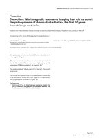
three groups. The corresponding values at day 1 and day 8 are
given in Table 4 and the CRP evolution is shown in Figure 1.
When the 15 excluded patients were added to the analysis,
there was still no difference between groups and the evolution
was the same.
Outcome
Within 3 to 5 days after the start of therapy, new endotracheal
samples were obtained from 70% of group C, 89% of group
C-A and 85% of group C-L: the same bacteria as those found
on day 1 were still present in the sputum of 8 patients in group
C, 4 in group C-A and 12 in group C-L. After 7 to 10 days, per-
sistence was documented in 4 patients out of 16 in group C,
5 out of 18 in group C-A, and 3 out of 13 in group C-L. New
bacteria strains requiring new treatment were found in one
patient in group C (one P. aeruginosa), three patients in group
C-A (two P. aeruginosa and one Enterobacter aerogenes with
extended spectrum beta-lactamase) and in three patients in
group C-L (one Proteus mirabilis, one Serratia marcescens
and one methicillin-resistant S. aureus).
The length of ICU stay after the occurrence of infection was
not different between the three groups: the medians (and 25th
to 75th percentile in parentheses) were 15 (7.5 to 24.75), 16
(9 to 21) and 14 days (9.5 to 21.5) for groups C, C-A and C-
L, respectively. There was also no difference between VFDs
Table 3
Percentage of bacteria found from endotracheal aspirates in each treatment group
Bacteria Cefepime - Cefepime-amikacin - Cefepime-levofloxacin -
S. pneumoniae 3.3 8.3 7.1
Haemophilus influenzae 6.7 4.2 7.1
S. aureus 23.3 (1) 25 28.6 (2)
E. coli 10 8.3 10.7
Klebsiella spp. 10 (1) 12.5 (1) 3.6 (1)
Proteus spp. 3.3 8.3 0
Enterobacter spp. 10 8.3 14.3 (1)
Serratia marcescens 6.712.57.1
P.aeruginosa 23.3 (1) 8.3 (1) 17.9
Acinetobacter baumannii 3.3 4.2 3.6
Numbers in parentheses correspond to the number of patients having positive blood culture with the same organism.
Table 4
Evolution of inflammatory parameters and oxygenation
Group Day PaO
2
/FiO
2
(mmHg) CRP (mg/l) Temperature (°C) Leukocytosis (10
3
/
mm
3
)
C 1 173 ± 88 209 ± 100 38.6 ± 0.9 14.2 ± 5.8
8 304 ± 114 94 ± 62 37.8 ± 0.7 15.5 ± 5.1
C-A 1 194 ± 95 233 ± 115 38.3 ± 0.9 14.3 ± 6.3
8 253 ± 72 113 ± 61 37.7 ± 0.6 12.4 ± 6.4
C-L 1 176 ± 87 211 ± 90 38.8 ± 0.9 13.8 ± 6
8 243 ± 72 94 ± 51 37.6 ± 0.7 15.1 ± 7.9
Groups: C, cefepime; C-A, cefepime with amikacin; C-L, cefepime with levofloxacin. CRP, C-reactive protein. PaO
2
/FiO
2
: arterial oxygen tension/
inspiratory oxygen fraction
Available online />Page 5 of 7
(page number not for citation purposes)
within 28 days after infection: the number of VFDs for each
group was 16.1 ± 8.3 VFDs after C treatment, 12.6 ± 8.1 VFD
after C-A treatment and 12.6 ± 10.4 VFD after C-L treatment
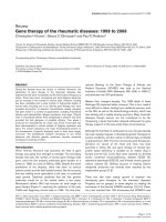
(p > 0.05). Figure 2 shows the Kaplan-Meier curves of
mechanical ventilation duration for the three groups (p > 0.05).
Ten patients died within 28 days, 2 in the C group (10%), 4 in
the C-A group (21%) and 4 in the C-L group (20%). Only one
death was clearly attributable to infection: one post-cardiac
surgery patient who developed bronchopneumonia due to P.
aeruginosa and died in septic shock with severe myocardial
depression at day 3 post infection in group C. Dying patients
had a more elevated CRP level at the eighth day compared to
survivors in the three groups: 145 ± 53 mg/l versus 93 ± 54.9
mg/l (p = 0.033). Among the 15 patients with no microbiologi-
cally confirmed VAP, there was 1 other death within 28 days,
in the C-A group.
Discussion
The aim of this study was to compare monotherapy versus
combination therapy for the treatment of VAP and to look for
differences in the clinical and inflammatory parameters, includ-
ing CRP level. This is the reason why we optimised the doses
of antibiotics as recently recommended [25], and took a highly
selected subgroup of ICU patients, represented mainly by
post-trauma, postoperative or post-intracranial haemorrhage
patients without infection at their entry into the ICU. This
choice was made in order to obtain as far as possible a group
of patients who did not have any life-threatening conditions
other than VAP.
The diagnosis of VAP remains controversial. Some experts
recommend obtaining quantitative culture of a protected spec-
imen of pulmonary secretions [18,19] while others accept the
use of the CPIS score based on clinical data [9,20,21]. How-
ever, this score is far from sensitive and also has a low specif-
icity, as recently published [22,23]. In particular, the recent
retrospective multicenter French study of Luyt and colleagues
[24] comparing CPIS and quantitative culture in patients with
VAP found a specificity as low as 47%. Due to the fact that
quantitative cultures are not routinely available in our hospital,
we used a modified CPIS to confirm the presence of VAP, in
which we incorporated the rapid recent increase of CRP level,
as it is one of our current diagnostic tools, and enrolled
patients before obtaining the culture results. As such, all 59
patients with confirmed VAP had a CPIS above 6 with at least
a positive non-quantitative culture. It is interesting to note that
in this study, patients fulfilling the clinical criteria of VAP but
with an absence of pathogens in their lung secretions had a
CPIS significantly lower than the others and, in contrast, all
patients with quantitative cultures had a CPIS of more than 6.
It would be worth validating this 'new' CPIS in another pro-
spective study.
The association of two drugs in the empirical treatment of VAP
is recommended in order to increase the spectrum of activity,
to decrease the emergence of resistance during treatment,
and to improve the bactericidal activity of therapy [25]. If the
first reason is obvious because of the presence of potential
nosocomial resistant pathogens and cannot be questioned,
the two others remain a matter of debate. Cefepime, a fourth
generation cephalosporin, possesses bactericidal activity
against the great majority of nosocomial pathogens in our hos-
pital and covers almost 100% of non-inducible Enterobacte-
riaceae, more than 90% of inducible Enterobacteriaceae and
nearly 90% of P. aeruginosa. We decided, therefore, to use it
as a first line therapy for VAP, in association or not with either
amikacin or levofloxacin, two other antibiotics with excellent
activity against nosocomial pathogens. The combination of an
anti-Pseudomonas beta-lactam with either an aminoglycoside
or a quinolone such as levofloxacin or ciprofloxacin is indeed
Figure 2
Kaplan-Meier curves of mechanical ventilation durationKaplan-Meier curves of mechanical ventilation duration.
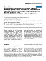
Figure 1
CRP time courseCRP time course. C, cefepime group; C-A, cefepime with amikacin
group; C-L, cefepime with levofloxacin group
Critical Care Vol 10 No 2 Damas et al.
Page 6 of 7
(page number not for citation purposes)
part of the published guidelines for the empirical treatment of
VAP [25].
Among inflammatory parameters, CRP was chosen because
of its routine use and its well known time course evolution.
Interleukin 6 or procalcitonin, two other inflammatory parame-
ters, well described as markers of severity, have not been val-
idated as markers of response to antibiotic therapy [26,27].
CRP, however, which is used as a non-specific marker of
inflammatory events and of bacterial infection, has been used
to monitor response to antibiotic treatment [11]. Recently, the
evolution of CRP levels has been described after CAP. Hans-
son and colleagues [16] showed that the mean time to a 50%
decrease in CRP level was 3.3 days after CAP. Our results
recorded in VAP patients, however, show that the decrease in
CRP level is much slower (six days to obtain a 50% reduction).
Of course, critically ill patients often have causes other than
just a lung infection that increase their CRP levels. The vast
majority of patients included in this study were either post-
trauma or postoperative and their infections were a new insult
but not the first they had encountered; however, the time delay
of more than nine days between the hospital entry and the
occurrence of infection (Table 2) should have been long
enough to allow the CRP level, which usually peaks at 48
hours after trauma or surgery, to decrease. Figure 1 shows
that the CRP levels peaked the second day after inclusion for
VAP and confirms the new insult caused by the infection. The
plasma half-life of CRP is about 19 hours and seems to be
constant whatever the disease [28]; this means that the sole
determinant of CRP level is the synthesis rate, which can be
sustained after infections complicating trauma or surgery. The
circulating CRP concentration decreases if the infectious
stimulus ceases, however, and this should fall more rapidly
with a better treatment than with a weaker one. This is what we
were expecting to find in this study, but this was not the case:
CRP levels failed to show any differences between mono-
therapy and combination therapy. The CRP levels decreased
in the same manner in all three groups.
With regard to temperature, leukocytosis and PaO
2
/FiO
2
,
Dennesen and colleagues [29] already described the resolu-
tion of infectious parameters after initiation of appropriate ther-
apy for VAP in 27 patients. They showed that improvements in
temperature, leucocytosis and PaO
2
/FiO
2
were slow and most
evident during the first six days [29].
There was no difference in terms of mortality, length of stay
and VFDs between the three groups. Moreover, persistence of
bacteria at the end of treatment was not reduced by combina-
tion therapy and recurrence of infection was as frequent in the
three groups. The persistence of bacteria in tracheal aspirates
confirms the observation of Dennesen and colleagues [29],
who observed 50% persistence for Enterobacteriaceae and
100% persistence for P. aeruginosa in spite of appropriate
therapy.
Weaknesses of this study include a lack of blinding of admin-
istration to therapy and the relatively small number of patients.
We enrolled only 74 patients within 21 months, although we
had to treat more than 300 ICU acquired pulmonary infections
during this period. This was mainly due to the exclusion crite-
ria, which did not admit patients receiving antibiotic treatment
for another infection or infection already treated before ran-
domisation. These criteria excluded all patients ventilated for
CAP or for abdominal sepsis and all immunocompromised
patients receiving antibiotics, among others. If this study failed
to demonstrate any advantage of combination therapy over
monotherapy, the use of empirical combination therapy
remains probably relevant in patients with potentially multire-
sistant strains, as recommended by Chastre [30]. Importantly,
the size of the samples in this study does not allow us to draw
definitive conclusions regarding this issue.
Conclusion
We failed to demonstrate any advantage of combination ther-
apy over monotherapy. None of the recorded parameters,
including time course of clinical and inflammatory parameters,
duration of mechanical ventilation, ICU length of stay, bacteri-
ological response and mortality, were influenced by the type of
treatment. The mean time to obtain a 50% decrease in CRP
levels after VAP was about six days, twice that seen after CAP,
reflecting in part the complexity of the different insults ICU
patients often may suffer from.
Competing interests
The authors declare that they have no competing interests.
Authors' contributions
PD and MM originated the study and contributed to the analy-
sis of data. PD, JCP and CG prepared the paper. CG and MN
collected the data, and JLC, DL and JCP enrolled the patients.
All authors read and approved the final manuscript.
References
1. Rello J, Diaz E: Pneumonia in the intensive care unit. Crit Care
Med 2003, 31:2544-2551.
2. Vincent JL, Bihari DJ, Suter PM, Bruining HA, White J, Nicolas-
Chanoin MH, Wolff M, Spencer RC, Hemmer M: Results of the
European Prevalence of Infection in Intensive Care (EPIC)
Study. EPIC International Advisory Committee. JAMA 1995,
274:639-644.
3. Rello J, Ollendorf DA, Oster G, Vera-Llonch M, Bellm L, Redman
R, Kollef MH, VAP Outcomes Scientific Advisory Board Group:
Epidemiology and outcomes of ventilator-associated pneu-
monia in a large US database. Chest 2002, 122:2115-2121.
Key messages
• The beneficial effects of an empirical combination anti-
biotic therapy for VAP were not confirmed in terms of
the evolution of clinical and biological parameters.
• CRP plasma levels decreased by 50% within six days
after antibiotic treatment of VAP.
Available online />Page 7 of 7
(page number not for citation purposes)
4. Sadfar N, Dexfulian C, Collard HR, Saint S: Clinical and eco-
nomic consequences of ventilator-associated pneumonia.
Crit Care Med 2005, 33:2184-2193.
5. Iregui M, Ward S, Sherman G, Fraser VJ, Kollef MH: Clinical
importance of delays in the initiation of appropriate antibiotic
treatment for ventilator-associated pneumonia. Chest 2002,
122:262-268.
6. Kollef MH, Ward S: The influence of mini-BAL cultures on
patient outcomes : Implications for the antibiotic management
of ventilator-associated pneumonia. Chest 1998,
113:412-420.
7. Paul M, Benuri-Silbiger I, Soares-Weiser K, Leibovici L: Beta
lactam monotherapy versus beta lactam-aminoglycoside
combination therapy for sepsis in immunocompetent patients
: systematic review and meta-analysis of randomised trials.
BMJ 2004, 328:668.
8. Pugin J, Auckenthaler R, Mili N, Janssens JP, Lew PD, Suter PM:
Diagnosis of ventilator-associated pneumonia by bacterio-
logic analysis of bronchoscopic and nonbronchoscopic "blind"
bronchoalveolar lavage fluid. Am Rev Respir Dis 1991,
143:1121-1129.
9. Luna CM, Blanzaco D, Niederman MS: Resolution of ventilator-
associated pneumonia: prospective evaluation of the clinical
pulmonary infection score as an early clinical predictor of out-
come. Crit Care Med 2003, 31:676-682.
10. Pepys MB, Baltz ML: Acute phase proteins with special refer-
ence to C-reactive protein and related proteins (pentaxins)
and serum amyloid A protein. Adv Immunol 1983, 34:141-212.
11. Pepys MB, Hirschfield GM: C-reactive protein: a critical update.
J Clin Invest 2003, 111:1805-1812.
12. Sierra R, Rello J, Bailen MA, Benitez E, Gordillo A, Leon C, Pedraza
S: C-reactive protein used as an early indicator of infection in
patients with systemic inflammatory response syndrome.
Intensive Care Med 2004, 30:2038-2045.
13. Kosmas EN, Baxevanis CN, Papamichail M, Kordossis T: Daily
variation in circulating cytokines and acute-phase proteins
correlates with clinical and laboratory indices in community-
acquired pneumonia. Eur J Clin Invest 1997, 27:308-315.
14. Garcia Vazquez E, Martinez JA, Mensa J, Sanchez F, Marcos MA,
de Roux A, Torres A: C-reactive protein levels in community-
acquired pneumonia. Eur Respir J 2003, 21:702-705.
15. Almirall J, Bolibar I, Toran P, Pera G, Boquet X, Balanzo X, Sauca
G, Community-Acquired Pneumonia Maresme Study Group: Con-
tribution of C-reactive protein to the diagnosis and assess-
ment of severity of community-acquired pneumonia. Chest
2004, 125:1335-1342.
16. Hansson LO, Hedlund JU, Ortqvist AB: Sequential changes of
inflammatory and nutrtional markers in patients with commu-
nity-acquired pneumonia. Scand J Clin Lab Invest 1997,
57:111-118.
17. Moreno R, Vincent JL, Matos R, Mendonca A, Cantraine F, Thijs L,
Takal J, Sprung C, Antonelli M, Bruining H, Willatts S: The use of
maximum SOFA score to quantify organ dysfunction/failure in
intensive car. Results of a prospective multicentre study.
Intensive Care Med 1999, 25:686-696.
18. Chastre J, Combes A, Luyt CE: The invasive (quantitative) diag-
nosis of ventilator-associated pneumonia. Respir Care 2005,
50:797-812.
19. Chastre J, Fagon JY: Ventilator-associated pneumonia. Am J
Respir Crit Care Med 2002, 165:867-903.
20. Yu VL, Singh N: Excessive antimicrobial usage causes meas-
urable harm to patients with suspected ventilator-associated
pneumonia. Intensive Care Med 2004, 30:735-738.
21. Singh N, Rogers P, Atwood CW, Wagener MM, Yu VL: Short
course empiric antibiotic therapy for patients with pulmonary
infiltrates in the intensive care unit. A proposed solution for
indiscriminate antibiotic prescription. Am J Respir Crit Care
Med 2000, 162:505-511.
22. Scurink CA, Van Nieuwenhoven CA, Jacobs JA, Rozenberg-Arska
M, Joore HCA, Buskens E, Hoepelman AIM, Bonten MJM: Clinical
pulmonary infection score for ventilator-associated pneumo-
nia: accuracy and inter-obserer variability. Intensive Care Med
2004, 30:217-224.
23. Fartoukh M, Maitre B, Honoré S, Cerf C, Zahar J-R, Brun-Buisson
C: Diagnosing pneumonia during mechanical ventilation. Am
J Respir Crit Care Med 2003, 168:173-179.
24. Luyt CE, Chastre J, Fagon JY: Value of the clinical pulmonary
infection score for the identification and management of ven-
tilator-associated pneumonia. Intensive Care Med 2004,
30:844-852.
25. American Thoracic Society: Guidelines for the management of
adults with hospital-acquired, ventilator-associated and
healthcare-associated pneumonia. Am J Respir Crit Care Med
2005, 171:388-416.
26. Damas P, Ledoux D, Nys M, Vrindts Y, De Groote D, Franchimont
P, Lamy M: Cytokine serum level during severe sepsis in
human. IL-6 as a marker of severity. Ann Surg 1992,
215:356-362.
27. Oberhoffer m, Karzai w, Meier-Hellmann A, Bogel D, Fassbinder J,
Reinhart : Sensitivity and specificity of various markers of
inflammation for the prediction of tumor necrosis factor-alpha
and interleukin-6 in patients with sepsis. Crit care Med 1999,
27:1814-1818.
28. Vigushin DM, Pepys MB, Hawkins PN: Metabolic and scinti-
graphic studies of radioiodinated human C-reactive protein in
health and disease. J Clin Invest 1993, 91:1351-1357.
29. Dennesen PJ, van der Ven AJ, Kessels AG, Ramsay G, Bonten MJ:
Resolution of infectious parameters after antimicrobial ther-
apy in patients with ventilator-associated pneumonia. Am J
Respir Crit Care Med 2001, 163:1371-1375.
30. Chastre J: Antibiotic prescribing for ventilator-associated
pneumonia: get it right from the beginning but be able to rap-
idly deescalate. Intensive Care Med 2005, 31:1463-1465.