Báo cáo y học: "Effects of thoraco-pelvic supports during prone position in patients with acute lung injury/acute respiratory distress syndrome: a physiological study" potx
Bạn đang xem bản rút gọn của tài liệu. Xem và tải ngay bản đầy đủ của tài liệu tại đây (1.28 MB, 9 trang )
Open Access
Available online />Page 1 of 9
(page number not for citation purposes)
Vol 10 No 3
Research
Effects of thoraco-pelvic supports during prone position in
patients with acute lung injury/acute respiratory distress
syndrome: a physiological study
Davide Chiumello
1
, Massimo Cressoni
2
, Milena Racagni
2
, Laura Landi
2
, Gianluigi Li Bassi
2
,
Federico Polli
2
, Eleonora Carlesso
2
and Luciano Gattinoni
1,2
1
Dipartimento di Anestesia e Rianimazione, Fondazione IRCCS – 'Ospedale Maggiore Policlinico, Mangiagalli, Regina Elena', Via F. Sforza 35, 20122
Milan, Italy
2
Istituto di Anestesia e Rianimazione Università degli Studi di Milano, Via F. Sforza 35, 20122 Milan, Italy
Corresponding author: Luciano Gattinoni,
Received: 2 Feb 2006 Revisions requested: 23 Feb 2006 Revisions received: 2 Apr 2006 Accepted: 2 May 2006 Published: 8 Jun 2006
Critical Care 2006, 10:R87 (doi:10.1186/cc4933)
This article is online at: />© 2006 Chiumello et al.; licensee BioMed Central Ltd.
This is an open access article distributed under the terms of the Creative Commons Attribution License ( />),
which permits unrestricted use, distribution, and reproduction in any medium, provided the original work is properly cited.
Abstract
Introduction This study sought to assess whether the use of
thoraco-pelvic supports during prone positioning in patients
with acute lung injury/acute respiratory distress syndrome (ALI/
ARDS) improves, deteriorates or leaves unmodified gas
exchange, hemodynamics and respiratory mechanics.
Methods We studied 11 patients with ALI/ARDS, sedated and
paralyzed, mechanically ventilated in volume control ventilation.
Prone positioning with or without thoraco-pelvic supports was
applied in a random sequence and maintained for a 1-hour
period without changing the ventilation setting. In four healthy
subjects the pressures between the body and the contact
surface were measured with and without thoraco-pelvic
supports. Oxygenation variables (arterial and central venous),
physiologic dead space, end-expiratory lung volume (helium
dilution technique) and respiratory mechanics (partitioned
between lung and chest wall) were measured after 60 minutes
in each condition.
Results With thoraco-pelvic supports, the contact pressures
almost doubled in comparison with those measured without
supports (19.1 ± 15.2 versus 10.8 ± 7.0 cmH
2
O, p ≤ 0.05;
means ± SD). The oxygenation-related variables were not
different in the prone position, with or without thoraco-pelvic
supports; neither were the CO
2
-related variables. The lung
volumes were similar in the prone position with and without
thoraco-pelvic supports. The use of thoraco-pelvic supports,
however, did lead to a significant decrease in chest wall
compliance from 158.1 ± 77.8 to 102.5 ± 38.0 ml/cmH
2
O and
a significantly increased pleural pressure from 4.3 ± 1.9 to 6.1
± 1.8 cmH
2
O, in comparison with the prone position without
supports. Moreover, when thoraco-pelvic supports were added,
heart rate increased significantly from 82.1 ± 17.9 to 86.7 ±
16.7 beats/minute and stroke volume index decreased
significantly from 37.8 ± 6.8 to 34.9 ± 5.4 ml/m
2
. The increase
in pleural pressure change was associated with a significant
increase in heart rate (p = 0.0003) and decrease in stroke
volume index (p = 0.0241).
Conclusion The application of thoraco-pelvic supports
decreases chest wall compliance, increases pleural pressure
and slightly deteriorates hemodynamics without any advantage
in gas exchange. Consequently, we stopped their use in clinical
practice.
Introduction
Prone positioning is used and recommended as a rescue
maneuver to improve arterial oxygenation in adult patients with
acute lung injury (ALI), acute respiratory distress syndrome
(ARDS) [1,2] or chronic obstructive pulmonary disease [3],
although its benefits with regard to outcome are not proven
[4,5].
Improved oxygenation implies, by definition, improvement of
the ventilation/perfusion ratio. This can be achieved through
different mechanisms, not mutually exclusive, each
ALI = acute lung injury; ARDS = acute respiratory distress syndrome; BSA = body surface area; EELV = end-expiratory lung volume; PEEP = positive
end-expiratory pressure.
Critical Care Vol 10 No 3 Chiumello et al.
Page 2 of 9
(page number not for citation purposes)
documented in the literature: (1) a more uniform distribution of
alveolar inflation/ventilation, due to the lower gradient of
transpulmonary pressure resulting from the changes in chest
wall mechanics, with perfusion being less affected [6-9]; (2) a
greater recruitment of the dorsal lung regions in comparison
with the derecruitment of the ventral lung regions when chang-
ing from the supine to the prone position [10]; (3) an overall
increase in end-expiratory lung volume (EELV) as a result of
the more favorable position of the diaphragm [11].
Douglas and colleagues [12] used supports under the ribcage
and the pelvis of patients with respiratory failure, to prevent
their abdomen from bearing the entire weight of the torso.
Indeed, some authors have advocated the use of thoraco-pel-
vic supports to avoid an increase in intra-abdominal pressure,
which could limit diaphragm excursion and, consequently,
alveolar ventilation in the most dependent lung regions
[13,14]. A survey study, in 29 intensive care units, found that
thoraco-pelvic supports were routinely applied in 18 of them
[15].
However, the use of thoraco-pelvic supports in the prone posi-
tion has potential drawbacks, such as the possibility of devel-
oping pressure sores at the contact surfaces [16]. Because
the effectiveness of this intervention is debated, in the present
study we set out to investigate whether the use of thoraco-pel-
vic supports on patients with ALI/ARDS improves, worsens, or
has no effect on respiratory mechanics, gas exchange, and
hemodynamics.
Materials and methods
Study population
Eleven consecutive intubated patients with ALI/ARDS,
defined in accordance with standard criteria [17], were
included in the study. None of them had a history of chronic
obstructive pulmonary disease, heart failure or severe head
trauma. Their main clinical characteristics are summarized in
Table 1. After completing the study and analyzing the data we
realized the possible importance of the contact pressures. We
therefore measured the contact pressures directly in four
healthy volunteers with or without the thoraco-pelvic supports.
The study was approved by the Institutional Review Board of
our hospital. Informed consent, because the patients were
incompetent, was obtained in accordance with Italian national
regulations (waived consent).
Study design
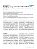
The patients were first studied in the supine position (1-hour
baseline). Subsequently, they were studied in the prone posi-
tion for 2 hours, for 1 hour with supports and for 1 hour with-
out, in a randomized manner (see flow diagram in Figure 1) for
a total duration of 3 hours study time.
The patients were lying on air-cushioned beds (Total Care
®
;
Hill Rom Services Inc., Batesville, IN, USA). In the supine posi-
tion and in the prone position without supports, the body of
each patient was in direct contact with the mattress. In prone
position with supports, a roll was placed under the cranial part
of the ribcage and a pillow under the pelvic region, so that
most of the body weight rested on them. The thoraco-pelvic
supports were placed so as to allow free abdominal move-
ments (see Figure 2 and Table 2).
The patients were studied while sedated with fentanyl (1.5 to
5.5 µg/kg per hour) and midazolam (4 to 8 mg/hour), para-
lyzed with pancuronium bromide (0.05 to 0.1 mg/kg per hour)
and ventilated in volume-control mode with a Servo Ventilator
300 C (Siemens, Solna, Sweden). Mechanical ventilation was
set by the attending physician on a clinical basis and remained
unchanged throughout the study periods. The baseline mean
tidal volume was 565.3 ± 160.5 ml (7.2 ± 1.4 ml/kg
IBW
, where
IBW stands for ideal body weight; means ± SD), respiratory
rate was 17.1 ± 3.5 breaths/minute, inspiratory oxygen frac-
tion was 0.43 ± 0.04, positive end-expiratory pressure (PEEP)
was 10.8 ± 1.8 cmH
2
O, and plateau pressure was 22.4 ± 4.3
cmH
2
O.
Fluids, drug infusions and ventilator settings remained
unchanged throughout the whole study period.
Measurements
Contact pressures
The pressures between the air-cushioned beds or thoraco-pel-
vic supports and the body (namely, the contact pressures)
were measured in four healthy volunteers (age 28.7 ± 4.9
years, weight 66.2 ± 11.8 kg, body mass index 22.1 ± 2.0 kg/
m
2
), in the same three conditions and body positions in which
the patients were studied. A plastic bag with a volume of 250
ml containing 100 ml of water and equipped with a pressure
transducer (Transpec IV L974; Abbott Ireland, Sligo, Ireland)
was used. The zero of the pressure transducer was at the level
of the plastic bag. In the supine position, pressure transducers
were placed under the shoulders, the lumbar spine, and the
sacrum. In the prone position, with and without the thoraco-
pelvic supports, pressure transducers were placed in the cor-
responding positions, under the upper chest, the mesogas-
trium, and the pelvic region (Figure 2).
Gas exchanges and hemodynamics
All variables were recorded at the end of each study period.
Blood gas tensions in the arterial and central venous blood
were analysed with a blood gas analyzer (IL-1312 Blood Gas
Manager; Instrumentation Laboratory, Milan, Italy). Minute met-
abolic carbon dioxide production, partial pressure of CO
2
in
mixed expired air, and end-tidal concentration of carbon diox-
ide were measured with a respiratory function monitor
(CO
2
SMO™; Novametrix Medical Systems Inc., Wallingford,
CT, USA). The venous admixture (estimated from the central
Available online />Page 3 of 9
(page number not for citation purposes)
venous blood values), the physiological dead space, and the
alveolar dead space were computed from standard formulae.
Blood pressures (central and arterial) were measured with dis-
posable pressure transducers (Transpec IV L974) positioned
at the mid-axillary line. Cardiac output was measured with the
thermo-dilution method, using a Swan–Ganz Oximetry Pace-
port Thermo-dilution Catheter (Edwards Lifesciences, Irvine,
CA, USA) in five patients, and by pulse contour analysis
(PiCCO System™ version 4.1; Pulsion Medical System,
Munich, Germany) in four. In the five patients with a Swan–
Ganz catheter, pulmonary artery and wedge pressures were
also recorded. The stroke volume index was computed as the
stroke volume divided by the body surface area (BSA). The
BSA was obtained with the formula BSA [m
2
] = 0.20247 ×
height [m]
0.725
× weight [kg]
0.425
[18].
End-expiratory lung volume and respiratory mechanics
EELVs at PEEP were measured with a simplified closed-circuit
helium-dilution method, during an end-expiratory pause [19].
An anesthesia bag, filled with 1.5 liters of a known gas mixture
(13% helium in oxygen) was connected to the airway opening
previously clamped at end-expiration to maintain the PEEP
level. Ten manual breaths were subsequently performed. The
helium concentration in the bag was then measured with a
helium analyzer (PK Morgan Ltd, Chatham, UK) and EELV was
computed from the formula EELV = (V
i
× [He]
i
/[He]
f
) - V
i
,
where V
i
is the initial gas volume in the anesthesia bag and
[He]
i
and [He]
f
are the initial and final concentrations of helium
in the bag, respectively.
Airway pressures were measured proximally to the endotra-
cheal tube with a dedicated pressure transducer (MPX 2010
DP; Motorola, Phoenix, AZ, USA). Mean airway pressures
were calculated as the area under the airway pressure–time
trace, divided by the duration of each breath. Esophageal and
gastric pressures were measured with two radio-opaque bal-
loons inflated with 0.5 to 1.0 ml of air (SmartCath; Bicore,
Irvine, CA, USA) connected to a pressure transducer (Bentley
Trantec; Bentley Laboratories, Irvine, USA). The esophageal
and gastric balloons were both positioned in the stomach with
the use of an endotracheal tube inserted through the mouth as
a guide through the pharynx. The esophageal balloon was then
retracted until it reached the upper third of the esophagus. In
addition, to ensure the correct position of the catheters, an
inspiratory occlusion was made, so that a check for concord-
ant changes in airway, esophageal, and gastric pressures
could be made.
Respiratory flow rates were measured with a heated pneumo-
tachograph (Fleisch no. 2; Fleisch, Lausanne, Switzerland)
inserted between the proximal tip of the endotracheal tube and
the Y-piece of the breathing circuit. Flow and pressure signals
were recorded on a personal computer for subsequent analy-
sis with dedicated software (Colligo; Elekton, Milan, Italy).
Tidal volumes were obtained by mathematical integration of
the measured flow signal. The static compliance of each com-
ponent of the respiratory system – respiratory system, chest
wall, and lung – was calculated as a chord compliance, using
standard formulae, with the rapid occlusion method [20]. The
end-inspiratory pause button of the ventilator was actioned
until airway, esophageal, and gastric pressures decreased
from their maximum value to an apparent plateau. Similarly,
Table 1
Patients' main characteristics
Patient Sex (M/F) Age (years) Measured
weight (kg)
BMI (kg/m
2
) PEEP (cmH
2
O) PaO
2
/FiO
2
(Torr) Diagnosis Days of ALI/
ARDS
Outcome
1 M 73 75 23,2 9.4 180 Sepsis (from peritonitis) 2 S
2 F 55 55 19,9 10.9 245 Sepsis (from peritonitis) 9 S
3 M 76 85 23,3 8.3 138 Community-acquired pneumonia 4 S
4 M 43 90 23,3 10.6 225 Pneumonia (ab ingestis) – sepsis 2 S
5 M 80 70 23,0 11.3 210 Nosocomial pneumonia 13 S
6 M 48 85 24,2 12.8 265 Polytrauma 8 S
7 M 44 80 24,7 9.3 178 Nosocomial pneumonia 6 S
8 M 38 92 23,3 8.8 225 Pneumonia (ab ingestis)7S
9 M 77 55 22,1 14.0 204 Idiopathic pneumonia in bone
marrow transplantation
1D
10 M 27 80 23,3 12.7 237 Nosocomial pneumonia 4 S
11 M 59 93 23,3 10.3 160 Sepsis 2 S
Overall 10 M, 1 F 56.4 ± 18.0 78.2 ± 13.4 23.1 ± 1.2 10.8 ± 1.81 206.2 ± 38.7 – 5.2 ± 3.7 1D, 10S
BMI, Body mass index; PEEP, positive end-expiratory pressure; PaO
2
/FiO
2
, ratio of arterial oxygen tension to fraction of inspired oxygen; ALI, acute
lung injury; ARDS, acute respiratory distress syndrome; S, survived; D, died. Overall results are means ± SD.
Critical Care Vol 10 No 3 Chiumello et al.
Page 4 of 9
(page number not for citation purposes)
end-expiratory airway, esophageal, and gastric pressures were
recorded after an end-expiratory hold maneuver.
Transpulmonary pressure was computed as the difference
between airway pressure and esophageal pressure, and the
transdiaphragmatic pressure as the difference between
esophageal pressure and gastric pressure. Pleural pressure
change, gastric pressure change, and transpulmonary pres-
sure change were calculated as the differences between end-
inspiratory and end-expiratory esophageal pressure, gastric
pressure, and transpulmonary pressure, respectively.
Intra-abdominal pressure was estimated by measuring the
bladder pressure by the method of Cheatham and Safcsak
[21].
Statistical analysis
Data are shown as means ± SD. All data were analyzed with
SAS software (version 8.2; SAS Institute, Cary, NC, USA).
The study design included a baseline condition (supine) and
two treatments (prone without supports and prone with sup-
ports). The treatments were administered to each patient in a
randomized order, in accordance with a crossover design.
The effect of the two treatments and of the sequence of their
administration was evaluated with an analysis of variance for
repeated measures, performed with the SAS MIXED proce-
dure. In addition, each study treatment (prone with and without
supports) was compared with baseline (supine) by using
paired t tests.
To explore the possible association between pleural pressure
change and several tested variables, we used the SAS MIXED
procedure, building a mixed-effect linear model, in which each
patient was treated as a random coefficient. This procedure
yielded the parameters of a global regression model, as well
as an indication (p value) of the significance of the association
itself.
Results
Contact pressures
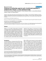
Contact pressures recorded in four healthy subjects in the
supine and in the prone position with and without supports are
summarized in Figure 2. As shown, in shifting the subjects
from the supine to the prone position without thoraco-pelvic
supports, the contact pressures at thorax and sacrum/pubis
did not change significantly, whereas pressures recorded at
the abdominal wall surface increased (11.0 ± 1.8 versus 5.8
± 2.9 cmH
2
O for the supine position). After application of the
thoraco-pelvic supports the contact pressures at thorax and
Figure 1
Flow chart of the study protocolFlow chart of the study protocol.
Figure 2
Patients' positions and contact pressuresPatients' positions and contact pressures. Patients' positions used in
the study: supine (top), prone without supports (center) and prone with
thoraco-pelvic supports (bottom). The mean contact pressures (meas-
ured with pressure transducers in four healthy volunteers) are also indi-
cated by white arrows. Table 2 shows detailed contact pressures at
different sites and global values.
30
0
Supine
15 8
5
navel
pubes
inter-nipple
line
sacrum
Prone
without
support
Contact Pressure (cmH O)
2
30
17 11
4.5
Prone
with
support
0
30
2829 0
0
15
8
5
17
11
4.5
29
28
Available online />Page 5 of 9
(page number not for citation purposes)
pubis increased significantly compared with those in the prone
position without supports (29.0 ± 6.5 versus 17.0 ± 7.4
cmH
2
O and 28.3 ± 8.9 versus 4.5 ± 4.2 cmH
2
O, respectively)
whereas the contact pressure at the abdominal wall surface
was zero because the abdomen remained suspended.
End-expiratory lung volume and respiratory mechanics
The EELVs and the mechanics of the respiratory system, par-
titioned into the chest wall and lung components, are summa-
rized in Table 3. Shifting the patients from the supine to the
prone position, without supports, led to a decreasing trend of
chest wall compliance and to a significant increase in lung
compliance. Adding the thoraco-pelvic supports in the prone
position led to a further significant decrease in chest wall com-
pliance and a significant increase in pleural pressure. We
found no sequence effect (that is, prone after supine or supine
after prone; see Figure 1) on lung volumes and respiratory
mechanics variables.
Gas exchange
Table 4 summarizes the gas exchange variables in the supine
and in the prone position with and without thoraco-pelvic sup-
ports. As shown, the oxygenation-related variables in the arte-
rial and central venous blood improved significantly in shifting
the patients from supine to prone without thoraco-pelvic sup-
ports. The application of thoraco-pelvic supports did not lead
to any further significant change. No significant differences
were observed in CO
2
-related variables between the supine
and the prone position with or without thoraco-pelvic sup-
ports. We found no sequence effect on gas exchange
variables.
Hemodynamics
The application of thoraco-pelvic supports caused a signifi-
cant increase in heart rate and a decrease in stroke volume
index and in pulmonary artery pressures, in comparison with
the prone position without supports. The other hemodynamic
variables (notably cardiac index and systemic vascular resist-
Table 2
Detailed contact pressures at different sites and global values
Position Units Supine Prone without supports Prone with supports
Thorax cmH
2
O 15.4 ± 4.1 17.0 ± 7.4 29.0 ± 6.5
a,b
Abdomen cmH
2
O 5.8 ± 2.9 11.0 ± 1.8
a
0.0 ± 0.0
a,b
Sacrum/pubis cmH
2
O 8.0 ± 5.7 4.5 ± 4.2 28.3 ± 8.9
a,b
Global cmH
2
O 9.7 ± 5.8 10.8 ± 7.0 19.1 ± 15.2
a,b
Results are means ± SD.
a
p ≤ 0.05 compared with supine;
b
p ≤ 0.05 compared with prone without supports.
Table 3
Lung volumes and respiratory mechanics
Variable Units Supine Prone without support Prone with support
Tidal volume (V
T
) ml 565.3 ± 160.5 577.6 ± 185.3 593.4 ± 200.7
Tidal volume per kg IBW (V
T
/kg
IBW
) ml/kg 7.2 ± 1.4 7.4 ± 1.6 7.6 ± 1.8
EELV l 1.12 ± 0.49 1.00 ± 0.26 1.07 ± 0.31
Mean airway pressure cmH
2
O 15.1 ± 2.1 15.6 ± 2.3 15.7 ± 2.1
a
Plateau pressure (P
plat
)cmH
2
O 22.4 ± 4.3 22.1 ± 3.8 23.6 ± 4.5
a,b
Respiratory system compliance ml/cmH
2
O 52.1 ± 17.6 52.9 ± 18.8 49.3 ± 18.1
b
Lung compliance ml/cmH
2
O 71.5 ± 23.8 93.5 ± 47.3
a
102.0 ± 47.0
a
Chest wall compliance ml/cmH
2
O 235.2 ± 152.5 158.1 ± 77.8 102.5 ± 38.0
a,b
Transpulmonary pressure change
c
cmH
2
O 8.4 ± 2.2 7.1 ± 2.2
a
6.6 ± 2.3
a
Pleural pressure change
c
cmH
2
O 3.2 ± 1.9 4.3 ± 1.9 6.1 ± 1.8
a,b
Gastric pressure cmH
2
O 13.4 ± 4.0 14.3 ± 3.5 13.2 ± 4.3
Gastric pressure change
c
cmH
2
O 2.6 ± 0.8 3.4 ± 1.1
a
4.5 ± 1.9
a,b
Transdiaphragmatic pressure cmH
2
O 0.6 ± 2.0 0.9 ± 1.6 1.6 ± 1.7
Bladder pressure cmH
2
O 12.0 ± 2.8 14.5 ± 3.4
a
14.5 ± 3.7
a
Results are means ± SD. IBW, ideal body weight; EELV, end-expiratory lung volume.
a
p ≤ 0.05 compared with supine;
b
p ≤ 0.05 compared with
prone without supports;
c
difference between end-inspiration and end-expiration
Critical Care Vol 10 No 3 Chiumello et al.
Page 6 of 9
(page number not for citation purposes)
ance) were not affected by the application of thoraco-pelvic
supports. There was no sequence effect on hemodynamic var-
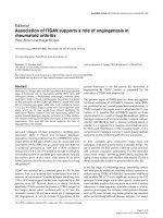
iables. We observed a significant association between the
level of pleural pressure change and heart rate (p = 0.0003)
and between pleural pressure change and stroke volume index
(p = 0.0241) (see Figure 3 and Table 5).
Discussion
In the present study we found that the prone position with tho-
raco-pelvic supports, as compared with the prone position
without supports, did not affect gas exchange and lung volume
but decreased the chest wall compliance, increased the pleu-
ral pressure and slightly modified the hemodynamic pattern
(heart rate and stroke volume index). In addition, we confirmed
Table 4
Gas exchanges
Variable Units Supine Prone without supports Prone with supports
PaO
2
/FiO
2
Torr [kPa] 206.2 ± 38.7 [27.5 ± 5.2] 261.8 ± 41.2
a
[34.9 ± 5.5] 265.0 ± 40.0
a
[35.3 ± 5.3]
PaO
2
Torr [kPa] 87.7 ± 10.2 [11.7 ± 1.4] 112.5 ± 16.0
a
[15.0 ± 2.1] 113.4 ± 12.0
a
[15.1 ± 1.6]
SaO
2
% 95.7 ± 1.0 96.6 ± 0.6
a
96.7 ± 0.6
a
PvO
2
Torr [kPa] 44.6 ± 4.0 [5.9 ± 0.5] 49.0 ± 6.3
a
[6.5 ± 0.8] 47.7 ± 5.6
a
[6.4 ± 0.7]
SvO
2
% 77.2 ± 4.4 80.3 ± 5.4
a
79.6 ± 5.7
a
pH
a
7.37 ± 0.06 7.37 ± 0.05 7.36 ± 0.06
Ve l/minute 9.4 ± 2.5 9.6 ± 2.8 9.9 ± 3.1
PaCO
2
Torr [kPa] 43.9 ± 4.2 [5.9 ± 0.6] 43.6 ± 4.2 [5.8 ± 0.6] 44.3 ± 6.1 [5.9 ± 0.8]
PvCO
2
Torr [kPa] 51.0 ± 6.6 [6.8 ± 0.9] 52.1 ± 6.8 [6.9 ± 0.9] 52.2 ± 6.4 [7.0 ± 0.9]
pH
v
7.35 ± 0.05 7.34 ± 0.06 7.34 ± 0.05
VCO
2
ml/minute 145.5 ± 38.8 143.3 ± 40.9 140.4 ± 42.9
V
d
/V
t
0.62 ± 0.10 0.63 ± 0.11 0.63 ± 0.13
V
d
/V
t
(alv) 0.18 ± 0.15 0.19 ± 0.15 0.18 ± 0.16
Results are means ± SD. PaO
2
/FiO
2
, ratio of arterial oxygen tension to fraction of inspired oxygen; SaO
2
, arterial oxygen saturation; PvO
2
, mixed-
venous oxygen tension; SvO
2
, mixed-venous oxygen saturation; pH
a
, arterial blood pH; Ve, minute ventilation; PaCO
2
, arterial carbon dioxide
tension; PvCO
2
, mixed-venous carbon dioxide tension; pH
v
, venous blood pH; VCO
2
, minute metabolic carbon dioxide production; V
d
/V
t
, dead
space; V
d
/V
t
(alv), alveolar dead space.
a
p ≤ 0.05 compared with supine.
Table 5
Hemodynamics
Variable Units Supine Prone without supports Prone with supports
CI (l/minute)/m
2
BSA
3.2 ± 0.7 3.2 ± 0.6 3.1 ± 0.5
SVI ml/m
2
BSA
38.1 ± 7.2 37.8 ± 6.8 34.9 ± 5.4
a,b
HR min
-1
79.6 ± 17.3 82.1 ± 17.9 86.7 ± 16.7
a,b
Mean BP mmHg 83.7 ± 8.0 89.2 ± 9.4 85.2 ± 11.8
Systolic BP mmHg 123.0 ± 16.5 132.0 ± 18.4
a
123.8 ± 19.8
Diastolic BP mmHg 64.0 ± 7.6 67.8 ± 9.8 65.9 ± 9.8
Mean PAP
c
mmHg 27.4 ± 3.0 31.0 ± 6.6 27.1 ± 9.0
b
Systolic PAP
c
mmHg 38.6 ± 5.1 41.8 ± 11.3 36.8 ± 11.9
b
Diastolic PAP
c
mmHg 21.8 ± 1.9 25.6 ± 4.7 22.2 ± 7.5
b
WP
d
mmHg 22.0 ± 5.7 24.4 ± 6.6 23.2 ± 7.5
CVP mmHg 12.0 ± 2.3 13.0 ± 2.8 12.7 ± 4.3
Diuresis ml 111 ± 71 90 ± 64
a
91 ± 58
a
CI, cardiac index; BSA, body surface area; SVI, stroke volume index; HR, heart rate; BP, arterial blood pressure; PAP, pulmonary artery pressure;
WP, wedge pressure; CVP, central venous pressure.
a
p ≤ 0.05 compared with supine;
b
p ≤ 0.05 compared with prone without supports;
c
cardiac
output in nine patients only;
d
Swan–Ganz in five patients only.
Available online />Page 7 of 9
(page number not for citation purposes)
the positive effects of the prone position on oxygenation when
shifting ALI/ARDS patients from the supine to the prone posi-
tion, as largely documented in the literature [4,5].
Mechanics of the respiratory system
When, in previous studies, we directly investigated chest wall
displacements by optoelectronic plethysmography we found
that both in spontaneously breathing subjects and in paralysed
patients with ALI/ARDS in the supine position, the ribcage
accounted for about 37% of the chest wall displacement and
the abdomen for 63% (that is, the ribcage compliance and
abdominal wall compliance were 37% and 63%, respectively,
of the whole chest wall compliance) [22,23]. When the sub-
jects were moved to the prone position without pelvic sup-
ports, the ribcage accounted for 46.5% of the chest wall
displacement, and the abdomen for 53.5% [23]. In experimen-
tal animals, too, with a computed tomography scan we found
a more even distribution of chest wall displacement [24] when
shifting from supine to prone.
If we apply these figures to our actual patients we can estimate
that in the supine position the ribcage compliance would have
been 86.8 ± 56.3 ml/cmH
2
O and the abdominal wall compli-
ance 148.4 ± 96.2 ml/cmH
2
O (total chest wall compliance
235.2 ± 152.5 ml/cmH
2
O), whereas in the prone position they
would have been 73.5 ± 36.2 and 84.6 ± 41.6 ml/cmH
2
O,
respectively (total chest wall compliance 158.1 ± 77.8 ml/
cmH
2
O). This suggests that, in shifting from supine to prone
without thoraco-pelvic supports, the decrease in abdominal
wall compliance accounts for most of the decrease in chest
wall compliance. If so, the use of thoraco-pelvic supports,
which allows free movement of the abdominal wall, should be
mostly indicated. In contrast, we found that applying the tho-
raco-pelvic supports led to a further decrease in chest wall
compliance. Thus, at least in patients with ALI/ARDS, when
using thoraco-pelvic supports, the possible improvement in
abdominal chest wall compliance may be offset by the greater
decrease of the ribcage compliance, possibly as a result of the
increased contact pressures at the ribcage. In addition, the
expected improvement in abdominal wall compliance with tho-
raco-pelvic supports could be lower than expected because of
the greater baseline distension of the unsupported abdominal
wall and the possible effects of pelvic supports on the lower
abdominal mechanics.
Lung volumes, gas exchange and hemodynamics
In the present study, as shown previously [4,5], the oxygena-
tion variables increased significantly when the patients were
shifted from supine to prone without thoraco-pelvic supports,
but did not change when the supports were added. The aver-
age EELVs did not change in any position. However, the lung
volume increased in some patients after being moved from the
supine to the prone position, whereas in others it decreased,
suggesting different individual interactions between the
opposite effects of the prone position on recruitment
(increased lung gas volume) and on increased pleural pres-
sure (decreased transpulmonary pressure). We found that
several hemodynamic variables changed significantly between
the use and the non-use of supports in the prone position.
Although we do not have direct evidence, we speculate that
the independent variable that caused the hemodynamic
changes is the increase in intrathoracic pressure associated
with the use of thoraco-pelvic supports. The hemodynamic
changes, in fact, are compatible with the homeostatic
response to an initial decrease in effective circulating volume
induced by an increase in pleural pressure. The correlation we
found between the progressive increase in pleural pressure
Figure 3
Intrathoracic pressure and hemodynamicsIntrathoracic pressure and hemodynamics. Top panel, association
between pleural pressure change (delta Ppl) and heart rate (HR); bot-
tom panel, association between pleural pressure change and stroke
volume index (SVI). Each patient is represented with a different symbol
and the values recorded in the three different conditions are all indi-
cated, together with the regression line about these points for each
individual patient. A regression line for the whole model of association
is also depicted (thick line). Pleural pressure change is significantly
associated with heart rate (p = 0.0003) and with stroke volume index (p
= 0.0241).
Critical Care Vol 10 No 3 Chiumello et al.
Page 8 of 9
(page number not for citation purposes)
change and the decrease in stroke volume and increase in
heart rate supports this hypothesis.
Clinical implications
One of the major complications related to the prone position
are pressure sores, usually located at the weight-bearing sites
such as the bony prominences where the contact pressures
are the highest [4,5,25]. A relationship between pressure
sores and the duration and magnitude of the contact pressure
has been shown [26]. In patients in the prone position, a sig-
nificantly higher number of new or worsening pressure sores
has been found in comparison with the supine position
[4,5,27]. The use of thoraco-pelvic supports, by increasing the
contact pressures, because of a lower contact surface com-
pared with lying with the body directly on the air-cushioned
beds, could potentially increase skin-tissue damage.
Conclusion
This study suggests that the prone position primarily induces
changes in pleural pressure, probably by modifying the geom-
etry and mechanics of the chest wall. Adding the thoraco-pel-
vic supports does not provide any advantage in oxygenation
but increases the pleural pressure. Moreover, although not
investigated in this short-term study, increased contact pres-
sures at the interface between the thoraco-pelvic supports
and the body may increase, with time, the likelihood of pres-
sure sores. Indeed, in clinical practice, we have stopped using
thoraco-pelvic supports in the prone position.
Competing interests
LG is a member of the paid KCI Advisory Board. All other
authors declare that they have no competing interests.
Authors' contributions
DC conceived the study, participated in its design and coordi-
nation, performed the measurements and wrote a first draft of
the manuscript. MC participated in the study design and coor-
dination and performed the measurements. MR participated in
the study design and coordination and performed the meas-
urements. LL participated in the study design and coordination
and performed the measurements. GLB participated in the
study design and coordination and performed the measure-
ments. FP performed the statistical analysis and helped draft
the manuscript. EC performed the statistical analysis and
helped draft the manuscript. LG conceived the study, partici-
pated in its design and coordination, coordinated the final
analysis of collected data, and revised the manuscript in writ-
ing its final version. All authors read and approved the final
manuscript.
Acknowledgements
The authors thank all who participated in the study and in the care of the
patients enrolled. Special thanks go to Angelo Colombo MD PhD, with
a degree in statistics, of the Terapia Intensiva Neuroscienze (Fondazione
Ospedale Maggiore Policlinico, Mangiagalli e Regina Elena) for statisti-
cal advice, and to the nursing staff of the general Intensive Care Unit of
the Fondazione Ospedale Maggiore Policlinico, Mangiagalli e Regina
Elena and to all the physicians, without whom this study would not have
been possible. This study received financial support from Fondazione
IRCCS 'Ospedale Maggiore Policlinico, Mangiagalli e Regina Elena di
Milano'.
References
1. Dellinger RP, Carlet JM, Masur H, Gerlach H, Calandra T, Cohen
J, Gea-Banacloche J, Keh D, Marshall JC, Parker MM, et al.: Sur-
viving Sepsis Campaign guidelines for management of severe
sepsis and septic shock. Crit Care Med 2004, 32:858-873.
2. Slutsky AS: The acute respiratory distress syndrome, mechan-
ical ventilation, and the prone position. N Engl J Med 2001,
345:610-612.
3. Mentzelopoulos SD, Zakynthinos SG, Roussos C, Tzoufi MJ,
Michalopoulos AS: Prone position improves lung mechanical
behavior and enhances gas exchange efficiency in mechani-
cally ventilated chronic obstructive pulmonary disease
patients. Anesth Analg 2003, 96:1756-1767.
4. Gattinoni L, Tognoni G, Pesenti A, Taccone P, Mascheroni D,
Labarta V, Malacrida R, Di Giulio P, Fumagalli R, Pelosi P, et al.:
Effect of prone positioning on the survival of patients with
acute respiratory failure. N Engl J Med 2001, 345:568-573.
5. Guerin C, Gaillard S, Lemasson S, Ayzac L, Girard R, Beuret P,
Palmier B, Le QV, Sirodot M, Rosselli S, et al.: Effects of system-
atic prone positioning in hypoxemic acute respiratory failure: a
randomized controlled trial. JAMA 2004, 292:2379-2387.
6. Gattinoni L, Pelosi P, Vitale G, Pesenti A, D'Andrea L, Mascheroni
D: Body position changes redistribute lung computed-tomo-
graphic density in patients with acute respiratory failure.
Anesthesiology 1991, 74:15-23.
7. Pelosi P, Tubiolo D, Mascheroni D, Vicardi P, Crotti S, Valenza F,
Gattinoni L: Effects of the prone position on respiratory
mechanics and gas exchange during acute lung injury. Am J
Respir Crit Care Med 1998, 157:387-393.
8. Mutoh T, Guest RJ, Lamm WJ, Albert RK: Prone position alters
the effect of volume overload on regional pleural pressures
and improves hypoxemia in pigs in vivo. Am Rev Respir Dis
1992, 146:300-306.
9. Richter T, Bellani G, Scott HR, Vidal Melo MF, Winkler T, Venegas
JG, Musch G: Effect of prone position on regional shunt, aera-
tion, and perfusion in experimental acute lung injury. Am J
Respir Crit Care Med 2005, 172:480-487.
10. Gattinoni L, Pelosi P, Valenza F, Mascheroni D: Patient position-
ing in acute respiratory failure. In Principles and Practice of
Mechanical Ventilation Edited by: Tobin M. New York: McGraw-
Hill; 1994:1067-1076.
Key messages
• The prone position is a recognized rescue therapy for
severe hypoxemia in ARDS.
• Allowing free abdominal movement should improve lung
mechanics and gas exchange on a theoretical basis.
• This hypothesis was tested by studying respiratory
mechanics (partitioned into lung and chest wall compo-
nents), gas exchange and hemodynamics with and with-
out thoraco-pelvic supports.
• We could not show any benefit from using thoraco-pel-
vic supports.
• Thoraco-pelvic supports are useless in ARDS patients
in the prone position and merely increase the likelihood
of pressure sores, as a result of increased contact
pressures.
Available online />Page 9 of 9
(page number not for citation purposes)
11. Pelosi P, Bottino N, Chiumello D, Caironi P, Panigada M, Gam-
beroni C, Colombo G, Bigatello LM, Gattinoni L: Sigh in supine
and prone position during acute respiratory distress
syndrome. Am J Respir Crit Care Med 2003, 167:521-527.
12. Douglas WW, Rehder K, Beynen FM, Sessler AD, Marsh HM:
Improved oxygenation in patients with acute respiratory fail-
ure: the prone position. Am Rev Respir Dis 1977, 115:559-566.
13. Pelosi P, Croci M, Calappi E, Cerisara M, Mulazzi D, Vicardi P, Gat-
tinoni L: The prone positioning during general anesthesia min-
imally affects respiratory mechanics while improving
functional residual capacity and increasing oxygen tension.
Anesth Analg 1995, 80:955-960.
14. Pelosi P, Croci M, Calappi E, Mulazzi D, Cerisara M, Vercesi P,
Vicardi P, Gattinoni L: Prone positioning improves pulmonary
function in obese patients during general anesthesia. Anesth
Analg 1996, 83:578-583.
15. Leonet S, Fontaine C, Moraine JJ, Vincent JL: Prone positioning
in acute respiratory failure: survey of Belgian ICU nurses.
Intensive Care Med 2002, 28:576-580.
16. Messerole E, Peine P, Wittkopp S, Marini JJ, Albert RK: The prag-
matics of prone positioning. Am J Respir Crit Care Med 2002,
165:1359-1363.
17. Bernard GR, Artigas A, Brigham KL, Carlet J, Falke K, Hudson L,
Lamy M, LeGall JR, Morris A, Spragg R: Report of the American-
European consensus conference on ARDS: definitions, mech-
anisms, relevant outcomes and clinical trial coordination. The
Consensus Committee. Intensive Care Med 1994, 20:225-232.
18. Du Bois D, Du Bois EF: A formula to estimate the approximate
surface area if height and weight be known. Arch Int Med
1916, 17:863-871.
19. Damia G, Mascheroni D, Croci M, Tarenzi L: Perioperative
changes in functional residual capacity in morbidly obese
patients. Br J Anaesth 1988, 60:574-578.
20. Polese G, Rossi A, Appendini L, Brandi G, Bates JH, Brandolese
R: Partitioning of respiratory mechanics in mechanically venti-
lated patients. J Appl Physiol 1991, 71:2425-2433.
21. Cheatham ML, Safcsak K: Intraabdominal pressure: a revised
method for measurement. J Am Coll Surg 1998, 186:594-595.
22. Aliverti A, Dellaca R, Pelosi P, Chiumello D, Pedotti A, Gattinoni L:
Optoelectronic plethysmography in intensive care patients.
Am J Respir Crit Care Med 2000, 161:1546-1552.
23. Aliverti A, Dellaca R, Pelosi P, Chiumello D, Gatihnoni L, Pedoti A:
Compartmental analysis of breathing in the supine and prone
positions by optoelectronic plethysmography. Ann Biomed
Eng 2001, 29:60-70.
24. Valenza F, Guglielmi M, Maffioletti M, Tedesco C, Maccagni P,
Fossali T, Aletti G, Porro GA, Irace M, Carlesso E, et al.: Prone
position delays the progression of ventilator-induced lung
injury in rats: does lung strain distribution play a role? Crit
Care Med 2005, 33:361-367.
25. McLeod AG: Principles of alternating pressure surfaces. Adv
Wound Care 1997, 10:30-36.
26. Kosiak M: Etiology and pathology of ischemic ulcers. Arch
Phys Med Rehabil 1959, 40:62-69.
27. Johannigman JA, Davis K Jr, Miller SL, Campbell RS, Luchette FA,
Frame SB, Branson RD: Prone positioning for acute respiratory
distress syndrome in the surgical intensive care unit: who,
when, and how long? Surgery 2000, 128:708-716.