Báo cáo y học: "Absence of evidence of Xenotropic Murine Leukemia Virus-related virus infection in persons with Chronic Fatigue Syndrome and healthy controls in the United States" ppt
Bạn đang xem bản rút gọn của tài liệu. Xem và tải ngay bản đầy đủ của tài liệu tại đây (1.08 MB, 13 trang )
Switzer et al. Retrovirology 2010, 7:57
/>Open Access
RESEARCH
© 2010 Switzer et al; licensee BioMed Central Ltd. This is an Open Access article distributed under the terms of the Creative Commons
Attribution License ( which permits unrestricted use, distribution, and reproduction in
any medium, provided the original work is properly cited.
Research
Absence of evidence of Xenotropic Murine
Leukemia Virus-related virus infection in persons
with Chronic Fatigue Syndrome and healthy
controls in the United States
William M Switzer*
1
, Hongwei Jia
1
, Oliver Hohn
2
, HaoQiang Zheng
1
, Shaohua Tang
1
, Anupama Shankar
1
,
Norbert Bannert
2
, Graham Simmons
3
, R Michael Hendry
1
, Virginia R Falkenberg
4
, William C Reeves
4
and
Walid Heneine
1
Abstract
Background: XMRV, a xenotropic murine leukemia virus (MuLV)-related virus, was recently identified by PCR testing in
67% of persons with chronic fatigue syndrome (CFS) and in 3.7% of healthy persons from the United States. To
investigate the association of XMRV with CFS we tested blood specimens from 51 persons with CFS and 56 healthy
persons from the US for evidence of XMRV infection by using serologic and molecular assays. Blinded PCR and
serologic testing were performed at the US Centers for Disease Control and Prevention (CDC) and at two additional
laboratories.
Results: Archived blood specimens were tested from persons with CFS defined by the 1994 international research case
definition and matched healthy controls from Wichita, Kansas and metropolitan, urban, and rural Georgia populations.
Serologic testing at CDC utilized a Western blot (WB) assay that showed excellent sensitivity to MuLV and XMRV
polyclonal or monoclonal antibodies, and no reactivity on sera from 121 US blood donors or 26 HTLV-and HIV-infected
sera. Plasma from 51 CFS cases and plasma from 53 controls were all WB negative. Additional blinded screening of the
51 cases and 53 controls at the Robert Koch Institute using an ELISA employing recombinant Gag and Env XMRV
proteins identified weak seroreactivity in one CFS case and a healthy control, which was not confirmed by
immunofluorescence. PCR testing at CDC employed a gag and a pol nested PCR assay with a detection threshold of 10
copies in 1 ug of human DNA. DNA specimens from 50 CFS patients and 56 controls and 41 US blood donors were all
PCR-negative. Blinded testing by a second nested gag PCR assay at the Blood Systems Research Institute was also
negative for DNA specimens from the 50 CFS cases and 56 controls.
Conclusions: We did not find any evidence of infection with XMRV in our U.S. study population of CFS patients or
healthy controls by using multiple molecular and serologic assays. These data do not support an association of XMRV
with CFS.
Background
Chronic fatigue syndrome (CFS) is a complex illness that
affects between 0.5 and 2 percent of adults in the U.S.
[1,2]. CFS is characterized by a severe debilitating fatigue
lasting at least six consecutive months that is not allevi-
ated with rest. Individuals with CFS also report various
cognitive, sleep and musculoskeletal pain disturbances,
and symptoms similar to those of infectious diseases [3].
At least a quarter of those suffering from CFS are unem-
ployed or receiving disability because of the illness; the
average affected family forgoes $20,000 annually in lost
earnings and wages; and, the annual value of lost produc-
tivity in the United States is at least $9 billion [2,4-6].
Diagnostic, treatment, and prevention strategies have
* Correspondence:
1
Laboratory Branch, Division of HIV/AIDS Prevention, National Center for HIV/
AIDS, Viral Hepatitis, STD, and TB Prevention, Centers for Disease Control and
Prevention, Atlanta, GA 30333, USA
Full list of author information is available at the end of the article
Switzer et al. Retrovirology 2010, 7:57
/>Page 2 of 13
proven difficult to devise because the etiology,
pathophysiology and risk factors for CFS remain unclear
[3,7].
Because the symptoms characterizing CFS resemble
those of infectious diseases, many studies have investi-
gated a viral etiology in CFS. However, involvement of
several viruses including human herpes virus-6 (HHV-6),
Epstein-Barr virus (EBV), various enteroviruses, and the
human T-lymphotropic virus type 2 (HTLV-2) has not
been conclusively proven [3,7-10]. In October 2009,
Lombardi et al. reported finding a gammaretrovirus
called xenotropic murine leukemia virus-related virus
(XMRV) in peripheral blood mononuclear cell (PBMC)
DNA from about 67% (68/101) of CFS patients compared
to only 3.6% (5/218) of healthy persons using PCR testing
[11]. Virus isolation and antibody detection were also
reported in some CFS patients [11].
XMRV is phylogenetically related to the xenotropic
murine leukemia viruses (MuLV) sharing about 94%
nucleotide identity across the viral genome [12]. XMRV
was initially identified in prostate tissues from about 10%
of prostate cancer patients using microarray and PCR
analysis [12]. XMRV prevalence in this study was higher
in patients with an inherited mutation in the RNase L
gene [12]. More recent studies examining XMRV preva-
lence in prostate tissues of patients with prostate cancer
from the US and Europe have reported both negative and
positive findings [13-15], highlighting the need for more
studies to assess the role of XMRV in prostate cancer.
Confirmation of an association and etiologic role of
XMRV in CFS is important because it could provide a
useful diagnostic test and might lead to new treatment
interventions. However, two recent studies of CFS
patients from the United Kingdom using PCR testing
alone or together with serologic testing reported negative
XMRV results in 186 and 170 CFS patients, respectively
[16,17]. XMRV was also not found by PCR testing of 32
CFS patients and 43 matched controls from the Nether-
lands [18]. Additional studies of different patient cohorts,
including those from the US, are critical to better evalu-
ate both a possible association of XMRV with CFS and a
potential geographic link.
We describe here results from the first US study follow-
ing the initial report by Lombardi et al. [11]. Testing of 51
specimens from CFS patients and 56 matched and
healthy controls from the US was performed indepen-
dently in three laboratories for XMRV DNA by using sev-
eral PCR tests and for anti-XMRV antibodies using
different serological assays.
Results
Absence of XMRV antibodies in persons with CFS and
healthy controls
Serologic testing at CDC was performed with a newly
developed WB assay using a strategy employed success-
fully for assessing human infection with other zoonotic
retroviruses [19,20]. The WB test used lysate from poly-
tropic MuLV (PMLV)-infected HeLa cells as antigen.
PMLV and XMRV are highly related. They share between
87 and 93% nucleotide identity across the genome with
XMRV and also have 88 - 97% and 88 - 91% amino acid
identity to XMRV Gag and Env proteins, respectively.
Partial Gag (123 aa) and Env (55 aa) sequences from our
polytropic HeLa isolate share 96% and 90% identity to
XMRV, respectively. Thus, excellent antigenic cross-reac-
tivity between XMRV and our polytropic HeLa isolate is
expected. Specimens were tested for reactivity in parallel
against control antigens from uninfected HeLa cell
lysates. Positive seroreactivity was defined as detection of
bands in the infected lysates corresponding to known
viral antigens and a lack of similar reactivity in uninfected
lysates to exclude nonspecific reactivity. Four available
antisera demonstrated good antigenic reactivity to Gag
and/or Env proteins (Figures 1 and 2): Goat anti-MuLV
polyclonal antisera to whole virus and to p69/71 Env pro-
teins, rabbit anti-XMRV polyclonal antiserum to whole
virus, and rat monoclonal antibody to the Env of spleen
focus forming virus (SFFV), a polytropic MuLV, that
reacts with gp69/71 Env of polytropic and xenotropic
MuLV [21]. The anti-XMRV antiserum was used previ-
ously to detect XMRV in prostate cancer tissues by
immunohistochemistry [13]. The anti-SFFV antibody
was used by Lombardi et al. in a flow-based antibody
competition assay to detect antibodies to XMRV Env in
CFS patients [11]. All positive control antisera were reac-
tive at high titers to various Gag and/or Env proteins (Fig-
ures 1 and 2). The anti-MuLV whole virus antiserum and
the anti-XMRV polyclonal antiserum both reacted to the
p68/p80 Gag precursor and p30 Gag proteins at titers of
1:32,000 and 1:64,000 respectively (Figures 1 and 2). The
polyclonal anti-gp69/71 Env antiserum and the anti-
SFFV monoclonal antibody reacted with the Env gp69/71
doublet proteins (Figures. 1 and 2) at a titer of 1:8,000 and
1:32,000, respectively (Figures. 1 and 2). The same pat-
tern of reactivity was seen using both the anti-MuLV
whole virus and anti-XMRV antisera though a higher
level of nonspecific reactivity was observed to the HeLa
lysates with the XMRV antisera (Figures 1 and 2). No spe-
cific reactivity was observed for the pre-immune goat
sera and to uninfected HeLa lysates (Figures 1 and 2).
1:500 dilutions of the whole virus and gp69/71 antisera
and a 1:50 dilution of pre-immune goat sera were then
used as positive and negative controls for testing patient
samples in the WB assay, respectively.
Plasma samples from 51 CFS cases and 53 healthy con-
trols were diluted 1:50 and examined for seroreactivity to
bands corresponding to Gag (p30 or p68/80) and/or Env
(gp69/71 or p15E) proteins present in only the infected
lysate and not the uninfected lysate. We also tested sera
from 26 retrovirus-positive specimens (13 HTLV-1/2,
Switzer et al. Retrovirology 2010, 7:57
/>Page 3 of 13
seven HIV-1, and six dual HIV-1/HIV-2 seropositive
patients) and observed no reactivity to XMRV proteins
(data not shown) confirming a lack of cross-seroreactiv-
ity. In addition, we tested archived sera from 121 anony-
mous US blood donors; all were negative (data not
shown). Plasma samples from the 51 CFS patients and 53
healthy controls all tested negative for XMRV antibodies
in this assay. Plasma samples were not available from
three healthy controls. Typical WB results of CFS persons
are shown in Figure 3. Every plasma specimen demon-
strated some level of background reactivity, but without
evidence of specific reactivity to Gag and/or Env proteins
(Figure 3). For example, plasma from a CFS person
showed reactivity to two proteins about 65 and 69 kD in
size in the infected cell lysate but reacted non-specifically
to proteins of the same size in the uninfected antigen and
was thus considered seronegative (lane 2 of Figure 3).
There were no clear differences in nonspecific WB sero-
reactivity observed in healthy persons compared to per-
sons with CFS (data not shown).
Blinded serologic testing of these same CFS and control
specimens was also performed at the Robert Koch Insti-
tute (RKI) in Germany using ELISAs containing recombi-
nant XMRV Gag and Env proteins [14]. Plasma from 51
CFS cases and 53 healthy controls were not reactive in
the recombinant XMRV Gag ELISA using either the N-
or the C-terminus of the protein [14]. Two specimens,
one each from a CFS patient (G9) and healthy control
(G6), were weakly reactive in the recombinant XMRV
Env ELISA with optical densities (OD) slightly above the
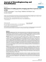
Figure 1 Titration of polyclonal MuLV goat antisera in Western blot (WB) assay. Antibody titers of positive control anti-sera and reactivity of pre-
immune sera to polytropic MuLV-infected (upper panel) and uninfected (lower panel) HeLa cell crude cell lysates in WB testing. Specific antisera tested
are located at the bottom of each WB. Arrows indicate observed titers for each antiserum. Fr, Friend; Ra, Rauscher. Locations of reactivity to specific
viral proteins are indicated. Env (gp69/71), envelope; TM (p15E), transmembrane; MA (p15), matrix; Gag (pr68/80); CA (p30), capsid. Molecular weight
markers (kD) are provided on the left of the WBs in the upper panels. Sizes of expected viral proteins are provided in each WB in the upper panels.
InfectedUninfected
α Rauscher MuLV (gp69/71)
250
500
1000
4000
2000
8000
16,000
32,000
64,000
Pre-immune
gp69/71
100
80
60
50
40
30
20
α Friend MuLV (whole virus)
250
500
1000
4000
2000
8000
16,000
32,000
64,000
Pre-immune
p30
pr68
120
200
Switzer et al. Retrovirology 2010, 7:57
/>Page 4 of 13
assay cutoff of 0.2 OD units (Figure 4) [14]. However,
both specimens were negative by IFA testing using 293T
cells expressing either XMRV Gag or Env proteins and
were thus considered negative. Two blinded positive con-
trol specimens each consisting of goat polyclonal MuLV
whole virus antisera diluted 1:100 in pre-immune goat
sera both tested positive in the recombinant Gag ELISAs
but were negative in the Env ELISA. These results are
consistent with the seroreactivity of these polyclonal anti-
sera to only Gag proteins in the WB assay. Five undiluted
pre-immune goat sera all tested negative in both the Gag
and Env ELISAs. These "external" positive and negative
controls were included as a separate set of specimens and
were all correctly detected in a blinded fashion. Testing of
the blinded human and goat control specimens was per-
formed separately since different secondary antibody
conjugates are used for these different specimens. Inter-
nal positive and negative controls were also included in
each run and performed as expected. Like the WB test-
ing, the goat anti-MuLV whole virus and anti-MuLV p70
polyclonal antisera gave titers of 1:64,000 and 1:6,400 in
the Gag and Env ELISAs, respectively.
Absence of XMRV sequences in PBMC DNA from persons
with CFS and healthy controls
We used two PCR assays at CDC to detect XMRV DNA.
The first assay was a nested gag PCR test used previously
to identify XMRV sequences in prostate cancer patients
and CFS patients [11,12]. The second nested PCR assay
was designed on highly conserved polymerase (pol)
sequences within xenotropic and other MuLV strains.
Serial, ten-fold dilutions of full-length XMRV(VP62)
plasmid (kindly provided by Robert Silverman) in a back-
ground of human DNA (PBMC or whole blood) showed
that the nested gag and pol PCR tests each detected 10
XMRV copies in different experiments on subsequent
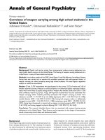
Figure 2 Titration of polyclonal XMRV rabbit and monoclonal spleen focus forming virus (SFFV) envelope rat antisera in Western blot (WB)
assay. Antibody titers of positive control anti-sera and reactivity of pre-immune sera to polytropic MuLV-infected (upper panel) and uninfected (lower
panel) HeLa cell crude cell lysates in WB testing. Specific antisera tested are located at the bottom of each WB. Arrows indicate observed titers for each
antiserum. Fr, Friend; Ra, Rauscher. Locations of reactivity to specific viral proteins are indicated. Env (gp69/71), envelope; TM (p15E), transmembrane;
MA (p15), matrix; Gag (pr68/80); CA (p30), capsid. Molecular weight markers (kD) are provided on the left of the WBs in the upper panels. Sizes of ex-
pected viral proteins are provided in each WB in the upper panels.
InfectedUninfected
α Fr MuLV (1:500)
Pre-immune
α Ra MuLV (1:500)
250
500
1000
4000
2000
8000
16,000
32,000
64,000
Rat α SFFV Env (7C10)
gp69/71(Env)
100/120
80
60
50
40
30
20
200
250
500
1000
4000
2000
8000
16,000
32,000
α Fr MuLV (1:500)
64,000
Pre-immune
α Ra MuLV (1:500)
100/120
80
60
50
40
30
20
α XMRV (whole virus)
p30(CA)
p15E(TM)
p15(MA)
gp69/71(Env)
pr68(Gag)
200
Switzer et al. Retrovirology 2010, 7:57
/>Page 5 of 13
days (34/34 (100%) and 32/34 (94.1%), respectively).
These results show that both PCR assays have an excel-
lent sensitivity for detecting XMRV in one ug of DNA
specimen. PBMC DNA from 41 anonymous US blood
donors was also tested and found to be negative in both
PCR assays. These 41 blood donors are distinct from the
US blood donors whose plasmas were tested in the WB
test.
PCR testing of β-actin sequences was positive for all
clinical specimens confirming the integrity of the DNA
and an absence of PCR inhibitors. Representative β-actin
PCR results are shown in Figure 5. Subsequent XMRV
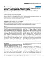
Figure 3 Absence of XMRV antibodies in CFS patients by Western blot (WB) analysis. Representative WB results for CFS cases from Wichita and
Georgia identified after unblinding. Determination of MuLV specific reactivity is determined by comparison of observed seroreactivity to polytropic
MuLV-infected HeLa antigens and uninfected HeLa antigens in upper and lower panels, respectively. Lanes 1 - 4 and 5 - 8 are plasma from CFS cases
from the population based studies in Georgia and Wichita, respectively; lanes 9 - 12 are physician-referred CFS cases from the Georgia Registry study.
MuLV positive and negative goat serum controls are labelled.
Pre-immune
α Ra MuLV (1:500)
α Fr MuLV (1:500)
123456 789101112
100/120
80
60
50
40
30
200
20
100/120
80
60
50
40
30
200
20
p30(CA)
gp69/71(Env)
pr68(Gag)
InfectedUninfected
Switzer et al. Retrovirology 2010, 7:57
/>Page 6 of 13
testing showed that XMRV gag and pol sequences were
not detected in 1 ug of PBMC (n = 31) or whole blood (n
= 19) DNA from the CFS patients or in 1 ug PBMC DNA
from the 56 healthy controls. A representative Southern
blot of the XMRV pol PCR testing of persons with CFS is
shown in Figure 5. Matching DNA was not available from
one CFS case.
Blinded PCR testing performed at an independent
institution (Blood Systems Research Institute (BSRI), CA)
using a second nested PCR assay for XMRV gag DNA
sequences, with a sensitivity of 3 copies per reaction, was
also negative using 100 ng DNA specimens from all 50
CFS cases and 56 healthy controls (data not shown). 250
ng of DNA from the Georgia Registry patients also tested
negative using this nested gag PCR test (Figure 6). Four
blinded, "external" control specimens, included with the
panel of human specimens and spiked with 4, 40, 400, and
4000 XMRV plasmid copies in 100 ng of human DNA,
were all detected by this testing (data not shown).
Discussion
We found no evidence of infection with XMRV among
persons with CFS or matched healthy controls from the
US by testing with multiple serologic and PCR assays per-
formed independently in three laboratories blinded to the
clinical status of the study participants. Our results con-
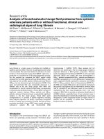
Figure 4 Absence of XMRV antibodies in CFS patients and healthy persons by ELISA using recombinant XMRV proteins. Representative XMRV
Envelope (Env) ELISA results for 50 CFS cases and 49 healthy persons identified after unblinding. Specimens coded with W and G1-G50 are from the
population-based study in Wichita and Georgia, respectively; specimens G59 - G75 are from physician-referred CFS cases from the Georgia Registry
study. Specimens from a healthy control and a person with CFS, coded as G6 and G9 respectively, were weakly seroreactive in this test but were not
confirmed by either Western blot or immunofluorscence testing. Human sera were diluted 1:200. The human negative control serum was obtained
from a healthy volunteer previously determined to be seronegative. The polyclonal mouse Env antiserum was diluted 1:100. Assay cut-off was deter-
mined by the mean of the test samples plus three standard deviations.
0,6
0,7
0,8
OD CFS
cut-off
OD healthy
OD positive control
0,3
0,4
0,5
OD 492/620
0
0,1
0,2
0,7
0,8
W1
W3
W4
W6
W7
W9
W10
W11
W12
W13
W14
W15
W16
W17
W18
W19
W20
W21
W22
W23
W25
W26
W27
W28
W29
W30
W31
W32
W33
W34
W35
W36
W37
G1
G2
G3
G4
G5
G6
G7
G8
G9
G10
G11
G12
G13
G14
G15
G16
mouse sera
0,4
0,5
0,6
O
D 492/620
0
,
1
0,2
0,3
O
0
,
G17
G19
G20
G21
G22
G23
G24
G25
G26
G27
G28
G29
G30
G31
G32
G33
G34
G35
G36
G37
G38
G39
G40
G41
G42
G44
G45
G46
G47
G48
G49
G50
G58
G59
G60
G61
G62
G63
G64
G65
G66
G67
G68
G69
G70
G71
G72
G73
G74
G75
mouse sera
Switzer et al. Retrovirology 2010, 7:57
/>Page 7 of 13
trast with the high rate of XMRV detection reported by
Lombardi et al. among both CFS patients and controls,
but are in agreement with recent data reported in two
large studies in the UK and a smaller study in the Nether-
lands that could not detect XMRV sequences in CFS
patients and one UK study that also failed to detect spe-
cific XMRV neutralizing antibody responses in CFS
[11,16-18]. Combined, these negative data do not support
XMRV as the etiologic agent of the majority of CFS cases.
Several possibilities could explain these discordant
results, including technical differences in assays used for
the testing in each study. However, the inability of four
independent laboratories to replicate the high XMRV
prevalence in CFS cases reported by Lombardi et al. can-
not be explained by minor differences in assays used in
each study. In addition, testing at CDC utilized the nested
XMRV gag PCR assay used by Lombardi et al. and Uris-
man et al. to identify XMRV infection in CFS and pros-
tate cancer patients, respectively [11,12]. Further, to
improve assay sensitivity, we used 1 ug of input DNA
which is 4-5 times higher than that used by others [11-
13,16,17], all while maintaining an assay sensitivity of 10
copies. To ensure that our testing would not miss geneti-
cally diverse XMRV or MuLV strains, we also used a sen-
sitive nested PCR assay with conserved pol gene primers
and found that this testing was also negative confirming
the absence of XMRV/MuLV sequences. While PBMC
DNA was used in the majority of specimens, 1 ug whole
blood DNA was also used in testing 19 CFS cases. This
input DNA represents about 350 ng of PBMC DNA
which is similar to the amount used by others [11-
13,15,16], thus not affecting the sensitivity of our results.
The negative PCR findings were confirmed by an inde-
pendent laboratory with a second nested gag PCR assay
which provided additional evidence for the absence of
XMRV sequences among CFS cases and controls. The
primary PCR amplification used in this second test is also
that used by Lombardi et al. which when combined with
a nested PCR step has a 3-copy detection threshold.
Antibody responses particularly to Gag and Env pro-
teins are hallmarks of immune responses to retroviral
infections including experimental XMRV infection of
macaques [22]. We used a new WB assay to test for anti-
XMRV antibodies and showed by using both monoclonal
antibodies and polyclonal antisera that this assay
detected specifically, and with high titers, reactivity to
both XMRV and MuLV Gag and Env proteins. We were
unable to detect antibodies to XMRV Gag and Env in any
of the CFS and controls specimens by using this WB
assay. Likewise, negative results were obtained in a sec-
ond, independent laboratory by using XMRV-specific
ELISA-based and IFA assays. Thus, the observed negative
serologic results for all CFS patients reflect an absence of
antibody responses and active XMRV infection. Although
limited, the negative WB serology observed in 56 healthy
controls and 121 blood donors also suggests that the
XMRV seroprevalence in this population is not high.
Screening of larger numbers of US blood donors using a
high throughput ELISA followed by confirmation in a
WB test also showed uncommon seropositivity (~0.1%)
[22]. More studies, however, are needed to determine the
prevalence of XMRV in healthy populations.
One current limitation of our study, and of others per-
forming serologic and PCR testing for XMRV, is the
absence of bona fide positive and negative control speci-
mens from infected and uninfected humans to determine
Figure 5 Absence of XMRV polymerase (pol) sequences in CFS pa-
tients. A. Representative nested pol PCR results using PBMC DNA spec-
imens from persons with CFS identified after unblinding. Lanes 1 - 5, 6
- 10, and 11 - 14 are results for persons with CFS from Wichita, Georgia,
and the Georgia registry studies, respectively; lanes 15 and 16, water
only controls; lane 17, negative human PBMC DNA control; lanes 18
and 19, assay sensitivity controls consisting of 10
1
and 10
3
copies of
XMRV VP62 plasmid DNA diluted in a background of 1 ug of human
PBMC DNA, respectively. B. Semi-quantitative β-actin PCR results for
PBMC DNA specimens above in lanes 1 - 14; lane 15, water control;
lanes 16 - 19, 10-fold dilutions of blood donor PBMC DNA starting at 0.1
ug as a positive assay control.
1 2 3 4 5 6 7 8 9 10 11 12 13 14 15 16 17 18 19
A.
1° PCR
2
°
PCR
2
°
PCR
ß
-
actin
B.
ß
actin
Figure 6 Absence of XMRV gag sequences in CFS patients. A. Rep-
resentative nested gag PCR results from patients from the Georgia
Registry identified after unblinding. Lanes 1 and 20, 100-bp ladder;
lanes 2 - 15 are results from CFS patients; lanes 16 - 18 assay sensitivity
controls consisting of 10, 3 and 1 copies of XMRV VP62 plasmid DNA
diluted in a background of 250 ng of human PBMC DNA; lane 19, water
control. B. GAPDH PCR results for same PBMC DNA specimens above.
A.
1 2 3 4 5 6 7 8 9 1011 12 1314 15 16 1718 19
20
2° PCR
B.
GAPDH
Switzer et al. Retrovirology 2010, 7:57
/>Page 8 of 13
the analytical sensitivity and specificity of the detection
assays. Until panels of well-characterized clinical speci-
mens become available, assay validation will be limited to
reagents generated experimentally, such as polyclonal
and monoclonal antibodies, XMRV plasmids, and
XMRV-infected cells.
The selection criteria with which persons with CFS
were included in these various studies may also help to
explain the incongruent XMRV findings. The study by
Lombardi et al. used samples from the Whittemore Peter-
son Institute National Tissue Repository reported to con-
tain specimens from well-characterized cohorts of CFS
[11]. Yet, the paper provides no information regarding the
repository or concerning the nature of these cohorts
other than that they were collected from private medical
practices in several regions of the U.S. where clusters of
CFS have been documented [11]. An absence of details of
the CFS cases and controls in this report makes it difficult
to replicate and interpret their findings. In contrast,
patients in the UK and Netherland studies were typical of
CFS patients seen in specialist clinical services in those
countries and resemble persons seen in other specialist
CFS services in the US and Australia [16-18]. Almost half
of the UK CFS patients described onset of their illness as
related to an acute viral disease [16,17]. Thus, they would
appear quite comparable to those in the study by Lom-
bardi et al. Similarly, our study also failed to detect
XMRV infection in 18 CFS patients referred to a fatigue
registry by health care providers in Georgia and included
three persons who reported sudden onset to their illness.
Our study is the first to evaluate XMRV infection in per-
sons with CFS and healthy controls from the general pop-
ulations of Wichita and Georgia. These CFS cases are
different from CFS patients seen in general practice and
referral clinics; of the participants from the population-
based study in Georgia, only half had consulted a physi-
cian because of their fatigue, about 16% had been diag-
nosed with CFS, and 75% described an insidious onset to
their illness that had no obvious relation to an acute
infectious disease. Nonetheless, results from our general
population cohort extend the examination of XMRV in
CFS to persons whose illness developed gradually, for the
most part, rather than acutely. Our negative findings, in
conjunction with those in Europe [16-18], indicate no dis-
cernable association of XMRV with a wide spectrum of
CFS cases. The negative results for CFS patients and con-
trols from the US in the current study also do not support
a continental clustering of XMRV infection suggested by
the absence of infection in the UK and Netherlands [16-
18]. However, our findings may not be generalizable
beyond our study populations because XMRV infection
rates may vary in different regions or locales.
CFS is a diagnosis of exclusion based on self-reported
symptoms and requires careful medical and psychiatric
evaluations to rule out conditions with similar clinical
presentation. Our study and the negative reports from
the UK and the Netherlands evaluated patients for exclu-
sionary conditions and defined CFS according to criteria
of the 1994 International CFS Research Case Definition
[23] or the earlier Oxford case definition [24]. The Lom-
bardi et al. study specifies that samples were selected
from patients fulfilling the 1994 international CFS case
definition [23] and the 2003 Canadian Consensus Criteria
for CFS/ME [25]. Lombardi et al. did not specify if
patients were evaluated for exclusionary conditions, or if
the study subjects met both definitions, or which patients
met either CFS definition. The 1994 International CFS
case definition and the Canadian Consensus Criteria are
different and do not necessarily identify similar groups of
ill persons. Most notably, the Canadian Criteria include
multiple abnormal physical findings such as spatial insta-
bility, ataxia, muscle weakness and fasciculation, restless
leg syndrome, and tender lymphadenopathy. The physical
findings in persons meeting the Canadian definition may
signal the presence of a neurologic condition considered
exclusionary for CFS and thus the XMRV positive per-
sons in the Lombardi et al. study may represent a clinical
subset of patients [11].
CFS is a complex disease with various clinical subtypes
proposed which could also account for differences in the
results obtained in each study [11,16-18]. While there is
still no universal agreement on a precise clinical presen-
tation encompassing CFS illness, defining patient charac-
teristics in studies of CFS etiology or pathogenesis
remains crucial for making comparisons across various
research conclusions.
Conclusions
In our study population of CFS and healthy persons from
the US, we did not find any evidence of infection with
XMRV using PCR and serologic methods performed
independently in three laboratories blinded to the clinical
status of the study participants. These results do not sup-
port an association of XMRV with CFS.
Methods
Study population and specimen preparation
The CDC Institutional Review Board reviewed and
approved all study protocols. All participants were volun-
teers and provided informed consent. Laboratory testing
of the samples was performed anonymously and blinded
to clinical status.
Details of our two study populations have been
described previously [2,26,27]. Briefly, between 2002 and
2003 we sampled adults 18 to 59 years old from Wichita,
Kansas [26,27] and between 2008 and 2009 we sampled
adults 18 to 59 years old from metropolitan, urban, and
rural Georgia [2]. In both studies, we used random digit-
dial screening interviews to classify household residents
as either well or having symptoms of CFS. A follow-up
Switzer et al. Retrovirology 2010, 7:57
/>Page 9 of 13
detailed telephone interview was administered to all indi-
viduals with symptoms and to a probability sample of
those without symptoms. Based on the detailed inter-
view, those meeting criteria of the 1994 International CFS
Research Case Definition [23] were classified as CFS-like
and other respondents classified as either unwell (not
CFS-like) or well. All CFS-like individuals were recruited
and a random sample of those who were unwell but not
CFS-like, and a set of matched (sex, age, race/ethnicity,
geographic) well people were recruited for a 1-day clinical
evaluation.
We also tested specimens from CFS cases identified in
a CDC Health Care Provider-based Registry of Unex-
plained Fatiguing Illnesses and CFS (unpublished).
Between October 2008 and December 2009, healthcare
providers practicing in Bibb County, GA referred adoles-
cents and adults 12 - 59 years old who met criteria for
unexplained fatiguing illness (fatigue for > 1 month), and
having at least one other core CFS symptom during that
period (unrefreshing sleep, problems with cognition or
memory, joint or muscle pain in extremities), and did not
have an exclusionary medical or psychiatric condition.
All referred patients underwent a telephone screening
interview to document fatigue lasting > 6 months, and
the presence of at least one core symptom and no exclu-
sionary conditions. Patients meeting these criteria under-
went the same 1-day clinical evaluation as persons from
our population-based studies, described in detail below.
Clinical assessment
Clinical evaluations involved: 1. Administration of stan-
dardized questionnaires to measure the 3 domains of the
1994 CFS case definition [23]: the Multidimensional
Fatigue Inventory (MFI) to measure 5 dimensions of
fatigue [28] the Medical Outcomes Survey Short Form 36
(SF-36) to evaluate 8 dimensions of functional impair-
ment [29]; and the CDC Symptom Inventory to evaluate
occurrence/frequency/severity of the 8 CFS-accompany-
ing symptoms [30]; 2. A standardized physical examina-
tion conducted by a specifically trained physician who
also reviewed past medical history, review of systems,
and current medications/supplements; 3. Collection of
blood and urine for routine clinical analyses [23,31]; 4. A
standardized psychiatric evaluation conducted by specifi-
cally trained psychiatric interviewers - Diagnostic Inter-
view Schedule (DIS) in Wichita [32] and the Structured
Clinical Interview for DSM-IV Disorders (SCID) in Geor-
gia [33].
The physician's evaluation and routine clinical labora-
tory tests served to identify medical conditions consid-
ered exclusionary for CFS, specified in the 1994 case
definition [23] as further clarified by the International
CFS Study Group in 2003 [31]. The psychiatric interview
served to identify current psychiatric disorders consid-
ered exclusionary for CFS, which included current mel-
ancholic depression, current or lifetime bipolar disorder
or psychosis, substance abuse within 2 years and eating
disorders within 5 years [23,31].
Illness classification
Following clinical evaluation, participants who had no
exclusionary medical or psychiatric conditions were diag-
nosed with CFS if they met criteria of the 1994 interna-
tional case definition [23] as quantified by the CDC
Symptom Inventory and ancillary criteria of the MFI and
SF-36 [26,31]. We used the MFI to assess fatigue status
[28]. For classification as CFS, those with a score ≥ well-
population medians on the general fatigue or reduced
activity scales of the MFI were considered to meet fatigue
criteria of the 1994 international case definition. Func-
tional impairment was assessed by the medical outcomes
survey short form-36 (SF-36) [29]. For classification as
CFS, those with a score ≤ 25th percentile of population
norms in the physical function or role physical, or social
function, or role emotional subscales of the SF-36 were
considered to have substantial reduction in activities as
specified in the 1994 definition. Those who met at least
one but not all 1994 criteria were considered unwell not
CFS. Those who met none of the criteria were considered
well.
Specimens were available from 89 persons (33 CFS and
56 well controls) from the population-based case-control
studies and 18 CFS persons from the Registry study
described above. Subjects were included based on avail-
ability of specimens, and comprised 11 of 43 persons with
CFS and 26 of 53 healthy controls from Wichita, KS and
22 of 32 persons with CFS and 30 of 51 healthy controls
from Georgia. Persons with CFS and healthy controls had
similar mean ages, similar predominance of females and
white race, and had a similar mean body mass index
(BMI) (Table 1). Subjects with CFS had been ill on aver-
age 13.9 years (median 11.15 yrs, range 3 - 40 yrs), were
severely fatigued (MFI General Fatigue 16.5, range 10 -
20; MFI Reduced Activity 12.8, range 4 - 20) and severely
impaired (SF-36 physical functioning 65.5, range 10-100);
SF-36 bodily pain 48.8, range 12 - 84), and 3/33 (9%)
reported sudden onset to their illness. Clinical and demo-
graphic characteristics of subjects with specimens avail-
able for this study did not differ from those persons who
did not have ample specimen volumes and case-control
matching was maintained.
18 of 38 persons enrolled in the Registry study had a
diagnosis of CFS and were available for the current study.
These provider-referred CFS patients had a mean age of
42.8 years (SEM = 2.85 years), and were predominantly
white [17/18, (94.4%)] and female [16/18 (88.99%)]. They
had suffered fatigue for an average of 9.4 years (range: 1 -
35 years) and 3/18 (16.7%) reported sudden onset to their
illness.
Switzer et al. Retrovirology 2010, 7:57
/>Page 10 of 13
Specimen collection, processing, storage
Fresh whole blood was collected in either CPT Vacu-
tainer tubes containing sodium citrate and a blood sepa-
ration reagent (Becton Dickinson, NJ, USA) for the
Georgia and Wichita studies or in PAXgene tubes for the
Georgia CFS Registry study and transported to CDC.
Blood was also collected in PAXgene tubes for two per-
sons from the Georgia population-based study. PAXgene
tubes were gently inverted 5 times, stored overnight at -
20°C, and then transferred to -70°C until DNA isolation
was performed. PBMCs and plasma were immediately
isolated by centrifugation of the CPT tubes. PBMCs were
stored in liquid nitrogen under conditions designed to
maintain viability. Plasma was aliquoted and stored at -
80°C within 4 hours of blood collection. For samples col-
lected from persons living in Wichita, KS and from the
Georgia CFS Registry study, whole blood was also col-
lected in EDTA Vacutainer tubes. Plasma was recovered
from the EDTA-treated blood by centrifugation at 15,000
× g for 20 minutes and aliquoted and frozen at -80°C until
use. Plasma samples were aliquoted again when thawed
for WB testing; the remaining aliquots were re-frozen at -
80°C.
DNA was extracted from cryopreserved PBMCs or fro-
zen whole blood with the Qiagen blood DNA minikit or
Qiagen PAXgene Blood DNA kit (Qiagen, Valencia, CA),
respectively, then aliquoted and stored frozen at -80°C.
All PBMC samples had viabilities > 90% when they were
thawed for DNA isolation. Nucleic acid concentrations
were determined by spectrophotometry using the Nano-
drop instrument (Thermo Scientific, Wilmington, DE).
For the PCR testing at CDC, 1 ug of PBMC or whole
blood DNA was used. Integrity of the DNA specimens
was determined using β-actin PCR as previously
described [34]. Matching plasma or DNA was not avail-
able from three healthy persons from Wichita, KS and
one CFS case from Georgia, respectively. All specimen
preparation, tissue culture, and PCR testing was done in
physically isolated rooms to prevent contamination of
specimens.
Serologic Assays
HeLa cells were infected with supernatant from the
murine macrophage cell line RAW264.7 (ATCC, Manas-
sas, VI) known to express polytropic and ecotropic MuLV
(PMLV and EMLV, respectively). To characterize the iso-
late that replicated in HeLa cells, a 166-bp RNA sequence
containing the variable region C of the envelope (Env)
surface protein was PCR-amplified from infected HeLa
cell tissue culture supernatants. Phylogenetic analysis of
Table 1: Distribution of demographic variables by CFS case-control status among persons from the combined Wichita and
Georgia case-control population-based studies.
CFS Well
Demographic Factor Wichita, KS
(N = 11)
Atlanta, GA
(N = 22)
Wichita, KS
(N = 26)
Atlanta, GA
(N = 30)
p-value
2,3,4
Age
Mean ± SEM
1
46.7 ± 3.32 47.7 ± 4.69 51.6 ± 5.1 46.1 ± 5.48 p = 0.51
Sex [n (%)]
Female 8 (72.7) 20 (90.9) 21 (80.8) 25 (83.3) p = 0.74
Male 3 (27.3) 2 (9.1) 5 (19.2) 5 (16.7)
Race [n (%)]
White 10 (90.9) 18 (81.8) 25 (96.2) 27 (90)
Black 0 (0) 3 (13.6) 1 (3.8) 3 (10) p = 0.69
Other 1 (9.1) 1 (4.6) 0 0 (0)
Body Mass Index
Mean ± SEM 27.6 ± 3.3 28.2 ± 4.7 29.2 ± 5.1 26.3 ± 5.5 p = 0.76
1. SEM, standard error of the mean
2. t-test was used to compute probabilities for comparisons of mean age and mean body mass index between study groups.
3. Chi square test was used to compute the probability for comparison of the distribution of sex between cases and controls.
4. Fisher's exact test was used to compute the probability for comparison of the distribution of race between the study groups, and was based
on Blacks and Whites only.
Switzer et al. Retrovirology 2010, 7:57
/>Page 11 of 13
the env sequence showed that the isolate was a PMLV by
clustering tightly with other PMLV, and not EMLV (data
not shown). XMRV and PMLV are highly related sharing
between 87 - 94% nucleotide identity across their
genomes and 88 - 97% and 88 - 91% amino acid identity
to complete Gag and Env proteins, respectively. Indeed,
partial Gag (123 aa) and Env (55 aa) sequences from our
polytropic HeLa isolate share 96% and 90% identity to
XMRV, respectively. Thus, the high amino acid related-
ness supports the use of this isolate for WB serologic test-
ing. Infected and uninfected HeLa crude cell lysates were
prepared for WB testing as previously described [35].
Protein concentrations of the lysates were determined
using the BioRad DC Protein Assay (Hercules, CA).
Plasma or serum samples were diluted 1:50 and reacted
separately to 150 ug of infected and uninfected cell lysates
overnight at 4°C after protein separation through 4-12%
polyacrylamide gels and transfer to nytran membranes,
as previously described [35,36]. Seroreactivity in human
specimens was detected using peroxidase-conjugated
protein A/G (Pierce, Rockford, IL) and chemilumines-
cence (Amersham, Uppsala, Sweden) [35,36].
Since validated XMRV-positive human sera are not
currently available, we used experimentally derived poly-
clonal antisera and monoclonal antibodies to assess anti-
genic reactivity of the WB assay. These reagents included
goat polyclonal antisera to MuLV (whole virus and gp69/
71Env, respectively) available at ATCC (VR-1537 and VR-
1521, respectively), and a rabbit anti-XMRV polyclonal
antiserum (kindly provided by Ila Singh) and a rat anti-
SFFV (7C10) monoclonal antibody (kindly provided by
Sandra Ruscetti) used previously to detect XMRV protein
expression and antibodies in prostate cancer and CFS
patients, respectively [11,13,21]. Peroxidase-conjugated
protein A/G or anti-rat antibody (Sigma, St. Louis, MS)
was used to detect bound goat, rabbit, and rat antibodies,
respectively. Sensitivity of the assay was estimated using
two fold serial dilutions of the MuLV, XMRV, and SFFV
polyclonal and monoclonal antibodies. Cross-reactivity
of the WB assay on HIV and HTLV positive plasma was
evaluated on 13 HTLV-1/2 positive, 7 HIV-1-positive,
and six HIV-1/HIV-2 dual positive plasma. In addition,
sera from 121 HIV and HTLV seronegative anonymous
US blood donors collected in 1998 were tested.
An aliquot of coded plasma from the CFS and healthy
controls was tested at RKI by an ELISA using recombi-
nant Gag and Env proteins used recently to investigate
XMRV infection in German prostate cancer patients [14].
Briefly, recombinant proteins were coated overnight on
microtiter plates at room temperature in equimolar
amounts. The plates were blocked with 2% Marvel milk
powder in phosphate buffered saline (PBS) for 2 h at
37°C, washed three times with PBS, 0.05% Tween 20.
Patient plasma diluted 1:200 in PBS with 2% milk powder
and 0.05% Tween20 were added into each well and incu-
bated for 1 hour at 37°C. Each well was again washed
three times and a 1:1000 dilution of a goat anti-human
IgG-HRP conjugate (Sigma Aldrich, Munich, Germany)
in PBS, 2% milk powder, 0.05% Tween 20 (Serva, Heidel-
berg, Germany) was added. Following incubation for 1
hour at 37°C, each well was again washed three times,
and chromogen ortho-phenylendiamin (OPD) in 0.05 M
phosphate-citrate buffer, pH 5.0 containing 4 μl of a 30%
solution of the hydrogen peroxide substrate per 10 ml
was added. After 5-10 minutes the color development
was stopped by addition of sulphuric acid and the absor-
bance at 492 nm/620 nm was measured in a microplate
reader. Positive controls included mouse anti-Gag and
Env antisera and pre-immune sera diluted 1:50 in PBS
with 2% milk powder and 0.05% Tween20. In addition, a
separate set of goat sera was also tested in a blinded fash-
ion and included external positive and negative controls
consisting of dilutions of the MuLV whole virus, gp69/71
goat polyclonal antisera, or pre-immune goat sera,
respectively. Detection of antibody reactivity in the goat
sera was done by using rabbit anti-goat HRP conjugate
(Dako, Hamburg, Germany).
Samples reactive by ELISA testing were then re-tested
using an immunofluorescence assay (IFA) [14]. Briefly,
plasma specimens were diluted 1:200 in blocking buffer
and tested against 293T cells expressing codon optimized
synthetic full-length genes of the XMRV env or gag under
control of the CMV promoter and bound to glass slides,
as described in detail previously [14]. Following incuba-
tion for 60 min at 37°C, the slides were washed exten-
sively with PBS and secondary antibodies conjugated to
fluorophores were added for 30 min. After thorough
washing steps with PBS, the cells were mounted in Mow-
iol and viewed on a Zeiss (LSM510) confocal laser-scan-
ning microscope.
Detection of XMRV sequences
DNA specimens were screened by PCR at the CDC with
an XMRV-specific gag and a polymerase (pol) assay that
detects xenotropic and polytropic MuLV. The XMRV
specific assay uses the primers GAG-O-F and GAG-O-R
and GAG-I-F and GAG-I-R for the primary and nested
PCRs, respectively, and conditions as previously
described [11,12]. This is the same nested PCR test used
by Urisman et al. and Lombardi et al. to detect 413-bp
XMRV gag sequences in prostate cancer and CFS
patients, respectively [11,12]. The primers and probes of
the generic pol PCR assay were designed from an align-
ment of complete XMRV and prototypical xenotropic,
polytropic, and ecotropic MuLV genomes available at
GenBank (accession numbers: xenotropic (XMLV):
XMRV VP35 = DQ241301
, XMRV VP62 = DQ399707,
XMRV VP42 = DQ241302
, XMRV WPI-1106 =
Switzer et al. Retrovirology 2010, 7:57
/>Page 12 of 13
GQ497344, XMRV WPI-1178 = GC497343, MuLV DG-
75 = AF221065
; MuLV MTCR = NC_001702, mERV Chr
9 = AC121813
, mERV Chr 4 = AL627077, mERV Chr 1 =
AC083892
; polytropic (PMLV): mERV Chr 7 =
AC167978
, mERV Chr 7 = AC127565, mERV Chr 12 =
AC153658
; ecotropic (EMLV): MuLV AKV = J01998,
MuLV BM5eco = AY252102.1
, Moloney MuLV = J02255,
Rauscher MuLV = NC_001819
, Friend MuLV = X02794).
The external external XPOLOF (5' CCG TGC CCA ACC
CTT ACA ACC TCT 3') and XPOLOR (5' CCG AGG
TTC CCT AGG GTT TGT AAT 3') and internal primers
XPOLIF (5' TCC ACC CCA CCA GTC AGC CTC TCT
3') and XPOLIR (5' AAG TGG CGG CCA GCA GTA
AGT CAT 3') were used to generically detect 216-bp
XMLV/XMRV pol sequences. All assays were optimized
to achieve the highest sensitivity in detecting XMRV
VP62 plasmid DNA in one ug of genomic DNA. One ug
of human DNA was used as input for the PCR tests. PCR
products were visualized by electrophoresis in an ethid-
ium bromide-stained 1.8% agarose gel. To further
increase the sensitivity and specificity of the PCR assays,
amplified gag and pol sequences were confirmed by
Southern blot analysis using the biotinylated oligoprobes
XGAGP2 (5' ACC TTG CAG CAC TGG GGA GAT GTC
3'), and XPOLP (5' TTG ATG AGG CAC TGC ACA
GAG ACC 3') and chemiluminescence detection. The
detection limit of the assays was evaluated using 10-fold
dilutions of XMRV VP62 plasmid diluted in a back-
ground of one ug of genomic human DNA. Assay speci-
ficity was evaluated using PBMC DNA from 41
anonymous US blood donors screened negative for HIV
and HTLV.
Nested PCR was also performed at BSRI using double
blinded genomic DNA specimens in order to indepen-
dently test for XMRV gag sequences. The first round was
performed as previously described to detect XMRV in
PBMC DNA of CFS patients [11]. Briefly, 100 - 250 ng of
genomic DNA was amplified using outer gag primers
419F (5' ATC AGT TAA CCT ACC CGA GTC GGA C 3')
and 1154R (5' GCC GCC TCT TCT TCA TTG TTC TC
3') at a final concentration of 0.3 μM, HotStart-IT Fideli-
Taq Master Mix (USB Corporation, Cleveland, OH) and 1
mM magnesium chloride. PCR was performed using an
initial denaturation step at 94°C for 4 minutes followed by
45 cycles of 94°C for 30 seconds, 57°C for 30 seconds and
72°C for 1 minute and a final extension step at 72°C for 2
minutes. Nested PCR was conducted using 1 μl of the
first round DNA in the second round reaction. Nested
primers 488F (5' GGG GAC GAG AGA CAG AGA CA
3') and 1107R (5' CAG AGG AGG AAG GTT GTG CT 3')
were used at a final concentration of 0.3 μM and amplifi-
cation was performed using HotStart-IT FideliTaq. PCR
was performed using an initial denaturation step at 95°C
for 90 seconds followed by 40 cycles of 95°C for 20 sec-
onds, 58°C for 30 seconds and 72°C for 40 seconds and a
final extension step at 72°C for 2 minutes. PCR contami-
nation occurring during nested PCR was evaluated by
including at least one third as many water controls as test
samples in each PCR experiment and were always nega-
tive.
Using serial dilutions of a cloned fragment of XMRV
gag as a positive control, the nested PCR assay could reli-
ably detect at least 3 copies of DNA per reaction, even
when spiked into genomic DNA prepared either from
293FT cells or donor PBMCs previously validated to be
negative for XMRV. Controls of GAPDH (forward - 5'
CAT GTT CCA ATA TGA TTC AC 3'; reverse - 5' CCT
GGA AGA TGG TGA TG 3'; 75 ng genomic DNA, 3 min-
utes at 95°C followed by 45 cycles of 95°C for 20 seconds,
55°C for 45 seconds and 72°C for 30 seconds, followed by
1 cycle of 72°C for 2 minutes) were performed to ensure
similar levels of genomic DNA input in each PCR reac-
tion.
Competing interests
The authors declare that they have no competing interests.
Authors' contributions
WMS, WCR, RMH and WH conceived and designed the study. WCR and VRF
provided specimens and data on study population. HJ, HZ, ST, AS, GS, NB, and
OH performed specimen testing and data analysis with WMS and WH. WMS,
WCR and WH wrote the manuscript. All authors read and approved the final
manuscript.
Acknowledgements
We are grateful to Dr. Robert Silverman at the Cleveland Clinic for the VP62
XMRV plasmid, Dr. Ila Singh at the University of Utah for the rabbit anti-XMRV
polyclonal sera, and Dr. Sandra Ruscetti at the National Cancer Institute for the
rat anti-SFFV (7C10) monoclonal antibody. We also thank Ben Capon and Beth
Slikas for development of the gag PCR assay used at the Blood Systems
Research Institute. Use of trade names is for identification only and does not
imply endorsement by the U.S. Department of Health and Human Services, the
Public Health Service, or the Centers for Disease Control and Prevention. The
findings and conclusions in this report are those of the authors and do not
necessarily represent the views of the Centers for Disease Control and Preven-
tion.
Author Details
1
Laboratory Branch, Division of HIV/AIDS Prevention, National Center for HIV/
AIDS, Viral Hepatitis, STD, and TB Prevention, Centers for Disease Control and
Prevention, Atlanta, GA 30333, USA,
2
Robert Koch-Institute, Centre for
Biological Safety 4, Nordufer 20, 13353 Berlin, Germany,
3
Blood Systems
Research Institute and Department of Laboratory Medicine, UCSF, 270 Masonic
Ave., San Francisco, CA 94118, USA and
4
Chronic Viral Diseases Branch, Division
of Viral and Rickettsial Diseases, National Center for Zoonotic, Vector-Borne and
Enteric Diseases, Centers for Disease Control and Prevention, Atlanta, GA
30333, USA
Received: 26 March 2010 Accepted: 1 July 2010
Published: 1 July 2010
This article is available from: 2010 Switzer et al; licensee BioMed Central Ltd. This is an Open Access article distributed under the terms of the Creative Commons Attribution License ( which permits unrestricted use, distribution, and reproduction in any medium, provided the original work is properly cited.Retrovirology 2010, 7:57
Switzer et al. Retrovirology 2010, 7:57
/>Page 13 of 13
References
1. Jason LA, Richman JA, Rademaker AW, Jordan KM, Plioplys AV, Taylor RR,
McCready W, Huang CF, Plioplys S: A community-based study of chronic
fatigue syndrome. Archives of Internal Medicine 1999, 159:2129-2137.
2. Reeves WC, Jones JF, Maloney E, Heim C, Hoaglin DC, Boneva RS,
Morrissey M, Devlin R: Prevalence of chronic fatigue syndrome in
metropolitan, urban, and rural Georgia. Popul Health Metr 2007, 8:5.
3. Devanur LD, Kerr JR: Chronic fatigue syndrome. Journal of Clinical
Virology 2006, 37:139-150.
4. Jason LA, Benton MC, Valentine L, Johnson A, Torres-Harding S: The
economic impact of ME/CFS: individuals and societal level costs.
Dynamic Medicine 2008, 7:6.
5. Reynolds KJ, Vernon SD, Bouchery E, Reeves WC: The economic impact of
chronic fatigue syndrome. Cost Eff Resour Alloc 2004, 2:4.
6. Solomon L, Nisenbaum R, Reyes M, Papanicolaou DA, Unger ER, Reeves
WC: Functional status of persons with chronic fatigue syndrome in the
Wichita population. Health Qual Life Outcomes 2003, 1:48.
7. Afari N, Buchwald D: Chronic fatigue syndrome: a review. Am J
Psychiatry 2003, 160:221-236.
8. DeFreitas E, Hilliard B, Cheney PR, Bell DS, Kiggundu E, Sankey D,
Wroblewska Z, Palladino M, Woodward JP, Koprowski H: Retroviral
sequences related to human T-lymphotropic virus type II in patients
with chronic fatigue immune dysfunction syndrome. Proc Natl Acad Sci
USA 1991, 88:2922-2926.
9. Heneine W, Woods TC, Sinha SD, Khan AS, Chapman LE, Schonberger LB,
Folks TM: Lack of evidence for infection with known human and animal
retroviruses in patients with chronic fatigue syndrome. Clin Infect Dis
1994, 18(Suppl 1):S121-125.
10. Khan AS, Heneine WM, Chapman LE, Gary HE, Woods TC Jr, Folks TM,
Schonberger LB: Assessment of a retrovirus sequence and other
possible risk factors for the chronic fatigue syndrome in adults. Ann
Intern Med 1993, 118:241-245.
11. Lombardi VC, Ruscetti FW, Das Gupta J, Pfost MA, Hagen KS, Peterson DL,
Ruscetti SK, Bagni RK, Petrow-Sadowski C, Gold B, Dean M, Silverman RH,
Mikovits JA: Detection of an infectious retrovirus, XMRV, in blood cells
of patients with chronic fatigue syndrome. Science 2009, 326:585-589.
12. Urisman A, Molinaro RJ, Fischer N, Plummer SJ, Casey G, Klein EA, Malathi
K, Magi-Galluzzi C, Tubbs RR, Ganem D, Silverman RH, DeRisi JL:
Identification of a novel Gammaretrovirus in prostate tumors of
patients homozygous for R462Q RNASEL variant. PLoS Pathog 2006,
2:e25.
13. Schlaberg R, Choe DJ, Brown KR, Thaker HM, Singh IR: XMRV is present in
malignant prostatic epithelium and is associated with prostate cancer,
especially high-grade tumors. Proceedings of the National Academy of
Sciences of the United States of America 2009, 106:16351-16356.
14. Hohn O, Krause H, Barbarotto P, Niederstadt L, Beimforde N, Denner J,
Miller K, Kurth R, Bannert N: Lack of evidence for xenotropic murine
leukemia virus-related virus(XMRV) in German prostate cancer
patients. Retrovirology 2009, 6:92.
15. Fischer N, Hellwinkel O, Schulz C, Chun FK, Huland H, Aepfelbacher M,
Schlomm T: Prevalence of human gammaretrovirus XMRV in sporadic
prostate cancer. J Clin Virol 2008, 43:277-283.
16. Erlwein O, Kaye S, McClure MO, Weber J, Wills G, Collier D, Wessely S,
Cleare A: Failure to detect the novel retrovirus XMRV in chronic fatigue
syndrome. PLoS One 2010, 5:e8519.
17. Groom HC, Boucherit VC, Makinson K, Randal E, Baptista S, Hagan S, Gow
JW, Mattes FM, Breuer J, Kerr JR, Stoye JP, Bishop KN: Absence of
xenotropic murine leukaemia virus-related virus in UK patients with
chronic fatigue syndrome. Retrovirology 2010, 7:10.
18. van Kuppeveld FJ, Jong AS, Lanke KH, Verhaegh GW, Melchers WJ,
Swanink CM, Bleijenberg G, Netea MG, Galama JM, van der Meer JW:
Prevalence of xenotropic murine leukaemia virus-related virus in
patients with chronic fatigue syndrome in the Netherlands:
retrospective analysis of samples from an established cohort. BMJ
2010, 340:c1018.
19. Paradis K, Langford G, Long ZF, Heneine W, Sandstrom P, Switzer WM,
Chapman LE, Lockey C, Onions D, Otto E, Grp XS: Search for cross-species
transmission of porcine endogenous retrovirus in patients treated with
living pig tissue. Science 1999, 285:1236-1241.
20. Switzer WM, Bhullar V, Shanmugam V, Cong ME, Parekh B, Lerche NW, Yee
JL, Ely JJ, Boneva R, Chapman LE, Folks TM, Heneine W: Frequent simian
foamy virus infection in persons occupationally exposed to nonhuman
primates. J Virol 2004, 78:2780-2789.
21. Wolff L, Koller R, Ruscetti S: Monoclonal antibody to spleen focus-
forming virus-encoded gp52 provides a probe for the amino-terminal
region of retroviral envelope proteins that confers dual tropism and
xenotropism. J Virol 1982, 43:472-481.
22. Qiu X, Swanson P, Luk KC, Gupta J, Onlamoon N, Silverman R, Villinger F,
Devare S, Schochetman G, Hackett JJ: XMRV: Examination of Viral
Kinetics, Tissue Tropism, and Serological Markers of Infection.
Conference on Retroviruses and Opportunistic Infections. San Francisco, CA
2010.
23. Fukuda K, Straus SE, Hickie I, Sharpe MC, Dobbins JG, Komaroff A: The
chronic fatigue syndrome - a comprehensive approach to its definition
and study. Annals of Internal Medicine 1994, 121:953-959.
24. Sharpe MC, Archard LC, Banatvala JE, Borysiewicz LK, Clare AW, David A,
Edwards RH, Hawton KE, Lambert HP, Lane RJ, et al.: A report-chronic
fatigue syndrome: guidelines for research. J R Soc Med 1991,
84:118-121.
25. Carruthers BM, Jain AK, DeMeirleir DL, Peterson DL, Klimas NG, Lerner AM,
Bested AC, Flor-Henry P, Joshi P, Powles ACP, et al.: Myalgic
encephalomyelitis/chronic fatigue syndrome: clinical working case
definition, diagnostic and treatment protocols. Journal of Chronic
Fatigue Syndrome 2003, 11:7-36.
26. Reeves WC, Wagner D, Nisenbaum R, Jones JF, Gurbaxani B, Solomon L,
Papanicolaou DA, Unger ER, Vernon SD, Heim C: Chronic fatigue
syndrome-a clinically empirical approach to its definition and study.
BMC Medicine 2005, 3:19.
27. Reyes M, Nisenbaum R, Hoaglin DC, Unger ER, Emmons C, Randall C,
Stewart JA, Abbey S, Jones JF, Gantz N, Minden S, Reeves WC: Prevalence
and incidence of chronic fatigue syndrome in Wichita, Kansas. Archives
of Internal Medicine 2003, 163:1530-1536.
28. Smets EMA, Garssen B, Bonke B, Dehaes JCJM: The multidimensional
fatigue inventory (MFI) psychometric qualities of an instrument to
assess fatigue. Journal of Psychosomatic Research 1995, 39:315-325.
29. Ware JE, Sherbourne CD: The Mos 36-item short-form health survey (Sf-
36) .1. Conceptual-framework and item selection. Medical Care 1992,
30:473-483.
30. Wagner D, Nisenbaum R, Heim C, Jones JF, Unger ER, Reeves WC:
Psychometric properties of a symptom-based questionnaire for the
assessment of chronic fatigue syndrome. BMC HlthQuality Life Outcomes
2005, 3:8.
31. Reeves WC, Lloyd A, Vernon SD, Klimas N, Jason LA, Bleijenberg G,
Evengard B, White PD, Nisenbaum R, Unger ER, Study ICFS: Identification
of ambiguities in the 1994 chronic fatigue syndrome research case
definition and recommendations for resolution. Bmc Health Services
Research 2003, 3:25.
32. Robbins L, Cottler L, Bucholz K, Compton W: Diagnostic Interview Schedule
for DSM-IV (DIS-IV) St. Louis, MO: Washington University; 1995.
33. First MB, Spitzer RL, Gibbon M, Williams JBW: Structured Clinical
Interview for DSM-IV-TR Axis I Disorders, Research Version. New York:
Biometrics Research, New York State Psychiatric Institute; 2002.
34. Busch MP, Switzer WM, Murphy EL, Thomson R, Heneine W: Absence of
evidence of infection with divergent primate T-lymphotropic viruses in
United States blood donors who have seroindeterminate HTLV test
results. Transfusion 2000, 40:443-449.
35. Hussain AI, Shanmugam V, Bhullar VB, Beer BE, Vallet D, Gautier-Hion A,
Wolfe ND, Karesh WB, Kilbourn AM, Tooze Z, Heneine W, Switzer WM:
Screening for simian foamy virus infection by using a combined
antigen Western blot assay: evidence for a wide distribution among
Old World primates and identification of four new divergent viruses.
Virology 2003, 309:248-257.
36. Matthews AL, Brown J, Switzer W, Folks TM, Heneine W, Sandstrom PA:
Development and validation of a Western immunoblot assay for
detection of antibodies to porcine endogenous retrovirus.
Transplantation 1999, 67:939-943.
doi: 10.1186/1742-4690-7-57
Cite this article as: Switzer et al., Absence of evidence of Xenotropic Murine
Leukemia Virus-related virus infection in persons with Chronic Fatigue Syn-
drome and healthy controls in the United States Retrovirology 2010, 7:57