Báo cáo y học: "Selective killing of human immunodeficiency virus infected cells by non-nucleoside reverse transcriptase inhibitor-induced activation of HIV proteas" doc
Bạn đang xem bản rút gọn của tài liệu. Xem và tải ngay bản đầy đủ của tài liệu tại đây (802.3 KB, 14 trang )
RESEARC H Open Access
Selective killing of human immunodeficiency
virus infected cells by non-nucleoside reverse
transcriptase inhibitor-induced activation of
HIV protease
Dirk Jochmans
1,3
, Maria Anders
2
, Inge Keuleers
1
, Liesbeth Smeulders
1
, Hans-Georg Kräusslich
2
, Günter Kraus
1
,
Barbara Müller
2*
Abstract
Background: Current antiretroviral therapy against human immunodeficiency virus (HIV-1) reduces viral load and
thereby prevents viral spread, but it cannot eradicate proviral genomes from infected cells. Cells in immunological
sanctuaries as well as cells producing low levels of virus apparently contribute to a reservoir that maintains HIV
persistence in the presence of highly active antiretroviral therapy. Thus, accelerated elimination of virus producing
cells may represent a complementary strategy to control HIV infection. Here we sought to exploit HIV protease (PR)
related cytotoxicity in order to develop a strategy for drug induced killing of HIV producing cells. PR processes the
viral Gag and Gag-Pol polyproteins during virus maturation, but is also implicated in killing of virus producing cells
through off-target cleavage of host proteins. It has been observed previously that micromolar concentrations of
certain non-nucleoside reverse transcriptase inhibitors (NNRTIs) can stimulate intracellular PR activity, presumably by
enhancing Gag-Pol dimerization.
Results: Using a newly developed cell-based assay we compared the degree of PR activation dis played by various
NNRTIs. We id entified inhibitors showing higher potency with respect to PR activation than previously described
for NNRTIs, with the most potent compounds resulting in ~2-fold increase of the Gag processing signal at 250 nM.
The degree of enhancement of intracellular Gag processing correlated with the compound’s ability to enhance RT
dimerization in a mammalian two-hybrid assay. Compounds were analyzed for their potential to mediate specific
killing of chronically infe cted MT-4 cells. Levels of cytotoxicity on HIV infected cells determined for the different
NNRTIs corresponded to the relative degree of drug induced intracellular PR activation, with CC
50
values ranging
from ~0.3 μM to above the tested concentration range (10 μM). Specific cytotoxicity was reverted by addition of
PR inhibitors. Two of the most active compounds, VRX-480773 and GW-678248, were also tested in primary human
cells and mediated cytotoxicity on HIV-1 infected peripheral blood mononuclear cells.
Conclusion: These data present proof of concept for targeted drug induced elimination of HIV producing cells.
While NNRTIs themselves may not be sufficiently potent for therapeutic application, the results provide a basis for
the development of drugs exploiting this mechanism of action.
Background
Current highly active antiretroviral therapy (HAART),
involving combination treatment with three or more
antiviral drugs, allows the efficient control of human
immunodeficiency virus (HIV) replication. Un der opti-
mal conditions, suppression of plasma viral load below
the detection limit of standard diagnostic assays (50
RNA copies/ml) can be ac hieved for prolonged periods
of time [1]. However, persistent viremia at very low
levels is d etected even in these cases usi ng highly sensi-
tive methods [2-4], and treatment interruption, even
after years of successful therapy, results in viral rebound
* Correspondence:
2
Department of Infectious Diseases, Virology, University of Heidelberg,
Germany
Full list of author information is available at the end of the article
Jochmans et al. Retrovirology 2010, 7:89
/>© 2 010 Jochmans et al; licensee BioMed C entral Ltd. This is an Open Access article distributed under the terms of the Creative
Commons Attribution License ( which perm its unrestricted use, distribution, and
reproduction in any medium, provided the original work is properly cited.
[5-8]. Targeted eradication of latently infected cells and
of virus producing cellular reservoirs appears to be
essential to cure HIV infection, which represents the
ultimate goal of antiretroviral therapy.
HIV has evolved mechanisms to influence the balance
of death a nd survival o f the host cell in order to pro-
mote efficient virus replication [9]. By directly and indir-
ectly destroying cells of th e immune system t he virus
undermines host defense mechanisms. On the other
hand, activ ation and temporary survival of infected
immune cells is also essential for productive virus repli-
cation. Tipping this delicate balance by drug induced
enhancement of HIV mediated cytotoxicity could poten-
tially be exploited as a means for rapid elimination of
infected cells. To explore this strategy we focused on
the viral protease (PR). While several other HIV
encoded proteins, in particular Vpr, Tat, Nef and Vpu,
have been reported to play complex roles in cell activa-
tion and cell destruction, mainly through induction or
inhibition of apoptosis [9], the intricate processes
mediated by thes e accessory proteins are not restric ted
to the infected cell itself, but can exert bystander effects
on non infected cells. In contrast, a more direct role in
killing of the infected cell has been suggested for HIV
PR. Overexpression of PR in various systems or prema-
ture activation of PR in virus producing cells, respec-
tively, has been shown to result in cell death,
presumably by off-target cleavage of cellular protein s
[10-13]. PR is an aspartic protease expressed as part o f
the viral Gag-Pol polyprotein precursor. It is encoded in
the viral genome as an enzymatica lly inactive monomer,
whose dimerization is required for formation of the
active site. Although the mechanism of HIV PR activa-
tion in the course of the viral replication cycle is cur-
rently not fully underst ood, it is believed tha t PR dimer
formation through dimerization of the Gag-Pol precur-
sor does play a role in this process.
PR is essential for proteolytic processing of the viral
Gag and Gag-Pol precursor proteins into their func-
tional subunits. This process occurs concomitant with
or shortly after particle release [14] and results in mor-
phological maturation of the virion into its infectious
form. Enhanced or premature processing of precursor
proteins prevents their assembly into an immature viral
particle [ 12,15-17]; the temporal regulation o f proteoly-
tic maturation is thus crucial for HIV replicatio n. This
involves an ordered series of cleavage events at distinct
processing sites within the Gag and Gag-Pol polypro-
teins, which differ in amino acid sequence and suscept-
ibility to PR p rocessing [18-20]. Due to the relaxed
substrate specificity of HIV PR the enzyme does not
exclusively recognize the viral polyproteins, but is al so
able to catalyze the cleavage of a number of host cell
proteins including actin [21], vimentin [22], Bcl-2 [13],
poly A binding pro tein [23], eIF4G [24] and procaspase
8 [25]. Proteolysis of host cell factors offers an explana-
tion for the cytotoxic effect of the HIV PR protein,
which has been observed in various cell types upon
overexpression of PR [10,11] or upon premature activa-
tion of PR through artificial joining of two monomeric
PR domains [16]. The relevance of PR clea vage of parti-
cular host cell proteins for HIV infection is currently
unclear. However, it has been reported that PR mediated
cleavage of procaspase 8 can be responsible for specific
killing of HIV infected T-cells [26].
Based on these data, augmenting intracellular PR
activity, e.g. by increasing Gag-Pol dimer formation,
should result in enhancement of HIV mediated cytotoxi-
city and thus selective killing of i nfected cells. To test
this hypothesis we made use of the fact that drug
induced enhancement of HIV-1 PR activity has already
been described for one class of currently used antiretro-
viral drugs, namely non-nucleoside inhibitors of HIV-1
reverse transcriptase (NNRTIs) [27]. NNRTIs are an
integral part of modern HAART regimens [28]. They
bind to a hydrophobic pocket within the palm subdo-
main of HIV-1 reverse transcriptase (RT) and inhibit its
DNA polymerase activity in a n allosteric manner. Like
PR, RT is encoded as part of the Gag-Pol polyprotein
and needs to dimerize in order to display enzymatic
activity [29,30]. The mature enzyme consists of p66,
comprising the polymerase and RNase H active sites,
and its 51 kDa subfragment lacking the C-terminal
RNase H domain. Mutational analyses indicate that RT
residues close to the NNRTI binding r egion are impor-
tant for RT dimer stability [31]. Using yeast two-hybrid
ass ays or biochemic al methods, respectively, it has been
shown that binding of some NNRTI compounds can
shift the monomer-dime r equi librium of p66 containing
proteins towards the dimeric form [27,32-35]. This cor-
relates with the observation that these NNRTIs lead to
an increase in intracellular Gag-Pol a nd Gag processing
by PR, suggesting that this is du e to an enhancement of
Gag-Pol dimerization. Since premature Gag proteolysis
results in reduced or abolished particle formation
[12,15-17], it has been proposed that this mechanism
could be an alternative principle of H IV inhibition by
NNRTIs. However, NNRTIs induce only partial inhibi-
tion of virion release and the drug concentrations
required are several orders of magnitude higher than
those resulting in efficient inhibition of RT activity [27].
Here, we investigate whether drug mediated PR activa-
tion can be exploited to induce specific killing of HIV
infected cells. Applying a newly developed cell based
assay system we compared the efficacy of various
NNR TIs with respect to the enhancement of intracellu-
lar Gag and Gag-Pol processing. Using the two most
potent compounds tested, we showed specific killing of
Jochmans et al. Retrovirology 2010, 7:89
/>Page 2 of 14
HIV producing T-cell lines or primary T-cells, which
was dependent on PR act ivity. The results obtained pro-
vided proof o f principle valid ation of this strategy and
can serve as a basis t o search for more potent small
molecule enhancers of Gag-Pol dimer formation.
Results
Development of a cell based assay to measure
intracellular Gag processing
In previous studies, high concentrations of NNRTI
(5 μ M) were required to observe NNRTI mediated acti-
vation of intracellular HIV PR activity [27]. Further-
more, not all NNRTI co mpounds tested were found to
be equally active: while 5 μM of efavirenz (EFV), etravir-
ine (ETV) or TMC-120, respectively, have been reported
to resulted in a similar enhancement of processing activ-
ity, nevirapine (NVP) or delavird ine (DLV) d id not sti-
mulate Gag or Gag-Pol processing under the conditions
used [27]. Hence, before testi ng the potential of NN RTI
compounds for HIV infected cell killing we wanted to
identify the most potent compound available. Towards
this end, we developed a bio chemical assay for gel inde-
pendent quantitation of intracellular Gag proc essing by
HIV PR in the context of a virus producing cell. We
had previously shown that additional protein domains,
consisting of small epitope tags or even the 27 kDa
green fluorescent protein (EGFP), can be inserted
between the MA and CA domains of the Gag and Gag-
Pol polyproteins without affecting polyprotein produc-
tion or processing by HIV PR [36]. Based on this, we
designed a HIV reporter construct which contained a
small N-terminal fragment (‘alpha peptide’)ofEscheri-
chia coli beta-galactosidase (b-Gal), flanked by two HIV
PR recognition sites, between the MA and CA coding
sequences of Gag (Figure 1A). Co-expression of the
alpha peptide together with the larger C-terminal por-
tion (‘omega subunit’)ofb-Gal results in restoration of
enzymatically active tetrameric b-Gal through the intra-
cellular association of the two enzymatically inactive
fragments. This so called alpha complementation princi-
ple can be exploited for use in mammalian cells [37,38]
and has been employed for the establishment of vario us
cell based biochemical assay systems [39]. We reasoned
that embedding of the small alpha peptide within the
multi-domain polyproteins Gag or Gag-Pol, respectively,
should impair its productive association with the omega
subunit, while proteolytic release of the alpha peptide
from the polyprotein by PR would allow the formation
of enzymatically active b-Gal. This should allow us to
monitor intracellular Gag and Gag-Pol processing
through increased b-Gal activity.
The reporter virus was generated by inserting the cod-
ing sequence for amino acids 1-51 of b-Gal (defined as
the minimal complementary peptide in [40]) at the 3’
end of the MA coding region of proviral plasmid
pNLC4-3, resulting in plasmid pNLC4-3. MAa.Inorder
to allow specific release of the alpha peptide from this
modified polyprotein by HIV-1 PR, the peptide sequence
was flanked by short linker sequences and two SQNY-
PIV motifs (Figure 1A, underlined) based on the PR
recogni tion site between HIV-1 MA and CA. Processing
by HIV PR at these sites would yield free alpha peptide
flanked by short linker sequences, the authentic CA pro-
tein, as well as MA extended by a 9 amino acid linker
insertion ( SQGSIGAQV) at its C-terminus (Figure 1A).
Construct pCHIV.MAa was based on the non-infectious
pNL4-3 derivativ e pCHIV, which express es all viral pro-
teins except Nef, but cannot repli cate due to the lack of
both viral long terminal repeat regions [41]. Particles
were prepared from the superna tant of 293T cells trans-
fected with pCHIV.MAa in the presence and absence of
PR inhibitor (PI) and analyzed for the presence of the
modified Gaga protein by immunoblot. Gag containing
particles were released from pCHIV.MAa transfected
cellswithcomparableefficiencyaswildtypepCHIV
derived particles and processing was blocked by the spe-
cific PI lopinavir (LPV) (Figure 1B). A slightly reduced
electrophoretic mobility of the Gag precursor in the
pCHIV.MAa transfected cells, as well as the reactivity
of the polyprotein with antiserum against b-Gal indi-
cated the presence of the alpha peptide. Processing pro-
ducts of the modified Gag precursor were identical to
those o f wild-type Gag, with the exception of a slightly
slower migrating form of MA (MA*), presumably repre-
senting mature MA extended by the 9 amino acid linker
sequence preceding t he cleavage site betwee n MA and
the alpha peptide retained only on a part of the MA
molecules. The free alpha peptide was not detectable by
immunoblot analyses. When the alpha peptide was
inserted in the context of the replication competent pro-
virus HIV-1
NL4-3
, no impairment of virus replication was
observed compared to wild-type HIV-1 (see Additional
file 1 for infectivity data).
Having established that the MAa modification did not
affect the properties of the virus in tissue culture, we
tested whether Gag processing could be measured via
proteolytic release of the alpha peptide and subsequent
reconstitution of b-Gal activity by association with the
omega fragment. 293T cells were co-transfected with
pCHIV.MAa and pCMVω, which encodes an inactive
fragment of b-Gal lacking amino acids 11-41 under the
control of the CMV promoter. Reco nstituted b-Gal
activity in cell lysates was measured b y cleavage of the
chromogenic substrate CPRG [42] as described in Meth-
ods. As shown in Figure 1C, lysates from untransfected
cells (filled circles) lacked detectable activity, while
lysates from cells co-transfected with pCMVω and
pCHIV.MAa (filled triangles) displayed b-Gal activity.
Jochmans et al. Retrovirology 2010, 7:89
/>Page 3 of 14
To test whether the enzymatic activity measured
reflected HIV-1 PR mediated release of t he alpha pep-
tide from the Gaga precursor, transfected cells were
incubated in the presence of 2 μMLPV,whichnearly
completely blocked Gag a processing as determined by
immunoblot. This treatment reduced, but did not abo l-
ish, b-Gal activity in the cell lysates (Figure 1C, open tri-
angles); a similar level of residual activity was also
observed when PR activity and Gag processing was com-
pletely blocked by a D25A mutation in the PR active site
(not shown), suggesting that some complementation by
the alpha peptide can occur when the peptide is inserted
within an extended and flexible region of the Gag-Pol
polyprotein. Nevertheless, PR inactivation resulted in
significantly reduced relative b-Gal activities of cell
lysatesascomparedtotheDMSOcontrol(p=0.0006
for the da ta shown i n Figure 1C, analyzed by a paired
two-tailed t-test).
Effect of different NNRTIs on intracellular Gag processing
In order to characterize NNRTI induced PR activation,
conditions were optimized for detection of i ncreased,
rather than decreased Gag processing. Assuming that the
degree of stimulation of Gag-Pol dimer formation is
inversely correlated with the intracellular concentration
of Gag-Pol [17], b-Gal activity and Gag processing of
cells were measured in cells expressing different amounts
of HIV derived proteins in the presence o r absence o f
5 μM EFV as a pro totype NNRTI. No effect of EFV was
seen at high Gag and Ga g-Pol concentrations, whereas
transfection of lower amounts of pCHIV.MAa resulted
in detectable increase of b-Gal activity in lysates of EFV
treated cells (see Additional file 2 for titration data).
Under optimized conditions (equal microgram amounts
of pCHIV.MAa and pCMVω) enhancement of intracellu-
lar Gag processing and a significant i ncrease in b-Gal
activity were induced by the a ddition of 5 μMEFV
A
75
50
100
37
25
20
Gag
Gag.MAα
GagPol/
GagPol.MAα
MA
CA
αMA αCA
- + - + - + - +
2μM LPV
BC
pCHIV pCHIV.MAα pCHIV pCHIV.MAα
MA CA NC p6
pol
NNSQGSIGAQVSQNYPIVGGSGTDSLAV RPSQQSAGSIVSQNYPIVQNL
gag
α peptide
MA*
75
50
37
25
20
pCHIV pCHIV.MAα
αbeta-Gal
- + - +
2μM LPV
Gag.MAα
0 5 10 15 20
0.0
0.5
1.0
1.5
2.0
2.5
time [min]
OD592
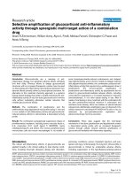
Figure 1 Construction and characterization of an HIV derivative carrying the b-Gal alpha peptide. (A) Thecodingsequenceforamino
acids 1-51 of b-Gal (gray box) was inserted into the gag open reading frame of plasmid pCHIV. Amino acids displayed in bold represent
authentic sequences from HIV Gag or b-Gal, respectively, while introduced linker sequences are displayed in italics. Arrowheads indicate cleavage
sites for HIV PR. (B) Immunoblot analysis of HIV.MAa particles. 293T cells transfected with the indicated constructs were grown in the absence (-)
or presence (+) of 2 μM LPV. At 44 h post transfection, particles were purified by ultracentrifugation and analyzed by immunoblotting using the
indicated antisera. Molecular mass standards (in kDa) are shown on the left, specific protein products are identified on the right. (C) b-Gal activity
in lysates of transfected 293T cells dependent on HIV PR activity. Cell lysates from untransfected 293T cells (filled circles), or from 293T cells
transfected with a mixture of pCMVω and pCHIV.MAa and incubated in the presence of DMSO (filled triangles) or 2 μM LPV (open triangles,
respectively, were prepared at 48 h post transfection and b-Gal activity was determined in vitro through cleavage of the colorimetric substrate
CPRG by measuring changes in OD592 over time. The graph shows mean values and standard deviations from five independent experiments.
Relative rates of CPRG cleavage were determined by linear regression, yielding an average value of 0.109 min
-1
for the DMSO controls and 0.054
min
-1
for the LPV treated samples, respectively
Jochmans et al. Retrovirology 2010, 7:89
/>Page 4 of 14
(Figure 2A, left panels). Cells transfected with a pCHIV.
MAa vari ant in which PR was inact ivated due to a D25A
mutation in the PR active site (PR-) displayed no increase
in Gag processing or b-Gal activity when grown in the
presence of 5 μM EFV (Figure 2A, middle panels). As a
control mimicking enhanced PR activity we used an
HIV-1 derivative expressing an artificially lin ked PR
dimer (2PR). Duplicating the PR monomer coding region
in the proviral context and connecting the two PR mono-
mers by a flexible 8 amino acid linker leads to premature
activation of HIV PR resulting in greatly enhanced intra-
cellular Gag processing and prevention of virus forma-
tion. Low PI doses, which interfere with infectivity of
wild-type HIV, partially rescue HIV(2PR) replication by
restoring an appropriate level of Gag processing, while
high concentrations of PI completely block the activity of
the artificially activated PR and lead to the production of
non-infectious virus [12,16]. Transfection of a construct
encoding the 2PR coding sequence in the context of
pCHIV.MAa led to nearly complete intracellular Gag
processing (Figur e 2A, right panels), while very low levels
of CA were released into the supernatant (not shown).
No effect of EFV on b-Gal activity was observed in this
case, presumably because Gag and Gag-Pol were already
completely processed in the absence of EFV (Figure 2A,
right panels). Taken together, these results indicate that
the EFV mediated increase in b-Gal activity was PR
dependent.
In order to identify the most potent available compound
we next employed the established assay for a detailed com-
parison of a series of NNRTIs. We included NNRTIs pre-
viously compared qualitatively with respect to activation of
Gag processing [27], namely EFV, ETV, NVP and TMC-
120 [43], as well as second generation NNRTIs not cur-
rently in clinical use: IDX-12899 [44], GW-678248 [45]
VRX-480773 [46] and UK-453061 [47]. 293T cells
B
DMSO
EFV
ETV
IDX-12899
GW-678248
VRX-480773
TMC-120
UK-453061
CA
Gag
A
C
Gag
CA
co EFV co EFV co EFV
0,0
0,2
0,4
0,6
0,8
1,0
1,2
1,4
1,6
1
2
3
4
5
6
7
8
0.0
1.0
0.2
0.4
1.2
1.4
1.6
0.8
0.6
relative β-Gal activity
1 2 3 4 5 6
co EFV co EFV co EFV
CA
GW678248
IDX-12899
EFV
ETV
VRX-480773
TM-120
UK-453061
NVP
VRX-480773
IDX-12899
GW-678248
Efavirenz
Etravirine
TMC-120
Nevirapine
UK-453061
-1.5 -1.0 -0.5 0.0 0.5
0.0
0.5
1.0
1.5
2.0
2.5
log [µM NNRTI]
relative
β
-Gal activity
pCHIV.MAα pCHIV.MAα(PR-) pCHIV.MAα2PR
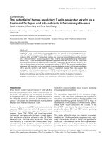
Figure 2 Effect of NNRTIs on alpha complementation and intracel lular Gag processing effici ency. (A) 293T cells trans fected wi th a
mixture of pCMVω and pCHIV.MAa (lanes 1-2), pCHIV.MAa(PR-) (lanes 3-4), or pCHIV.MAa2PR (lanes 5-6), respectively, were incubated in the
presence of DMSO (lanes 1, 3 and 5), or 5 μM EFV (lanes 2, 4 and 6). At 44 h post transfection, cell lysates were harvested and analyzed by
immunoblot using antiserum raised against HIV CA (top), as well as for relative b-Gal activity (bottom). CPRG cleavage rates determined as
described in materials and methods were normalized to the value obtained for the respective solvent control. (B) 293T cells transfected with a
mixture of pCHIV.MAa and pCMVω were grown in the presence of DMSO or 0.25 to 10 μM of the indicated NNRTI, respectively. At 44-48 h post
transfection, cell lysates were harvested and analyzed for b-Gal activity. The graph shows mean CPRG cleavage rates and standard deviations
from 3-5 transfections each out of three independent experiments. Values were normalized to the cleavage rate obtained for the corresponding
solvent control (indicated by a gray line). (C) Lysates of transfected cells grown in the presence of 0.5 μM of the respective inhibitor were
analyzed for Gag processing by quantitative immunoblot using antiserum against HIV CA. Data from one representative replicate are shown.
Jochmans et al. Retrovirology 2010, 7:89
/>Page 5 of 14
co-transfected with pCHIV.MAa and pCMVω were
grown in the presence of the respective NNRTI at concen-
trations ranging from 0.03 to 10 μM. At 44 h post trans-
fection, cell lysateswereanalyzedforb-Gal activity. As
showninFigure2B,compounds varied in their effect:
NVP, TMC-120 and UK-453061 displayed little or no
enhancem ent of alpha complementation, while the other
compounds tested enhanced b- Gal activity up to 2.5 fold
relative to the DMSO control. The most efficient com-
pounds IDX-12899, GW-678248 and VRX-480773 showed
strong b-Gal activity enhancement at ~ 250 nM, while ~ 1
μM o f ETV or EFV was required to achieve the maximal
effect (Figure 2B). At high NNRTI concentrations (5 μM
and above) microscopically detectable impairment of cell
growth, accompanied by a decrease in b-G al activity and
high signal variability between replicates indicative of cyto-
toxic effects was observed, and concentrations above 2.5
μM NNRTI were therefore excluded from the analysis
shown here; this eff ect was most pronounced for TMC-
120, ETV and VRX-480773. The cytotoxicity observed for
TMC-120 under the conditions used, which was con-
firmed by CC
50
determination using a T-cell line (see
below), likely presents an explanation for a discrepancy
between our findings and those of Fig ueiredo et al. [27],
who had repo rted a stimulation of Gag processing upon
shorter incubation of cells with 5 μMTMC-120.Under
our experimental conditions we could not measure repro-
ducible b-Gal activities at this concentration due to cell
death; we can also not exclude that cytotoxicity might
have obscured stimulatory effects of TMC-120 at lower
concentrations. The ranking in the efficacy of compounds
was confirmed by immunoblot analysis of lysates from
cells incubated with 0.5 μM of the respective inhibitors
(Figure 2C), which showed clear differences between the
compounds with respect to the enhancement of Gag pro-
cessing directly paralleling the results obtained in the
alpha complementation assay.
Selective PR dependent killing of HIV expressing T-cells
by NNRTIs
The described drug induced PR activation might be
exploited to selectively kill HIV infected cells. In order to
test this hypothesis, we established the persistently
infected T-cell lines MT4-IIIB and MT4-LTR-EGFP-IIIB,
where the expression of HIV encoded proteins in >9 9%
of cells could be detected by intracellular p24 staining
(not shown). In MT4-LTR-EGFP-IIIB cells, HIV expres-
sion could additionally be detected through long terminal
repeat (LTR) driven exp ression of t he gfp marker gene.
As a control we used uninfected MT-4 cel ls or MT4-
CMV -EGFP ce lls, consti tutively expressing EGFP from a
CMV promoter, respectively. The use of persistently
infected cells enabled us to study the effects of NNRTIs
on virus producing cells regardless of their effect on
reverse transcription, since t he proportio n of virus pro-
ducing cells in this system does not depend on infection
of new host cells. Immunoblot analysis of cell lysates
after treatment with two of the more potent NNRTIs,
VRX-480773 and GW-678248, confirmed that NNRTI
mediated enhancement of Gag processing also occurred
in virus producing cells, as apparent from the decreased
ratio of Gag to intermediate and fully mature processing
products (Figure 3A, compare lanes 2 and 5 to lane 1). In
order to investigate the effect of NNRTIs on viability of
chronically infected cells, MT4-LTR-EGFP-IIIB cell s as
well as MT4-LTR-EGFP parental cells were treated with
1 μM VRX-480773 for 6 days. Quantification of live cells
by microscopic evaluation of trypan blue stained samples
revealed a significant decrease in live cell numbers for
the HIV infect ed MT4-LTR-EGFP-IIIB cells, whereas the
number of uninfected control cells remained constant
(Figure 3B). In order to test whether the observed cyto-
toxic effect on virus producing cells was due to enhanced
HIVPRactivityweadded200nMofthePIdarunavir
(DRV) to infected and uninfected cells in the presence
and absence of VRX-480773. DRV treatment impaired
Gag processing (Figure 3A, lanes 3, 4 and 6) and comple-
tely reversed the cytotoxic effect of VRX-480773 in
MT4-LTR-EGFP-IIIB cells, supporting the interpretation
that the observed NNRTI induced cell killing was
mediated by HIV PR.
By quantification of intracellular GFP fluorescence of
drug treated MT4-CMV-EGFP and MT4-TR-EGFP-IIIB
cells, resp ectively, we compared the relat ive effect of dif-
ferent NNRTIs on viability of infected versus uninfected
cells (Figure 3C and Table 1). Differential effects, corre-
lating with the biochemical data obtained on 293T cells,
were revealed (Table 1). The most potent comp ounds,
IDX-12899, GW-6 78248 and VRX-480773, display ed
CC
50
values in the submicromolar range on MT4-LTR-
EGFP-IIIB cells. Cytotoxicity on uninfected MT4-CMV-
EGFP control cells was undetectable f or IDX-12899 an d
GW-678248 in the tested range; VRX-480773, displayed
detectable unspecific toxicity, albeit with a ~10 fold
higher CC
50
than on virus producing cells. EFV was less
cytotoxic on the inf ected cells, b ut this ef fect was again
specific as indicated by the observation that MT4-CMV-
EGFP cells were no t affected. The remaining compounds
showed no specific effect in the tested concentration
range: TMC-120 displayed toxicity on the virus produ-
cing cells, but also showed comparable toxicity on unin-
fected control cells, while the remaining compounds had
no detecta ble effect on total E GFP expres sion on either
cell line. In all cases the specific NNRTI induced cyto-
toxicity on virus producing cells was comp letel y reverted
by addition of DRV (Table 1).
These results support the hypothesis that NNRTIs can
exert a dose dependent, inhibitor specific activation o f
Jochmans et al. Retrovirology 2010, 7:89
/>Page 6 of 14
MT4-LTR-EGFP-IIIBMT4-LTR-EGFP
2.0x10
6
0
3.0x10
6
1.0x10
6
cells / ml
n.s.
n.s.
p=0.0052
p=0.036
B
C
-1.5 -1.0 -0.5 0.0 0.5 1.0
0
50
100
log [µM GW-678248]
GFP intensity [%]
-1.5 -1.0 -0.5 0.0 0.5 1.0
0
50
100
log [µM NVP]
GFP intensity [%]
-1.5 -1.0 -0.5 0.0 0.5 1.0
0
50
100
log [µM EFV]
GFP intensity [%]
A
CA
MA-CA
Gag
GagPol
75
50
37
25
20
1 2 3 4 5 6
Figure 3 Intracellular PR activatio n and NNRTI induced killing of M T-4 cells persistently infected with HIV. (A) NNRTI induced
enhancement of intracellular Gag processing in chronically infected MT-4 cells . MT-4-IIIB cells were cultured in the presence of DMSO (lane 1),
1 μM GW-678248 (lane 2), 200 nM DRV (lane 3), 1 μM GW-678248 + 200 nM DRV (lane 4), 1 μM VRX-480773 (lane 5), or 1 μM VRX-480773 + 200
nM DRV (lane 6), respectively. Cell lysates were harvested and analyzed by immunoblot using antiserum raised against HIV-1 CA. Positions of Gag
and Gag-Pol processing products are marked at the right, molecular mass standards are indicated to the left (in kDa). Lysates shown here were
harvested at day 2 post addition of compounds; longer incubation periods (6 days) resulted in a more pronounced accumulation of
unprocessed Gag in the DRV treated samples, but the pattern in the NNRTI treated samples became difficult to detect due to cell death. (B)
NNRTI induced killing of chronically infected MT-4 cells. The MT4-LTR-EGFP parental cell line or its persistently HIV-1 infected derivative MT4-LTR-
EGFP-IIIB, respectively, were seeded at a density of 1.5 × 10
5
cells/ml and incubated for 6 days in the presence of 0.1% DMSO (white bars), 200
nm DRV (gray bars), 1 μM VRX-480773 (black bars) or 1 μM VRX-480773 + 200 nM DRV (hatched bars), respectively. Live cells were counted after
trypan blue staining. Data represent mean values and standard deviations from three parallel cultures. P-values were calculated with GraphPad
Prism using an unpaired two-tailed t-test. n.s., non significant. (C) MT4-CMV-EGFP (circles) or MT4-LTR-EGFP-IIIB (triangles) cells were seeded in
96-well plates at a density of 10
5
cells/ml and incubated for 4 days in the presence of various concentrations of the indicated NNRTI, either with
(open symbols) or without (filled symbols) the addition of 100 nM DRV. EGFP intensity per well was quantitated at the end of the incubation
period by measuring total fluorescence intensity per well based on analysis of microscopic images as described in Methods. The graphs show
exemplary data for three NNRTIs. Mean values and standard deviations from three independent wells of one representative experiment are
shown. Lines represent fits of the data to a standard dose response equation (4 parameters), yielding CC
50
values on virus producing cells in the
absence of DRV (filled triangles) of 0.35 μM for GW-678248 and 2.44 μM for EFV, respectively. Data from several independent experiments for
these compounds as well as for the other NNRTIs were used to calculate the CC
50
values summarized in Table 1.
Jochmans et al. Retrovirology 2010, 7:89
/>Page 7 of 14
intracellular HIV PR by stabilizing Gag-Pol dimers. In
order to obtain further evidence for this model, we ana-
lyzed the effect of the various NNRTIs on RT dimeriza-
tion in a mammalian two-hybrid system [48]. We found
that, while lower absolute concentrations were required
in this context, the relative effects of the various com-
pounds on RT dimer formation paralleled their effects on
intracellular Gag processing: IDX-12899, GW-678248
and VRX-480773 promoted RT dimerization in the low
nM range, whereas a fivefold higher concentration was
required for EFV, and EC
50
values for the remaining
compounds were higher than 100 nM (Table 1; see Addi-
tional file 3 for exemplary primary data). This correlation
lends further support to the proposed mechanism of
action.
To validate our results obtained for the persistently
infected cell line in a more relevant cell system we per-
formed additional infection experiments using human
peripheral blood mononuclear cells (PBMC). In these
experiments we focused on two of the most potent
compounds, GW-678248 and VRX-480773, which dis-
played CC
50
values in the sub-micromolar range on
virus producing MT-4 cells (Table 1). PBMC isolated
from healthy blood donors were activated and infected
with a replication competent HIV-1 derivative which
carries a gfp gene in the nef locus [49]. The co-receptor
antagonist AMD-3100 was added at day 2 post infection
to prevent further viral spread. This was done to distin-
guish the proposed killing of infected cells from the
inhibitory effect of NNRTIs and PIs on virus replication.
At the time of AMD-3100 addition, individual samples
were further treated with solvent only, 1 μM NNRTI,
200 nM DRV, or a mixture of both. The percentage of
infected cells was determined following incubation for
5 days by flow cytometry (Figure 4 A) yielding values
between 2 and 6% for the control samples. Analogous to
our results with the MT-4 cell line (compare Figure 3B)
we observed a significant reduction of infected primary
cells upon treatment with VRX-480773 or GW-678248
as compared with the control. This effect was partially
reversed by addition of PI and thus dependent on PR
activity (Figure 4A). Rescue was incomplete, however,
despiteacompleteblockageofGagprocessingbyDRV
under these conditions (see Additional file 4 for immu-
noblot analysis). Similar results we re obtained upon
infection of CD4-positive primary T-cells with an EGFP-
expressing virus (Figure 4B). In this case, AZT was used
to prevent ongoing viral spread, but the same PR depen-
dent cytotoxicity was observed upon addition of either 1
μM GW-678248 or 1 μM VRX-480773. In this case, the
addition of DRV c ompletely reversed the NNRTI effect,
indicating that the induced cytotoxicity was largely
dependent on PR activity.
Discussion
Triggered by previous reports that certain NNRTIs can
enhance HIV-1 PR activity, the present study provides
proof of principle that this effect can be exploited for
the specific killing of HIV producing cells in tissue cul-
ture. Applying a newly developed enzymatic assay mea-
suring intracellular HIV PR activation we compared
relative activities of various NNRTIs on intracellular
Gag and Gag-Pol pro cessing. Thes e activities correlated
with the potency of the respective compounds to
enhance intracellular RT heterodimerization and, more
importantly, with their efficacy regarding specific killing
of HIV producing cells. Similar effects were obtained for
chronically HIV-1 infected MT-4 cells and for acutely
Table 1 Comparison of NNRTI efficacies in various assay systems
Inhibition of HIV
replication in
vitro EC
50
[nM]
Enhancement
of Gag
processing
(Fig. 2)
Cytotoxicity on
MT4-CMV-EGFP
control cells CC
50
[μM]
Cytotoxicity on MT4-
LTR-EGFP-IIIB HIV-1
producing cells CC
50
[μM]
Ctotoxicity on MT4-LTR-
EGFP-IIIB cells in presence
of 0.1 μM DRV CC
50
[μM]
Enhancement
of RT-
Dimerization
EC
50
[μM]
IDX-
12899
1.9 ± 1.3 ++ > 10 0.29 ± 0.21 > 10 0.0046
GW-
678248
0.84 ± 0.25 ++ > 10 0.63 ± 0.29 > 10 0.0032
VRX-
480773
1.6 ± 0.81 ++ 5.82 ± 1.44 0.68 ± 0.34 6.33 ± 0.08 0.0040
EFV 1.9 ± 0.9 + > 10 1.71 ± 0.43 > 10 0.020
ETV 3.2 ± 5 + > 10 > 10 > 10 0.27
UK-
453061
7.5 ± 1.4 - > 10 > 10 > 10 0.15
NVP 42 ± 20 - > 10 > 10 >10 18
TMC-
120
1.7 ± 1.4 - 3.02 ± 0.90 2.56 ± 0.74 4.33 ± 0.81 ND
*mean values and standard deviations from three or more independent measurements are shown; ND, not done.
Jochmans et al. Retrovirology 2010, 7:89
/>Page 8 of 14
infected PBMC, indicating that the observed effects are
not cell-type dependent and may occur at different
levels of HIV-1 gene expression.
Efficient intracellular PR act ivation is apparently not a
general property of NNRTIs. The relative efficacies varied
and three NNRTIs tested did not display detectable
effects under the conditions used here. The structural
basis for these differences in PR activating potential
betweenthevariousNNRTIsiscurrentlynotclear.The
fact that this potential did not correlate with the relative
antiviral efficacies of the respe ctive compounds at lower
concentrations medi ated by inhi bition of RT enzymatic
activity suggests that the two activities are structurally
distinct. This may be related to the relative affinities of
the compounds to mono- or dimeric forms of the
enzyme [32] and these features may be exploited for the
development of derivatives with increased activity.
Anti-infective d rugs acting not, or not exc lusively, on
viral replication, but rather affecting virus producing
cells may be considered for strategies aimed at HIV era-
dication from the infected organism. Despite efficient
long term suppression of H IV by current the rapies,
virus eradication is not achieved, most likely because of
reservoirs of long-lived latently infected cells [50-52].
HIV gene expression is an obvious requirement for the
NNRTI enhanced PR cytotoxicity described in the cur-
rent study, and transcriptionally silent cells harbouring
HIV proviral DNA can thus not be directly targeted.
This approach may be synergistic, however, with the
proposed activation of latent reservoirs by small mole-
cules (e.g. affecting chromatin structure). The activation
should induce HIV expression in the absence o f global
T-cell activation, while the spread of infection to new
target cells is prevented by available antiretroviral drugs
[53]. A combination of this strategy with targeted PR
activation would of course require the use of PI sparing
HAART regimens [54] for prevention of viral spread; a
regimen lacking PI and containing NNRTIs with a high
potential for PR activation may be optimal to exploit the
observed cytotoxic activity in such a situation. Induced
killing o f HIV-1 infected cells may also be exploited to
target persistent res ervoirs of HI V producing cells. The
existence of such reservoirs that differ from latently
infected cells is suggested by the conti nuous presence of
very low viral loa ds unde r therapy, which do not
respond to HAART treatment intensification [3,55,56].
While the nature of these reservoirs is uncertain, a strat-
egy for targeted PR activation may contribute to dimin-
ish or eliminate these virus producing cells.
Previous studies had reported EFV to be the most effi-
cient NNRTI with respect to PR activation. Although we
were able to identify inhibitors in clinical development
displaying a higher efficacy than EFV and showed that
these higher efficacies transl ated into a detectable speci-
fic cytotoxicity on HIV producing cells i n tissue culture,
CC
50
values determined were still in the high nanomolar
0
1
2
3
4
A
proportion of infected cells
normalized to solvent control
p < 0.0001
0.0
0.2
0.4
0.6
0.8
1.0
p = 0.025
VRX-480773 GW-678248
B
infected cells [%]
VRX-480773 GW-678248
** *
Figure 4 NNRTI induced selective killing of HIV-1 infected primary human cells. (A) PBMC prepared from buffy coats of healthy blood
donors were infected with HIV-1AGFP. At day 2 post infection, 100 ng/ml AMD-3100 was added to all samples to prevent further infection.
Individual samples were incubated in addition with DMSO (white bars), 200 nm DRV (gray bars), 1 μM of the indicated NNRTI (black bars) or 1
μM NNRTI + 200 nM DRV (hatched bars), respectively. After further incubation for 5 days, cells were harvested and analyzed for the proportion of
infected GFP expressing cells by flow cytometry. The figure shows mean values and standard deviations from three independent experiments
(VRX-480773) or one experiment (GW-678248), respectively, each comprising three parallel cultures using different donor pools. P-values were
calculated using a two-tailed unpaired t-test (GraphPad Prism). Values were normalized to the respective solvent control. (B) CD4 positive cells
isolated from PBMC were infected with HXB2D-EGFP. At day 7 post infection 1 μM AZT (white bars), 1 μM of the indicated NNRTI (striped bars),
1 μM of the indicated NNRTI + 1 μM AZT (black bars) or 1 μM NNRTI + 1 μM AZT + 100 nM DRV (hatched bars), respectively, were added. After
further incubation for 3 days, cells were harvested and analyzed for the proportion of infected cells by flow cytometry. The figure shows mean
values and standard deviations of values from one representative experiment (three parallel infections). Asterisks: non-infected controls.
Jochmans et al. Retrovirology 2010, 7:89
/>Page 9 of 14
range. Peak serum levels of EFV are in the micromolar
range [57], suggesting that the proposed mechanism of
NNRTI induced killing of HIV-1 producing T -cells
might already occur in vivo under therapy. Nevertheless,
the therapeutic window between specific and unspecific
cytotoxicity is likely t o be rather narrow for most
NNRTIs and thus more potent compounds will be
required for development of this inhibitory mechanism
into an applicable therapeutic strategy. A peptid e (P
AW
)
which stabilizes RT dimers and displays potent antiviral
activity in vitro has also been described [58]. Sinc e P
AW
appears to interact with a site not overlapping the
NNRTI binding pocket, it points to another potential
target site for enhancers of Gag-Pol dimer stabilization.
However, P
AW
has so far only been rep orted to inter act
with the dimeric forms o f RT; it remains to be investi-
gated whether this peptide - or compounds targeting
the same bindi ng site on RT - could also promote Gag-
Pol dimer formation.
Conclusion
In summary, the results presented here are consistent
with the following model, which we propose as a work-
ing hypothesis as a basis for further investigation: cer-
tain NNRTIs can increase intracellular Gag-Pol dimer
concentration upon binding to the RT domain of Gag-
Pol and thereby stimulate intracellular PR activity.
Enhanced activation of PR reduces v irion formation
through depletion of the assembly competent Gag and
Gag-Pol precursor protei ns, as shown in earlier studies
[12,16,17,27], but furthermore leads to the death of the
virus expressing cell, as presented in this study. Based
on the proposed mechanism, a small m olecule com-
pound w hich efficiently enhanc es Gag-Pol dimerization
would have a dual and synergistic effect on HIV spread
in directly preventing virusproductionononesideand
accelerating the death of virus producing cells on the
other. The data presented here provide proof of concept
for a drug induced killing of HIV producing cells, but
more potent inducers of Gag-Pol dimerization will likely
be required for therapeutic application, especially for
targeting cells expressing low amounts of Gag-Pol. The
current incomplete knowledge of the Gag-Pol dimeriza-
tion process and of other mechanisms involved in PR
activation prevents a rational search for PR activating
compounds; however, the gel independent assay
described here may provide a basis for screening of
compound libraries for such activities. Alpha comple-
mentation has successfully been used in various high
throughput screening approaches [39] and it appears
likely that more potent enhancers of Gag-Pol dimeriza-
tion and PR activation can be identified based on this
method. Such novel compounds may ultimately render
selective killing of HIV-1 infected cells by increased PR
toxicity a feasible therapeutic approach.
Methods
Plasmids
HIV-1 proviral constructs were based on plasmid
pNLC4-3 [59] and non-in fectious viru s vari ants were
derived from the previously described plasmid pCHIV, a
CMV promoter driven derivative o f NL4-3 lacking both
HIV LTR regions [41]. The coding sequence for amino
acids 1-51 of b -Gal from Escherichia coli, amplified by
PCR from plasmid pCMVbeta (Invitrogen) and flanked at
the N-terminus by a coding sequence for a HIV-1 PR
recognition site, was cloned into engineered unique
BspEI and AfeI restriction sites which had been inserted
into pCHIV between codon s 128 and 129 of MA (see
Figure 1A for resulting amino acid sequences). The 2PR
derivatives of pCHIV and pCHIV.MAa were cloned by
exchange of an ApaI fragment against the respective frag-
ment from plasmid pNL4- 3.2PR [16]. Plasmid pCMVω
was constructed by amplifying the b-Gal encoding
sequence from plasmid pCMVbeta by PCR, using an N-
terminal primer that introduced a deletion of codons
11-41 (primer sequence: GGCGCCATGGGCGTGAT-
CACCGACAGCCTGGCCGTGGAGGCCCGCACCG
ATCGCCC). The resulting ω-fragment encoding PCR
fragment was cloned into the EcoRV site of pcDNA3.1-
Zeo by blunt end ligation. Expression of a protein of the
expected molecular mass was confirmed by immunoblot
using polyclonal antiserum against b-Gal (Abcam ab 616;
not shown).
Cells and viruses
MT4-CMV-EGFP and MT4-LTR -EGFP cells were
obtained by transfection of MT-4 cells with a selectable
construct comprising the egfp gene under the control of
a CMV promoter or the HIV-1 long terminal repeat
(LTR) region, respectively, and subsequent selection of
stably transfected c ells. Persistently infected MT4-IIIB
and MT4-LTR-EGFP-IIIB cells were generated by infec-
tion of parental MT-4 or MT4-LTR-EGFP cells, respec-
tively, with HIV-1IIIB at an MOI of 0.1. The cytopathic
effect of HIV led to a dramatic cell loss early after infec-
tion, but persistently infected MT4-IIIB and MT4-LTR-
EGFP-IIIB cells, displaying a similar morphology as the
parental cells and only slightly delayed proliferation
could be selected within 2-3 weeks post infect ion. Persis-
tent productive infection with HIV-1 was demonstrated
by the detection of infectious virus in the tissue culture
supern atant and int racellular anti-p24 staining, as well as
by syncytia formation upon m ixing with non-infected
MT-4 cells. All MT-4 derived cell lines as well as
C8166 cells were maintained in RPMI 1640 medium
Jochmans et al. Retrovirology 2010, 7:89
/>Page 10 of 14
supplemented with 10% heat-inactivated fetal calf
serum, 2 mM L-glutamine, 0 .1% NaHCO3, and 0.02%
gentamycin.
Peripheral blood mononuclea r cells (PBMC) were pur-
ified from buffy coats of HIV-negative blood donors,
grown in supplemented RPMI 1640 and stimulated by
the addition of 10 ng/ml IL-2 (Biomol) and 2 μg/ml
PHA (Sigma). PBMC pooled from two donors each
were used for infection. CD4 positive cells from the
PBMC pool activated as previously described (Division
of AIDS, National Institute of Allergy and Infectious Dis-
eases, National Institutes of Health, and Collaborating
Investigators. 1997. Virology manual for HIV labora-
tories. Publication NIH-97-3828. U.S. Department of
Health and Human Services,Washington D.C.) were iso-
lated by magnetic sorting using anti-CD4 magnetic
microbeads (Miltenyi Biotec) according to the manufac-
turer’s instructions. For infection of PBMC, the HIV-1
derivatives HIV-1-AGFP [49] carrying the gfp gene fused
to the codon for amino acid 16 of Nef in pNL4-3, or
HXB2D-EGFP [60], which carries an egfp gene in the
place of the viral ne f open reading frame, wer e used as
indicated. Virus stocks were prepared by transfection of
the respective proviral plasmids in 293T cells.
Inhibitors
EFV, LPV, DRV, ETV, NVP and AMD-3100 were
obtained through t he AIDS Research an d Referenc e
Reagent Program, Division of AIDS, NIAID, NIH. IDX-
12899 [44], GW-678248 [45], VRX-480773 [46],
UK-453061 [47] and TMC-120 [43] were synthesized at
Tibotec. Compounds we re dissolved and stored as 10
mM stock solutions in 100% DMSO and diluted with tis-
sue culture medium to the final concentration immedi-
ately before use.
Analysis of Gag expression, processing and particle
release
293T cells were seeded in 6-well plates and transf ected
with the indicated constructs using FuGene6 (Roche)
according to the manufacturer’s instructions. Cell lysates
and tissue culture supernatants were harvested at 44-48
h post transfection. Virus was purified by ultracentrifu-
gation through a 20% (w/w) sucrose cushion. Cell
lysates, tis sue culture supernatants or pelleted viral par -
ticles were separated by SDS-PAGE (17.5% acrylamide;
acrylamide:bisacrylamide 200:1). Proteins were trans-
ferred to nitrocellulose by semi-dry blotting and
detected usi ng polyclonal antisera raised against recom-
binant HIV-1 CA or MA, or a commercial antiserum
against b-Gal (Abcam, ab616), respectively. Detec tion of
bound antibody by quantitative immunoblot was carried
out with a LiCor Odyssey system using protocols and
secondary antibodies suggested by the manufacturer and
evaluated using Odyssey v2.0 detection software.
Measurement of b-Gal activity in cell lysates
The activity of b-Gal in cell lysates from transfected 293T
cells was measured by enzymatic cleavage of the chromo-
genic b-Gal substrate chlorphenolred-b-D-galactopyrano-
side (CPRG, Roche; [42]). At 44 h post transfection , cells
were briefly rinsed with PBS and suspended in reporter
gene assay lysis buffer (Roche, 600 μl per 6-well dish) sup-
plemented with a protease inhibitor mix (Roche). Cell sus-
pensions were incubated for 10 min at room temperature
and cell debris was subsequently removed by brief centri-
fugation. Five μl of supernatant were diluted in 96-well
plates with 95 μl CPRG reaction buffer (50 mM potassium
phosphate, pH 7.5, 1 mM MgCl
2
)andpre-warmedfor
5 min to 37°C. 100 μl of pre-warmed reaction mix (100
μM CPRG in CPRG reaction buffer supplemented with
protease inhibitor cocktail and 40 μM b-mercaptoethanol)
were added and b-Gal mediated cleavage of CPRG was
monitored by recording absorption at 592 nm every 2 min
for 20 m in at 37°C using a TECAN Safire multi-well
reader. OD592 values were plotted over time and relative
reaction rates (OD592/min) were determined from the
initial linear velocities.
Determination of direct antiviral activity and cytotoxicity
MT4-LTR-EGFP cells were seeded at a density of 1.5 ×
10
5
cells/ml and infected with HIV-1IIIB at a multiplicity
of infection of 0.01 in the presence of different NNRTI
concentrations. After 3 days of incubation, infected cells
were quantified by determination of total EGFP fluores-
cence per well based on microscopy and subsequent
image analysis. Threshold values were determined from
the average pixel value plus 6 standard deviations from the
uninfected control wells, an d the median thresho ld from
all control wells on a plate was defined as baseline G FP
expression. Intensity values for the sample wells were then
determined by subtracting the background threshold from
each pixel value obtained from the image of the respective
well and calculating the sum of net pixel intensities. Per-
cent inhibition was calculated as 100 * (1 - (Sample - CC)/
(VC - CC)). The 50% effective concentration (EC
50
)was
calculated by fitting the data to a standard dose response
equation and is defined as the concentration that reduced
virus induced fluorescence by 50% as compared to the
DMSO control. Data shown i n Table 1 represent mean
values of at least three independent experiments.
The cytotoxicity of inhibitors was determined in parallel
on MT4-CMV-EGFP control cells and on MT4-LTR-
EGFP-IIIB virus producing cells, respectively. Cells were
seeded into 96-well plates at a density of 1.5 × 10
5
cells/ml
and grown for 4 days in the presence or absence of
Jochmans et al. Retrovirology 2010, 7:89
/>Page 11 of 14
different compound concentrations. Cell proliferation was
quantified by measuring the EGFP fluorescence per well
based on microscopy followed by image analysis as
described above and expressed as CC
50
values calculated
by fitting the data to a standard dose response equation
(drug concentration which led to reduction of cell asso-
ciated fluorescence by 50%).
Determination of enhancement of RT dimerization
RT heterodimer formation was monitored using a mam-
malian two hybrid system described previously [48]. In
brief, the bait protein (p66) was fused to the C-terminus
of a chimeric receptor consisting of the extracellular
part of the erythropoietin receptor and the i ntracellular
part of the leptin receptor incapable of STAT activation.
The prey protein (p51) was coupled to a part of the
cytoplasmic tail of the gp130 chain carrying several
STAT3 recruitment domains. Interaction of bait and
prey protein leads to functional complementation of
STAT3 activity, which results in Epo dependent induc-
tion of a STAT3-responsive luciferase reporter gene.
Enhancement of this interaction by the addition of com-
pounds can thus be measured by an increase of lucifer-
ase expression. The compound concen tration which
resulted in enhancement of the signal by 50% was
reported as EC
50
in Table 1.
Additional material
Additional file 1: Infectivity of HIV.MAa (A) HIV
NL4-3
and HIV
NL4-3
MAa
harvested from transfected 293T cells were used to infect C8166 cells. At
days 3 to 7 post infection, samples from the tissue culture supernatant
were harvested and the amount of p24 CA was determined by
quantitative immunoblot. The graph shows mean values and standard
deviations from three independent infections from one representative
experiment (wild-type HIV, filled triangles; HIV.MAa , open triangles; mock
infected cells, open circles), respectively. (B) Integrity of the reporter virus
after several rounds of replication was verified by immunoblot of lysate
from infected cells. At day 7 post infection, cell lysates from the infection
experiment shown in (A) were harvested and analyzed by immunoblot
using the indicated antisera. The presence of the slower migrating form
of MA carrying the linker sequence (MA*) as well as of a slightly slower
migrating form of Gag (Gag.MAa) indicates that the peptide insertion
was retained.
Additional file 2: Effect of EFV on Gag processing (A) and b-Gal
activity (B) in cell lysates 293T cells were seeded in 6-well plates,
transfected with the indicated ratio of pCHIV.MAa and pCMVω and
incubated in the absence (-, white bars) or presence (+, black bars) of 5
μM EFV, respectively. (A) At 44 h post transfection, cell lysates and virus
particles pelleted from the supernatant by ultracentrifugation were
harvested and analyzed by immonoblot using antiserum raised against
HIV-1 CA. Data from one representative experiment are shown. (B) In
parallel, samples of cell lysates were analyzed for b-Gal activity as
described in methods. The graph shows mean values and standard
deviations from three independent transfections from one representative
experiment.
Additional file 3: Enhancement of RT heterodimer formation by
NNRTIs RT heterodimer formation in cells treated with different
concentrations of NNRTIs was assayed using a mammalian two-hybrid
system (MAPPIT, [48]) as described in Methods. Enhancement of
luciferase reporter gene activities relative to the DMSO control was
plotted and used to calculate EC
50
values, defined as an enhancement of
50% over the control value. The graph shows representative dat sets for
titrations with NVP (diamonds), EFV (triangles) and VRX-480773 (c ircles),
respectively. Several independent experiments for each NNRTI tested
were performed to calculate CC
50
values summarized in Table 1.
Additional file 4: Efficacy of PR inhibitor treatment on infected
PBMC Representative samples from the experiment shown in Figure 4
were analyzed by immunoblot of cell lysates harvested at the end of the
experiment using antiserum raised against HIV-1 CA. The figure shows
samples of unifected cells (lane 1), as well as infected cells treated with
AMD-3100 (lane 2), AMD-3100 + DRV (lane 3), AMD-3100 + VRX-480773
(lane 4) and AMD-3100 + VRX-480773 + DRV (lane 5), respectively.
Samples corresponding to equal tissue culture volumes were loaded.
Acknowledgements
EFV, LPV, DRV, ETV, NVP and AMD-3100 were obtained through the AIDS
Research and Reference Reagent Program, Division of AIDS, NIAID. We thank
Daniel Boden for providing HXB2D-EGFP.
Author details
1
Tibotec-Virco BVBA, Mechelen, Belgium.
2
Department of Infectious Diseases,
Virology, University of Heidelberg, Germany.
3
Rega Institute for Medical
Research, Katholieke Universiteit Leuven, Minderbroedersstraat 10, B-3000
Leuven, Belgium.
Authors’ contributions
DJ and BM performed initial experiments and cooperated in study design
and coordination. MA carried out alpha complementation assays and
experiments on specific cytotoxicity in infected MT-4 cells and PBMC. IK and
LS performed experiments on sorted PBMC and comparison of the
complete NNRTI panel with respect to cytotoxicity levels on MT-4 cells and
RT dimerization. GK and HGK participated in study design, discussion and
coordination. BM drafted the manuscript with help of DJ and HGK. All
authors read and approved the final manuscript.
Competing interests
This study was supported by Tibotec Pharmaceuticals, Ltd. DJ, IK, LS and GK
are employed by Tibotec-Virco BVBA, Mechelen, Belgium.
Received: 24 August 2010 Accepted: 15 October 2010
Published: 15 October 2010
References
1. Hammer SM, Saag MS, Schechter M, Montaner JS, Schooley RT,
Jacobsen DM, Thompson MA, Carpenter CC, Fischl MA, Gazzard BG, et al:
Treatment for adult HIV infection: 2006 recommendations of the
International AIDS Society-USA panel. Jama 2006, 296:827-843.
2. Dornadula G, Zhang H, VanUitert B, Stern J, Livornese L Jr, Ingerman MJ,
Witek J, Kedanis RJ, Natkin J, DeSimone J, Pomerantz RJ: Residual HIV-1
RNA in blood plasma of patients taking suppressive highly active
antiretroviral therapy. Jama 1999, 282:1627-1632.
3. Maldarelli F, Palmer S, King MS, Wiegand A, Polis MA, Mican J, Kovacs JA,
Davey RT, Rock-Kress D, Dewar R, et al: ART suppresses plasma HIV-1 RNA
to a stable set point predicted by pretherapy viremia. PLoS Pathog 2007,
3:e46.
4. Palmer S, Maldarelli F, Wiegand A, Bernstein B, Hanna GJ, Brun SC,
Kempf DJ, Mellors JW, Coffin JM, King MS: Low-level viremia persists for at
least 7 years in patients on suppressive antiretroviral therapy. Proc Natl
Acad Sci USA 2008, 105:3879-3884.
5. Davey RT Jr, Bhat N, Yoder C, Chun TW, Metcalf JA, Dewar R, Natarajan V,
Lempicki RA, Adelsberger JW, Miller KD, et al: HIV-1 and T cell dynamics
after interruption of highly active antiretroviral therapy (HAART) in
patients w ith a history of sustained viral suppression. Proc Natl Acad Sci
USA 1999, 96:15109-15114.
6. Garcia F, Plana M, Vidal C, Cruceta A, O’Brien WA, Pantaleo G, Pumarola T,
Gallart T, Miro JM, Gatell JM: Dynamics of viral load rebound and
immunological changes after stopping effective antiretroviral therapy.
Aids 1999, 13:F79-86.
Jochmans et al. Retrovirology 2010, 7:89
/>Page 12 of 14
7. Ruiz L, Carcelain G, Martinez-Picado J, Frost S, Marfil S, Paredes R, Romeu J,
Ferrer E, Morales-Lopetegi K, Autran B, Clotet B: HIV dynamics and T-cell
immunity after three structured treatment interruptions in chronic HIV-1
infection. Aids 2001, 15:F19-27.
8. Ruiz L, Martinez-Picado J, Romeu J, Paredes R, Zayat MK, Marfil S,
Negredo E, Sirera G, Tural C, Clotet B: Structured treatment interruption in
chronically HIV-1 infected patients after long-term viral suppression. Aids
2000, 14:397-403.
9. Gougeon ML: Apoptosis as an HIV strategy to escape immune attack.
Nat Rev Immunol 2003, 3:392-404.
10. Baum EZ, Bebernitz GA, Gluzman Y: Isolation of mutants of human
immunodeficiency virus protease based on the toxicity of the enzyme in
Escherichia coli. Proc Natl Acad Sci USA 1990, 87:5573-5577.
11. Blanco R, Carrasco L, Ventoso I: Cell killing by HIV-1 protease. J Biol Chem
2003, 278:1086-1093.
12. Kräusslich HG: Specific inhibitor of human immunodeficiency virus
proteinase prevents the cytotoxic effects of a single-chain proteinase
dimer and restores particle formation. J Virol 1992, 66:567-572.
13. Strack PR, Frey MW, Rizzo CJ, Cordova B, George HJ, Meade R, Ho SP,
Corman J, Tritch R, Korant BD: Apoptosis mediated by HIV protease is
preceded by cleavage of Bcl-2. Proc Natl Acad Sci USA 1996, 93:9571-9576.
14. Kaplan AH, Manchester M, Swanstrom R: The activity of the protease of
human immunodeficiency virus type 1 is initiated at the membrane of
infected cells before the release of viral proteins and is required for
release to occur with maximum efficiency. J Virol 1994, 68:6782-6786.
15. Karacostas V, Wolffe EJ, Nagashima K, Gonda MA, Moss B: Overexpression
of the HIV-1 gag-pol polyprotein results in intracellular activation of HIV-
1 protease and inhibition of assembly and budding of virus-like
particles. Virology 1993, 193:661-671.
16. Kräusslich HG: Human immunodeficiency virus proteinase dimer as
component of the viral polyprotein prevents particle assembly and viral
infectivity. Proc Natl Acad Sci USA 1991, 88:3213-3217.
17. Park J, Morrow CD: Overexpression of the gag-pol precursor from human
immunodeficiency virus type 1 proviral genomes results in efficient
proteolytic processing in the absence of virion production. J Virol 1991,
65:5111-5117.
18. Pettit SC, Lindquist JN, Kaplan AH, Swanstrom R: Processing sites in the
human immunodeficiency virus type 1 (HIV-1) Gag-Pro-Pol precursor are
cleaved by the viral protease at different rates. Retrovirology 2005, 2:66.
19. Pettit SC, Moody MD, Wehbie RS, Kaplan AH, Nantermet PV, Klein CA,
Swanstrom R: The p2 domain of human immunodeficiency virus type 1
Gag regulates sequential proteolytic processing and is required to
produce fully infectious virions. J Virol 1994, 68:8017-8027.
20. Wiegers K, Rutter G, Kottler H, Tessmer U, Hohenberg H, Krausslich HG:
Sequential steps in human immunodeficiency virus particle maturation
revealed by alterations of individual Gag polyprotein cleavage sites. J
Virol 1998, 72:2846-2854.
21. Shoeman RL, Kesselmier C, Mothes E, Honer B, Traub P: Non-viral cellular
substrates for human immunodeficiency virus type 1 protease. FEBS Lett
1991, 278:199-203.
22. Shoeman RL, Honer B, Stoller TJ, Kesselmeier C, Miedel MC, Traub P,
Graves MC: Human immunodeficiency virus type 1 protease cleaves the
intermediate filament proteins vimentin, desmin, and glial fibrillary
acidic protein. Proc Natl Acad Sci USA 1990, 87:6336-6340.
23. Alvarez E, Castello A, Menendez-Arias L, Carrasco L: HIV protease cleaves
poly(A)-binding protein. Biochem J 2006, 396:219-226.
24. Ventoso I, Blanco R, Perales C, Carrasco L: HIV-1 protease cleaves
eukaryotic initiation factor 4G and inhibits cap-dependent translation.
Proc Natl Acad Sci USA 2001, 98:12966-12971.
25. Nie Z, Bren GD, Vlahakis SR, Schimnich AA, Brenchley JM, Trushin SA,
Warren S, Schnepple DJ, Kovacs CM, Loutfy MR, et al: Human
immunodeficiency virus type 1 protease cleaves procaspase 8 in vivo. J
Virol 2007, 81:6947-6956.
26. Nie Z, Bren GD, Rizza SA, Badley AD: HIV Protease Cleavage of Procaspase
8 is Necessary for Death of HIV-Infected Cells. Open Virol J 2008, 2:1-7.
27. Figueiredo A, Moore KL, Mak J, Sluis-Cremer N, de Bethune MP,
Tachedjian G: Potent nonnucleoside reverse transcriptase inhibitors
target HIV-1 Gag-Pol. PLoS Pathog 2006, 2:e119.
28. de Bethune MP: Non-nucleoside reverse transcriptase inhibitors (NNRTIs),
their discovery, development, and use in the treatment of HIV-1
infection: a review of the last 20 years (1989-2009). Antiviral Res 85:75-90.
29. Restle T, Müller B, Goody RS: Dimerization of human immunodeficiency
virus type 1 reverse transcriptase. A target for chemotherapeutic
intervention. J Biol Chem 1990, 265:8986-8988.
30. Restle T, Müller B, Goody RS: RNase H activity of HIV reverse
transcriptases is confined exclusively to the dimeric forms. FEBS Lett
1992, 300:97-100.
31. Figueiredo A, Zelina S, Sluis-Cremer N, Tachedjian G: Impact of residues in
the nonnucleoside reverse transcriptase inhibitor binding pocket on
HIV-1 reverse transcriptase heterodimer stability. Curr HIV Res 2008,
6:130-137.
32. Braz VA, Holladay LA, Barkley MD: Efavirenz binding to HIV-1 reverse
transcriptase monomers and dimers. Biochemistry 49:601-610.
33. Tachedjian G, Moore KL, Goff SP, Sluis-Cremer N: Efavirenz enhances the
proteolytic processing of an HIV-1 pol polyprotein precursor and reverse
transcriptase homodimer formation. FEBS Lett 2005, 579:379-384.
34. Tachedjian G, Orlova M, Sarafianos SG, Arnold E, Goff SP: Nonnucleoside
reverse transcriptase inhibitors are chemical enhancers of dimerization
of the HIV type 1 reverse transcriptase. Proc Natl Acad Sci USA 2001,
98:7188-7193.
35. Venezia CF, Howard KJ, Ignatov ME, Holladay LA, Barkley MD: Effects of
efavirenz binding on the subunit equilibria of HIV-1 reverse
transcriptase. Biochemistry 2006, 45:2779-2789.
36. Müller B, Daecke J, Fackler OT, Dittmar MT, Zentgraf H, Kräusslich HG:
Construction and characterization of a fluorescently labeled infectious
human immunodeficiency virus type 1 derivative. J Virol 2004,
78:10803-10813.
37. Möhler WA, Blau HM: Gene expression and cell fusion analyzed by lacZ
complementation in mammalian cells. Proc Natl Acad Sci USA 1996,
93:12423-12427.
38. Moosmann P, Rusconi S: Alpha complementation of LacZ in mammalian
cells. Nucleic Acids Res 1996, 24:1171-1172.
39. Olson KR, Eglen RM: Beta galactosidase complementation: a cell-based
luminescent assay platform for drug discovery. Assay Drug Dev Technol
2007, 5:137-144.
40. Wehrman TS, Casipit CL, Gewertz NM, Blau HM: Enzymatic detection of
protein translocation. Nat Methods 2005, 2:521-527.
41. Lampe M, Briggs JA, Endress T, Glass B, Riegelsberger S, Kräusslich HG,
Lamb DC, Bräuchle C, Müller B: Double-labelled HIV-1 particles for study
of virus-cell interaction. Virology 2007, 360:92-104.
42. Eustice DC, Feldman PA, Colberg-Poley AM, Buckery RM, Neubauer RH: A
sensitive method for the detection of beta-galactosidase in transfected
mammalian cells. Biotechniques 1991, 11:739-740, 742-733.
43. Fletcher P, Harman S, Azijn H, Armanasco N, Manlow P, Perumal D, de
Bethune MP, Nuttall J, Romano J, Shattock R: Inhibition of human
immunodeficiency virus type 1 infection by the candidate microbicide
dapivirine, a nonnucleoside reverse transcriptase inhibitor. Antimicrob
Agents Chemother 2009, 53:487-495.
44. Klibanov OM, Kaczor RL: IDX-899, an aryl phosphinate-indole non-
nucleoside reverse transcriptase inhibitor for the potential treatment of
HIV infection. Curr Opin Investig Drugs 11:237-245.
45. Ferris RG, Hazen RJ, Roberts GB, St Clair MH, Chan JH, Romines KR,
Freeman GA, Tidwell JH, Schaller LT, Cowan JR, et al: Antiviral activity of
GW678248, a novel benzophenone nonnucleoside reverse transcriptase
inhibitor. Antimicrob Agents Chemother 2005, 49:4046-4051.
46. Zhang Z, Xu W, Koh YH, Shim JH, Girardet JL, Yeh LT, Hamatake RK,
Hong Z: A novel nonnucleoside analogue that inhibits human
immunodeficiency virus type 1 isolates resistant to current
nonnucleoside reverse transcriptase inhibitors. Antimicrob Agents
Chemother 2007, 51:429-437.
47. Allan G, Davis J, Dickins M, Gardner I, Jenkins T, Jones H, Webster R,
Westgate H: Pre-clinical pharmacokinetics of UK-453,061, a novel non-
nucleoside reverse transcriptase inhibitor (NNRTI), and use of in silico
physiologically based prediction tools to predict the oral
pharmacokinetics of UK-453,061 in man. Xenobiotica 2008, 38:620-640.
48. Pattyn E, Lavens D, Van der Heyden J, Verhee A, Lievens S, Lemmens I,
Hallenberger S, Jochmans D, Tavernier J: MAPPIT (MAmmalian Protein-
Protein Interaction Trap) as a tool to study HIV reverse transcriptase
dimerization in intact human cells. J Virol Methods 2008, 153:7-15.
49. Welker R, Harris M, Cardel B, Kräusslich HG: Virion incorporation of human
immunodeficiency virus type 1 Nef is mediated by a bipartite
Jochmans et al. Retrovirology 2010, 7:89
/>Page 13 of 14
membrane-targeting signal: analysis of its role in enhancement of viral
infectivity. J Virol 1998, 72:8833-8840.
50. Chun TW, Carruth L, Finzi D, Shen X, DiGiuseppe JA, Taylor H,
Hermankova M, Chadwick K, Margolick J, Quinn TC, et al: Quantification of
latent tissue reservoirs and total body viral load in HIV-1 infection.
Nature 1997, 387:183-188.
51. Chun TW, Finzi D, Margolick J, Chadwick K, Schwartz D, Siliciano RF: In vivo
fate of HIV-1-infected T cells: quantitative analysis of the transition to
stable latency. Nat Med 1995, 1:1284-1290.
52. Finzi D, Hermankova M, Pierson T, Carruth LM, Buck C, Chaisson RE,
Quinn TC, Chadwick K, Margolick J, Brookmeyer R, et al: Identification of a
reservoir for HIV-1 in patients on highly active antiretroviral therapy.
Science 1997, 278:1295-1300.
53. Richman DD, Margolis DM, Delaney M, Greene WC, Hazuda D,
Pomerantz RJ: The challenge of finding a cure for HIV infection. Science
2009, 323:1304-1307.
54. Hammer SM, Eron JJ Jr, Reiss P, Schooley RT, Thompson MA, Walmsley S,
Cahn P, Fischl MA, Gatell JM, Hirsch MS, et al: Antiretroviral treatment of
adult HIV infection: 2008 recommendations of the International AIDS
Society-USA panel. Jama 2008, 300:555-570.
55. Dinoso JB, Kim SY, Wiegand AM, Palmer SE, Gange SJ, Cranmer L, O’Shea A,
Callender M, Spivak A, Brennan T, et al: Treatment intensification does not
reduce residual HIV-1 viremia in patients on highly active antiretroviral
therapy. Proc Natl Acad Sci USA 2009, 106:9403-9408.
56. McMahon D, Jones J, Wiegand A, Gange SJ, Kearney M, Palmer S,
McNulty S, Metcalf JA, Acosta E, Rehm C, et al: Short-course raltegravir
intensification does not reduce persistent low-level viremia in patients
with HIV-1 suppression during receipt of combination antiretroviral
therapy. Clin Infect Dis 50:912-919.
57. Csajka C, Marzolini C, Fattinger K, Decosterd LA, Fellay J, Telenti A, Biollaz J,
Buclin T: Population pharmacokinetics and effects of efavirenz in
patients with human immunodeficiency virus infection. Clin Pharmacol
Ther 2003, 73:20-30.
58. Agopian A, Gros E, Aldrian-Herrada G, Bosquet N, Clayette P, Divita G: A
new generation of peptide-based inhibitors targeting HIV-1 reverse
transcriptase conformational flexibility. J Biol Chem 2009, 284:254-264.
59. Bohne J, Kräusslich HG: Mutation of the major 5’ splice site renders a
CMV-driven HIV-1 proviral clone Tat-dependent: connections between
transcription and splicing. FEBS Lett 2004, 563:113-118.
60. Chen BK, Feinberg MB, Baltimore D: The kappaB sites in the human
immunodeficiency virus type 1 long terminal repeat enhance virus
replication yet are not absolutely required for viral growth. J Virol
1997,
71:5495-5504.
doi:10.1186/1742-4690-7-89
Cite this article as: Jochmans et al.: Selective killing of human
immunodeficiency virus infected cells by non-nucleoside reverse
transcriptase inhibitor-induced activation of HIV protease. Retrovirology
2010 7:89.
Submit your next manuscript to BioMed Central
and take full advantage of:
• Convenient online submission
• Thorough peer review
• No space constraints or color figure charges
• Immediate publication on acceptance
• Inclusion in PubMed, CAS, Scopus and Google Scholar
• Research which is freely available for redistribution
Submit your manuscript at
www.biomedcentral.com/submit
Jochmans et al. Retrovirology 2010, 7:89
/>Page 14 of 14