Báo cáo y học: "A nuclear export signal within the structural Gag protein is required for prototype foamy virus replication" pdf
Bạn đang xem bản rút gọn của tài liệu. Xem và tải ngay bản đầy đủ của tài liệu tại đây (986.02 KB, 11 trang )
RESEARC H Open Access
A nuclear export signal within the structural
Gag protein is required for prototype foamy
virus replication
Noémie Renault
1
, Joelle Tobaly-Tapiero
1
, Joris Paris
1
, Marie-Lou Giron
1
, Audrey Coiffic
1
,
Philippe Roingeard
2
, Ali Saïb
1,3*
Abstract
Background: The Gag polyproteins play distinct roles during the replication cycle of retroviruses, hijacking many
cellular machineries to fulfill them. In the case of the prototype foamy virus (PFV), Gag structural proteins undergo
transient nuclear trafficking after their synthesis, returning back to the cytoplasm for capsid assembly and virus
egress. The functional role of this nuclear stage as well as the molecular mechanism(s) responsible for Gag nuclear
export are not understood.
Results: We have identified a leptomycin B (LMB)-sensitive nuclear export sequence (NES) within the N-terminus of
PFV Gag that is absolutely required for the completion of late stages of virus replication. Point mutations of
conserved residues within this motif lead to nuclear redistribution of Gag, preventing subsequent virus egress. We
have shown that a NES-defective PFV Gag acts as a dominant negative mutant by sequestrating its wild-type
counterpart in the nucleus. Trans-complementation experiments with the heterologous NES of HIV-1 Rev allow the
cytoplasmic redistribution of FV Gag, but fail to restore infectivity.
Conclusions: PFV Gag-Gag interactions are finely tuned in the cytoplasm to regulate their functions, capsid
assembly, and virus release. In the nucleus, we have shown Gag-Gag interactions which could be involved in the
nuclear export of Gag and viral RNA. We propose that nuclear export of unspliced and partially spliced PFV RNAs
relies on two complementary mechanisms, which take place successively dur ing the replication cycle.
Introduction
Retroviral Gag proteins are involved in early stages of
infection such as trafficking of incomi ng viruses and
nuclear import (reviewed in [1]). Additionally , during the
late phases of infection, they coordinate the assembly of
viral particles, selecting the viral genome for encapsida-
tion and directing the incorporation of the envelope gly-
coproteins [2]. For most retroviruses, expression of Gag
alone is sufficient to induce the formation and release of
virus like particles. For that purpose, retroviruses hijack
the cellular endosomal machinery, enrolling components
of the class E vacuolar protein sorting (VPS) machinery
that induce topologically analogous membrane fission
events [3,4]. In addition to these defined assembly
domains, independent s ubcellular trafficking and/or
retention signals that provide important functions in the
virus life cycle have been identified (for a review, see [5]).
Foamy viruses (FVs) are complex exogenous animal ret-
roviruses that differ in many aspects of their lif e cycle
from orthoretroviruses such as the human immunodefi-
ciency viruses (HIV) [6]. For example, Gag and Pol pro-
teins of FVs are expressed independently of one another
[7], and both proteins undergo a single cleavage event [8].
Hence, the structural Gag protein is not cleaved into the
matrix, capsid, nucleocapsid sub-units as in most retro-
viruses, but is C-terminally cleaved by the viral protease,
leading to the production of a G ag doublet during viral
replication. Moreover, FV Gag is not myristoylate d, and
none of the conventional Gag landmarks of exogenous ret-
roviru ses, such as the major homology region or Cys-His
motifs, are found in this protein [6]. Instead, prototype
foamy virus (PFV) Gag harbors conserved C-terminal
* Correspondence:
1
CNRS UMR7212, Inserm U944, Université Paris Diderot, Institut Universitaire
d’Hématologie, Paris, France
Full list of author information is available at the end of the article
Renault et al. Retrovirology 2011, 8:6
/>© 2011 Renault et al; licensee BioMed Central Ltd. This is an Open Access article distributed under the terms of the Creati ve Commons
Attribution License ( which permits unrestricted use, distribution, and reproductio n in
any med ium, provided the original work is properly cited.
basic motifs, referred to as Gly-Arg (GR) boxes [9].
Although the first GR (GRI) box binds viral nucleic acids
and is required for viral genome packaging [10], the sec-
ond (GRII) harbors a nuclear localization sequence (NLS)
at its C-terminus, targeting Gag to the nucleus early after
infection [7,11]. Although this NLS is not absolutely
required for productive infection, since other NLSs in
Pol are likely involved in nuclear import of pre-integra-
tion complexes [12], it determines multiple integration
events [13]. GRII also contains a chromatin binding
sequence (CBS) in its N-terminus, tethering the PFV
incoming pre-integration complex onto host chromo-
somes prior to integration [14]. Therefore, depending
upon the stage of the viral cycle and thanks t o these
motifs, PFV Gag harbors distinct sub-cellular localiza-
tions. Of note, PFV does not encode a post-transcrip-
tional regulator such as Rev or Rex from HIV or HTLV,
respectively [15]; and therefore the mechanisms responsi-
ble for nuclear export of singly spliced or unspliced viral
mRNA, such as the one encoding for the structural Gag
proteins, are still not known. Similarly, where in the
infected cell Gag initially interacts with the viral genome,
is not known.
Similar to Mason-Pfizer monkey virus (MPMV) [16],
PFV assembles into capsids intracellularly at a pericentrio-
lar site [17]. Cytoplasmic PFV capsid assembly, which only
requires the expression of Gag proteins, as for other retro-
viruses, is mediated by a motif akin to a cytoplasmic tar-
geting and retention signal (CTRS) [18], also f ound in
MPMV Gag [ 19]. Both domains harbor a conserved and
indispensable arginine residue. However, unlike MPMV,
budding of PFV is absolutely dependent upon the presence
of cognate Env protein, implying a specific interaction
between the Gag and Env proteins that may occur at the
trans-Golgi network [17]. The unusually long leader
peptide of PFV Env is likely involved in this specific inter-
action with the respective Gag domains located in the
N-terminus of the protein, which are distinct from the
CTRS [20]. Finally, PFV Gag was shown to interact with
components of the VPS machinery for virus egress
[21-23].
During viral replication, PFV Gag shows distinct sub-
cellular localizations. During early stages of infection,
incoming Gag can be found near the microtubule-
organizing center (MTOC) and in the nucleus [24,25],
similar to incoming HIV-1 Gag [26]. During the late
stages of infection, followi ng its synthesis in the cyto-
plasm, PFV Gag displays a transient nuclear localization
triggered by the NLS present within its C-terminus [11].
Since PFV capsid assembly occurs near the centrosome
[17] and the presence of Gag is required for Pol packa-
ging [10], nuclear export of Gag i s an absolute prerequi-
site for the completion of the retroviral cycle. The role of
this nuclear stage as well as the molecular mechanism(s)
responsible for nuclear export of PFV Gag are not yet
understood.
Although this transient nuclear localization was initially
thought to be a specific feature of PFV, other retroviral
Gag proteins were shown to display a similar distribution
during the late stages of infection. T his is the case f or
example for HIV-1 [27] or Rous Sarcoma Virus (RSV) [28]
Gag. For RSV, the nuclear stage of Gag proteins contri-
butes to viral genomic RNA packaging [29], while the
exact role of nuclear Gag is not clear in the case of HIV-1.
Remarkably, both Gag proteins harbor a short hydropho-
bic motif that actively directs their nuclear export [27,28].
These so called leucine-rich nuclear export signals (NES)
are recognized by exportin 1, also named CRM1, a mem-
ber of the b importin superfamily of soluble nuclear trans-
port receptors (reviewed in [30,31]). The first viral ligand
of CRM1 identified was the HIV-1 Rev protein, w hich
serves as an adaptor for the export of the unspliced and
singly spliced viral mRNA that would otherwise be
restricted from leaving the nucleus [32]. Leptomycin B
(LMB) binds specifically to the central domain of CRM1,
preventing interaction with the NES and inhibiting subse-
quent nuclear export [33-35].
Here, we identify a LMB-sensitive n uclear export
sequence within the N-terminus of the PFV Gag. Point
mutations of residues conserved among primate foamy
viruses enhance nuclear distribution of the corresponding
Gag mutants. Consequently, recombinant viruses pro-
duced in the presence of NES-defective G ag mutants
were non-infectious. NES-defective Gag proteins behave
as dominant negative mutant s over their wild-type coun-
terpart, preventing viral particle release. Finally, substitut-
ing the LMB- sensitive NES of PFV Gag with that of
HIV-1 Rev lead to nucleocytoplasmic redistribution of
the chimeric Gag protein, but failed to restore infectivity.
Methods
Cells and drugs
HeLa and 293T cells were cultured in Dulbecco’smodi-
fied Eagles’ s medium supplemented with 10% fetal
bovine serum, 2 mM L-glutamine, 20 mM H epes and
antibiotics (1% penicillin and streptomycin). Leptomycin
B (LMB) (Sigma) was added to culture medium of trans-
fected cells to a fin al concentration of 40 nM for
6 hours.
Vector production
Vector stocks were produced by transfection of 293T
cells using Polyfect (Qiagen) with equimolar quantity of
the PFV pMD9 vector together with Gag (pCZIgag4),
Pol (pCZIpol1) and Env (pCZHFVenvEM02) expressing
plasmids kindly pro vided by A. Rethwilm [36]. Twenty-
four hours post-transfection, CMV promoter transcrip-
tion was enhanced by addition of 10 mM of sodium
Renault et al. Retrovirology 2011, 8:6
/>Page 2 of 11
butyrate for 6 h. Twenty-four hours later, supernatants
were clarified, filtrated through 0.45-μm-pore-size
filters, concentrated by centrifugati on on filter Amicon
(Millipore) and conserved at - 80°C until use.
Viral stocks titration
Infectious titers were determined by transduction of
293T cells with dilutions o f vector stocks by spinocula-
tion at 1, 200 g for 1 h 30 minutes at 30°C. Forty-eight
hours later, the cells were harvested and fixed in 1%
paraformaldehyde (PFA), and the amounts of GF P-
positive cells were determined by fluorescence-activated
cell sorting on a FACScan device with CellQuest
software (Becton Dickinson). The titer was calculated
as follows: T =(F xC/V)xD (F is the frequency of
GFP-positive cells, C is the number of cells at the time
of infection, V is the volume of the inoculum, and D is
the factor of dilution), expressed as transducing units
(tu)/milliliter.
Constructs
The full-length green fluorescent protein (GFP)-Gag
expression plasmid (pGFP-Gag) was previously
described [24]. Concerning Gag-RevNES, amino acids
95 to 112 were substituted by the 11aa of the HIV-1
RevNES in pCZIgag4 by t wo-steps procedure: deletion
of aa 95-112 to generate GagΔ95-112 and then insertion
of 11aa of RevNES to obtain Gag-RevNES. The G FP-
NES expression plasmids were generated by inserting
the annealing products of appropriate complementary
oligonucleotides into the SacI-EcoRI sites of the pEGFP-
C3 vector (Clontech). The tagged His-HA Gag expres-
sion plasmid, pCZIGagPGCLHH (noted as GagHH), was
kind ly provided by D. Lindemann. Mutations of the dif-
ferent expression plasmi ds were created using the
QuickChange site-directed mutagenesis protocol accord-
ing to the manufacturer’s specifications (Stratagene). All
PCR-generate d clones were confirmed by sequencing.
Primer sequences are available upon request.
Immunocytochemistry
Cells, grown on glass coverslips, were transfected with
wild-type expression plasmids or derived mutants using
Polyfect reagent (Qiagen). Twenty-four hours post-
transfection, the cells were rinsed with phosphate-
buffered saline (PBS), fixed with 4% PFA for 15 minutes
at 4°C, and permeabilized with methanol for 5 minutes
at 4°C. After blocking (0.1% Tween 20, 3% bovine serum
albumin in PBS), coverslips were successively incubated
with mouse monoclonal anti-HA 12CA5 (Roche) serum
overnight at 4°C (1/2000). Cells were then washed and
incubated for 30 min with a 1/800 dilution of the appro-
priate fluorescent-labeled secondar y antibody. Finally,
nuclei were stained with 4,6-diamidino-2-phenylindole
(DAPI), and the coverslips were mounted in Moviol.
Confocal micr oscopy observations were performed with
a laser-scanning confocal microsc ope (LSM510 Me ta;
Carl Zeiss) equipped with an Axiovert 200 M inverted
microscope, using a Plan Apo 63_/1.4-N oil immersion
objective.
Immunoprecipitation and Western blotting
Cells were lysed in Chaps buffer (10 mM Tris, pH 7.4,
0.15M NaCl, 0.1% (3cholamidopropyl)-dimethylamonio]-
1-propanesulfonate (Chaps) in the presence of 1 mM
Protease Inhibitor Cocktail (Roche) for 30 min 4°C.
Cells lysates were centrifuged at 12,000 g for 5 min
(supernatant: cytoplasmic fraction). Pelleted nuclei were
lysed in Chaps buffer containing 0.85M NaCl (nuclear
fraction). For co-immunoprecipitation experiments,
cytoplasmic and nuclear fractions were incubated over-
night at 4°C with anti-HA or anti-GFP mouse monoclo-
nal antibodies (Roche), captured on protein A Sepharose
(GE Healthcare), aft er 20 min treatment with 1.6 μg/ml
cytochalasine D (Sigma). Immune co mplexes were
washed 4 times with 0.85M NaCl Chaps lysis buffer and
solubilised in Laemmli buffer.
Western-blotting was performed as follows: Samples
were migrated on a SDS-10% polyacrylamide gel, proteins
were transferred onto cellulose nitrate membrane
(Optitran BA-S83; Schleicher-Schuell), and incubated
with appropriate antibodies before being detected by
enhanced chemoluminescence (Amersham). Rabbit poly-
clonal anti-PFV Gag, rabbit polyclonal anti-actin (Sigma),
and mouse monoclonal anti-LDH (Sigma) were used.
Electron microscopy
For electron miscroscopy (EM), tr ansfect ed 293T cells
were fixed in situ by incubation for 48 h in 4% parafor-
maldehyde and 1% glutaraldehyde in 0.1 M phosphate
buffer (p H 7.2), and were then post-fixed by incubation
for 1 h with 2% osmium tetroxide (Electron Microscopy
Science, Hatfield, PA). They were dehydrated in a
graded ethanol series, cleared in propylene oxyde, and
then embedded in Epon resin (Sigma), which was
allowed to polymerize for 48 h at 60°C. Ultrathin sec-
tions were cut, stained with 5% uranyl acetate 5% lead
citrate, and then placed on EM grids coated with collo-
dion membrane. They were then observed with a Jeol
1010 transmission electron microscope (Tokyo, Japan).
Results
A point mutation in the N-terminus of Gag inhibits capsid
assembly and virus egress
To decipher the implication of highly conserved residues
among PFV Gag proteins on the sub-cellular localiza-
tions of this structural protein and their respective roles
during viral replication, a series of point mutations was
Renault et al. Retrovirology 2011, 8:6
/>Page 3 of 11
introduced into the N-terminus part of the protein. The
corresponding Gag constructs were used to produce
PFV-derived recombinant viruses in a vector system as
already reported [37]. Briefly, 293T cells were trans-
fected with a GFP encoding PFV-derived vector together
with homologous Pol, Env and Gag expression plasmids.
Twenty-four hours post -transfection, cell-free superna-
tants were used to transduce 293T cells, and the remain-
ing transfected cells were lysed for Western-blotting
analysis. Forty-eight hours post-transduction, GFP expres-
sion was monitored by flow cytometry. The use of the
wild-type (WT) Gag expressing plasmid led to efficient
production of infectious recombinant viruses. In contrast,
when a Gag mutant harboring a glycine to valine substitu-
tion at position 110 (GagG110V) was transfected instead
of its wild-type c ounterpart, GFP positive cells were not
detected by FACS following transduction (Figure 1A).
Western-blot analysis of the corresponding cell-free super-
natant demonstrated the absence of the characteristic
71/68 kDa Gag doublet, whereas intracellular Gag
proteins, efficiently cleaved, were similarly detected in
both producer cells (Figure1B).Theseobservations
demonstrate that the G110V substitution does not impair
expression and processing of the Gag polyprotein, but
precludes virus production.
Lack of virus production could either be due to
impairment of vir us release due to a Gag-Env interac-
tion defect and/or capsid assembly deficiency. Since it
wasreportedthattheGagdomaininvolvedinGag-Env
interaction is l ocated upstream of residue 92 [36], the
second hypothesis was assessed. For that purpose, elec-
tron microscopy analysis was performed on 293T cells
transfected with either wild-type Gag or GagG110V
expressing plasmids. As shown in figure 1C, normal
shaped viral capsids were easily detected in the cyto-
plasm from cells transfected with wild-type Gag. In con-
trast, no viral capsid was observed in cell cultures
transfected wit h a GagG110V expressing plasmid.
Therefore, the G110V substitution prevents capsid
assembly, impairing subsequent virus egress.
The GagG110V mutant is restricted to the nucleus
To understand the molecular basis of the defect in capsid
assembly observed with the GagG110V mutant, its sub-
cellular localization was analyzed in transfected Hela cells
in comparison with its wild-type counterpart. Twenty-four
hours post-transfection with wild-type or mutated Gag
expressing plasmids, cells were fixed, permeabilized and
Gag proteins were stained for indirect immunofluores-
cence using anti-Gag antibodies. Wild-type Gag proteins
were detected in the cytoplasm for 33% ± 2% of trans-
fected cells, including around the centrosome, within the
nucleus (28% ± 2%) or harbored a nucleocytoplasmic
distribution (39% ± 2%) (Figure 1D). Conversely,
GagG110V was mainly confined in the nucleus (77% ± 2%
of transfected cells), some GagG110V-positive cells exhi-
biting a nucleocytoplasmic staining (23% ± 2% of trans-
fected cells) (Figure 1 D). These sub-cellular localizations
were confirmed by western-blot following cell fractiona-
tion (Figure 1E). Note that wild-type Gag and GagG110V
were similarly maturated by viral protease (see Figure 1B).
Moreover, electron microscopy analysis of GagG110V
transfected cells did not reveal any Gag-derived nuclear
structures (Figure 1C).
Several hypotheses could explain this observation.
(i) First, the G110V mutation could lead to a conforma-
tional change which efficiently exposes the GRII NLS,
dominantly targeting the mutant protein in the nucleus.
(ii) In addition, this mutation could also unmask a cryp-
tic NLS in the N-terminus that may synergize with the
GRII NLS. (iii) This mutation could also create a second
nuclear retention motif, the first one being th e CBS in
GRII [14], trapping more efficiently Gag in the nuclear
compartment. (iv) This mutation could indirectly affect
a region necessary to maintain Gag in the c ytoplasm,
such as the CTRS. (v) Finally, the G110V substitution
could affect a nuclear export signal that allows cytoplas-
mic redistribution of Gag following its nuclear import.
The G110 is part of a leucine rich nuclear export motif
Interestingly , the G110 a mino-acid is loc ated within a
stretch of conserved hydrophobic residues, between aa 95
and 112 (Figure 2A), that is predicted to constitute a
leucine-rich NES by the NetNES Prediction method [38].
To direc tly asses s the last assumption, amino acids 95 to
112 from PFV Gag was cloned in frame to the C-terminus
of the green fluorescent protein (GFP-Gag 95-112) and
the sub-cellular localization of the corresponding fusion
protein was analyzed following transfection of Hela cells
in the presence or absence of leptomycin B (LMB), a spe-
cific inhibitor of the CRM1-dependent nuclear export
pathway. The prototypic NES of HIV-1 Rev, fused to the
C-terminus of GFP (GFP-RevNES), was used as a positive
control. As shown in figure 2B, GFP-RevNES showed a
nucleocytoplasmic distribution in the absence of LMB,
probably due to passive diffusion through the nuclear
pores. As expected, under LMB treatment, GFP-RevNES
concentrated in the nucleus. A nucleocytoplasmic distri-
bution was also observed for GFP-Gag 95-112 in the
absence of LMB. Remarkabl y, GFP-Gag 95-112 mainly
concentrated in the nucleus following LMB treatment. In
the context of GFP-Gag 95-112, the G110V mutation led
to a nuclear localization of the co rresponding mutant,
with or without LMB treatment. Note that the sub-cellular
distribution of wild-type GFP alone, used as negative
control, was not affected by LMB treatment (Figure 2B).
Renault et al. Retrovirology 2011, 8:6
/>Page 4 of 11
Furthermore, deleting amino acids 95 to 112 on the full
length PFV Gag, and to a lesser extent, point mutations
of conserved residues, led to nuclear redistribution of
the corresponding mutants (Figure 2C). GagF109A,
GagL95A/F97A and GagΔ95-112 mutants, which each
showed a similar distribution as the G110V mutant, were
furth er examined for release particle and infectivity (data
not shown) and behaved as G110V (see Figure 1).
Therefore, the PFV Gag domain encompassing aa 95
to 112 constitutes an effective LMB-sensitive nuclear
export signal. This sequence will be referred to the
Gag NES. Consequently, the lack of viral capsids in
Gag
WT
Gag
G110V
Supernatan
t
A
B
Titers (tu/mL)
1,0E+01
1,0E+02
1,0E+03
1,0E+04
1,0E+05
1,0E+06
AntiͲGag
Cell extract
NT GagWT GagG110V
C
1,0E+00
12
GagWT GagG110V
GagWT Merge GagG110V Merge
D
33%
23%
CNCN
GagWT
GagG110V
E
28%
77%
AntiͲLDH
AntiͲGag
39%
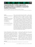
Figure 1 Characteriza tion of the GagG110V mutant. (A) Transduction rate of viruses harboring either GagWT or GagG110 V. 293T cells were
transfected for 48 h with FV vector encoding for GFP together with plasmids expressing Env, Pol and GagWT or GagG110V. Cell free
supernatants were used to transduce 293T cells and the viral titer was determined from the number of GFP-positive cells by FACS analysis 48 h
post-transduction. No infectivity was detected in the supernatant of GagG110V transfected cells, as observed in five independent experiments.
(B) Western blotting performed on 293T cellular extracts and cell free supernatants shows the absence of viral particles in the supernatant of
GagG110V transfected cells whereas intracellular GagG110V is normally produced. (C) Electron microscopy revealed, furthermore, the absence of
intracellular capsids in 293T cells transfected with GagG110V. Bar: 0.5 μm. (D) Subcellular localization of GagWT and GagG110V in Hela
transfected cells with GagWT or GagG110V and analyzed, 24 h post-transfection, by confocal microscopy following indirect immunofluorescence
using rabbit polyclonal anti-PFV. GagWT is either nucleocytoplasmic, cytoplasmic or nuclear whereas GagG110V is mainly nuclear, as observed in
three independent experiments (approximately 200 cells were counted in each preparation). (E) Western blotting performed on fractionated
Hela cell extracts of Gag WT and GagG110V. Detection of the human lactate dehydrogenase (LDH) in cytoplasmic extracts only attests the
validity of the fractionation assay (C: Cytoplasm, N: Nucleus).
Renault et al. Retrovirology 2011, 8:6
/>Page 5 of 11
GagG110V transfected cells relies on efficient nuclear
confinement of the mutant proteins.
The GagG110V mutant harbors dominant negative
properties by sequestrating wild-type Gag in the nucleus
We then asked whether a NES-defective Gag mutant
could negatively interfere with the replication of wild-
type PFV Gag. This was assessed by quantifying recombi-
nant virus production in the presence or in the absence
of a NES-defective Gag mutant in the same system as the
one used in figure 1. PFV-derived vector encoding for
GFP, together with Pol, Gag and Env expressing plasmids
were transfected in 293T cells. Increasing amounts of
either wild-type Gag or a GagG110V expressing plasmids
were co-transfected in parallel experiments. For this
experiment, we used a GagG110V expressing plasmid in
which the Gag open reading frame was fused to the His
and HA tags (named GagHHG110V), since its presence
was easily detected in cell extracts as a higher molecular
size band by Western-blot. Forty-eight hours later, cell
free supernatants were collected and viral titers were
evaluated by FACS following transduction of 293T cells.
Whereas co-transfection of the wild-type GagHH plas-
mid had only a minor effect on virus production, the pre-
sence of GagHHG110V impaired virus release in a dose
dependent manner (Figure 3A). Biochemical an alysis
confirmed this observation since the 71/68 kDa Gag
doublet in cell-free supernatants decreased concomi-
tantly with increasing amounts of GagHHG110V, the lat-
ter was detected as a higher molecular band (Figure 3B).
Note that similar to GagG110V, GagHHG110V was
never detected in cell free supernatants (Figure 3B).
These results demonstrated that the NES-defective Gag
mutant dominantly interferes with viral particle release.
Since PFV Gag-Gag interactions were demonstrated in
the nucleus [39] and given that GagG110V is mainly
confined in the nucleus, we wondered whether the
dominant negative effect of the GagG110V protein relies
on nuclear retention of wild-type Gag proteins via intra-
nuclear Gag-Gag i nteractions. To substantiate this,
HeLa cells were transfected with wild-type Gag fused
with His and HA tags (GagHH) and GFP-GagG110V
expression plasmids, and their respective sub-cellular
localizations were studied by indirect immunofluores-
cence followed by confocal analysis, forty-eight hours
post-transfection. Whereas wild-type Gag expressed
alone showed distinct localizations (data not shown), as
previously reported (Figure 1D), it was mainly restricted
in the nucleus in the presence of GFP-GagG110V
(80% ± 4% of transfect ed cells, Figure 3C). These obser-
vations were confirmed at the biochemical level by
co-immunoprecipitation assays. Whereas wild-type Gag
was detected in both the nuclear and cytoplasmic frac-
tions when expressed alone, i t was mainly restricted in
the nucleus when co-expressed with GFP-GagHHG110V
(Figure 3D).
Altogether, these results demonstrated that the domi-
nant negative property of GagG110V mainly relies on
nuclear retention of wild-type Gag, precluding Gag
nuclear export and subsequent capsid assembly.
The NES of HIV-1 Rev could only partially trans-
complement that of PFV Gag
To assess whether an heterologous LMB-sensitive NES
could functionally trans-complement that of PFV G ag,
the latter was replaced by the NES of HIV-1 Rev. The
A
PFVGag(PrototypeFoamyvirus)
SFVͲ1Gag(SimianFoamyvirus1)
SFVͲ3Gag(SimianFoamyvirus3)
HIVͲ1Rev
RSV Gag
95
LAFQDLDLPEGPLRFGPL
112
89
QAFEDLDVAEGTLRFGPL
10
6
91
LAFDNIDVGEGTLRFGPL
108
73
LQLPPLERLTL
83
L
TD
W
AR
V
REE
L
B
RSV
Gag
LMB(40nM)
+
B
GFPͲGag95Ͳ112 GFPͲGag95Ͳ112/G110V
Ͳ
+
Ͳ
GFP
2
19
L
TD
W
AR
V
REE
L
229
GFP
Merge
GFPͲRevNES
+
Ͳ
+
Ͳ
LMB(40nM)
Merge
GFP
C
GagWT
95
LAFQDLDLPEGPLRFGPL
112
GagL95AAͲͲͲͲͲͲͲͲͲͲͲͲͲͲͲͲͲͲͲͲͲͲͲͲͲͲͲͲͲ
GagF97AͲͲͲͲAͲͲͲͲͲͲͲͲͲͲͲͲͲͲͲͲͲͲͲͲͲͲͲͲͲ
C
NC
N
33%
30%
31%
39%
40%
34%
28%
30%
35%
GagF109AͲͲͲͲͲͲͲͲͲͲͲͲͲͲͲͲͲͲͲͲͲͲͲͲAͲͲͲͲͲ
GagG110VͲͲͲͲͲͲͲͲͲͲͲͲͲͲͲͲͲͲͲͲͲͲͲͲͲVͲͲͲͲ
GagL95A/F97AAͲAͲͲͲͲͲͲͲͲͲͲͲͲͲͲͲͲͲͲͲͲͲͲͲͲͲͲ
Gagȴ95Ͳ112
GagRevNES LQLPPLERLTL
1%
0%
2%
0%
7%
34%
23%
33%
22%
61%
65%
77%
65%
78%
32%
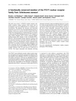
Figure 2 Identification of a functional NES in PFV Gag.
(A) Sequence alignment of a N-terminal region within Gag protein
of primate foamy viruses. (B) Subcellular localization of GFP-Gag
95-112 and derived G110V mutant in Hela cells in the presence or
the absence of LMB (40nM). GFP-RevNES and GFP alone were used
respectively as positive and negative controls. Representative
fluorescence images of the vast majority of cells expressing the
indicated fusion proteins are shown by confocal microscopy.
(C) Amino acid(s) important for Gag nuclear export. Point mutations
or deletion were generated in the context of full length Gag and
the resulting mutants were tested for sub-cellular localization after
24 h transfection using rabbit polyclonal anti-PFV antibodies. Results
concerning Gag-RevNES localization were included. The numbers
shown are the means of three independent experiments by
counting 200 cells each (N: nuclear, NC: nucleocytoplasmic, C:
cytoplasmic localization).
Renault et al. Retrovirology 2011, 8:6
/>Page 6 of 11
resulting Gag chimeric construct, named Gag-RevNES,
was transfected in 293T cells, and its sub-cellular locali-
zation was analyzed. As shown in figure 4A and 2C,
Gag-RevNES displayed a predominant nucleocytoplas-
mic distribution (61% ± 2%). As a control, GagΔNE S
was mainly detected in the nucleus (78% ± 2% of trans-
fect ed cells). Since the NE S of HIV-1 Rev was shown to
restore the nucleocytoplasmic distribution of PFV Gag,
we next assessed whet her the chimeric Gag protein was
able to restore infectivity of recombinant viruses. For
that purpose, wild-type Gag, GagG110V, GagΔNES or
Gag-RevNES expressing plasmids were used to produce
recombinant viruses following transfection o f 293T cells
with a GFP expressing PFV vector together with Env
and Pol expressing plasmids. Forty-eight hours post-
transfection, cell-free supernatant was used to transduce
293T cells, and GFP expression was monitored by flow
cytometry forty-eight hours later. Remarkably, only the
use of wild-type Gag led to the production of infectious
viruses (Figure 4B). Western-blot analysis of cell-free
supernatants from transfected 293T cells demonstrated
the presence of the Gag doublet when wild-type Gag
was used and their absence when using GagG110V or
GagΔNES to produce recombinant viruses, as expected.
Importantly, no Gag doublet was detected when using
the Gag-RevNES construct, whereas these proteins were
efficiently expressed in producer cells (Figure 4C). These
results demonstrated that the heterologous leucine rich
NES of HIV-1 Rev, whic h allowed ef ficient nucleocyto-
plasmic redistribution of PFV Gag deleted from its own
NES, failed to restore infectivity of the corresponding
recombinant viruses.
A
B
Titers(tu/mL)
0248μg
GagHHG110
V
AntiͲGag
GagHHG110V
Gagdoublet
Supernatant
Cellextract
GagHH
GagHHG110V
C
QuantityofDNA(μg)
AntiͲactin
MergeGFP AntiͲHA
80%
GFPͲGagG110V
GagHH
100
75
50
GagHH
GFPͲGagG110V
AntiͲGag
GagHH ++Ͳ Ͳ ++
GFPͲGagG110VͲ Ͳ ++++
D
CNCNCN
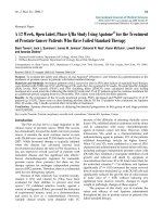
Figure 3 Dominant-negative properties of the GagG110V mutant. (A ) Virus titers. Viral particles were produced in the supernatant of 293T
cells transfected with the four-plasmid PFV vector system in the presence of increasing amounts of GagHH or GagHHG110V. Target 293T cells
were transduced with cell free supernatants and titers were determined by FACS analysis 48 h post-transduction. Viral titers were dramatically
reduced following addition of increasing amounts of GagHHG110V. This result is representative of three independent experiments. (B) Western
blotting also shows a decrease in the amount of Gag proteins in supernatants whereas they are efficiently produced in 293T cells extracts.
Therefore, GagG110V mutant negatively interferes with WT Gag impairing particles production. (C) Co-localization of GagHH and GFP-GagG110V.
Hela cells were co-transfected with indicated plasmids and analyzed, 48 h post-transfection, by confocal microscopy following indirect
immunofluorescence. GagWT colocalizes with GFP-GagG110V in the nucleus in 80 ± 4% of transfected cells in three independent experiments
with approximately 100 cells counted each time. (D) Sequestration of GagWT by GagG110V in the nucleus. Nuclear interaction of GagHH and
GFP-GagG110V revealed by co-immunoprecipitation of nuclear extracts of transfected Hela cells, using mouse anti-HA or anti-GFP antibodies
followed by western-blotting performed with rabbit polyclonal anti-Gag antibodies (N : nucleus and C : cytoplasm).
Renault et al. Retrovirology 2011, 8:6
/>Page 7 of 11
Discussion
The late occurring nuclear targeting of Gag proteins,
which was initially thought to be a specific feature of
PFV [11], was also demonstrated for distinct retroviruses,
such as Rous Sarcoma Virus (RSV) [28] and also for the
retrotransposon Tf1 [40]. Hence, for RSV and PFV, fol-
lowing proviral integration, the late stages of infection
can be divided into an early (synthesis of Gag and its
nuclear translocation) and late (nuclear export of Gag,
capsid assembly and virus egress) phases [41]. We show
here that nuclear export o f PFV Gag proteins relies on a
LMB-sensitive leucine-rich nuclear export sequence
(NES) within the N-terminus of the structural protein.
NES-defective Gag proteins are mainly located in the
nucleus when compared to the ir wild-type counterpart.
Using NES-defective Gag mutants, production of PFV-
derived recombinant viruses was unsuccessful, their
nuclear localization preventing the formation of viral
capsids in the cytoplasm and subsequent virus egress.
Moreover, NES-defective Gag proteins behave as
dominant negative (DN) mutants by sequestrating wild-
type Gag in the nuclear compartment. This DN effect is
reminiscent to what has been already reported in the
case of DN mutants for HIV-1 Rev [ 42-44] or for HIV-1
Gag [45]. Note that the sub-cellular distribution of a
chimeric PFV Gag prot ein, in which the NES of Gag was
replaced with that of HIV-1 Rev, efficiently induces the
nucleocytoplasmic redistribution of the fusion protein.
Remarkably, no extracellular virus was detected when the
Gag chimera was used instead of its wild-type counter-
part for the production of PFV-derived recombinant
viruses (Figure 4C). This substitution could alter the tri-
dimensional structure of PFV Gag, preventing essential
Env-Gag interactions required for virus egress. Alterna-
tively but not exclusively, nuclear export driven by the
NES of HIV-1 may trigger a cytoplasmic localization of
the chimeric Gag pro tein distinct from that of its wild-
type counterpart, preventing subsequent late stag es of
the viral cycle.
Sequential dimerization, oligomerization, and multi-
merization of Gag proteins are finely tuned to regulate
their functions, in particular for proper capsid assembly
and subsequent virus release [1]. PFV Gag-Gag interac-
tions mainly occur via distinct motifs along this polypro-
tein [36,46], including a coiled-coil domain (called CC2)
located in the N-terminal part [39]. We show here that a
NES-defective Gag could retain its wild-type counterpart,
in the nucleus, confirming the existence of Gag-Gag
interactions in this compartment, as recently demon-
strated for RSV Gag [41]. These results are consistent
with our previous observations. Indeed, when PFV Gag
was fused to the promyelocytic leukemia protein (PML),
the chimera was restricted onto PML-nuclear bodies
(NBs), structures belonging to the nuclear matrix [39].
When wild-type Gag, but not a CC2-deleted mutant
which was defective for Gag-Gag interaction, was
expressed in these cells, it delocalized the PML-Gag
fusion from NBs to a diffuse but nuclear staining,
demonstrat ing the existence of nuclear Gag-Gag interac-
tions. These nuclear interactions were demonstrated also
at the biochemical level by co-immunoprecipitation. Of
course, this does not exclude the existence of interactions
that could take place in the cytoplasm, as is also the case
for RSV Gag [41].
What is the role of PFV Gag nuclear stage? In higher
eukaryotic cells, pre-mRNAs are retained in the
nucleus until they are fully spliced (for a review [47]).
Therefore, to overcome this quality control, retro-
viruses have developed different strategies to export
their unspliced or partly spliced mRNAs, hijacking
cellular nuclear export machineries (reviewed in [48]).
Simple retroviruses generally harbor cis-acting
sequences involved in viral RNA nuclear export [49].
In contrast, in most of co mplex retroviruses, small reg-
ulatory proteins deal with this cellular restriction. For
example, HIV-1 encodes Rev, a nucleocytoplasmic
shuttling protein that bridges unspliced and incomple-
tely spliced viral RNAs on the Rev-responsive element
GagȴNES
GagRevNES
Merge
Merge
A
1,0E+06
1,0E+07
B
GagGagGagGag
WT G110V
ȴ
NES R NES
78%
C
40%
60%
61%
L
)
1,0E+00
1,0E+01
1,0E+02
1,0E+03
1,0E+04
1,0E+05
1234
WT
G110V
ȴ
NES
R
ev
NES
Supernatan
t
Cellextract
Gag
WT
Gag
G
11
0
V
Gag
ȴNE
S
Gag
R
e
vNE
S
Titers (tu/m
L
Figure 4 HIV-1-RevNES fails to restore infectivity. (A) Subcellular
localization of GagΔNES and Gag-RevNES in Hela cells analyzed 24h
post-transfection with PFV antibodies by confocal microscopy following
indirect immunofluorescence in three independent experiments
(approximately 200 cells counted each time). (B) Transduction rat e of
viruses harboring eit her GagWT, Ga gG110V, GagΔNES or Gag-Rev NES.
Cell fr ee supern at ants were used to transduce 293T cells an d the vira l
titer w as dete r mined from the n umber of GFP-positiv e cells by FACS
analysis 48h post-transduction. No infectivity was detected in the
supernatants of GagG110V, Gag ΔNES and Gag-RevNES transfected cells
in four independent experi m ents. (C ) Western blotting performed on
293T cell extracts an d cell-free supern atants sh o ws the absence o f v i ral
particles in the supernatants of GagG110V, GagΔNES and Gag-RevNES
transfected cells whereas the intracellular Gag mutants are normally
produced and matured.
Renault et al. Retrovirology 2011, 8:6
/>Page 8 of 11
(RRE) -a cis-acting element located within the env
gene- to CRM1, thanks to its leucine-rich nuclear
export sequence [32]. For theJaagsiekteSheepRetro-
virus (JSRV), an unusually long Env leader peptide
contributes to viral nuclear export [50]. PFV, although
harboring a complex genomic organization, does not
encode a functional Rev-like protein [15] and i ts Env
leader peptide was n ot implicated in nuclear export
but was shown to be involved in Env-Gag interactions
required for virus budding [20].
In the case of RSV, Gag dimerization is promoted by
binding to viral RNA, as already propos ed for other ret-
roviruses [51]. This, which mainly occurs in the nucleus,
triggers a conformational change that unmasks an effi-
cient NES within the p10 domain of the Gag polypro-
tein, resulting in nuclear export of Gag-RNA complexes
[52,53]. Remarkably, prior to Gag synthesis, nuclear
export of intron-containing RNA likely relies on cis-act-
ing direct repeat sequences located in the 3’ end of the
viral genome, involving the cellular TAP/NXF1 and
Dbp5 export factors [54]. The cytoplasmic fate of the
viral genome could rely on the use of one of these two
pathways, leading either to its packaging following Gag-
dependent nuclear export or translation if based on cis-
acting sequences. Indeed, there is a m echanistic link
between retroviral RNA trafficking, in particular the way
it is exported from the nucleus, and viral protein activ-
ities in the cytoplasm, affecting distinct late cytoplasmic
stages such as capsid assembly, genome packaging and/
or virus budding [49,55 -58]. Of note, upon inclusion of
Gag sequences from more distantly related FV species,
such as the one from the feline isolate into the align-
ment, the C-terminal part contains a highly conserved
shor t motif with the PFV Gag G110 residue being 100%
conserved throughout. However, the Gag protein from
the feline foamy virus (FeFV), although detected close to
perinuclear regions, seems to be excluded from the
nucleus [59]. Either nuclear export of FeFV Gag is
extremely efficient and therefore the nuclear stage is not
easily discernible or, alternatively during infection, other
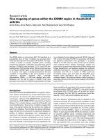
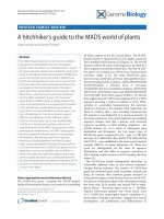
Figure 5 Model for the p ossible nuclear role of FV Gag during the late stages of infection. (1) Full le ngth viral RNA export is still
unknown. (2) After synthesis in the cytoplasm, Gag protein enters the nucleus via its NLS domain (located within the GRII box). In the nucleus,
Gag could interact with the full length viral RNA via its GRI box favoring Gag-Gag interaction and subsequently unmasking Gag NES. (3) The
nuclear export factor, CRM1, also called exportin 1, would then be able to interact with this ribonucleoprotein complex leading to its efficient
nuclear export. (4) In the cytoplasm, Gag proteins will multimerize for capsid assembly near the MTOC. In the absence of Gag proteins, the initial
nuclear export of unspliced PFV RNA could rely on another export mechanism independent of these proteins.
Renault et al. Retrovirology 2011, 8:6
/>Page 9 of 11
viral components are required for nuclear export of
unspliced or partly spliced mRNAs.
Therefore, based on our results, it would be interest-
ing to assess whether PFV Gag proteins could be
involved in this crit ical step, in a way similar to what
was reported for RSV Gag. According to this model,
PFV Gag proteins would bridge the nuclear intro n-con-
taining viral RNAs thanks to the GRI box to CRM1 via
the leucine-rich NES we identified, promoting their
nuclear export (Figure 5). In this context, PFV Gag pro-
teins were effectively shown to interact with CRM1 in
the presence of the PFV RNA packaging signal (preli-
minary results). Interaction b etween Gag and the viral
RNA c ould occur e ither prior to Gag nuclear import or
within the nucleus. In the cytoplasm, following nuclear
export, Gag might transport viral RNAs towards the
MTOC where capsid assembly and Pol packaging take
place [17]. In a viral context, predominant nuclear loca-
lization of a PFV Gag protein deleted from its GR1 box
[9], which was shown to be essential for viral nucleic
acids binding, is in agreement with this working model.
Before Gag synthesis, initial nuclea r export of intron-
containing RNA c ould rely on cis-acting sequences on
viral RNA, as already reported for RSV [54]. Remark-
ably, in that case, it seems that nuclear export is depen-
dent on a structured RNA element and the cellular
RNA-binding protein HuR as well as the adapter mole-
cules ANP32A and B (pp32 and April) [60]. Thus, we
propose that nuclear export of unspliced and partially
spliced PFV RNAs relies on two complementary
mechanisms, which take place successively during the
replication cycle.
Noteaddedinproof:Since the acceptation of this
manuscript, the initial nuclear export pathway of mRNA
PFV has been recently published online ahead of print
on 15 December 2010 by Bodem J et al. [61].
Acknowledgements
We thank Christelle Doliger and Niclas Setterblad at the Imagery and Cell
sorting Department of the Institut Universitaire d’Hématologie IFR 105 for
confocal microscopy. We thank Elisabeth Savariau for the photographic
work. This study is supported by CNRS, Inserm, Université Paris Diderot, ARC,
ANRS, SIDACTION, F. Lacoste. NR is supported by the French Research
Ministry. The authors thank Axel Rethwilm and Dirk Lindemann for providing
some FV reagents.
Author details
1
CNRS UMR7212, Inserm U944, Université Paris Diderot, Institut Universitaire
d’Hématologie, Paris, France.
2
Université François Rabelais- Inserm U966,
Tours, France.
3
Conservatoire National des Arts et Métiers, Paris, France.
Authors’ contributions
AS, NR, JTT conceived and designed the experiments; NR, JP, MLG, PR, AC,
JTT performed the experiments; AS, MLG, JTT analyzed the data; AS wrote
the manuscript.
Competing interests
The authors declare that they have no competing interests.
Received: 7 October 2010 Accepted: 21 January 2011
Published: 21 January 2011
References
1. Freed EO: HIV-1 gag proteins: diverse functions in the virus life cycle.
Virology 1998, 251:1-15.
2. Ganser-Pornillos BK, Yeager M, Sundquist WI: The structural biology of HIV
assembly. Curr Opin Struct Biol 2008, 18:203-217.
3. Martin-Serrano J: The role of ubiquitin in retroviral egress. Traffic 2007,
8:1297-1303.
4. Morita E, Sundquist WI: Retrovirus budding. Annu Rev Cell Dev Biol 2004,
20:395-425.
5. Klein KC, Reed JC, Lingappa JR: Intracellular destinies: degradation,
targeting, assembly, and endocytosis of HIV Gag. AIDS Rev 2007,
9:150-161.
6. Delelis O, Lehmann-Che J, Saib A: Foamy viruses - a world apart. Curr Opin
Microbiol 2004, 7:400-406.
7. Yu SF, Baldwin DN, Gwynn SR, Yendapalli S, Linial ML: Human foamy virus
replication: a pathway distinct from that of retroviruses and
hepadnaviruses. Science 1996, 271:1579-1582.
8. Flugel RM, Pfrepper KI: Proteolytic processing of foamy virus Gag and Pol
proteins. Curr Top Microbiol Immunol 2003, 277:63-88.
9. Yu SF, Edelmann K, Strong RK, Moebes A, Rethwilm A, Linial ML: The
carboxyl terminus of the human foamy virus Gag protein contains
separable nucleic acid binding and nuclear transport domains. J Virol
1996, 70:8255-8262.
10. Stenbak CR, Linial ML: Role of the C terminus of foamy virus Gag in RNA
packaging and Pol expression. J Virol 2004, 78:9423-9430.
11. Schliephake AW, Rethwilm A: Nuclear localization of foamy virus Gag
precursor protein. J Virol 1994, 68:4946-4954.
12. Imrich H, Heinkelein M, Herchenroder O, Rethwilm A: Primate foamy virus
Pol proteins are imported into the nucleus. J Gen Virol 2000, 81:2941-2947.
13. Meiering CD, Comstock KE, Linial ML: Multiple integrations of human
foamy virus in persistently infected human erythroleukemia cells. J Virol
2000, 74:1718-1726.
14. Tobaly-Tapiero J, Bittoun P, Lehmann-Che J, Delelis O, Giron ML, De The H,
Saib A: Chromatin tethering of incoming foamy virus by the structural
Gag protein. Traffic 2008, 9:1717-27.
15. Lee AH, Lee HY, Sung YC: The gene expression of human foamy virus
does not require a post-transcriptional transactivator. Virology
1994,
204:409-413.
16.
Sfakianos JN, LaCasse RA, Hunter E: The M-PMV Cytoplasmic Targeting-
Retention Signal Directs Nascent Gag Polypeptides to a Pericentriolar
Region of the Cell. Traffic 2003, 4:660-670.
17. Yu SF, Eastman SW, Linial ML: Foamy virus capsid assembly occurs at a
pericentriolar region through a cytoplasmic targeting/retention signal in
Gag. Traffic 2006, 7:966-977.
18. Eastman SW, Linial ML: Identification of a conserved residue of foamy
virus Gag required for intracellular capsid assembly. J Virol 2001,
75:6857-6864.
19. Choi G, Park S, Choi B, Hong S, Lee J, Hunter E, Rhee SS: Identification of a
cytoplasmic targeting/retention signal in a retroviral Gag polyprotein.
J Virol 1999, 73:5431-5437.
20. Lindemann D, Pietschmann T, Picard-Maureau M, Berg A, Heinkelein M,
Thurow J, Knaus P, Zentgraf H, Rethwilm A: A particle-associated
glycoprotein signal peptide essential for virus maturation and infectivity.
J Virol 2001, 75:5762-5771.
21. Patton GS, Morris SA, Chung W, Bieniasz PD, McClure MO: Identification of
domains in gag important for prototypic foamy virus egress. J Virol 2005,
79:6392-6399.
22. Stange A, Mannigel I, Peters K, Heinkelein M, Stanke N, Cartellieri M,
Gottlinger H, Rethwilm A, Zentgraf H, Lindemann D: Characterization of
prototype foamy virus gag late assembly domain motifs and their role
in particle egress and infectivity. J Virol 2005, 79:5466-5476.
23. Stange A, Luftenegger D, Reh J, Weissenhorn W, Lindemann D: Subviral
particle release determinants of prototype foamy virus. J Virol 2008,
82:9858-9869.
24. Petit C, Giron ML, Tobaly-Tapiero J, Bittoun P, Real E, Jacob Y, Tordo N, De
The H, Saib A: Targeting of incoming retroviral Gag to the centrosome
involves a direct interaction with the dynein light chain 8. J Cell Sci 2003,
116:3433-3442.
Renault et al. Retrovirology 2011, 8:6
/>Page 10 of 11
25. Saib A, Puvion-Dutilleul F, Schmid M, Peries J, de The H: Nuclear targeting
of incoming human foamy virus Gag proteins involves a centriolar step.
J Virol 1997, 71:1155-1161.
26. McDonald D, Vodicka MA, Lucero G, Svitkina TM, Borisy GG, Emerman M,
Hope TJ: Visualization of the intracellular behavior of HIV in living cells.
J Cell Biol 2002, 159:441-452.
27. Dupont S, Sharova N, DeHoratius C, Virbasius CM, Zhu X, Bukrinskaya AG,
Stevenson M, Green MR: A novel nuclear export activity in HIV-1 matrix
protein required for viral replication. Nature 1999, 402:681-685.
28. Scheifele LZ, Garbitt RA, Rhoads JD, Parent LJ: Nuclear entry and CRM1-
dependent nuclear export of the Rous sarcoma virus Gag polyprotein.
Proc Natl Acad Sci USA 2002, 99:3944-3949.
29. Garbitt-Hirst R, Kenney SP, Parent LJ: Genetic evidence for a connection
between Rous sarcoma virus gag nuclear trafficking and genomic RNA
packaging. J Virol 2009, 83:6790-6797.
30. Kobe B, Kajava AV: The leucine-rich repeat as a protein recognition motif.
Curr Opin Struct Biol 2001, 11:725-732.
31. Kutay U, Guttinger S: Leucine-rich nuclear-export signals: born to be
weak. Trends Cell Biol 2005, 15:121-124.
32. Neville M, Stutz F, Lee L, Davis LI, Rosbash M: The importin-beta family
member Crm1p bridges the interaction between Rev and the nuclear
pore complex during nuclear export. Curr Biol 1997, 7:767-775.
33. Kudo N, Matsumori N, Taoka H, Fujiwara D, Schreiner EP, Wolff B,
Yoshida M, Horinouchi S: Leptomycin B inactivates CRM1/exportin 1 by
covalent modification at a cysteine residue in the central conserved
region. Proc Natl Acad Sci USA 1999, 96:9112-9117.
34. Kudo N, Wolff B, Sekimoto T, Schreiner EP, Yoneda Y, Yanagida M,
Horinouchi S, Yoshida M: Leptomycin B inhibition of signal-mediated
nuclear export by direct binding to CRM1. Exp Cell Res 1998, 242:540-547.
35. Wolff B, Sanglier J, Wang Y: Leptomycin B is an inhibitor of nucleo-
cytoplasmic translocation of the human immunodeficiency virus type 1
(HIV-1) Rev protein and Rev-dependent mRNA. Chem Biol 1997, 4:139-147.
36. Cartellieri M, Herchenroder O, Rudolph W, Heinkelein M, Lindemann D,
Zentgraf H, Rethwilm A: N-terminal Gag domain required for foamy virus
particle assembly and export. J Virol 2005, 79:12464-12476.
37. Lehmann-Che J, Giron ML, Delelis O, Lochelt M, Bittoun P, Tobaly-Tapiero J,
de The H, Saib A: Protease-dependent uncoating of a complex retrovirus.
J Virol 2005, 79:9244-9253.
38. la Cour T, Kiemer L, Molgaard A, Gupta R, Skriver K, Brunak S: Analysis and
prediction of leucine-rich nuclear export signals. Protein Eng Des Sel 2004,
17
:527-536.
39. Tobaly-Tapiero J, Bittoun P, Giron ML, Neves M, Koken M, de The H, Saib A:
Human foamy virus capsid formation requires an interaction domain in
the N terminus of Gag. J Virol 2001, 75:4367-4375.
40. Balasundaram D, Benedik MJ, Morphew M, Dang VD, Levin HL: Nup124p is
a nuclear pore factor of Schizosaccharomyces pombe that is important
for nuclear import and activity of retrotransposon Tf1. Mol Cell Biol 1999,
19:5768-5784.
41. Kenney SP, Lochmann TL, Schmid CL, Parent LJ: Intermolecular
interactions between retroviral Gag proteins in the nucleus. J Virol 2008,
82:683-691.
42. Malim MH, Freimuth WW, Liu J, Boyle TJ, Lyerly HK, Cullen BR, Nabel GJ:
Stable expression of transdominant Rev protein in human T cells
inhibits human immunodeficiency virus replication. J Exp Med 1992,
176:1197-1201.
43. Szilvay AM, Brokstad KA, Kopperud R, Haukenes G, Kalland KH: Nuclear
export of the human immunodeficiency virus type 1 nucleocytoplasmic
shuttle protein Rev is mediated by its activation domain and is blocked
by transdominant negative mutants. J Virol 1995, 69:3315-3323.
44. Szilvay AM, Boe SO, Kalland KH: Co-expression of a trans-dominant
negative mutant of the human immunodeficiency virus type 1 (HIV-1)
Rev protein affects the Rev-dependent splicing pattern and expression
of HIV-1 RNAs. J Gen Virol 1999, 80(Pt 8):1965-1974.
45. Trono D, Feinberg MB, Baltimore D: HIV-1 Gag mutants can dominantly
interfere with the replication of the wild-type virus. Cell 1989, 59:113-120.
46. Mannigel I, Stange A, Zentgraf H, Lindemann D: Correct capsid assembly
mediated by a conserved YXXLGL motif in prototype foamy virus Gag is
essential for infectivity and reverse transcription of the viral genome.
J Virol 2007, 81:3317-3326.
47. Terry LJ, Shows EB, Wente SR: Crossing the nuclear envelope: hierarchical
regulation of nucleocytoplasmic transport. Science 2007, 318:1412-1416.
48. Cullen BR: Nuclear mRNA export: insights from virology. Trends Biochem
Sci 2003, 28:419-424.
49. Swanson CM, Malim MH: Retrovirus RNA trafficking: from chromatin to
invasive genomes. Traffic 2006, 7:1440-1450.
50. Caporale M, Arnaud F, Mura M, Golder M, Murgia C, Palmarini M: The signal
peptide of a simple retrovirus envelope functions as a
posttranscriptional regulator of viral gene expression. J Virol 2009,
83:4591-4604.
51. Muriaux D, Mirro J, Harvin D, Rein A: RNA is a structural element in
retrovirus particles. Proc Natl Acad Sci USA 2001, 98:5246-5251.
52. Scheifele LZ, Kenney SP, Cairns TM, Craven RC, Parent LJ: Overlapping roles
of the Rous sarcoma virus Gag p10 domain in nuclear export and virion
core morphology. J Virol 2007, 81:10718-10728.
53. Gudleski N, Flanagan JM, Ryan EP, Bewley MC, Parent LJ:
Directionality of
nucleocytoplasmic transport of the retroviral gag protein depends on
sequential binding of karyopherins and viral RNA. Proc Natl Acad Sci USA
2010, 107:9358-9363.
54. LeBlanc JJ, Uddowla S, Abraham B, Clatterbuck S, Beemon KL: Tap and
Dbp5, but not Gag, are involved in DR-mediated nuclear export of
unspliced Rous sarcoma virus RNA. Virology 2007, 363:376-386.
55. Jin J, Sturgeon T, Chen C, Watkins SC, Weisz OA, Montelaro RC: Distinct
intracellular trafficking of equine infectious anemia virus and human
immunodeficiency virus type 1 Gag during viral assembly and budding
revealed by bimolecular fluorescence complementation assays. J Virol
2007, 81:11226-11235.
56. Butsch M, Boris-Lawrie K: Destiny of unspliced retroviral RNA: ribosome
and/or virion? J Virol 2002, 76:3089-3094.
57. Sherer NM, Swanson CM, Papaioannou S, Malim MH: Matrix Mediates the
Functional Link between Hiv-1 Rna Nuclear Export Elements and Gag
Assembly Competency in Murine Cells. J Virol 2009, 83:8525-35.
58. Swanson CM, Puffer BA, Ahmad KM, Doms RW, Malim MH: Retroviral
mRNA nuclear export elements regulate protein function and virion
assembly. Embo J 2004, 23:2632-2640.
59. Bodem J, Zemba M, Flugel RM: Nuclear localization of the functional Bel
1 transactivator but not of the gag proteins of the feline foamy virus.
Virology 1998, 251:22-27.
60. Rethwilm A: Molecular biology of foamy viruses. Med Microbiol Immunol
2010, 199:197-207.
61. Bodem J, Schied T, Gabriel R, Rammling M, Rethwilm A: Foamy viral
nuclear RNA-export is distinct from other retroviruses. J Virol 2010, [Epub
ahead of print].
doi:10.1186/1742-4690-8-6
Cite this article as: Renault et al.: A nuclear export signal within the
structural Gag protein is required for prototype foamy virus replication.
Retrovirology 2011 8:6.
Submit your next manuscript to BioMed Central
and take full advantage of:
• Convenient online submission
• Thorough peer review
• No space constraints or color figure charges
• Immediate publication on acceptance
• Inclusion in PubMed, CAS, Scopus and Google Scholar
• Research which is freely available for redistribution
Submit your manuscript at
www.biomedcentral.com/submit
Renault et al. Retrovirology 2011, 8:6
/>Page 11 of 11