Báo cáo y học: "Highly conserved serine residue 40 in HIV-1 p6 regulates capsid processing and virus core assembl" potx
Bạn đang xem bản rút gọn của tài liệu. Xem và tải ngay bản đầy đủ của tài liệu tại đây (1.59 MB, 16 trang )
RESEARC H Open Access
Highly conserved serine residue 40 in HIV-1 p6
regulates capsid processing and virus core
assembly
Jörg Votteler
1†
, Liane Neumann
1†
, Sabine Hahn
1
, Friedrich Hahn
1
, Pia Rauch
1
, Kerstin Schmidt
1
,
Nicole Studtrucker
1
, Sara MØ Solbak
2
, Torgils Fossen
2
, Peter Henklein
3
, David E Ott
4
, Gudrun Holland
5
,
Norbert Bannert
5
, Ulrich Schubert
1*
Abstract
Background: The HIV-1 p6 Gag protein regulates the final abscission step of nascent virions from the cell
membrane by the action of two late assembly (L-) domains. Although p6 is located within one of the most
polymorphic regions of the HIV-1 gag gene, the 52 amino acid peptide binds at least to two cellular budding
factors (Tsg101 and ALIX), is a substrate for phosphorylation, ubiquitination, and sumoylation, and mediates the
incorporation of the HIV-1 accessory protein Vpr into viral particles. As expected, known functional domains mostly
overlap with several conserved residues in p6. In this study, we investigated the importance of the highly
conserved serine residue at position 40, which until now has not been assigned to any known function of p6.
Results: Consistently with previous data, we found that mutation of Ser-40 has no effect on ALIX mediated rescue
of HIV-1 L-domain mutants. However, the only feasible S40F mutation that preserves the overlapping pol open
reading frame (ORF) reduces virus replication in T-cell lines and in human lymphocyte tissue cultivated ex vivo.
Most intriguingly, L-domain mediated virus release is not dependent on the integrity of Ser-40. However, the S40F
mutation significantly reduces the specific infectivity of released virions. Furth er, it was observed that mutation of
Ser-40 selectively interferes with the cleavage between capsid (CA) and the spacer peptide SP1 in Gag, without
affecting cleavage of other Gag products. This deficiency in processing of CA, in consequence, led to an irregular
morphology of the virus core and the formation of an electron dense extra core structure. Moreover, the defects
induced by the S40F mutation in p6 can be rescued by the A1V mutati on in SP1 that generally enhances
processing of the CA-SP1 cleavage site.
Conclusions: Overall, these data support a so far unrecognized function of p6 mediated by Ser-40 that occurs
independently of the L-domain function, but selectively affects CA maturation and virus core formation, and
consequently the infectivity of released virions.
Background
The Gag polyprotein Pr55 of HIV-1 comprises the main
structural components that are essential and sufficient
for the formation of virus like particles (VLPs). Follow-
ing synthesis in the cytoplas m, the Gag polyproteins are
targeted to the plasma membrane where they assemble
into immature budding particles. Concurrent with
assembly and release of nascent virions, the Pr55 Gag
precursor is cleaved by the autocatalytically activated
viral protease (PR), generating the matrix (MA, p17),
capsid(CA,p24),nucleocapsid(NC,p7),andthep6
protein. This processing ultimately leads to structural
rearrangement of Gag molecules within the virion and
the formation of the typical cone shaped core structure,
characteristic for a mature infectious particle [1]. MA
mediates the plasma membrane targeting of Gag poly-
proteins and, after cleavage, lines the inner shell of the
mature virion. CA forms the conical core shell encasing
NC, which regulat es packaging and condensation of the
viral genome [2-6]. The C-terminal p6 domain of Pr55,
* Correspondence:
† Contributed equally
1
Institute of Virology, Friedrich-Alexander-University, Erlangen, Germany
Full list of author information is available at the end of the article
Votteler et al. Retrovirology 2011, 8:11
/>© 2011 Votteler et al; licensee BioMed Central Ltd. This is an Open Access article distribu ted under the terms of the Creative Commons
Attribu tion License ( which permits unrestricted use, distribution, and reproduction in
any medium, provided the original work is prope rly cited.
the smallest known lentiviral protein, containing 52
amino acids, comprises a quite complex structural and
functional organization and contains two distinct late
assembly (L-) domains that regulate efficient separation
of assembled virions from the cell surface. L-domains
function as docking sites for components of ESCRT
(endosomal sorting compl ex required for transport), cel-
lular multi-protein complexes that are normally involved
in the endocytotic recycling of cell surface receptors and
in cytokinesis [4,7-17].
The L-domain activity of p6 is mainly driven by the
7
PTAP
10
motif that is responsib le for the recruitment of
the primary budding factor Tsg101 (tumor susceptibility
gene 101) to the viru s assembly site [ 15,18-20]. Another
region of p6 involves the residues
36
YPLASL
41
, compris-
ing a cryptic YPX
n
L-type L-domain, which forms a
degenerated version of the YPDL L-domain motif
(YPX
n
L, n = 3) found in equine infectious anemia virus
(EIAV). This secondary L-domain in p6 represents a
binding site for another cellular budding factor, AIP1/
ALIX (ALG-2 interacting protein 1/X, ALIX is used
hereafter), a multifunctional ESCRT-associated regulator
of intracellular protein transport [13,21]. In addition to
the interaction with cellular ESCRT c omponents, p6
mediates the incorporation of the HIV-1 accessory pro-
tein Vpr into virus particles. This incorporation was
shown to be dependent on three motifs in p6, the
15
FRFG
18
motif, the
34
ELY
36
motif, and the
41
LXXLF
45
motif [22-24].
Besides these well characterized interactions, p6 con-
tains several highly conserved amino acids, some of
which were shown or proposed to undergo posttransla-
tional modification. In this study, we show that muta-
tion of the highly conserved Ser residue in position 40
(Ser-40) leads to an irregular core assembly of the
released virions, a reduced infectivity, and, thus, a dis-
turbed virus replication capacity. The results support a
novel function of p6 in virus maturation that occurs
independently of L-domain function.
Results
Mutation of Ser-40 has no effect on folding of p6
HIV-1 p6 is located within one of the most polymorphic
regions of the HIV-1 gag gene, yet the 52 amino acid
peptide harbors one of the densest region of protein
interacting domains in HIV-1. Figure 1A gives an over-
view of the previously reported binding domains of cel-
lular (Tsg101, ALIX, ERK-2, SUMO-1, ubiquitin) and
viral (Vpr) proteins within p6 from HIV-1
NL4-3
and their
relationship to the primary and secondary structures
[25]. An alignment of consensus sequences, derived
from all HIV-1 group M subtypes, revealed that the
known f unctional domains overlap - as expected - with
conserved residues in p6.
While the PTAP L-domain is highly conserved, the
conservation within the A LIX binding site varies to
some extent (Figure 1). The ALIX-binding motif has
been defined as (L)[FY]PX
1-3
LXX[IL] [21,26,27] and cor-
responds in p6 derived from HIV-1
NL4-3
[28] to
35
LYPLASLRSL
44
, in which essential residues are in
bold. Interestingly, the three amino acid motif
35
LYP
37
at the start of the b inding region is only poorly con-
served (Figure 1) while both, Leu-41 and Leu-44, are
highly conserved. These two residues together, with the
downstream Phe-45, comprise the LXXLF binding
domain for the HIV -1 accessory protein Vpr. From pre-
vious structural and mutational analysis, it can be con-
cluded that Thr-39 and Ser-40 do not directly
participate in the binding of p 6 to ALIX [21,27,29].
Consistently, Thr-39 is not conserved, while in contrast,
Ser-40 is highly conserved among HIV-1 group M iso-
lates, indicating a function other than ALIX binding.
To investigate a potential function of Ser-40 in p6, the
residue was mutated in the infectious molecular clone
HIV-1
NL4-3
[28] and an otherwise isogenic R5-tropic
derivative thereof carrying the 005pf135 V3 loop region
in Env [30]. In order to obtain replication competent
viruses, the mutation was introduced in a way that does
not affect the overlapp ing pol-ORF. In particular, Ser-40
of p6 overlaps in the pol-ORF with the cleavage site
between the transframe p6*proteinandPR.Theonly
possibilitytoleavethepol-ORF unaffected was to
exchange Ser-40 for Phe, creating the mutant S40F.
Considering this limitat ion in mutating p6, we wanted
proof that the non-conservative S40F exchange does not
disturb the secondary structure of p6 in the respective
region. To ascertain whether the S40F mutation alters
the C-terminal helix of p6, the synt hetic (s)p6
23-52
S40F
peptide was characterised by
1
H NMR spectro scopy
[25]. Recently, we solved the structure of sp6
23-52
by
NMR and found that the C-terminal fragment adopts
the same structure as the full length molecule sp6
1-52
.
Therefore, it was legitimate to analyse the structure of
the m utant and compare it with the wt molecule. After
complete assignment of the
1
H resonances of the NMR
spectra of the peptide, NOE cross peaks important for
secondary structure identification were identified. The
observation of NH
i
-NH
i+1
,NH
i
-NH
i+2
, aH
i
-NH
i+ 2
,
aH
i
-NH
i+ 3
, aH
i
-NH
i+ 4
and aH
i
- bH
i+3
NOEs,
which are indicative of helical secondary structure,
showed that, similarly to wt sp6
23-52
, sp6
23-52
S40F has a
preference for an a-hel ical structure invol ving residues
Ile-31 - Asp-48 under hydrophobic membranous condi-
tions (50% aqueous trifluorethanol (TFE) solution ). Sub-
stitution of Ser-40 with Phe did not change the position
or number of residues included in the C-terminal helix
compared with wt sp6
23-52
, and their structures appear
to be similar. This is confirmed fro m a comp arison of
Votteler et al. Retrovirology 2011, 8:11
/>Page 2 of 16
plots of the a-proton chemical shifts rel ative to those of
random coil values (Figure 2), which indicates that the
substitution slightly stabilizes the region of the
C-terminal helix comprised of residues Leu-41 - Leu-44
proximal to Phe-40.
Mutation of Ser-40 in HIV-1 compromises virus replication
The finding that Ser-40 in p6 is highly conserved raises
the question whether this amino acid is important for
the function of p6 in HIV-1 replication. To investigate
this, comprehensive replication studies of HIV-1 carry-
ing mutation of Ser-40 in p6 were conducted in T cells
and primary lymphocytes. Purified virus stocks of wt
HIV-1
NL4-3
and the S40F mutant were generated in
293T cells and normalized for p24 content. First, paral-
lel cultures of CEM T-cells were infected with 20 and
50 ng of input virus, respectively, and samples of culture
supernatants collected every other day were analyzed for
secretion of virus particles by measuring the virus asso-
ciated reverse transcriptase (RT) activity. The resulting
replication profiles are shown in Figure 3A and 3B.
Mutation of Ser-40 resulted in a diminished rep licatio n
capacity of H IV-1, which is observed as a delayed onset
of virus replication (Figure 3A). Furthermore, increase
of the input virus to 50 ng resulted in a forward shift of
the p eak of virus replication. Yet, the overall virus pro-
duction was still reduced, indicating altogether a com-
promised replication capacity of the Ser-40 mutant of
p6 (Figure 3B). To further evaluate the relevance of
Ser-40 in p6, replication studies were conducted in lym-
phoid cells, derived from human tonsillary tissue, culti-
vated as aggregate cultures (HLAC) [31]. The HLAC
system has been reported to be of comparable value to
that of previo usly described human lymphoid tissue cul-
tures (HLT) where virus replication is studied in tissue
blocks [32,33]. Generally, both HLACs and HLTs, sup-
port productive HIV-1 replication independently of the
corecepto r tropism and do not require artificial exogen-
ous activation of host cells [32,33]. Parallel cultures of
HLAC were infected with X4-tropic or R5-tropic HIV-
1
NL4-3
carrying mutation of Ser-40. Samples of culture
supernatant were harvested every third day, and secre-
tion of viral particles was determined by measuring the
virus associated RT activity. The resulting replication
profiles obtained in tissue samples, derived from two
diff erent donors for X4-tropic strains, are shown in Fig-
ure 3C and 3E, and for R5-tropic variants in Figure 3D
and 3F. Consistent with results obtained in CEM cells
(Figure 3A, 3B), mutation of Ser-40 resulted in a
reduced replication capacity of both X4-tropic and R5-
tropic viruses.
Mutation of Ser-40 in p6 does not affect virus release but
disturbs CA maturation
To analyze whether the decreased replication capacity of
the Ser-40 mutant is due to either a reduction of virus
ERK-2
I S-L L
TTTPSQKQEP
30
N
L4-3
A
1
A
2
B
C
D
I SPP Q
AP
I
-
Q
PEESFRFGEE
20
-A-I-GM
-A-NL-M
-A
-
A
G
SQIDKELY.PLAS
40
LRSLFGSDPS
50
K-R-QDP V-
KTR-P-N-AI-
K-R T-
K
T-
-K N L
-K N L
N
-K L
-
K
N
L
P
P T
N
LQSRPEPTAP
10
P
i
I PR T
M P L
D
F
1
G
H
K
I
.
Q
I P Q
IA P Q
AG
-A G-R
-A G
-A G
-A G
K
.
T
K-EG P
KE
K P
K QGP T-
KNL
-K N
-K
N L
-K N L
N
LY PLAS LLRS
4
0
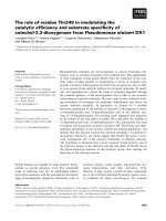
Figure 1 Location and conservation of important functional sites in HIV-1 p6. Primary sequence of p6 derived from the isolate HIV-1
NL4-3
[28] and structural domains according to previous work [25]. Indicated are previously identified (ERK-2) phosphorylation sites [55], attachment
sites for ubiquitin (Ub) [56] and SUMO-1 [57], and binding domains for Tsg101 [15,18,20,58], ALIX [13] and Vpr [22]. The consensus sequences of
p6 proteins derived from the M-group viruses were aligned and conserved residues are boxed in grey.
Votteler et al. Retrovirology 2011, 8:11
/>Page 3 of 16
particle production or to a loss of infectivity of the
released virions, we first investigated whether Ser-40 is
somehow involved in the L-domain mediated assembly
and release of virus particles. Virus release kinetics were
studied by pulse chase m etabolic labeling experiments.
Parallel cultures of HeLa cells were transfected with the
env-deleted HIV-1
NL4-3
subgenomic expression vector
pNLenv1 [34,35], encoding either wt p6 or the S40F
mutant. Cells were pulse labeled with [
35
S]-methionine
for15minutesandchasedforupto4hours.Ateach
time point indicated, samples of cells were harvested,
and V LPs released into the supernatants were collected
by centrifugation and processed fo r immunoprecipita-
tion with Gag-specific antibodies. Immunoprecipitated
proteins we re separated by SDS-PAGE and analyzed by
fluorography (Figure 4A). T he amounts of Gag recov-
ered from virus and cell lysates were quantified by
image analysis. The release of VLPs was calculated as
percentage of Gag proteins found in the virus fraction,
relative to the tot al amount of Gag recovered from virus
and cell fraction s and plotted as a function of time
(Figure 4B). Clearly, virus release was not affected by
mutation of Ser-40. However, quantification of the CA
processing products p25 and p24 revealed a significant
reduction in the processing rate of p25 to p24 for the
S40F mutant (Figure 4C). Thus, Ser-40 somehow regu-
lates the proteolytic maturation of the CA-SP1 Gag pro-
cessing intermediate, without affecting the overall
efficiency of virus release.
S40F mutation reduces specific infectivity of the virions
Having shown that mutation of Ser-40 in p6 does not
affect L-domain mediated virus release but disturbs
maturation of CA, we analyzed the role of this Ser residue
in maturation of progeny virions. To measure the specific
infectivity of the mutant, HeLa TZM-bl cells were infected
with individual virus stocks standardized for p24 content
and infectivity was determined by b-galactosidase assay.
Consistent with virus replication data, mutation of Ser-40
reduced the infectivity by approximately 6-10 fold when
compared to the wt virus (Figure 5).
Mutation of Ser-40 has no effect on ALIX mediated virus
release
Ser-40 residue is located within the previously identified
36
YPLASL
41
ALIX binding sequence, although shown
0,400
0
,5
00
s
p
6
23-52
wt
-0,300
-0,200
-0,100
0,000
0,100
0,200
0,300
D-shift ppm
p
Į2
-0,500
-0,400
T PSQKQEP I DKELYPLAS LRSLFGSDPSSQ
0,100
0,200
0,300
0,400
0,500
hift ppm
sp6
23-52
S40F
23 5
2
Į2
-0,500
-0,400
-0,300
-0,200
-0,100
0,000
TPSQKQEPI DKELYPLAF LRSLFGSDPSS
Q
D-s
23 5
2
Figure 2 Mut ation of Ser-40 does neither affect the secondary structure of p6, nor the interaction with ALIX. A) Chemical shift
differences (ppm) of the a-protons between the experimental values and those for residues in a random coil for sp6
23-52
wt and sp6
23-52
S40F in
50% aqueous TFE at pH 3 at 300 K. All positive values for N-terminal residues adjacent to proline residues at positions 24, 30, 37 and 49 arise
from an inherent effect of proline and not out of a structural perturbation. This was explained in detail previously [25].
Votteler et al. Retrovirology 2011, 8:11
/>Page 4 of 16
Mock
S
4
0
F
w
t
13579111315
0
1
2
3
4
5
3691215
0, 0
0, 1
0, 2
0, 3
0, 4
135791113
0
2
4
6
8
10
12
14
3691215
0,0
0,2
0,4
0,6
0,8
1,0
3691215
0, 0
0, 1
0, 2
0, 3
0, 4
3691215
0,0
0,1
0,2
0,3
CEM, 20 ng
virus input
CEM, 50 ng
virus input
HLAC donor A
X4-tropic
HLAC donor A
R5-tropic
HLAC donor B
X4-tropic
HLAC donor B
R5-tropic
AB
CD
EF
RT activity [cCPM x 10
3
]
days post infection
RT activity [cCPM x 10
3
]
days post infection
RT activity [cCPM x 10
3
]
days post infection
RT activity [cCPM x 10
3
]
days post infection
RT activity [cCPM x 10
3
]
days post infection
RT activity [cCPM x 10
3
]
days post infection
3691215
0, 0
0, 1
0, 2
0, 3
0, 4
Figure 3 Replication of HIV-1
NL4-3
wt and S40F mutant in CEMs and HLAC. CEM cells were inoculated with purified virus equivalent to
20 ng (A) or 50 ng (B) of p24 bearing indicated mutations, and medium was collected every two days. For replication analyses in HLAC, X4
tropic (C and E) and R5 tropic (D and F), 1 ng of p24 of purified virus carrying indicated mutations was used for infection and release of viral
particles was determined in the supernatant every third day (C and D: donor A, E and F: donor B).
Votteler et al. Retrovirology 2011, 8:11
/>Page 5 of 16
not to be involved in the interaction with ALIX directly
[21,29]. It was further reported that mutation of either
Leu-41 or Leu-44 directly adjacent to Ser-40 exhibit a
similar phenotype to that observed for the S40F mutant
in terms of CA processing [36]. Therefore, it was legiti-
mate to investigate whether mutation of Ser-40 affects
the functional interaction between ALIX and p6. We
first examined the ability of ALIX to rescue the release
of an HIV-1
ΔPTAP
L-domain mutant b y overexpression
in the absence of the Ser-40 [21, 37]. The S40F mutation
was cloned into a variant of HIV-1
NL4-3
where the
PTAP motif was replaced by LIRL (HIV-1
ΔPTAP
[38]).
293T cells we re cotransfected with plasmids encoding
ALIX and either HIV-1
ΔPTAP
or HIV-1
ΔPTAP/S40F
.As
control, cells were cotransfected with HIV-1
ΔPTAP/ΔYP
,
where the ALIX binding site YPX
3
L was replaced by
SRX
3
L. As previously reported, this mutation completely
abrogates ALIX mediated rescue of release of HIV-
1
ΔPTAP
variants [21]. Gag processing and release of
infectious virions were determined 24 hours post trans-
fection by Western blot and single round infection of
TZM-bl cells. The results shown in Figure 6A demon-
strate that in the presence of the intact ALIX binding
site in p6, overexpression of ALIX dramatically stimu-
lated virus release by more than 10-fold as shown by
Western blot and single round infection of TZM-bl cells
(Figure 6A, lanes 1 and 2). Consistent with previous
results [21,27], the mutation of the minimal ALIX bind-
ing motif further reduced virus release even below the
residual budding of an HIV-1
ΔPTAP
mutant (Figure 6A,
lane 5), and completely prevents rescue of the HIV-
1
Δ PTAP
variant by overexpression of ALIX (Figure 6A,
lane 6) [21]. Mutation of Ser-40 also reduced the resi-
dual release of the HIV-1
ΔPTAP
mutant (Figure 6A, lane 3).
In contrast to the ΔYP mutation, in the presence of the
S40F mutation, overexpression of ALIX still efficiently res-
cued the release of the HIV-1
ΔPTAP
mutant (Figure 6A,
lane 4 upper panel). However, overexpression of ALIX did
AB
Pr55
Cell VLPs
00.51 2 4 00.51 24
[h]
wt
20
30
40
50
% VLP release
C
p24
p25
Cell VLPs
0
05
1
2
4
0
05
1
2
4
[h]
S40F
01234
0
10
wt
S40F
Time [h]
Pr55
0
0
.
5
1
2
4
0
0
.
5
1
2
4
[h]
5
10
15
20
25
o
cessing in VLPs (p24/25)
wt
S40F
p24
p25
01234
0
5
CA pr
o
Time [h]
Figure 4 Gag processing and release of virions. (A) Phosphorimages of SDS-PAG E gels of immunoprecipitations of
35
S pulse-chase-labeled
Gag protein immunoprecipitates are presented for cell and viral lysates from HeLa cells transiently transfected with either pNLenv or pNLenv
S40F. (B) Percentage of Gag released from the cell presented as the amount of Gag found in virus particles versus the total amount of Gag
recovered form cell and virus lysates. (C) The rate of CA processing was estimated by calculating the ratio of mature CA p24 versus the CA
precursor p25 detected in the cell and virus lysates at different time points.
Votteler et al. Retrovirology 2011, 8:11
/>Page 6 of 16
not restore the infectivity of the S40F mutant virions (Fig-
ure 6A, lane 4 lower panel), indicating that the loss of
infectivity induced by mutation of Ser-40 occurs indepen-
dently of the ALIX-p6 interaction.
To further uncouple the phenotype induced by S40F
mutation from the underlying L-domain function of the
ALIX binding site, we investigated whether ALIX can
rescue the infectivity of t he S40F mutant in the context
of a functional PTAP motif. To this end, 293T cells
were co-tranfected with HIV-1 encoding either wt p6 or
the S40F mutant and ALIX. Release of infectious vir ions
was determined 24 hours post transfection by single
round infection of TZM-bl cells. Consistent with pre-
vious results, the S40F mutation reduced the infectivity
of released virions by ~5-fold (Figure 6B, 1 and 3).
While overexpression of ALIX had no significant influ-
ence on the infectivity of wt HIV-1 (Figure 6B, 2), ALIX
also could not restore the reduced infectivity of the
S40F mutant (Figure 6B, 4).
In order to determine whether the ALIX mediated
rescue of the HIV-1
ΔPTAP
variant of the S40F is still
comparable to the control, we measured the require-
ment of ALIX for HIV-1
ΔPT AP
release at varying ALIX
concentrations. 293T cells were cotransfected with HIV-
1
ΔPTAP
and increasing amounts of ALIX expression
plasmids. S ubsequent determination of virus release by
Westernblot(Figure7A)showedthatinthepresence
of the S40F mutation similar amounts of ALIX were
required to stimulate virus release (Figure 7A and 7B).
Notably, ALIX substantially improved the processing of
CA of the HIV-1
ΔPTAP
mutant. In contrast, CA proces-
sing of the HIV-1
ΔPTAP/S40F
mutant was further
impaired, compared to the HIV-1
ΔPTAP
mutant, and was
/
S40F
/
'YP
A
FLAG-ALIX
RP0
Pr55
ALIX
NL4-3
'PTAP
'PTAP
/
'PTAP
/
-+++
Cell
anti-RP0
Cell
anti-FLAG
p25
p24
Cell
anti-Gag
p24
p24
Virus
anti-Gag
123456
B
Figure 6 S40F mutation has no effec t on ALIX mediated virus
release. (A) Rescue of budding of the Ser-40 mutant by ALIX. Virus
release, Gag processing, and exogenous expression of ALIX were
analyzed by Western blot (upper panel). Ribosomal P0 antigen (RP0)
was used as loading control. The amounts of infectious units
released from the cells were analyzed by b-galactosidase
quantification after infection of TZM-bl cells (lower panel). Shown
are virus infectivities relative to the HIV-1
ΔPTAP
control ± SD. Lane 1
shows HIV-1
ΔPTAP
mutant cotransfected with the empty control
vector, lane 2 shows HIV-1
ΔPTAP
mutant cotransfected with a FLAG-
ALIX expression plasmid, lanes 3 and 4 show the HIV-1
ΔPTAP/ΔYP
double mutant cotransfected with the empty control plasmid and
the vector expressing V5-POSH, respectively, lanes 5 and 6 show the
HIV-1
ΔPTAP/ΔYP
double mutant cotransfected with the empty control
plasmid and the vector expressing V5-POSH, respectively. (B)
Overexpression of ALIX does not rescue infectivity of the S40F
mutant. The amounts of infectious units released from the cells
analyzed were by b-galactosidase quantification after infection of
TZM-bl cells. Shown are virus infectivities relative to the HIV-1
control ± SD. 1: HIV-1 contransfected with the empty control vector,
2: HIV-1 cotransfected with a FLAG-ALIX expression plasmid, 3: HIV-
1
S40F
cotransfected with the empty control vector, 2: HIV-1
S40F
cotransfected with a FLAG-ALIX expression plasmid.
Figure 5 Specific infectivity of HIV-1
NL4-3
wt and p6-mutants.
TZM-bl cells were infected with purified virus preparations
standardized for p24. Infectious titers were determined by
measuring b-galactosidase activity as described in Materials and
Methods (RLU: relative light units).
Votteler et al. Retrovirology 2011, 8:11
/>Page 7 of 16
NL4-3
ALIX
1
2
4
8
1
2
4
8
'PTAP 'PTAP/S40F
A
ALIX
0
0.
1
0.
2
0.
4
0.
8
0
0.
1
0.
2
0.
4
0.
8
μg
Cell
anti-FLAG
Pr55
FLAG-ALIX
RP0
cell
anti-RP0
Cell
anti-Gag
CA
Virus
anti-Gag
CA
CA
B
C
Figure 7 Mutation of Ser-40 has no effect on ALIX mediated virus release. (A) 293T cells were cotransfected with HIV-1
ΔPTAP
or HIV-1
ΔPTAP/
S40F
and increasing amount of ALIX plasmid. (B) Quantification of Western blot data using AIDA Software (Raytest). Shown is the amount of Gag
release determined by the ratio of virus associated Gag/total Gag expressed. (C) The amount of infectious units released from the cells was
analyzed by b-galactosidase quantification after infection of TZM-bl cells.
Votteler et al. Retrovirology 2011, 8:11
/>Page 8 of 16
not rescued by overexpression of ALIX (Figure 7A).
This was further supported by the notion that the infec-
tivity of the S40F mutant virions was substantially
reduced and could not be restored by overexpression of
ALIX (Figu re 7C). Taken together, the data indicate that
the interaction of p6 with ALIX is not affect ed by repla-
cing the conserved Ser-40 by Phe, and the phenotype
induced by this mutation occurs independently of the
ALIX mediated L-domain function of p6 in this region.
The S40F mutation does neither affect cleavage of Gag
products, other than CA, nor incorporation of Env
In order to rule out the possibility that mutation of Ser-
40 also affects processing of Gag proteins other than
CA, virus preparations were analyzed by Western blot-
ting using antibodies specific for NC or MA. Virus par-
ticles prepared by transient transfection of 293T cells
were purified by centrifugation through a 20% sucrose
cushion and standardized for p24 content by ELISA.
Equal amounts of p24 were loaded on SDS-PAGE and
analyzed by Western blotting. The amount of MA and
NC proteins in S40F mutant viruses was similar to that
of wt virions, indicating that maturation of t hese Gag
proteins is not affected by the S40F mutation (Figure
8A). Even with the relativ ely low resolution of the SDS-
PAGE system, the disturbed processing of CA from p25
to p24 was detectable (Figure 8A).
It was shown previously that mutations of p6 in this
area, in particular, mutations of Tyr-36 and Leu-41 pro-
duce mutants that fail to package Env proteins into
virus particles [39]. Since Ser-40 is located directly adja-
cent to Leu-41, we wanted to exclude that the reduced
replication capacity and in fectivity of the S40F mutant is
due to reduce d Env incorporation. To this end, purified
virions standardized for p24 content were analyzed by
Western blotting using Env specific antibodies. As
shown in Figure 8B, the S40F mutation had no influence
on Env incorporation into virus particles.
Electron microscopy analysis of p6 S40F mutants
Next, we examined the effects of the S40F mutation on
assembly, release, and virion morphology by thin-section
electron microscopy (Figure 9). HeLa cells transiently
transfected with plasmids encoding HIV-1
NL4-3
and
mutants thereof were drawn into cellulose capillary
tubes 24 hours post transfection. The cellulose capillary
tubes retain secreted virions, thereby obviating the need
for centrifuga tion steps that usually affect the native vir-
ion structure. In agreement with our biochemical data,
accumulation of virions tethered at the cell membrane,
a phenot ype commonly observed for L-domain mutants
in p6, was not o bserved for the S40F mutant (data not
shown). However, and most intriguingly, mutation of
Ser-40 in p6 led to the formation of aberrant virus parti-
cles, c haracterized by irregularly shaped viral cores and
the formation of closely neighboring electron-dense lat-
eral bodies, as indicated in Figure 9A. These irregularly
shaped cores and lateral bodies were not observed in
the wt and the ΔYP mutant, again indicating that this
phenotype occurs independently of the ALIX-Gag inter-
action. As we observed a deficiency in CA processing of
p25 to p24 in the virions containing the S40F mutation
(Figure 9), we investigated whether this defect in virus
core assembly is a consequence of imperfect CA proces-
sing. A previously characterized CA5 mutan t was gener-
ated in HIV-1
NL4-3
that is incapable of processing p25
to p24 and was shown to exhibit a similar defect of core
assembly [40]. Indeed, the phenotype observed for CA5
M
ock
w
t
S
40F
w
t
S
40F
A
B
M
w
S
anti-CA
anti-MA
CA
MA
w
S
anti-Env
anti-CA
gp120
CA
ant
i
-N
C
NC
Figure 8 Analysis of Gag processing products and Env incorporation in wt HIV-1 and S40F mutant virions. (A) Analysis of Gag processing
in VLPs. VLPs produced in 293T cells transiently transfected with pΔR and pΔR
S40F
were purified and analyzed by Western blot using antibodies
against CA, MA and NC. (B) Analysis of Env incorporation in VLPs. VLPs produced in 293T cells transiently transfected with pΔR and pΔR S40F
were purified and analyzed by Western blot using antibodies against Gag and Env.
Votteler et al. Retrovirology 2011, 8:11
/>Page 9 of 16
A
B
B
t
YP
CA5
w
t
'
S40F
S40F
CA5
wt
'YP
Figure 9 Core morphology of wild type and mutant HIV-1 particle s. (A) Electron micrographs showing thin sections of HeLa SS6 deri ved
extra cellular particles of HIV-1
NL-43
wt, the S40F mutant, the CA-SP1 Gag cleavage deficient mutant CA5, and the ALIX binding site mutant ΔYP.
A higher-magnification view of representative particles illustrates the dominating core structures. (B) Quantitative assessment of the relative
amount of particles with regular and irregular core morphology. About 100-120 unselected particles of the wt virus and each mutant were
evaluated. The data are representative for four independent experiments.
Votteler et al. Retrovirology 2011, 8:11
/>Page 10 of 16
by analyzing the core structures of this mutant clearly
resembles that of the S40F mutant (Figure 9A).
To evaluate quantitatively this morphological phenom-
enon, cores of 100 - 120 viron s were counted and the
percentages of irregular core structures relative to wt
virions were calculated (Figure 9B). Obviously, mutatio n
of Ser-40 significantly increases the amount of virions
containing aberrant, irregularly shaped virus cores.
Moreover, the CA5 mutant, in which CA processing is
blocked completely, shows the same phenotype as that
of the S40F mutant, further supporting the notion that
Ser-40 governs the processing of CA by a yet unidenti-
fied mechanism.
Defect in CA maturation of the S40F mutant can be
rescued by mutation in the CA-SP1 cleavage site
As described above, the S40F mutation increases the ratio
of p25 to mature p24. Our data, together with previous
results from others, suggest that this disturbed CA pro-
cessing subsequently leads to an irregular morphology of
the virus core and thus, to reduced virus infecti vity of the
virions. This prompted us to investigate, whether restor-
ing the CA processing by introducing specific mutations
into the CA-SP1 cleavage site can rescue the defects in
core assembly and infectivity induced by the S40F muta-
tion. It is known that the affinity of the PR to the clea-
vage site between CA and SP1 is weak compared to other
proteolytic cleavage sites in Gag [41,42]. The A1V muta-
tion in the SP1, which previously was identified to confer
resista nce to the CA-maturation inhibitor Bevirimat [43],
enhances the affinity of the viral protease to the CA-SP1
cleavage site. Thus, the A1V mutation in SP1 was intro-
duced into the HIV-1
NL4-3
backbone in combination with
the S40F mutation in p6. Western blot analysis of puri-
fied virions revealed that, by introducing the A1V muta-
tion, CA processing is substantially enhanced in both, the
wt and the S40F mutant virions (Figure 10A). Further-
more, the A1V/S40F mutant displayed a similar CA pro-
cessing compared to the wt.
Consequently, we wanted to examine whether this
enhanced CA processing affects virus core assembly.
Therefore, virion structure of the A1V mutants was ana-
lyzed by thin-section electron m icroscopy. Cores of
100 - 120 virons were counted and the percentages of
irregular core structures relative to wt virions were
calculated (Figure 10B). The S40F mutation again sub-
stantially increases the amount of virions containing
aberrant, irregularly shaped v irus cores. The A1V muta-
tion had only marginal effects o n the core morphology
of wt HIV-1. However, in the case of the S40F mutant,
introducing the A1V mutation largely improves virus
core assembly (Figure 10B).
Since enhancing the CA processing rate rescues the
defect of viral core assembly, we subsequently wanted to
analyze whether this improved core form ation also
affects the infectivity of the virions. To measure the spe-
cific infectivity, HeLa TZM-bl cells were infected with
individual virus stocks standardized for p24 content and
infectivity was determined by b-galactosidase assay.
4
0F
A Western blot
wt
S40F
A1V
A1V/S
4
Mock
p25
p24
50
60
70
regular
irregular
B Electron microscopy
20
30
40
50
%
wt S40F A1V A1V/S40F
0
10
C Infectivity
wt
'YP
Figure 10 Rescue of CA matu ration by A1V m utation in CA-
SP1 cleavage site. (A) Western blot analysis of CA processing in
VLPs. VLPs derived from 293T cells transfected with pΔR and pΔR
S40F
in combination with the A1V mutation in SP1 were purified and
analyzed by Western blot using antibodies against CA. (B)
Quantitative assessment of the viral core morphology derived from
electron micrographs for the above indicated mutants. About 100-
120 unselected particles of the wt virus and each mutant were
evaluated. (C) Specific infectivity of HIV-1
NL4-3
wt and S40F mutant
in combination with the A1V mutation. TZM-bl cells were infected
with virions derived from pNL4-3 wt and S40F in combination with
SP1 A1V and infectious titers were determined by measurement of
the b-galactosidase activity (RLU: relative light units).
Votteler et al. Retrovirology 2011, 8:11
/>Page 11 of 16
The A1V mutation alone enhances the specific infectiv -
ity of the virons by 4-fold (Figure 10C) . Introducing the
A1V mutation enhances the sp ecific infectivity of the
virions of the otherwise attenuated S40F mutant to
almost wt levels (Figure 10C), indicating that the defi-
ciency in CA processing is the m ajor determinant for
the reduced infectivity of the S40F mutant.
Discussion
In this study we demonstrat e that mutation of the highly
conserved Ser-40 interferes with Gag processing and
virus core formation. Although Ser-40 in HIV-1 p6 is
highly conserved among HIV-1 isolates, it is not in volved
in any of the functional motifs described so far, neither
the ALIX nor the Vpr binding site. Therefore, it was
legitimate to speculate that Ser-40 is involved in another ,
until now unrecognized function of p6. However, it
should be noted that the position of Ser-40 in the nucleo-
tide sequence of HIV-1 overlaps with the p6*/PR cleavage
site in the overlapping pol-ORF, which limits the prob-
ability of mutations in this respective area and might
contribute to the high conservation of this amino a cid.
Nevertheless, our findings of a compromised replication
capacity and reduced infectivity of the Ser-40 mutant
viruses support the assumption that Ser-40 has an impor-
tant function directly associated with p6.
Notably, virus release kinetic was not reduced for t he
S40F mutant providing first evidence that the L-domain
function of p6 is not affected by mutation of Ser-40.
This was supported further by the observation that the
ability of ALIX to rescue HIV-1
ΔPTAP
L-domain mutant
viruses was not influenced by the S40F mutation.
Although Ser-40 is located within the ALIX b inding
region in p6 , previous structural investigations indicated
that Ser-40 itself does no t participate in the binding o f
p6 to ALIX [21,27,29]. The fact that the Ser-40 mutant
was still full y active in terms of L-domain function
further supports the notion, that the mutation
introduced into p6 did not disturb the overall
structure of the molecule in this respective region.
In consistency, structural calculat ions indicated that the
non-conservative exchange of Se r-40 to Phe does not
change the ability of the molecule to ado pt a helical
structure. In fact, the C-te rminal a-helix is conserved in
the S40F mutant, as it was established by NMR studies
of the C-terminal peptides sp6
23-52
.
Yet, as shown p reviously, the phenotypes induced by
mutations in the ALIX bind ing site are depending on
the type of amino acid that is mutated [36]. Mutation of
the
35
LYP
37
sequences in the ALIX binding site reduces
release and in fectivity of HIV-1 virions, which otherwise
exhibit normal Gag processing [27,36]. In contrast,
mutations of Leu-41 and Leu-44 have no impact on
virus relea se but increase the ratio of p25 to mature
p24, similar to the phenotype we observed for mutation
of Ser-40 [36]. Unlike the typical phenotype of L-
domain mutants, these mutants interfere somehow spe-
cifically with the final step in maturation of CA. It is
currently not clear, whether this phenotype is in some
way associated with the ALIX-p6 interaction. While
mutations of Leu-41 and Leu-45 disrupt binding to
ALIX, mutation of Ser-40 apparently has no influence
on this inte raction. Thus, it might also be possible, th at
this area in p6 harbors another function that, indepen-
dent of L-domain activity, requires a so far unrecognized
cellular interaction partner.
Maturation of the Gag processi ng intermediate p25 to
mature CA p24, e. g. the cleavage of the CA-SP1 junc-
tion by the P R, appears to be one of the last steps of
Gag processing [41,42]. It was previously demonstrated
that mutating the junction between CA and SP1, in
order to block cleavage of p25, leads to the production
of noninfectious viral particles with aberrant core mor-
phology [40]. In addition, treatment of virus producing
cells with 3-O-(3’ -3’ dimethylsuccinyl) betulinic acid
(BVM, Bevirimat, also known as PA-457 or DSB), a spe-
cific inhibitor that blocks PR-mediated cleavage between
CA and SP1, disturbed viral core formation [43]. Intri-
guingly, mutation of Ser-40 leads to an almost identical
aberrant virus core morphology as shown previous ly for
CA5 mutants. Both are characterized by misshapen co re
structures and the formation of an electron dense lateral
body near the viral membrane. Previous studies already
indicated that maturation of HIV-1 virions, l eading to
the typical cone shaped cores, is regulated by the
sequential, and highly ordere d proteolytic cleavage of
Gag [40]. Apparently, the last step of Gag processing -
the cleavage of the CA-SP1 junction - is required for
capsid condensation. However, pulse chase data indicate
that the kinetic of cleavage of the CA-SP1 junction is
delayed, but not completely blocked inasmuch as the
mature CA accumulates over time. This indicates a
rather dynamic process in which the mutation of Ser-40
somehow delays the kinetic of CA maturation. This phe-
nomenon correlates with deficiencies in CA processing,
infectivity, and core morphology.
Recently published results from Müller et al. demon-
strate that even low amounts of Gag processing inter-
mediates interfere with HIV particle maturation in a
trans-dominant manner, with the final cleavage between
p24 and SP1 being of particular importance [44]. This
explains why the rather subtle effect on CA maturation
detected for Ser-40 mutants by Western blotting and
pulse chase analysis results in a substantial reduction of
virus core formation.
Interestingly, the effect of the S40F mutation appears
to be specific for the CA-SP1 cleavage inasmuch as
i) no other Gag processing defi ciency could be detected
Votteler et al. Retrovirology 2011, 8:11
/>Page 12 of 16
(Figure 6) and ii) enhancing CA processing by introdu-
cing the A1V mutation could restore the deficient core
formation, and, consequently, enhanc e infectivity. Thus,
it can be concluded that Ser-40 somehow regulates the
cleavage of the CA-SP1 junction and the subsequent
capsid condensation.
The molecular mechanism behind how Ser-40 regu-
lates the processing of Gag, in part icular the cleavage of
the CA-SP1 junction, is still elusive so far. The pre-
viously described defects in Gag processing commonly
observed for L-domain mutants are believed to be linked
to the overall process of virus budding inasmuch as PR
activation and subsequent Gag processing occur conco-
mitantly with and shortly after release of virus particles
[45,46]. In the case of Ser-40, this can be excluded, as
the mutant S40F exhibits wt budding. However, Ser-40
in p6 and the CA-SP1 junction are separated by 123
amino acids in Pr55 and it remains elusive so far, how
both proteins can affect each other, either in the context
of Pr55 or after Gag processing. Our NMR experiments
demonstrated that the C-terminal structure of p6 is not
influenced by the S40F mutation. Therefore, one possi-
bility of the effect observed would be that t he mutation
affects the Gag structure prior to initiation of Gag pro-
cessing, thereby reducing the cleavage efficiency of the
weakest cleavage site in Pr55. A prerequisite for this
scenario would be that p6 represen ts a structured Gag
domain and thus influence the folding the Pr55 polypro-
tein. Even though the 283 residue N-terminal part of
HIV-1 Gag including MA and CA has been solved by
NMR [47], the structure of the complete Pr55 has not
been determ ined hitherto. Although S40F does not
appear to affect the folding of the mature p6 protein, we
can not exclude that this mutation indeed affect the
overall struct ure of the PR55 polyprotein, which in turn
would reduce the processing efficiency, a phenotype we
clearly observed for the S40F mutant as a novel function
of p6.
Currently, there is no evidence of an intra-molecular
interaction between these domains in the Pr55 polypro-
tein. The p6 domain of the Pr55 represents a docking
site of several cellular and viral factors. Thus, since an
intramolecular interaction between CA and p6 appears
to be unlikely, it is conceivable to hypothesize that p6
harbors another interaction domain of a yet unknown
factor that, independently of the L-domains, regulates
processing of CA.
Conclusions
Overall, these data support a so far unrecognized func-
tion of p6 that occurs independently of the L-domain
function, does not affect virus release, but selectively
affects CA maturation, virus core formation, and thus,
infectivity.
Methods
Peptide synthesis and purification
The synthesis, purification and molecular characteriza-
tion of p6 and the related fragments derived from HIV-
1
NL4-3
have been described in detail previously [25].
NMR Spectroscopy
2D
1
H Total Correlation Spectroscopy (TOCSY), Corre-
lation Spectroscopy (COSY) and Nuclear Overhauser
enhancement spectroscopy (NOESY) NMR experiments
were performed at 600.13 MHz on a Bruker Avance
600 MHz instrument equipped with an UltraShield Plus
magnet and a triple resonance cryoprobe with gradient
unit. Individual samples were dissolved in 600 μl50%
aqueous TFE-d2 at concentrations between 1-2 mM.
The 2D NMR experiments w ere performed at 300 K
without spinning with mixing times of 110 ms for the
TOCSY experiments and 250 ms for the NOESY
experiments, respectively. Efficient suppression of the
wat er signal was achieved with application of excitation
sculpting in the 1D
1
Handthe2D
1
HTOCSYand
NOESY NMR experiments.
1
H signal assignments of
theNMRspectrawereachievedbyidentificationofthe
individual spin systems in the 2D
1
H TOCSY spectra,
combined with observations of sequence-specific short-
distance crosspeaks (H
a
-HN i, i+1) in the 2D
1
H-
1
H
NOESY spectra [48,49]. Readily recognizable spin sys-
tems were used as starting points for correlation of the
individual spin systems observed in the TOCSY and
NOESY spectra with individual residues in the peptide
sequences. Acquisition of data, processing and spectral
analysis were performed wit h Bruker Topspin 1.3
software.
Antibodies
Antibody specific for FLAG was obtained from Sigma,
the ribosomal P antigen specific antiserum from Immu-
nov ision Inc., the CA specific antiserum from Seramun.
The p6 specific antibody was described earlier [25]. The
anti-mouse, anti-rabbit, and anti-human IgG antibodies
coupled to horser adish peroxidase (HRP) were obtained
from Amersham.
DNA mutagenesis
Amino acid exchanges at Ser-40 in p6 were introduced
by site-directed mutagenesis using oligonucleotides con-
taining t he indicated changes (S40F, ΔYP, and ΔPTAP)
and the Quick Change
®
site directed mutagenesis kit
(Stratagene). The mutations were introduced in the X4-
tropic HIV-1
NL4-3
infectious molecular clone [28] and
isogenic R5-tropic derivative thereof [30]. In order to
avoid taking biosafety measures, the mutations were also
introduced in two HIV-1
NL4-3
based subgenomic expres-
sion vectors giving rise to noninfectious VLPs: the
Votteler et al. Retrovirology 2011, 8:11
/>Page 13 of 16
pNLenv, in which env was deleted [50], and a an HIV-1
expression construct that carries a primer binding site
deletion, as well a s two point mutations in the active
site of the RT coding region (pΔR [51]). All introduce d
mutations did not lead to mutations in the overlapping
pol-ORF.
Cell culture
HeLa SS6, HeLa TZM-bl and 293T cells were cultured
in Dulbecco’s modified Eagle’ s medium (DMEM) sup-
plemented with 10% (v/v) inactivated fetal calf serum
(FCS), 2 mM L-glutamine, 100 U/ml penicillin and
100 μg/ml streptomycin. CEM cells were maintained in
RPMI 1640 supplemented with 10% (v/v) inactivated
FCS, 2 mM L-glutamine, 100 U/ml penicillin and
100 μg/ml streptomycin. All media and compounds
were provided by Gibco.
Preparation and cultivation of primary cells
Human tonsils, removed during routine tonsillectomy,
were received a few hours after excision from the Olga-
hospital, Stuttgart, Germany, prepared and infected as
described earlier [32,33]. After washing the tonsils,
human lymphocyte aggregate cul tures (HLAC) were
prepared by dividing the tonsils into tissue blocks of
2-3mmandgrindingthetissuethroughthesieveofa
cell straine r (70 μm, BD Falcon) with a syringe plunger.
Cells were seeded in a 96 well p late at a concentration
of 2 × 10
6
cells per well. HLACs we re cultured in RPMI
1640 supplemented with 15% (v/v) inactivated FCS, 2
mM L-glutamine, 100 U/ml penicill in and 100 μg/ml
streptomycin, 2.5 μg/ml Fungizone, 1 mM sodium pyru-
vate, 1% (v/v) MEM non-essential amino acid solution
and 50 μg/ml gentamicin.
Western blot for protein analysis
HeLa SS6 cells were transiently transfected with the
appropriate DNA using Lipofectamine 2000™ (Invitro-
gen) according to the manufacturer’ s protocol. For
ALIX cotransfection, 293T cells were transfected with
equal amounts of both DNAs and cells were harvested
24 h post transfection. Cells were lysed in cold RIPA
buffer (50 mM Tris-HCl pH 7.4, 150 mM NaCl, 1%
Nonidet P-40, 0.5% sodium deoxycholat, 0.1% Na-SDS,
5 mM EDTA, DNase, 1 mM PMSF and complete pro-
tease inhibitor cocktail (Boehringer Mannheim)), and
the lysates were cleared by centrifugation at 16000 × g
and 4°C for 10 min. RIPA-soluble proteins and VLPs
were separated in 10% SDS/PAA gels, according to
Laemmli [52], transferred onto PVDF membranes (GE
Healthcare) and probed with specific antibodies, fol-
lowed by enhanced chemiluminescence detection. For
internal controls, blots were stripped and re-incubated
with the appropriate antibody.
Metabolic labeling and immunoprecipitation
For pulse chase experiments, adherent cultures of trans-
fected HeLa SS6 cells were washed once with PBS and
starved for 30 min in methionine-free, serum-free RPMI
1640. Cells were pulse-labeled for 15 min with [
35
S]-
methionine (3 mCi/ml; Amersham Life Sciences) and
chased for up to 4 h while shaking at 37°C in D-MEM,
supplemented with 10% FCS and 10 mM methionine.
At the indicated time points, cells and supern atants
were collected by centrifugation for 1 min at 16000 × g.
Virions were pelleted through a 20% (w/v) sucrose cush-
ionandlysedinTritonwashbuffer(50mMTris-HCl
pH 7.4, 300 mM NaCl, 0.1% Triton X-100, 1 mM
PMSF). Cells were lysed in RIPA buffer as described
above, containing additionally 5 mM N-ethylmaleimide
and 20 μM carbobenzoxyl-Leu-Leu-leucinal (zLLL;
Sigma). Gag proteins from precleared cell lysates and
lysed VLPs were recovered by immunoprecipi tation
using a mixture of polyclonal rabbit anti-p6 and anti-
p24 antibodies prebound to protein G-Sepharose (GE
Healthcare). Samples were separated by SDS-PAGE on a
10% (w/v) acryl amide ProSieve gel (Cambrex
Bioscience), backed with Gel Bond film (FMC Biopro-
ducts). Following fixation for 1 h in 50% methanol and
10% acetic acid, gels were rinsed with water, soaked in
1 M sodium salicylic acid solution with 10% glycerol for
5 h and dried . Radioactivity in dried gels was quantif ied
using AIDA imaging software (Raytest).
Viruses
Virus containing cell culture supernatant was harvested
after 48 h and, after removal of residual cells by centri-
fugation, passed through a 0.45 μm pore-size filter.
Virus w as pelleted through 20% (w/v) sucrose (16000 ×
g, 4°C, 90 m in). Virus stocks were normalized for p24
content as quantified by a enzyme-linked immunosor-
bent assay (ELISA, Aalto, Dublin, Ireland) and aliquots
were stored at -80°C.
Infection of cells
For infection of T cell cultures, 1 × 10
7
cells were incu-
bated with 20 or 50 ng of p24, respectively, and super-
natant was collected every second day post infection.
Virus replication was assessed by quantification of the
virus-associated RT activity by [
32
P]-TTP incorporation
using an oligo(dT)-poly(A) template as described [53].
For testing each virus in the HLAC from one donor,
1 ng of p24 was applied to 2 × 10
6
cells in 96 well for-
mat, and virus replication was assessed, as described for
T cell cultures, every third day post infection.
Viral infectivity assay
HeLa TZM-bl cells were seeded in 96 well fo rmat (4000
cells per well) and infected with standardized amount
Votteler et al. Retrovirology 2011, 8:11
/>Page 14 of 16
of p24. The next day, fresh medium with 100 μg/ml
dextran sulphate was added to prevent further spread of
virus infectio n, and cells were incubated for further two
days. Infection was detected using a galactosidase screen
kit f rom Tropix as recommended by the manufacturer.
b-Galacto sidase activity was quantified as relative light
units per second using an Orion Microplate Lumin-
ometer (Berthold).
Transmission electron microscopy (TEM)
Transfected HeLa SS6 cells were processed for transmis-
sion electron microscopy in the following way: 24 h post
transfection, cells were placed in cellulose capillary tubes
[54], cultivated for one more day, then fixed in 2.5% glu-
taraldehyde for 1 h at 37°C and stored for further pre-
paration at 4°C. Tubes were collected by centrifugation
and sealed by immersion in low-melting-point agarose.
The samples were post fixed with OsO
4
(1% in distilled
water, 1 h), tannic acid (0.1% in Hepes 0,05 M, 30 min)
and uranyl acetate (1% in distilled water, 2 h) followed
by stepwise dehydration i n a graded ethanol series and
embedding in epon resin, which was subseq uently poly-
merized. Thin sections were prepared with an ultrami-
crotome (Ultracut S; Leica, Wetzlar, Germany) and
counterstained with uranyl acetate and lead citrate. The
sections were examined using a TEM 902 (Carl Zeiss
SMT AG) at 80 kV, and the images were digitized using
a slow-scan charge-coupled-device camera (Pro Scan;
Scheuring, Germany). The evaluation of the capsid mor-
phology was performed by using these images or directly
on the screen.
Acknowledgements
We thank Dr. Henning Heumann and the surgical staff of the Olgahospital,
Stuttgart, for generous assistance in obtaining post-tonsillectomy samples,
Raymond Sowder for HPLC purification, and Victor Wray for critical reading
of the manuscript. This work was supported by a grant IE-S08T06 from the
German Human Genome Research Project, by grants SFB 643-A1,
SCHU1125/3, and SCHU 1125/5, from the German Research Council to US.
Author details
1
Institute of Virology, Friedrich-Alexander-University, Erlangen, Germany.
2
Centre of Pharmacy, University of Bergen, Bergen Norway.
3
Institute of
Biochemistry, Humboldt University, Berlin, Germany.
4
SAIC-Frederick, Inc.,
National Cancer Institute, Frederick, USA.
5
Robert Koch-Institute, Berlin,
Germany.
Authors’ contributions
US designed the study. LN, SH, FH, PR, KS, NS and JV performed virus
replications, infectivity as well as Western blot and pulse chase analysis of
virions. SMØS and TF performed NMR studies, PH synthesized the peptides
and DEO supplied essential material. NB and GH did the electron
microscopy. JV and US wrote the manuscript. All authors read and approved
the final manuscript.
Competing interests
The authors declare that they have no competing interests.
Received: 5 November 2010 Accepted: 16 February 2011
Published: 16 February 2011
References
1. Wills JW, Craven RC: Form, function, and use of retroviral gag proteins.
Aids 1991, 5(6):639-654.
2. Turner BG, Summers MF: Structural biology of HIV. J Mol Biol 1999,
285(1):1-32.
3. Kräusslich HG, Welker R: Intracellular transport of retroviral capsid
components. Curr Top Microbiol Immunol 1996, 214:25-63.
4. Vogt VM: Ubiquitin in retrovirus assembly: actor or bystander? Proc Natl
Acad Sci USA 2000, 97(24):12945-12947.
5. Swanstrom R, Wills JW: Retroviral gene expression: synthesis, processing,
and assembly of viral proteins. In Retroviruses. Edited by: Coffin JM,
Hughes SH. Varmus HE: Cold Spring Harbor Laboratory Press, Cold Spring
Harbor, N.Y; 1997:263-334.
6. Vogt VM: Retroviral virions and genomes. In Retroviruses. Edited by: Coffin
JM, Hughes SH. Varmus HE: Cold Spring Harbor Laboratory Press, Cold
Spring Harbor, N.Y; 1997:27-70.
7. Pornillos O, Garrus JE, Sundquist WI: Mechanisms of enveloped RNA virus
budding. Trends Cell Biol 2002, 12(12):569-579.
8. Göttlinger HG, Dorfman T, Sodroski JG, Haseltine WA: Effect of mutations
affecting the p6 gag protein on human immunodeficiency virus particle
release. Proc Natl Acad Sci USA 1991, 88(8):3195-3199.
9. Freed EO: Viral late domains. J Virol 2002, 76(10):4679-4687.
10. Greene WC, Peterlin BM: Charting HIV’s remarkable voyage through the
cell: Basic science as a passport to future therapy. Nat Med 2002,
8(7):673-680.
11. Perez OD, Nolan GP: Resistance is futile: assimilation of cellular
machinery by HIV-1. Immunity 2001, 15(5):687-690.
12. Luban J: HIV-1 and Ebola virus: the getaway driver nabbed. Nat Med
2001, 7(12):1278-1280.
13. Strack B, Calistri A, Craig S, Popova E, Göttlinger HG: AIP1/ALIX is a binding
partner for HIV-1 p6 and EIAV p9 functioning in virus budding. Cell 2003,
114(6):689-699.
14. Carlton JG, Martin-Serrano J: Parallels between cytokinesis and retroviral
budding: a role for the ESCRT machinery. Science 2007,
316(5833):1908-1912.
15. Martin-Serrano J, Zang T, Bieniasz PD: HIV-1 and Ebola virus encode small
peptide motifs that recruit Tsg101 to sites of particle assembly to
facilitate
egress. Nat Med 2001, 7(12):1313-1319.
16. von Schwedler UK, Stuchell M, Müller B, Ward DM, Chung HY, Morita E,
Wang HE, Davis T, He GP, Cimbora DM, et al: The protein network of HIV
budding. Cell 2003, 114(6):701-713.
17. Pornillos O, Alam SL, Rich RL, Myszka DG, Davis DR, Sundquist WI: Structure
and functional interactions of the Tsg101 UEV domain. Embo J 2002,
21(10):2397-2406.
18. Garrus JE, von Schwedler UK, Pornillos OW, Morham SG, Zavitz KH,
Wang HE, Wettstein DA, Stray KM, Cote M, Rich RL, et al: Tsg101 and the
vacuolar protein sorting pathway are essential for HIV-1 budding. Cell
2001, 107(1):55-65.
19. Myers EL, Allen JF: Tsg101, an inactive homologue of ubiquitin ligase e2,
interacts specifically with human immunodeficiency virus type 2 Gag
polyprotein and results in increased levels of ubiquitinated Gag. J Virol
2002, 76(22):11226-11235.
20. VerPlank L, Bouamr F, LaGrassa TJ, Agresta B, Kikonyogo A, Leis J, Carter CA:
Tsg101, a homologue of ubiquitin-conjugating (E2) enzymes, binds the
L domain in HIV type 1 Pr55
Gag
. Proc Natl Acad Sci USA 2001,
98(14):7724-7729.
21. Fisher RD, Chung HY, Zhai Q, Robinson H, Sundquist WI, Hill CP: Structural
and biochemical studies of ALIX/AIP1 and its role in retrovirus budding.
Cell 2007, 128(5):841-852.
22. Kondo E, Göttlinger HG: A conserved LXXLF sequence is the major
determinant in p6gag required for the incorporation of human
immunodeficiency virus type 1 Vpr. J Virol 1996, 70(1):159-164.
23. Salgado GF, Marquant R, Vogel A, Alves ID, Feller SE, Morellet N, Bouaziz S:
Structural studies of HIV-1 Gag p6ct and its interaction with Vpr
determined by solution nuclear magnetic resonance. Biochemistry 2009,
48(11):2355-2367.
24. Zhu H, Jian H, Zhao LJ: Identification of the 15FRFG domain in HIV-1 Gag
p6 essential for Vpr packaging into the virion. Retrovirology 2004, 1:26.
25. Fossen T, Wray V, Bruns K, Rachmat J, Henklein P, Tessmer U, Maczurek A,
Klinger P, Schubert U: Solution structure of the human immunodeficiency
virus type 1 p6 protein. J Biol Chem 2005, 280(52):42515-42527.
Votteler et al. Retrovirology 2011, 8:11
/>Page 15 of 16
26. Munshi UM, Kim J, Nagashima K, Hurley JH, Freed EO: An Alix fragment
potently inhibits HIV-1 budding: characterization of binding to retroviral
YPXL late domains. J Biol Chem 2007, 282(6):3847-3855.
27. Zhai Q, Fisher RD, Chung HY, Myszka DG, Sundquist WI, Hill CP: Structural
and functional studies of ALIX interactions with YPX
n
L late domains of
HIV-1 and EIAV. Nat Struct Mol Biol 2008, 15(1):43-49.
28. Adachi A, Gendelman HE, Koenig S, Folks T, Willey R, Rabson A, Martin MA:
Production of acquired immunodeficiency syndrome-associated
retrovirus in human and nonhuman cells transfected with an infectious
molecular clone. J Virol 1986, 59(2):284-291.
29. Lazert C, Chazal N, Briant L, Gerlier D, Cortay JC: Refined study of the
interaction between HIV-1 p6 late domain and ALIX. Retrovirology 2008,
5:39.
30. Papkalla A, Münch J, Otto C, Kirchhoff F: Nef enhances human
immunodeficiency virus type 1 infectivity and replication independently
of viral coreceptor tropism. J Virol 2002, 76(16):8455-8459.
31. Eckstein DA, Penn ML, Korin YD, Scripture-Adams DD, Zack JA, Kreisberg JF,
Roederer M, Sherman MP, Chin PS, Goldsmith MA: HIV-1 actively replicates
in naive CD4
+
T cells residing within human lymphoid tissues. Immunity
2001, 15(4):671-682.
32. Glushakova S, Baibakov B, Margolis LB, Zimmerberg J: Infection of human
tonsil histocultures: a model for HIV pathogenesis. Nat Med 1995,
1(12):1320-1322.
33. Glushakova S, Baibakov B, Zimmerberg J, Margolis LB: Experimental HIV
infection of human lymphoid tissue: correlation of CD4
+
T cell depletion
and virus syncytium-inducing/non-syncytium-inducing phenotype in
histocultures inoculated with laboratory strains and patient isolates of
HIV type 1. AIDS Res Hum Retroviruses 1997, 13(6):461-471.
34. Clavel F, Hoggan MD, Willey RL, Strebel K, Martin MA, Repaske R: Genetic
recombination of human immunodeficiency virus. J Virol 1989,
63(3):1455-1459.
35. Maldarelli F, Martin MA, Strebel K: Identification of posttranscriptionally
active inhibitory sequences in human immunodeficiency virus type 1
RNA: novel level of gene regulation. J Virol 1991, 65(11):5732-5743.
36. Fujii K, Munshi UM, Ablan SD, Demirov DG, Soheilian F, Nagashima K,
Stephen AG, Fisher RJ, Freed EO: Functional role of Alix in HIV-1
replication. Virology 2009, 391(2):284-292.
37. Usami Y, Popov S, Göttlinger HG: Potent rescue of human
immunodeficiency virus type 1 late domain mutants by ALIX/AIP1
depends on its CHMP4 binding site. J Virol 2007, 81(12):6614-6622.
38. Huang M, Orenstein JM, Martin MA, Freed EO: p6
Gag
is required for
particle production from full-length human immunodeficiency virus type
1 molecular clones expressing protease. J Virol 1995, 69(11):6810-6818.
39. Ott DE, Chertova EN, Busch LK, Coren LV, Gagliardi TD, Johnson DG:
Mutational analysis of the hydrophobic tail of the human
immunodeficiency virus type 1 p6
Gag
protein produces a mutant that
fails to package its envelope protein. J Virol 1999, 73(1):19-28.
40. Wiegers K, Rutter G, Kottler H, Tessmer U, Hohenberg H, Kräusslich HG:
Sequential steps in human immunodeficiency virus particle maturation
revealed by alterations of individual Gag polyprotein cleavage sites. J
Virol 1998, 72(4):2846-2854.
41. Pettit SC, Moody MD, Wehbie RS, Kaplan AH, Nantermet PV, Klein CA,
Swanstrom R: The p2 domain of human immunodeficiency virus type 1
Gag regulates sequential proteolytic processing and is required to
produce fully infectious virions. J Virol 1994, 68(12):8017-8027.
42. Pettit SC, Henderson GJ, Schiffer CA, Swanstrom R: Replacement of the P1
amino acid of human immunodeficiency virus type 1 Gag processing
sites can inhibit or enhance the rate of cleavage by the viral protease. J
Virol 2002, 76(20):10226-10233.
43. Li F, Goila-Gaur R, Salzwedel K, Kilgore NR, Reddick M, Matallana C,
Castillo A, Zoumplis D, Martin DE, Orenstein JM, et al: PA-457: a potent HIV
inhibitor that disrupts core condensation by targeting a late step in Gag
processing. Proc Natl Acad Sci USA 2003, 100(23):13555-13560.
44. Müller B, Anders M, Akiyama H, Welsch S, Glass B, Nikovics K, Clavel F,
Tervo HM, Keppler OT, Kräusslich HG: HIV-1 Gag processing intermediates
trans-dominantly interfere with HIV-1 infectivity. J Biol Chem 2009,
284(43):29692-29703.
45. Kaplan AH, Swanstrom R: Human immunodeficiency virus type 1 Gag
proteins are processed in two cellular compartments. Proc Natl Acad Sci
USA 1991, 88(10):4528-4532.
46. Kaplan AH, Manchester M, Swanstrom R: The activity of the protease of
human immunodeficiency virus type 1 is initiated at the membrane of
infected cells before the release of viral proteins and is required for
release to occur with maximum efficiency. J Virol 1994, 68(10):6782-6786.
47. Tang C, Ndassa Y, Summers MF: Structure of the N-terminal 283-residue
fragment of the immature HIV-1 Gag polyprotein. Nat Struct Biol 2002,
9(7):537-543.
48. Bruns K, Fossen T, Wray V, Henklein P, Tessmer U, Schubert U: Structural
characterization of the HIV-1 Vpr N terminus: evidence of cis/trans-
proline isomerism. J Biol Chem 2003, 278(44):43188-43201.
49. Wüthrich K: NMR of Proteins and Nucleic Acids. New York: John Wiley &
Sons, Inc; 1986.
50. Schubert U, Clouse KA, Strebel K: Augmentation of virus secretion by the
human immunodeficiency virus type 1 Vpu protein is cell type
independent and occurs in cultured human primary macrophages and
lymphocytes. J Virol 1995, 69(12):7699-7711.
51. Gottwein E, Kräusslich HG: Analysis of human immunodeficiency virus
type 1 Gag ubiquitination. J Virol 2005, 79(14):9134-9144.
52. Laemmli UK: Cleavage of structural proteins during the assembly of the
head of bacteriophage T4. Nature 1970, 227(5259)
:680-685.
53. Willey RL, Smith DH, Lasky LA, Theodore TS, Earl PL, Moss B, Capon DJ,
Martin MA: In vitro mutagenesis identifies a region within the envelope
gene of the human immunodeficiency virus that is critical for infectivity.
J Virol 1988, 62(1):139-147.
54. Hohenberg H, Mannweiler K, Müller M: High-pressure freezing of cell
suspensions in cellulose capillary tubes. J Microsc 1994, 175(Pt 1):34-43.
55. Hemonnot B, Cartier C, Gay B, Rebuffat S, Bardy M, Devaux C, Boyer V,
Briant L: The host cell MAP kinase ERK-2 regulates viral assembly and
release by phosphorylating the p6gag protein of HIV-1. J Biol Chem 2004,
279(31):32426-32434.
56. Ott DE, Coren LV, Copeland TD, Kane BP, Johnson DG, Sowder RC,
Yoshinaka Y, Oroszlan S, Arthur LO, Henderson LE: Ubiquitin is covalently
attached to the p6
Gag
proteins of human immunodeficiency virus type 1
and simian immunodeficiency virus and to the p12
Gag
protein of
Moloney murine leukemia virus. J Virol 1998, 72(4):2962-2968.
57. Gurer C, Berthoux L, Luban J: Covalent modification of human
immunodeficiency virus type 1 p6 by SUMO-1. J Virol 2005, 79(2):910-917.
58. Demirov DG, Ono A, Orenstein JM, Freed EO: Overexpression of the N-
terminal domain of TSG101 inhibits HIV-1 budding by blocking late
domain function. Proc Natl Acad Sci USA 2002, 99(2):955-960.
doi:10.1186/1742-4690-8-11
Cite this article as: Votteler et al.: Highly conserved serine residue 40 in
HIV-1 p6 regulates capsid processing and virus core assembly.
Retrovirology 2011 8:11.
Submit your next manuscript to BioMed Central
and take full advantage of:
• Convenient online submission
• Thorough peer review
• No space constraints or color figure charges
• Immediate publication on acceptance
• Inclusion in PubMed, CAS, Scopus and Google Scholar
• Research which is freely available for redistribution
Submit your manuscript at
www.biomedcentral.com/submit
Votteler et al. Retrovirology 2011, 8:11
/>Page 16 of 16