Báo cáo y học: " HIV-1 Accessory Protein Vpr: Relevance in the pathogenesis of HIV and potential for therapeutic interventio" ppsx
Bạn đang xem bản rút gọn của tài liệu. Xem và tải ngay bản đầy đủ của tài liệu tại đây (915.17 KB, 20 trang )
REVIEW Open Access
HIV-1 Accessory Protein Vpr: Relevance in the
pathogenesis of HIV and potential for therapeutic
intervention
Michael Kogan and Jay Rappaport
*
Abstract
The HIV protein, Vpr, is a multifunctional accessory protein critical for efficient viral infection of target CD4
+
T cells
and macrophages. Vpr is incorporated into virions and functions to transport the preintegration complex into the
nucleus where the process of viral integration into the host genome is completed. This action is particularly
important in macrophages, which as a result of their terminal differentiation and non-proliferative status, would be
otherwise more refractory to HIV infection. Vpr has several other critical functions including activation of HIV-1 LTR
transcription, cell-cycle arrest due to DCAF-1 binding, and both direct and indirect contributions to T-cell
dysfunction. The interactions of Vpr with molecular pathways in the context of macrophages, on the other hand,
support accumulation of a persistent reservoir of HIV infe ction in cells of the myeloid line age. The role of Vpr in
the virus life cycle, as well as its effects on immune cells, appears to play an important role in the immune
pathogenesis of AIDS and the development of HIV induced end-organ disease. In view of the pivotal functions of
Vpr in virus infection, replication, and persistence of infection, this protein represents an attractive target for
therapeutic intervention.
Introduction
Human immunodeficiency virus type 1 (HIV-1) is a len-
tiviral family member that encodes retroviral Gag, Pol,
and Env proteins along with six additional accessory
proteins, Tat, Rev, Vpu, Vif, Nef, and Vpr. Viral protein
R (Vpr) is a 96 amino acid, 14 kDa protein that was ori-
ginally isolated almost two decades ago [1,2] and is
highly conser ved in both HIV-1 and simian immunode-
ficiency virus (SIV) [3-5]. Numerous investigations over
the last 20 years have shown that Vpr is multifunctional.
Vpr mediates many processes that aid HIV-1 infection,
evasion of the immune system, and persistence in the
host, thus contributing to the morbidity and mortality
of acquired immunodeficiency syndrome (AIDS). Vpr
molecular functions include nuclear import of viral pre-
integration complex (PIC), induction of G
2
cell cycle
arrest, modulation of T-cell apoptosis, transcriptional
coactivation of viral and host genes, and regulation of
nuclear factor kappa B (NF-B) activity. The numerous
functions of Vpr in the viral life cycle suggest that Vpr
would be an attractive target for therapeutic interven-
tion. A summary of the effects of Vpr on HIV-1 infec-
tivity and permissivness is provided in Figure 1.
Vpr mediates nuclear transport of the HIV-1 pre-
integration complex and enables macrophage
infection
In non-dividin g mammalian cells, free diffusion of cellu-
lar contents into t he nucleus is limited to components
that are less than 40 kDa [6]. Retrov iruses require entry
into the nucleus to replicate and are, therefore, naturally
restricted to those cells that undergo mitosis. Lenti-
viruses such as HIV-1, however, are unique among ret-
roviruses in that they able to infect non-dividing cells
[7,8]. Early studies have shown that the HIV-1 PIC can
enter the nucleus by an active process without causing
structural damage to the nuclear envelope [9,10].
Indeed, Vpr has been found to localize to the nucleus
when expressed alone or in the context of viral infection
[11-13]. Furthermore, Vpr has been demonstrated to
play an important role in the localization of the HIV-1
PIC to the nucleus and a critical role in the infection of
* Correspondence:
Department of Neuroscience, Department of Neuroscience, Center for
Neurovirology, Temple University School of Medicine, 3500 North Broad
Street, Philadelphia, PA 19140, USA
Kogan and Rappaport Retrovirology 2011, 8:25
/>© 2011 Kogan and Rappaport; licensee BioMed Central Ltd. This is an Open Access article distributed under the terms of the Creative
Commons Attribution Licen se (http://creative commons.org/license s/by/2.0), which permits unrestricted use, distribution, and
reproduction in any medium, provided the original work is properly cited.
non-dividing cells, as discussed in more detail later in
this review. The role of Vpr in the nuclear import of the
PIC is illustrated in Figure 1. The PIC is targeted to the
nucleus by Vpr via interaction with importin-a, ulti-
mately promoting binding to nuclear pore proteins.
In addition to Vpr, viral proteins matrix antigen (MA)
and integrase (IN), have been shown to participate in
nuclear entry. MA and IN both have a functional
nuclear localization sequence (NLS) and the nuclear
import function of these proteins requires the action of
cell ular partners importin-a and -b. Interesting ly, it was
reported that IN can be sufficient for import of PICs
when over expressed in the absence of Vpr or MA [14].
Furthermore, the HIV-1 central DNA flap and capsid
protein (CA) have also been reported to play a role in
PIC nuclear targeting [15,16]. Unlike Vpr, these compo-
nents appear to promote nuclear localization by a linked
mechanism involving t he uncoating of the PIC. It
appears that there are multiple and sometimes redun-
dant nuclear localization signals involved in nuclear
entry of the HIV PIC. Two classical pathways have been
characterized for the transport of proteins across the
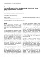
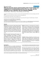
Figure 1 The role of Vpr in HIV-1 infection and host permissiveness. 1). HIV-1 enters human cells via interaction with cell-surface receptors
CD4 and co-receptors CXCR4 (T-cell tropic viruses) or CCR5 (macrophage tropic viruses). The virus fuses with the cell surface membrane
introducing genetic material and virion proteins, which include gag proteins that comprise the matrix and nucleocapsid, the latter containing
significant quantities of Vpr. 2). Vpr promotes the binding of the PIC (including MA, integrase (IN) and proviral DNA) to importins and
nucleoporins, thereby facilitating nuclear entry of HIV-1 provirus into the nucleus of non-dividing cells. 3). Vpr binds to the p300/transcription
factor initiation complex. This binding activity may recruit additional elements to the promoter, such as glucocorticoid receptor (GR).
Alternatively, Vpr may bind to GR bound to GRE elements in the promoter to recruit the p300/TF complex. This results in both increased HIV-1
production, and the regulation of cellular genes that may increase viral permissiveness. 4). Vpr induces G
2
cell-cycle arrest by promoting
phosphorylation of Chk1, which increases viral production. Interestingly, the biochemical properties that contribute to this effect may be
important in HIV-1 production in cells that do not divide. This property is dependent on the degradation of an unknown factor, which is
recruited to Vpr via DCAF-1 interaction. The factor(s) involved in G
2
arrest and viral permissiveness may be overlapping or unique. 5). HIV-1 buds
from the cell, promoting further infection and pathogenesis.
Kogan and Rappaport Retrovirology 2011, 8:25
/>Page 2 of 20
nuclear pore complex (NPC): the NLS and M9-depen-
dent pathways (for review see [17]). The former pathway
involves the binding of NLS signal containing peptide to
importin-a via central armadillo repetitive motifs.
Importin-a binds to importin-b via an amino-terminal
importin-b-binding (IBB) domain [18,19]. The binding
of the classical NLS to importin- a is not possible until
this IBB binding to importin-b occurs, which causes
importin-a to expose an internal NLS [19]. This multi-
protein structure then interacts with the NPC at which
point importin-b transports this NLS component into
the nucleoplasm.
Two other proteins, GTPase Ran/TC4 and NTF2, are
also involved in NLS mediated transport [20-24]. Impor-
tin-a serves as an adaptor molecule by bridging NLS
containing compounds to nuclear transport machinery.
It has been reported, however, that importin-a can facil-
itate nuclear entry of Ca
2+
/calmodulin-de pendent pro-
tein kinase type IV (CaMKIV) without importin-b [25].
Further, importin-b can transport cyclin B1/Cdc2 with-
out Ran, suggesting that mechanisms of import exist
that can utilize one or both importins [26]. In the M9-
dependent pathway, transportin facilitates both nuclear
import and export of RNA binding protein hnRNP A1
by recognizing an M9 signal sequence [27-31]. M9
mediated nuclear trafficking also depends on the func-
tion of Ran/TC4, just as in the classical NLS system
[32].
Vpr nuclear localization seems to utilize cellular
machinery in a unique way that is independent of the
classical NLS and M9 pathways. While viral MA is
inhibited by NLS blocking peptides and dominant-nega-
tive importin-a (residues 244-529), Vpr nuclear entry is
not affected by either treatment strongly supporting the
notion that Vpr functions in an NLS-independent man-
ner [14]. Vpr mediated import is also unaffected by
treatment with R anQ69L, a do minant-negative form o f
Ran, that inhibits both M9 and NLS pathways [32-34].
GTPgS, a nonhydrolyzable GTP that inhibits Ran func-
tion [23,35,36], has no effect on Vpr localization, further
suggesting that Vpr localizes in a non-conventional,
Ran-independent manner [37]. Vpr mediated karyophilic
activity is starkly contrasted to that of classical SV40
NLS, which requires the presence of importin-a/b and
RanGTP[38].Further,Vprnuclear localization appears
to be independent of energy, or at least requires less
energy than conventional transport. Addition of adeno-
sine triphosphate (ATP) or treatment with apyrase,
which lowers NTP levels, affected the localization of
classical NLS beari ng proteins but had no effect on Vpr
localization [34,37]. Another study suggested that Vpr
can enter the nucleus via two different mechanisms;
one involving importin-a and another involving
energy [39]. In summary, Vpr may use importin-a in a
non-conventional, energy independent manner, but m ay
also use a yet undetermined mediator in the absence of
importin-a in a process requiring ATP.
In accord with Vpr’s ability to promote nuclear locali-
zation of the PIC, Vpr has been shown to be essential
for productive HIV-1 and HIV-2 infection of macro-
phages [40-43]. While HIV-1 IN can compensate for
loss of Vpr at high MOI of HIV-1 [14,44], other studies
suggest that Vpr deficient HIV-1 is non-productive in
macrophages at least partly due to the inability to pene-
trate nuclei of non-dividing mononuclear cells
[38,41,45-50]. Further, it was shown that Vpr is directly
involved in targeting the HIV-1 PIC to the nuclear
envelope [51]. It appears that mucosal infection of HIV-
1 involves the transmission of likely a single virus per
patient, as determined by sequence analysis of founder
virus [52]. This claim from initial studies has been
greatly strengthened by a recent study following patients
early during acute infection and the analysis of HIV spe-
cific escape epitopes variants by deep sequencing [53].
Therefore, as the multiplicity of infection during trans-
mission is quite low, it would be expected that Vpr
would be required during this event. Later in infection,
when viremia is elevated, IN and MA may have appreci-
able effects on PIC entry, although this remains to be
proven. Interestingly, it was also reported that Vpr’ s
nuclear localizatio n and consequent G
2
arrest properties
are important in HIV-1 infection of primary CD4
+
T-
cells irrespect ive of proliferative status [54](reviewed in:
[55]). HIV-1 clearly infects resting T-cells in vivo, where
Vpr mediated transport of the PIC into the nucleus
would be expected to have importance. The action of
Vpr, however, appears to be required for CD4
+
T-cell
infection, even under conditions promoting proliferation
(i.e. in the presence anti-CD3 and IL-2 treatment [54]).
It is likely, therefore, that the transport of the PIC
across the nuclear envelope is i mportant in both T-cells
and macrophages in vivo.
In addition to Vpr, there are other requirements for
viral replication in non-dividing cells. The viral capsid
protein, CA, appear s to support this role in that muta-
tions in CA disrupt the cell cycle independence of HIV-
1 infection [56]. The role of CA appears to be indepen-
dent of nuclear import as one of the mutants in CA
exhibited a defect in repl ication in non-dividing cells
beyond the nuclea r entry point. The necessity of Vpr’s
karyophilic properties for the infection of actively divid-
ing cells suggests that the targeting of the PIC to the
NPC is a generally required aspect of lentiviral infection,
regardless of cell cycle progression. In an e volutionary
context, this may imply that lentiviruses evolved to
infect non-dividing macrophages a nd expanded later to
T-cells while retaining the use of already evolved infec-
tion machinery from the original, non-dividing, target
Kogan and Rappaport Retrovirology 2011, 8:25
/>Page 3 of 20
cell population. Indeed, macrophages are a common tar-
get of all known naturally occurring lentiviruses [57].
Furthermore, T-cell infection is common only to lenti-
viruses that cause immunodeficiency, further suggesting
that these cells were later targets of tropism during len-
tivirus evolution. In this model, Vpr may contribute to
nuclear localization in general, whereas other compo-
nents, such as CA, may facilitate additional processes
necessary for productiv e infection of cell cycle arrested
cells. In conclusion, Vpr seems to be an important med-
iator of human lentiviral infection, at least in part due
to nuclear localization properties. This effect may be
most important during periods of low HIV-1 plasma vir-
emia or transmission from person to person.
Correlations between Vpr’s structure and nuclear
localization function
Structural studies have been invaluable to understanding
HIV-1 viral interaction with host cells, including non-
dividing macrophages. Relatively recent structural stu-
dies have identified three alpha helical domains, a-H1
(13-33), a-H2 (38-50), and a-H3 (55-7 7) as well as
other structural features capable of mediating diverse
biological functions [58]. Indeed, Vpr’ s structure allows
for direct binding to many cellular proteins, which likely
enables Vpr to mediate functions such as nuclear import
and G
2
arrest. All three alpha helices have been impli-
cated in Vpr mediated nuclear localization [12,13,59-62],
while the G
2
arrest propert y has been attrib uted mainly
to the C-terminal region of Vpr [59]. However, as the
nuclear import, promoter transactivation, and G
2
arrest
properties of Vpr seem to not only be related, at least
on a structural level, they also may be jointly attributed
to specific physiological properties of Vpr in productive
HIV-1 infection of macrophages [63].
Vpr mediates nuclear localization by binding to impor-
tin-a via residues located within the al pha helices. Whil e
some studies initially reported a low affinity of Vpr for
importin-a [37], others have f ound that Vpr binds to
importin-a using other techniques [50,51,64]. Vpr/
importin-a binding was shown to be non-competitive
with that of the classical the NLS found on MA [65].
Kamata and others demonstrated that regions 17-34
(aH1) and 46-74 (aH2+aH3) can both independently
localize to the nucleus, alb eit to a lower ext ent than an
identified bona fide Vpr NLS consisting of residues 17-74
[66]. Mutations in aH1, aLA (L20,22,23,26A), as well as
in aH2+aH3, I60P and L69P, completely ablated the
ability of the individual peptides to localize to the
nucleus. Later, Kamata and others found that Vpr aH1
and aH3 both bind importin-a,thattheIBBdomainof
importin-a primarily d etermines this interaction, and
that the C-terminal domain of impo rtin-a, 393-462, is
necessary for nuclear localization of Vpr [39]. Although,
an importin-a lacking an IBB still facilitated import of
Vpr, a mutation in Vpr’sfirstalphahelix,aLA, impaired
importin-a binding and nuclear locali zation but still
showed perinuclear accumulation. In contrast, a muta-
tion in the third alpha helix, L67P, failed to localize to
both the nuclear and perinuclear areas, but still permitted
binding to importin-a. The authors concluded that bind-
ing to importin-a requires only the first alpha helix and
that the third alpha helix serves to localize Vpr to the
perinuclear area indepe ndently of importin binding. Pre-
vious findings from other inve stigators also showed that
the use of IBB peptides failed to inhibit Vpr mediat ed
nuclear localization. This suggests that importin-a may
be essential f or Vpr’s karyophilic properties but that the
direct interaction between importin-a and Vpr may not
be essential [34]. Hitahara-Kasahara and others showed
that im portin-a1, a3, and a5isoformsareallableto
induce Vpr mediated nuclear import [38]. Importin-a
was shown to be essential for HIV-1 replication in
macrophages, suggesting that importin-a nuclear import
is a vital process in the infection o f these cells. Further-
more,arecentstudyfoundthatVprdoesnotbindto
importin-a2 or importin-a2/b1 heterodimers, suggesting
that cell-line specific expression of importins may regu-
late Vpr’s
karyophilic properties [46]. In summary, these
studies suggest that importin-a is important for Vpr-
mediated nuclear translocation, but the exact nature of
this mechanism is still under investigation.
In addition to the reported binding interaction with
importin-a, Vpr has been demons trated to bind directly
to nuclear pore proteins [47,49-51,67]. Vpr mutants
F34I and H71R have been found to lose the ability to
localize to perinuclear areas, suggesting that these resi-
dues are involved in nuclear pore interaction [50].
These mutants were still found in the nucleus , which is
not surprising considering that Vpr is less than 40kDa.
The F34I mutant showed lower binding to importin-a
and Nsp1p, a member of the nuclear pore complex. WT
Vpr colocalizes with importin-b and nuclear pores in
perinuclear regions and binds both Pom 121 and very
weakly to Nsp1p [47]. An A30P mutant lacked these
abilities.
FXFG regions on nucleoporins, a form of phenylala-
nine-glycine (FG) repea t, have been reported to interact
with cytoplasmic proteins involved in nuclear import
[22,68,69]. Vpr was reported to bind to FXFG contain-
ing proteins p54 and p58 as well as to the FXFG region
of Nup1 [51]. Further, addition of Vpr was shown to
stabilize the binding of importin-a/b to Nup1 FXFG.
Another report failed to show interaction between Vpr
and FXFG of Pom121, but instead demonstrate d that
the alpha helices of V pr interact with hCG1 by binding
to a non-FG repeat region located in the N-terminal
region on residues 49-170 [67]. This area has no known
Kogan and Rappaport Retrovirology 2011, 8:25
/>Page 4 of 20
homology to bind ing motifs and has no known binding
partners. In a later study, it was found that four Vpr
mutants L23F, K27M, A30L, and F34I, which all occur
on one face of the first alpha helix, have impaired hCG1
binding and fail to show nuclear localizat ion [49]. Thus,
it seems t hat Vpr is able to bind to importin-a as well
as nucleoporin using the same residues on the first
helix. In both cases, there is evidence that Vpr binding
to nucleoporin components occurs in a way that is dis-
tinct from the classical NLS pathway.
The role of importin-b in the nuclear transport of Vpr
is an aspect of the mechanism of Vpr’s karyophilic prop-
erties that remains to be fully understood. Early studies
showed that Vpr fails to bind importin-b [65] or that it
binds at a low affinity [37]. Oddly, the latter study found
greater affinity of Vpr to importin-b than to -a.Subse-
quent studies argued that Vpr’s localization is importin -
a,butnot-b, dependent. Addition of importin-b to
digitonin permeabilized cells, which was required for the
classical SV40-NLS localization, was unnecessary for
Vpr N17C74, a construct containing the minimal region
for nuclear localization [38,66]. These studies also found
that ΔIBB importin-a, which is unable to bind to impor-
tin-b, still caused nuclear translocation of N17C74. Pre-
vious studies demonstrating that the use of IBB peptides
failed to inhibit Vpr localization also lend some support
to these findings [34]. Further, importin-b siRNA failed
to prevent N17C74 localization to the nucleus [38]. Vpr
has also b een shown to physiologically behave in ways
similar to importin-b, leading some authors to suggest
that Vpr replaces the role of importin-b,which,like
Vpr, also binds to both importin-a and nuclear pores,
in the nuclear translocation process [50]. Other studies,
however, suggest that importin-b is necessary for Vpr’s
karyophilic properties. Papov and others found that Vpr
prevents FXFG Nup 1 peptide mediated dissociation of
MA with importin-a/b complexes and increases the affi-
nity of importin-a to NLS [51,65]. Based on these find-
ings Papov and others proposed that Vpr stabilizes the
MA and IN NLS complex with importin-a/b to pro-
mote nuclear entry. A dominant negative form of
importin-b, residues 71-876 [70] has also been shown to
inhibit Vpr localization, further suggesting that impor-
tin-b plays a role in Vpr mediated nuclear targeting
[34]. Recent studies have clearly shown binding of Vpr
to importin-b3, but not to importin-b 1ortoimportin-
a2/b1 complexes [46]. This may explain discrepancies
in early findings that failed to find effects of isolated
importin-b which were not necessarily applicable to
other importin-b isoforms.
The respective roles of the alpha helices and the C-
terminal region in nuclear localization and G
2
arrest
remain controversial. Through extensive mutational ana-
lysis, Mahalingam and others put forth a hypothesis that
the nuclear localization function resides primarily in the
alpha helic es while the G
2
arrest property is determined
by the carboxyl-terminus [59]. Previous studies lend
support to this assertion as the al pha helices, but not N-
terminal or C-termina l regions were involved in nucleo-
porin binding by Vpr [67]. Other reports found that
N17C74 Vpr, which lacks the C and N terminal regions
and other Vpr constructs lacking the C-terminus are
unimpaired in nuclear localization [11,66]. Although the
C-terminal region closely resembles a classical NLS, this
region does not have NLS function and Vpr functions
independently of NLS binding [14,71]. Conversely, many
other studies found that the C-terminal is necessary or
sufficient for nuclear entry of Vpr [12,34,47,62,72]. The
disc repancy between these studies remains unexplained.
Interestingly, recent studies have shown that all three
alpha helices are involved in Vpr oligomerization [63].
The authors reported that mutatio ns that affected oligo-
merization did not prevent apoptosis induction by Vpr
(a G
2
arrest dependent property [73]). Nuclear lo caliza-
tion, however, was perturbed for these mutants. These
studies may suggest that karyophilic and cell cycle arrest
properties rely on multiple domains that may be separ-
able to some degree.
Vpr functions as a coactivator of the HIV-1 long
terminal repeat
While Vpr promotes infection of HIV-1 into non-
dividing cells, the ability of Vpr to activate both viral
and endogenous promoter activity likely contributes to
increased viral replication and pathogenesis. Initially, it
was observed that Vpr can reactivate cells latently
infected with HIV-1 [74,75]. Later studies demon-
strated more spec ifically that Vpr transacti vates the
HIV-1 long terminal repeat (LTR) as well as other pro-
moters [76-78]. The U3 region of the HIV-1 LTR has
several activating elements, which include NF-AT, glu-
cocorticoid response elements (GRE), NRF, NF-B,
Sp1, a Tat responsive RNA element (TAR), and a
TATA box [79-83]. Studies employing HIV-1 LTR
indicator constructs demonstrated that Vpr acts via
Sp1 sites [78]. Vpr binds to the Sp1/promoter complex
and it has been proposed that Vpr exerts its effects by
stabilizing promoter complexes containing multiple
bound Sp1 proteins. Other studies, however, support
the notion that Vpr transactivates primarily the -278
to -176 region of the LTR, which contains the GREs,
while the NF-B and Sp1 are utilized by Tat mediated
transact ivation [84].
Vpr appears to act as a coactivator in the presence of
other activating elements but not on a bare promoter
alone. Vpr was shown to bind transcription factor IIB
(TFIIB), suggesting that the effect of Vpr is indeed due
to coactivation rather than direct transcription factor
Kogan and Rappaport Retrovirology 2011, 8:25
/>Page 5 of 20
function [76]. Vpr has also been demonstrated to
potentiate the activation of the HIV-1 LTR by p300 [85]
and was shown to form a complex with p300 and TFIIH
to cooperatively induce GRE activation in a manner
independent of G
2
cell cycle arrest [86]. Consistent with
these findings, a Vpr mutant deficient in p300 binding,
I74,G75A, did not display this property. Several Vpr
mutants including R73S, C76S, and Q21P have also
been reported to lose HIV-1 LTR transactivation abil-
ities [87]. Intriguingly, the R73S mutation imparted a
dominant-negative phenotype with regard to transactiva-
tion. Vpr has also been reported to act cooperatively
with Tat, another LTR coactivator. Their cooperative
effect was disrupted by the Vpr R73S mutation [88].
Therefore, in the p resence of Vpr, viral production is
likely amplified via coactivation of the HIV-1 LTR by a
mechanism that appears to be dependent on multiple
binding sites within the viral LTR.
The glucocorticoid r eceptor (GR) has been a known
target of Vpr function for more than a decade [89]. Ori-
ginally, Vpr was shown to induce R-interacting protein
1 (Rip-1) nuclear translocation in a GR dependent man-
ner and along with Rip-1 form a complex with GR. A
later study showed that Vpr transactivates promoters
containing GREs [90]. T he authors also reported that
Vpr L64A, a mutant for a signature GR binding motif
LXXLL, was found to be defective for binding to GR
and in GRE transactivation, but like WT Vpr, Vpr L64A
retained the ability to bind TFIIB. A Vpr R80A mutant,
which lacked G
2
arrest, was unimpaired in GRE-
mediated transactivation. This study also reported that
Vpr/p300 synergy was amplified in the presence of dex-
amethasone. A later publication confirmed many of
these observations for LXXLL Vpr mutants in the first
and third alpha-helices, 22-26 and 64-68 respectively
[91]. The authors reported that mutations in both
helices were necessary to compl etely diminish GRE pro-
moter activation. Subsequently, Kino and others identi-
fied Vpr mutants, F72, R73A and I74,G75A, which were
unable to bind p300 and were therefore deficient in
GRE transactivation [92]. Unlike Vpr L64A, these
mutants were not reported to be transdominant, sug-
gesting that Vpr L64A competes with WT Vpr for p300
binding. It is noteworthy that while some subsequent
studies have found conflicting results [93], later research
has solidified the notion that GR and Vpr function
synergistically. Human Vpr interacting protein (hVIP/
Mov34), which binds to both Vpr and GR, translocates
to the nucleus following either dexamethasone or Vpr
treatment, further suggesting that Vpr and GR form an
functional complex within cells [94]. Vpr and GR also
have a gain of function in inhibiting poly (ADP-ribose)
polymerase 1 (PARP-1) nuclear transloca tion, which i s a
necessary event in NF-B transcription [95]. It is worth
noting that the effect of Vpr on NF- Bremainsacon-
troversial topic (discussed below in: “Vpr and immune
dysfunction” ). However, HIV-1 infection and NF-B
activation form a positive feedback loop [96,97], and Tat
is known to induce the HIV-1 LTR synergistically with
NF-B [98], highlighting the importance of the NF-B
pathway for HIV-1 replication. Considering that NF-B
signal ing is activated during HIV-1 infect ion, the role of
Vpr in the context of HIV-1 infection may or may not
be identical to studies using ectopic Vpr expression. In
summary, these studies suggest that Vpr and GR func-
tion in a cooperative manner through a mechanism that
involves direct binding, and this interaction is at least
partly responsible for the transctivation of the HIV-1
LTR by Vpr. The interaction of Vpr with GR and ele-
ments of the LTR transcription complex, including p300
is illustrated in Figure 1.
Although Vpr appears to coactivate the HIV-1 promo-
ter via GRE and generally behaves in a GR-dependent
manner (with respect to transcriptional activation), the
role of glucocortcoids o n HIV-1 viral replication
remains controversial. Several groups have reported
altered hypothalamic-pituitary-adrenal (HPA) axis func-
tion in HIV-1 infection [99-104]. Additional in vitro
molecular studies have reported that glucocorticoids
suppress the HIV-1 LTR [105-109]. Kino and others
reported that this effect depends on GR and is not influ-
enced by Vpr [105]. These reports are seemingly in con-
tradiction with aforementioned studies, which showed
that Vpr transactivates the HIV-1 LTR and that Vpr
enhancement of other promoter elements containing
GREs is potentiated by glucocorticoids. Intrigui ngly,
Laurence and others reported that the level of HIV-1
LTR activity in unstimulated cells is not diminished by
dexamethasone, while phorbol ester induction of the
HIV-1 LTR was attenuated by such treatment [106]. In
contrast, some investigators have reported that gluco-
corticoids have an enhancing effect on HIV-1 LTR
activity [110,111]. The latter study showed that this
effect was seen only in the context of interleukin (IL)-6
and tumor necrosis factor alpha (TNF-a). Interestingly,
a recent study found that extracellular Vpr was capable
of increasing IL-6 production in an NF-B and C/EBP-b
dependent manner by stimulating Toll-like receptor 4
(TLR4) signaling in macr ophages [112]. Glucocor ticoids
and TNF-a have also been shown to increase HIV-1
virus production [113]. Therefore, the effect of glucocor-
ticoids on the HIV-1 promoter may be influenced by the
presence or absence o f pro-inflammatory signals.
Increased levels of glucocorticoids have been associated
with HIV-1 progression, although some reports suggest
that these effects are due to immune system modulation
rather than a direct effect on viral replication
[12,11 4-116]. Subsequently, it was shown that RU486, a
Kogan and Rappaport Retrovirology 2011, 8:25
/>Page 6 of 20
GR and progesterone receptor (PR) inhibitor, can reduce
HIV-1 LTR activation by Vpr and attenuate virus pro-
duction in X4 infected PBMCs as well as R5 infected
macrophages [117]. In contrast, glucocorticoids can
incr ease the permissi veness to infection of unstimulated
PBMCs by HIV-1 [118]. These studies demonstrated
that the viral life-cycle was blocked at a stage of infec-
tion before proviral integration. Interestingly, a similar
block in HIV-1 replication was also shown t o be abro-
gated by Vpr, further suggesting GR/Vpr cooperativity
[41]. In summary, Vpr may have varying effects on the
HIV-1 LTR depending on the context of proinflamma-
tory and anti-inflammatory signals, in addition to GR
pathways.
The interrelationship of Vpr functions and their
relevance to macrophage permissiveness and
HIV-1 reservoirs
Numerous studies have focused on the role of Vpr in
macrophage infection and permissiveness to HIV-1.
However, the involve ment of multiple properties of Vpr
in these processes has made it difficult to exactly ascer-
tain which features are most important for macro phage
infection. Further, some studies have relied on mutation
of individual residues to discern these effects. However,
the mutants produced often show defects in multiple
properties, which are cle arly independent biologically,
making the analy sis of s tructu ral studies challenging. A
confusing issue in the literature is that the “so called”
G
2
arrest function of Vpr, which is likely irrelevant to
the status of terminally differentiated cells such as
macrophages, has been assoc iated in some studies with
HIV-1 infectivity of such differentiated cells. Recent
findings in the field, however, suggest the likelihood that
both G
2
arrest and another, yet unknown, cellular pro-
cess use similar machinery and that the factors involved
in these Vpr functions may have significant overlap.
Findings from mutational studies have suggested over-
lap in G
2
arrest and localization of the HIV PIC to the
nucleus. In a recent study the authors reported that the
G
2
arrest properties of Vpr depend on nuclear localiza-
tion [49]. Jacquot and others showed that four Vpr
mutations in the first alpha helix, Vpr L23F, K27M,
A30LandF34Iallexhibitbothatleastpartially
impaired G
2
arrest and defective nuclear localization
while Vpr mutants R80A and R90K were deficient in G
2
arrest alone. While previous studies confirmed some of
these results, they have also reported opposite results
for the same mutations or support the notion that the
two properties are independent [11,50,59]. It is note-
worthy to mention that these two properties are com-
pletely separated in HIV-2/SIV
SM
viruses which
accomplish nuclear localization by using accessory pro-
tein Vpx and G
2
arrest by using Vpr [119]. Vpr/Vpx
defective SIV virus, but not viruses defective in either
protein alone, have been shown to have a greatly attenu-
ated course with no progression to AIDS in rhesus
monkeys, suggesting that both of these properties play
significant roles in vivo [120]. Many studies also argue
that nuclear localization rather than G
2
arrest is impor-
tant in macrophage infection of HIV-1. For example,
HIV-1 transcripts in Vpr defective viruses lose the abil-
itytobedetectedatsometimebetweenthereverse
transcription and pro-viral DNA replication phases [41],
suggesting that in the absence of Vpr the viral life cycle
may be inhibited at the nuclear entry phase. The ability
of IN to compensate for Vpr loss also suggests that
nuclear localization plays a predominant role [14,44].
Therefore, there is ample evidence to support the notion
that Vpr can induce nuclear localization indep endent of
G
2
arrest. Mutation studies have not demonstrated such
independence, however, as the structure/function rela-
tionships have not proven separable.
As nuclear localization and G
2
arrest seem to be
related in some structural studies, it is not surprising
that both properties of Vpr have been linked to produc-
tive infection of macroph ages. Subbramanian and others
argued that Vpr’s ability to cause G
2
arrest may also
play a role in HIV-1 infection of macrophages [121].
Upon infecting macrophages with HIV-1 viruses that
were Vpr WT, ATG-Vpr (Vpr negative), Vpr R62P
(impaired in nuclear localization), and Vpr R80A
(impaired in G
2
arrest), the authors observed that unlike
the Vpr R62P mutant, which only inhibited viral growt h
at low MOI, the Vpr R80A and ATG-Vpr viruses were
the most impaired at higher MOI. However, R80A
mutant, as expected, showed no differences as compared
to the other mutants in the number of G
2
stage cells in
terminally differentiated macrophages, as these cells are
already arrested. These results suggest that the so cal led
G
2
arrest propert y of Vpr is impo rtant in different ways
than nuclear localization for productive viral infection in
myeloid cells. While the authors hypothesized that the
effect of G
2
arrest on viral replication is due to bio-
chem ical proper ties of the mutant protein, the indepen-
dence of these two properties in mutated Vpr constructs
remains to be fully ascertained.
It is very important to note that the G
2
arrest property
of Vpr has been recently attributed binding to damaged
DNA binding protein 1 and Cullin 4a-associated factor-
1 (DCAF-1) [122-128] (origin ally identified as a bind ing
partner called VprBP [129]), and is a result of subse-
quent induction of ataxia telangiectasia-mutated and
Rad3 related (ATR) kinase. While it is unknown how
Vpr/DCAF-1 binding promotes G
2
arrest, it has been
proposed that Vpr may recruit a particular factor to this
complex, promoting ubiquitinat ion and degradation of a
yet unknown cellular protein or, perhaps, several targets
Kogan and Rappaport Retrovirology 2011, 8:25
/>Page 7 of 20
[130,131]. Macrophages are non-dividing cells and are
therefore not subject to the cell-cycle arrest function of
Vpr and even lack the prerequisite ATR induction in
the presence of Vpr [132]. The findings that demon-
strate the importance of Vpr residues involved in G
2
arrest in promoting HIV-1 replication likely suggest that
the recruitment of native cellular factors to DCAF-1
promotes both propert ies. However, it is unknown what
bin ding partners mediate these effects or if they are the
same or overlapping for both G
2
arrest and cellular per-
missiveness. A synopsis of these three properties and
their effects on HIV-1 infection of macrophages is
found in Figure 1.
The G
2
arrest and HIV LTR promoter transactivation
properties of Vpr may also be dependent or independent
of each other. Many studies have shown that Vpr’sabil-
ity to cause G
2
arrest and increase viral production are
linked [62,75,85,133,134]. While G
2
cell cycle arrest may
make HIV-1 infected T-cells and oddly macrophages,
which are not dividing, more permissive to active infec-
tion, many studies have shown that Vpr constructs defi-
cient in G
2
arrest maintain the ability to function as a
coactivator [59,84,90-92]. While G
2
arrest and transacti-
vation properties of Vpr both impart positive effects on
viral replication, whether these effects represent inde-
pendent functions is a matter of debate.
As mentioned previously, Vpr is believed to allow for
permissive infection of HIV-1 in many cell types, but is
considered particularly important for the infection of
non-dividing cells such as macrophages and resting T-
cells. As such, Vpr is likely important in generating a
long lived reservoir for virus infection. Indeed, it has
been suggested based on results in non-human primate
studies, that mac rophages are likely the main producers
of virus in late stage simian/human immunodeficiency
virus(SHIV)atatimewhenCD4
+
T-cells have been
depleted [135]. In HIV-2/SIV
SM
virus, Vpr is hypothe-
sized to have duplicated, giving rise to Vpx [5,136]. Vpr
and Vpx have discrete functions in HIV-2/SIV
SM
viruses
causing G
2
arrest and nuclear localization respectively,
whereasVprhasbothpropertiesinHIV-1[119].
Recently,itwasshownthatSIV/HIV-2Vpxovercomes
a block to reverse transcription in macrophages, further
suggesting that HIV-1 Vpr may increase viral permis-
siveness in myeloid cells as well [137-139]. It is note-
worthy to mention that Vpx also has such an effect on
HIV-1 defective in Vpr, yet this effect is not seen with
Vpr treatment. This likely suggests that Vpx acts on cel-
lular targets that may be only partially in common to
those of Vpr. Interestingly, Vpx binds DCAF-1 in a way
similar to Vpr [125] and such interaction is necessary
for the permissive effects described above. It has been
suggested that Vpr and Vpx compete for binding to this
complex and perhaps recruit unique or only partly
overlapping binding partners [130]. Therefore, it is likely
that the particular macrophage restriction factor antago-
nized by Vpx is not a target of Vpr. In agreement with
this notion, previous studies have attributed Vpr to lift-
ing a post-reverse transcriptional block, whereas Vpx
seems to affect an earlier block in viral replication [41].
However, Vpr may use the same system to recruit other
factors that promote permissive infection of HIV-1 into
macrophages. It is unknown why HIV-1 Vpr does not
possess the same properties as seen with Vpx in SIV or
HIV-2, but obviously HIV-1 does not rely on these
effects for successful infection in vivo. Considering that
Vpr has small eff ects on macrophage permissiveness to
HIV-1 during single a round of infection [140], but
causes profound changes after long-term culture [40,41],
it is likely Vpr mediated macrophage permissiveness has
not been detected as compared to Vpx simply due to
the a smaller magnitude of it’ s effect or due to short-
term culture conditions.
HIV-1 virus is known to have anti-apoptotic proper-
ties in chronically infected macrophages and microglia
[141], and causes a reduction of pro-apoptotic Bax
expression in mitochondria of persistently infected cells
[142]. While Vpr promotes apoptosis [143,144], it also
exhibits anti-apoptotic properties [145]. It is noteworthy
to mention that no study of which we are aware has
ever shown toxicity of Vpr in macrophages. On the con-
trary, it has been argued that macrophages lack the
ATR mediated the cell stress response normally induced
by Vpr [132], which is required for the apoptotic activity
that has been reported in other cell types. Intriguingly,
Vpr was observed to inhibit apoptosis in a lymphoblas-
toid cell line by inducing Bcl-2, with concomitant down-
regulation of Bax in a manner seemingly contingent on
Vpr expression level [145]. Further, Vpr mediates resis-
tance to cell death from Fas ligand and TNF-a in these
cells. The G
2
arrest function of Vpr in these cells, how-
ever, is most likely defective since these clones exhibited
cell cycle characteristics similar to those of control-
transfected cells. As Vpr is toxic to non-myeloid cells,
such as T-cells, the possible anti-apoptotic effects of
Vpr that have been observed and attributed to Vpr in
the study may be due to a low level of Vpr expression
in the cell lines used. As such, the pro-survival effects of
Vpr may need to be evaluated further. If Vpr promotes
cell survival, it i s conceivable that the pro-survival
effects of HIV-1 may involve the action of Vpr, espe-
cially in macrophages, possibly in combination with
additional host-viral interaction. In combination with
the aforementioned abilities of Vpr to increase viral
replication by inducing G
2
arrest and a ctivating the
HIV-1 LTR, the potential of Vpr to promote infection
of and survival of macrophages could be a highly signifi-
cant factor in the development and/or maintenance of
Kogan and Rappaport Retrovirology 2011, 8:25
/>Page 8 of 20
macrophage viral reservoirs. The differential mechani sm
of pro-apoptotic/anti-apoptotic Vpr activity warrants
further investigation and may provide an avenue of ther-
apy as an additive to highly active antiretroviral therapy
(HAART), now renamed combination antiretroviral
therapy (cART).
Vpr and HIV dementia
HIV encephalopathy (HIV-E) is an associated underlying
pathological condition seen in autopsy of patients with
HIV-1 associated dementia (HIV-D), a disease charac-
terized by motor and cognitive deficits. The presence
HIV-1 virus in the brain is seemingly the cause of this
condition as it was detected by in situ hybridization in
patients with HIV-E but not i n HIV-1 patents who do
not exhibit this pathological condition [146]. Although
the introduction of cART initially reduced the preva-
lence of HIV-D, the prevalence of HIV associated neu-
rocognitive disorders (HAND) has been increasing (for
review see [ 147]). While it is unclear if the minor and
severe forms of HAND have common etiologic mechan-
isms, there is reason t o suspect t he importance of HIV
infection in macrophages in the central nervous system
(CNS) and/or the perip hery, as well as the r ole of Vpr.
Since Vpr has bee n implicated as both a direct and
indirect contributor to the development of dementia,
Vpr may also play a role in the more subtle forms of
neurologic disease (Figure 2).
Although the principle mechanism o f HIV-D pathol-
ogy is not known, there is a preponderance of evidence
suggesting that mononuclear cells play a critical role in
disease progression. The major sources of HIV-1 pro-
duction in the brain appear to be macrophages and
microglia [146,148-150]. Furthermore, in brains of ani-
mals infected with SIV, perivascular macrophages are
responsible for the majority of virus production, further
implicating these cells in the pathology of CNS disease
[151]. Macrophage/microglia numbers are more highly
correlated with the severity of HIV-D than the presence
of HIV in the CNS [152]. Patients with HIV-D also have
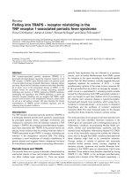
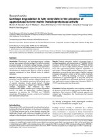
Figure 2 Summary of HIV-1 pathology involving Vpr. Vp r is likely important for both immune dysfunction as seen in AIDS and associated
diseases including HIV-D and HIVAN.
Kogan and Rappaport Retrovirology 2011, 8:25
/>Page 9 of 20
elevated numbers of CD14
+
/CD16
+
monocytes in the
periphery [153,154], which have neurotoxic properties in
vitro [154]. CD14
+
/CD16
+
, HIV-1 positive macrophages
have also been found in brains of patients suffering
from HIV-D [155]. The presence of TNF-a protein and
mRNA in patients with HIV-D has been reported to sig-
nificantly correlate with the severity of symptoms in
these patients, further suggesting that activated macro-
phage activity is directly involved in HIV-D pathology
[152,156]. The increased number of CNS macrophages/
microglia (in the absence of evidence for proliferation)
suggests that the accumulation of myeloid cells in the
brain is due to trafficking of peripherally derived macro-
phages [157], (reviewed in [158]). As mentioned pre-
viously, Vpr plays a significant role in the permissive
infection of HIV-1 into macrophages and may increase
the survival of infected myeloid cells; therefore, it is
indirectly related to HIV-D pathogenesis.
Vpr may be a direct effec tor of HIV-1 mediated HIV-
E pathology. Higher levels of Vpr have been found in
the CSF of patients with HIV associated cognitive defi-
cits. Vpr has been detected by immunofluorescence in
the basal ganglia and frontal cortex of brains with HIV-
E and is elevated in the serum and CSF of seropositive
HIV patients [74,159] and has been shown to cause
apoptosis in vitro [160]. The cells that contained Vpr in
HIV-E brains were either macrophages or neurons.
Transgenic mice that express Vpr in monocytoid cells
display neuronal injury in the basal ganglia and subcor-
tical area, which confirms in vitro findings [161].
Mechanistically, the neurotoxic effect of Vpr depends
on the 70-96 C-terminal region, which is essential for
the induction of neuronal apoptosis in striatal and corti-
cal cells [162]. In neurons, this effect is mediated by
activation of p53, caspase 9, and caspase 8 [161,163].
Although gp120 and Tat have also been shown to
induce apoptosis in neuronal cells [164,165], intracellu-
lar Vpr expression in NT2 cells seemed to be necessary
for the induction of apoptosis [166]. This effect many
have even greater clinical relevance considering that Vpr
and ethanol together cooperatively increase apopto sis in
brain microvascular endothelial cells, which may possi-
bly allow for greater blood brain barrier permeability to
virus and infected cells [ 167]. Most recently, Vpr was
shown to increas e reactive oxygen species production in
microglia and neuroblastoma cell lines, to lower ATP, to
lower plasma membrane Ca
2+
ATPase (PMCA) protein
levels, and increase cytoplasmic permeability in neuro-
blastoma cells [168]. By lowering PMCA levels, the
efflux of Ca
2+
would be expected to increase in neuronal
cells, which has been linked to cell death signaling in
these cells (for review see [169]). Vpr produced from
HIV-1 infected macrophages was found to impair axonal
growth of neuronal precursors independently of
apoptosis [170]. Vpr binds to CCAAT-enhancer binding
protein (C/EBP) sites on the HIV-1 LTR [171] and con-
sequently a C/EBP site with high affinity for Vpr, C/EBP
I, is associated with clinical progression to HIV-D [172].
It has b een proposed that Vpr a ctivat es C/EBP sites by
direct bindi ng to C/EBP I in the HIV-1 LTR, which has
low affinity for C/EBP, as well as indirectly by upregulat-
ing the expression of C/EBP in host cells [173]. Vpr and
Nef both induce RANTES/CCL5 chemokine in micro-
glia, causing activation of brain mononuclear cells,
which correlates with clinical dementia [174]. Therefore,
Vpr is a direct and in direct mediator of cell d eath and
neuronal impairment in HIV-1 patients as well as a
necessary factor for the infection and survival of HIV
infected macrophages, thereby further contributing to
the pathogenesis of HIV-D.
Vpr and HIVAN
HIV associated nephropathy (HIVAN) is a form of col-
lapsing focal segmental glomerulosclerosis, largely due
to HIV-1 protein toxicity to epithelial cells (for review
see [175]). The most significant incidence of the disease
is seen in HIV-1 positive patients of African descent,
likely due to a prevalence of the MYH9 allele in this
population [176]. As in HIV-D, macrophage trafficking
and expression of virus has been implicated in pathology
of HIVAN. Fibroblast growth factor 2 (FGF-2), which is
elevated in kidneys of children with HIVAN, increases
the attachment of uninfected and HIV-1 infected PBMC
to tissue culture plates coated with renal tubular epithe-
lium [177]. In vivo, FGF-2 likely increases the invasion
of inflammatory cells into renal tissue, leading to renal
injury. Interestingly, Vpr has been implicated in the
development of HIVAN (Figure 2). A c-fms/Vpr trans-
gene in mice produced focal glomerulosclerosis, suggest-
ing that macrophage specific Vpr expression might be
sufficient for kidney damage [178]. Further, it was
reported that FVB/N mice expressing Vif, Vpr, Nef, Tat,
and Rev in podocytes developed nephropathy and pro-
teinuria suggesting that viral proteins themselves have
toxic effects in the kidneys [179]. Vpr expressed in a
transgenic mouse model demonstrated that presence of
Vpr in podocytes is sufficient for glomerulosclerosis
[180]. Lentiviral experiments in vitro produced similar
find ings [181]. Vpr expression in combination with Nef,
however,resultsinseverekidneydamageintransgenic
mice [180]. Vpr expression combined with hemine-
phrectomy also resulted in far more profound nephrotic
changes [182]. The impact of heminephrectomy was
almost entirely prevented by including treatment with
angiotensin II type 1 (AT1R) receptor blocker olmesar-
tan. To date, however, no specific therapies targeting
Vpr/Nef nephrotoxicity or the attachment of affected
macrophages to the tubular epithelium have been
Kogan and Rappaport Retrovirology 2011, 8:25
/>Page 10 of 20
developed. It should be not ed that in the studies using
single or combined expression of viral proteins in parti-
cular cell types, such as in macrophages in the c-fms
driven Vpr model, it is unclear if these effects occurred
due to the secretion o f these products from cells traf-
ficking to the kidneys or due to other inflammatory
cytokines produced in these cells due to the expression
of these products.
Vpr and immune dysfunction
Vpr has profound inhibitory effects on many members
of the immune system involved in adaptive response
(Figure 2). Consequently, Vpr reduces the efficacy of
DNA and SIV-Nef vaccination in vivo, suggesting that
Vpr may aid in evasion of immune response during
HIV-1 [183,184]. The mechanism of immune dysfunc-
tion caused by Vpr appears to involve the induction of
apoptosis and cell cycle arrest in bystander T-cells,
contributing to the depletion of immune cells. While
Vpr is seemingly anti-apoptotic in HIV-1 infected cell
lines, in vitro studies suggest that bystander T-cells
may be induced to undergo apo ptosis in response to
extracellular or secreted Vpr [145,185,186]. Although
many studies argue that Vpr has effects outside of the
infected cell due to secretion, this point remains con-
troversial. However, in vivo, Vpr alone has been shown
to be contribute to HIV-1 mediated immune dysfunc-
tion by promoting depletion of thymic cells (reviewed
in [187]). Activation induced cell death by apoptosis
has been proposed as a mechanism of HIV-1 infected
CD4
+
lymphocyte depletion, although multiple
mechanisms distinct from Vpr likely contribute to this
process [188,189]. Vpr can increase Fas dependent cas-
pase-8 dependent cleavage i n T-cells to induce apopto-
sis, providing a potential mechanism for increased cell
death. CD4 promoter-Vpr transgenic mice do show T-
cell depletion in a Bcl-x, Bax, and caspase-1 dependent
and Fas-Fas ligand independent manner [190]. G
2
arrest precedes the induction of apoptosis by Vpr and
has been reported to be necessary for progression to
apoptosis [73], however, the latter findings remain con-
troversial [191]. R ecently, it was demonstrated that this
property depends on Vpr activated phosphorylation of
Chk1, an event that begins during the S phase of the
cell cycle [192]. Apoptosis oc curs via caspase-9 and
seems to cause apoptosis in cancer cell lines with
mutated p53, suggesting that this effect is independent
of p53 function [193-195]. Vpr has also been postu-
lated to increase the expression of TNF-a in dendritic
cells (DC)s and in this way may indirectly promote
apoptosis in CD8
+
T-cells [196]. The Vpr mediated
depletion of uninfected T-cell populations likely con-
tributes, in part, to the immune dysfunction observed
in AIDS.
Recent studies have identified additional mechanisms
of Vpr mediated T-Cell depletion. Vpr has been shown
to upregulate natural killer gr oup 2, member D
(NKG2D) ligands in CD4
+
lymphocytes, which resulted
in natural killer (NK) mediated toxicity to these cells
[197,198]. It is unclear what effect Vpr has on HIV-1
infected CD4
+
T-cell depletion in vivo, since Vpr alone
is sufficient to upregulate NKG2D ligand expression.
Vpr could induce bystander T-cell killing due to NK
mediated toxicity. It should also be mentioned, however,
that Vpr has been reported to inhibit NK function
[199,200], which would be predicted to oppose NK
mediated toxicity. If infected T-cells are depleted due to
NK function, this may suggest that the infection of
these targets is outwei ghed by the advantage conveyed
by immune suppression. Interestingly, the upregulation
of NKG2 ligands by Vpr is also related to DCAF-1 bind-
ing in an ATR related mechanism, which suggests that
these ligands may not be readily upregulated in macro-
phages that are reported to lack ATR response to Vpr
expression [132,197,198]. Considering that macrophages
have been reported to be the main viral reservoir during
late stage infection of rhesus macaques with an SIV/
HIV-1 chimeric virus (SHIV) [135], the depletion of T-
cells may not be a limitation to virus persisten ce due to
the availability of myeloid target cells. In summary, Vpr
has been reported to cause apoptosis of bystander T-
cells by multiple mechanisms, which may contribute to
decreased immune function and possibly impaired viral
clearance in the host.
Vpr may suppress cellular immunitybymodulating
antigen mediated activat ion and cytotoxic killing of sur-
viving T-cells. In vivo, Vpr promotes a shift toward a
Th2 response, likely by suppressing IFN-g,aTh1indu-
cing cytokine [183]. Other s tudies have also confirmed
that Vpr promotes Th2 cytokine IL-10 while suppres-
sing the expression of Th1 cytokine IL-12 [201-203],
presumably by modulating NF-B response (discussed
below). T-cell function also may b e perturbed by down-
regulation of CD28 and CTLA-4 which are required for
activation by antigen presenting cells and therefore
adaptive immune function [204]. Recombinant Vpr has
been shown to lower activation of macrophages and
maturation of DCs by inhibiting the expression of key
co-stimulatory molecules including CD40, CD80, CD83,
and CD86 [201,205]. This suggests that Vpr may dam-
pen antigen presentation by downregulation of partner
molecules on both presenter and effector cells. Vpr has
also been shown to suppress immune activation to
superantigens in vivo [206]. More recently, Vpr has also
been shown to modulate NK cell function, causing a
reduction in cytolyt ic killing and differential regulation
of IL-12 and TGF-b by Smad3 activation [200]. There-
fore, Vpr may significantly contribute to the immune
Kogan and Rappaport Retrovirology 2011, 8:25
/>Page 11 of 20
deficiency seen in AIDS by altering both adaptive and
innate immune cellular function.
Evidence from many studies suggests that Vpr’ s effect
on the im mune system seems to be mediated by intera c-
tion with the NF-B pathway by a mechanism involving
GR. Glucocorticoids have been shown to have immuno-
suppressive effects due to NF-B inhibition and induc-
tion of I kappa B alpha (IBa), which prevents NF-B
translocation into the nucleus thereby preventi ng cyto-
kine release and immune activation [207,208]. Vpr was
first shown to induce T-cell apoptosis in a TCR depen-
dent mechanism by inducing IB and reducing NF-B
activity [209]. Vpr downregulates NF-B inducible cyto-
kines, including IL-2, IL-12, TNF- a, and IL-4, and che-
mokines, MIP-1a,MIP-1b, and RANTES [209-211].
These effects were reversed with RU486 treatment, sug-
gesting that the inhibition of NF-BviaIBinduction
mechanistically involv es GR. Indeed, Vpr and GR coop-
erate to suppress NF-B mediated transcription [95]. The
cooperativity of Vpr with GR has been proposed as a
cause of the hypersensitivity to glucocorticoids seen in
HIV infected patients thus amplifying the GR induced
immunosuppressive effect [210]. Recent studies, however,
have reported that Vpr can increase NF-Bactivityby
inducing IB phosphorylation and subsequent degrada-
tion [112]. Indeed, other studies have also shown that
Vpr can induce NF-B activity [212,213], therefore, the
context in which these effects differ remains to be eluci-
dated. Vpr’seffectsontheimmunesystemseemtobe
carried out by several and possibly independent mechan-
isms. Therapeutic strategies targeting Vpr, therefore, may
impair virus replication directly and at the same time
serve promote functional antiviral immune responses.
Targeting Vpr’s effects as an adjuvant therapy to
cART for HIV
The actions of Vpr i n the virus life cycle and its role in
the pathogenesis of HIV induced immune dysfunction
and end-stage organ disease suggest the potential
importance of Vpr as a therapeutic target for the treat-
ment of HIV infection (Figure 3). Several additional
key observations have provided additional support for
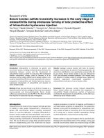
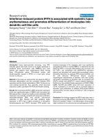
Figure 3 Proposed timeline for HIV-1 Vpr mediated pathology and resistance to therapy. Early in infection, Vpr allows for productive viral
infection of macrophages. These cells contribute to virus production and drug resistant reservoirs seen throughout the infection. During clinical
latency, Vpr contributes to the depletion of CD4
+
and CD8
+
T-cells, as well as interferes with antigen presentation. Such properties may
contribute to HIV-1 escape from immune surveillance, and effective humoral control of HIV infection. While it is yet unclear if neurocognitive
dysfunction and HIV-D are related pathologies, Vpr mediated immune dysregulation and neurotoxicity may contribute to early neurological
impairment in HIV-1 patients. Late in HIV-1 pathogenesis, increased expression of viral proteins including Vpr, contributes to the development of
associated pathologies, such as HIV-D and HIVAN.
Kogan and Rappaport Retrovirology 2011, 8:25
/>Page 12 of 20
this notion. Vpr/Vpx defective SIV virus has been
shown to have a greatly attenuated course with no
progression to AIDS in rhesus monkeys [120]. In HIV-
1, Vpx is absent and Vpr is thought to carry out Vpx
functions, suggesting that in humans a Vpr deletion
would have similar effects. Infection of Vpr defective
HIV-1 into tonsilar histocultures showed a fifty per-
cent reduction in HIV-1 production, even though
macrophages represented a small portion of total
infectable cells [214]. Further, an accidental infection
of a lab worker with HIV-1 containing a frame shift
mutation in codon 73 of the Vpr gene as well as infec-
tion of rhesus macaques with Vpr mutated virus
resulted in spontaneous reversion of the Vpr defective
virus to the WT phenotype, which implies that Vpr
containing virus obtained a selective advantage over
the Vpr mutant [134,215]. Vpr has also been shown to
reduce the efficacy of DNA and SIV-Nef vaccination in
vivo, suggesting that in the absence of Vpr a more
effective immune response to HIV would be possible
[183,184]. Finally, a recent study of six vertically
infected children that presented as long-term nonpro-
gressors reported that every patient had a mutated Vpr
gene in addition to mutations in other genes that were
not present in all patients [216]. Interestingly, all of
these mutations involved a decrease in Vpr’s apoptotic
effects, suggesting that the cytotoxic properties of Vpr
are of key clinical importance. However, another report
suggests that these effects are more related to nuclear
localization [217]. One of the major clinical conse-
quences of Vpr in HIV-1 infected patients is the exis-
tence of viral reservoirs in macrophages. Nucleoside
reverse transcriptase inhibitors (NRTIs) are more effec-
tive in macrophages than in CD4
+
T-cells for early
viral inhibition; non-NRTIs are equally effective in
macrophages and in CD4
+
T-cells for early infection
(for review see [218]). Protease inhibitors, however,
requireamuchhigherdosetoeffectivelycontrolHIV-
1 infection in macrophages than in CD4
+
T-cells, and
it is unknown if they achieve the concentrations
needed to inhibit macrophage mediated HIV-1 produc-
tion in compartments such as CNS or testes. While
NRTIs, non-NRTIs and protease inhibitors prevent the
cell to cell spread of HIV-1 infection, it is unknown
how efficiently these drugs address virus produced
from infected macrophages in vivo . There is currently
no therapeutic approach for eliminating macrophage
reservoirs that represent drug resistant reservoirs of
HIV-1 infection and contribute to the pathogenesis of
AIDS. Nanotechnology-based drug delivery systems
have been proposed as one method for delivering
drugs more effectively to macrophages, especially those
in relatively inaccessible body compartments [219,220].
These novel technologies offer ways to better del iver
currently available medications, but do not address the
survival of persis tently infected HIV-1 reservoirs.
A therapeutic approach to target HIV-1 infected
mononuclear cells would be to employ specific cytokines
or cellular kinase inhibitors. One candidate, TNF-related
apoptosis-inducing ligand (TRAIL), has been shown to
cause HIV-1 infected macrophages to undergo cell
death. However, M-CSF , which is upregulated in HIV-1
infected cells, downregulates TRAIL-R1/DR4 [221]. Ima-
tinib, a tyrosine kinase inhibitor that has some cross
reactivity to colony stimulating factor-1 receptor (CSF-
1R), the receptor for M-CSF, restores the effect of
TRAIL on infected MDM cells [221]. TRAIL has been
shown to act through the PI3/Akt pathway [222] and
consequently other PI3/Akt inhibitors have similar
effects on infected MDM cells [223]. Additionally, mor-
phine in combination with gp160 has been shown to
cause apoptosis in mononuclear cells [224]. In combina-
tion with cART therapy, a clinical approach to target
the anti-apoptotic pathways in HIV-1 infected macro-
phages may yield more effective therapies.
Another approach for targeting macrophage reservoirs
is to target the specific host mediators of Vpr function.
Hea t shock proteins have been proposed as cellular tar-
gets of Vpr and a mechanism of antiviral response (for
review see [225]). HSP 2 7 inhibits Vpr dependent G
2
arrest and c ell death in T-lymphocytes when expressed
exogenously, but does not seem to inhibit viral replica-
tion in macrophages [226]. Another heat shock protein,
HSP 70, can inhibit HIV-1 replication in a Vpr depen-
dent manner as well as reduce G
2
arrest in proliferating
cells [227]. HSP 70, however, can replace Vpr function
in Vpr defective viruses as well as have anti-viral proper-
ties in non-proliferating macrophages [228]. As heat
shock response is protective, increasing heat shock path-
ways could promote the survival of chronically infected
cells. In light of the recent findings suggesting that Vpr
mediated apoptotic effects are important in pathogen-
esis, and that G
2
arrest apoptosis and NK mediated
destruction of T-cells depends o n Vp r binding to
DCAF-1, targeting the Vpr ubiquitination pathways may
also be useful for clinical intervention. Additionally,
HAX-1 associates with Vpr, and suppresses Vpr pro-
apoptotic effects, suggesting that molecules that bind to
this site on Vpr may be used to neutralize Vpr’s immu-
nosuppressive effects [229]. Alternat ively, the anti-apop-
totic effects of Vpr in HIV-1 infected cells may
contribute to the persistence of viral reservoirs in vivo.
The Tat mediated upregulation of c-Flip, which prevents
TRAIL toxicity, has been proposed as one mechanism of
the differential effects of Vpr in infected and non-
infected cells and may prove to be a good target for
inducing apoptosis in chronically HIV-1 infected macro-
phages [230].
Kogan and Rappaport Retrovirology 2011, 8:25
/>Page 13 of 20
Several pharmacological approaches have already been
suggested to target Vpr pathways. As many Vpr
mediated effects depend on GR a ctivity, RU486 has
been proposed as a therapy for HIV-1 and has been
shown to suppress HIV-1 replication in infected mono-
nuclear cells and to suppress Vpr mediated downregula-
tion of IL-12 and other cyto kines [117,209]. Vpr is
necessary for viral PIC entry into the nucleus of non-
dividing cells and therefore this property of Vpr has also
been investigated as a potential avenue of therapy. CNI-
H0294, a specific inhibitor of HIV nuclear localization,
was shown to indeed inhibit viral production. It was
found to diminish infection in PBMCs and macro-
phages, which would not necessarily deplete viral reser-
voirs but may help prevent new macrophage infection
[231]. More recently, a study has demonstrated that
hematoxylin is a specific inhibitor of the Vpr/im portin-
a interaction and consequently prevented the nuclear
import of the HIV PIC complex [232]. In summary,
many studies have proposed targeting the cellular effects
of Vpr as a way of treating the consequences of Vpr
function in HIV-1 infection. In combinati on with estab-
lished cART regiments, these approaches may lower
viral loads, increase immune response, and even contri-
bute to the depletion of viral reservoirs thus improving
the clinical outcome in HIV patients.
Vpr as a pharmacotherapeutic and delivery agent
Vpr is a multifunctional protein that is able to efficiently
facilitate many HIV-1 functions. Some of these proper-
ties, however, lend themselves for use in the c linic.
Importantly, Vpr can traffic into cells [75] and is incor-
porated into HIV particles [12,233]. Further, the Vpr
peptide region from R14-88 has been used to introduce
other protein products into HIV-1 particles [234]. As a
result, Vpr has been explored as a vector system for
drug delivery by conjugation to apolipoprotein B mRNA
editing enzyme, catalytic peptide 3G (Vpr14-88-Apo-
bec3G) [235]. Apobec3G has strong antiviral effects in
Vif deficient viruses, but in the presence of Vif loses the
ability to incorp orate into virons and therefore its thera-
peutic efficacy [236,237]. The fusion of Vpr 14-88 to
Apobec3G facilitates packaging into the HIV-1 particles
and restores the ability of Apobec3G to inhibit viral
replication. These studies demonstrate that the use of
Vpr to amplify the effect of antiviral drugs or facilitate
drug delivery is a promising avenue for HIV therapy.
The discoveries of other properties of Vpr, including
induction of G
2
cell cycle arrest and apop tosis, have led
the argument that Vpr has efficacy as an anti-cancer
agent [238]. Further, Vpr induction of apoptosis seems
to be independent of p53 function, suggesting that
mutations in p53 commonly seen in var ious tumor
types will not prevent the potential therapeutic efficac y
of Vpr [193]. Other studies have also provided support
for the anti-cancer application of Vpr by showing that
Vpr induces greater apoptosis in cells underg oing active
replication, implying that this toxic effect would be par-
ticularly targeted to cancer cells [73,209]. However, Vpr,
like other chemotherapeutic agents, also possesses the
ability to transform cells as double stranded breaks and
aneuploidy have been reported in cell lines [239]. The
consequences of Vpr on mitogenic transformation in
vivo require further assessment and remain one poten-
tial limitation of such a therapeutic approach.
Conclusion
More than two decades of research on Vpr has greatly
contributed to the knowledg e scientists and clinicians
have available about HIV-1 pathogenesis. The findings
revealed that Vpr, while not essential for viral replica-
tion per se, is a biologically important, playing a critical
role in the infection of non-dividing target cells includ-
ing macrophages and resting T-cells. Vpr promotes
infection of dividing as well as non-dividing cells
through a variety of effects including, nuclear localiza-
tion, cell cycle arrest, apoptosis, and other effects due to
DCAF-1 binding, as well as transactivation of host and
viral genes. These activities of Vpr are likely responsi ble
for many aspects of HIV-1 infection as well as asso-
ciated pathology seen in AIDS. With the advent and
success of cART therapy, HIV-1 infection has trans-
formed from an untreatable disease to a more manage-
able chronic condition. Current investigations for new
therapies represent an ongoing area of basic science
research that holds great priority due to cART resistant
reservoirs of HIV-1 infection in vivo. Vpr mediated
pathogenesis is one avenue of investigation that holds
promise when combined with o ther therapeutic
approaches. Further basic and translational studies will
be required to generate future therapeutic advances tar-
geting Vpr function. Such studies could target Vpr as
well as a variety of host-virus interaction pathways.
Acknowledgements and Funding
The figures created used images found in Science Slides (Visi Science Inc.,
Chapel Hill, NC). This work was supported by the National Institutes of
Health under Ruth L. Kirschstein National Research Service Award
(T32MH079785, Interdisciplinary and Trans lational Research Training in
NeuroAIDS) providing support to Michael Kogan. Jay Rappaport is supported
by R01 grants from the NINDS and NIMH. The authors would also like to
thank Michael Jan, a fellow student in the MD/PhD program at Temple
University, for extensive help in proofreading of this manuscript.
Authors’ contributions
MK conducted a literature review and wrote the above material. MK also
created the figures presented in the paper. JR organized the ideas presented
in this review and helped to edit the content so that it is relevant to current
HIV research. JR also suggested ideas on how to illustrate the figures and
highlighted important concepts that needed to be included. Both authors
read and approved the final manuscript.
Kogan and Rappaport Retrovirology 2011, 8:25
/>Page 14 of 20
Competing interests
The authors declare that they have no competing interests.
Received: 25 October 2010 Accepted: 13 April 2011
Published: 13 April 2011
References
1. Cohen EA, Terwilliger EF, Jalinoos Y, Proulx J, Sodroski JG, Haseltine WA:
Identification of HIV-1 vpr product and function. J Acquir Immune Defic
Syndr 1990, 3:11-18.
2. Yuan X, Matsuda Z, Matsuda M, Essex M, Lee TH: Human
immunodeficiency virus vpr gene encodes a virion-associated protein.
AIDS Res Hum Retroviruses 1990, 6:1265-1271.
3. Emerman M: HIV-1, Vpr and the cell cycle. Curr Biol 1996, 6:1096-1103.
4. Planelles V, Jowett JB, Li QX, Xie Y, Hahn B, Chen IS: Vpr-induced cell cycle
arrest is conserved among primate lentiviruses. J Virol 1996, 70:2516-2524.
5. Tristem M, Marshall C, Karpas A, Hill F: Evolution of the primate
lentiviruses: evidence from vpx and vpr. Embo J 1992, 11:3405-3412.
6. Feldherr CM, Feldherr AB: The nuclear membrane as a barrier to the free
diffusion of proteins. Nature 1960, 185:250-251.
7. Lewis PF, Emerman M: Passage through mitosis is required for
oncoretroviruses but not for the human immunodeficiency virus. J Virol
1994, 68:510-516.
8. Roe T, Reynolds TC, Yu G, Brown PO: Integration of murine leukemia virus
DNA depends on mitosis. Embo J 1993, 12:2099-2108.
9. Lewis P, Hensel M, Emerman M: Human immunodeficiency virus infection
of cells arrested in the cell cycle. Embo J 1992, 11:3053-3058.
10. Bukrinsky MI, Sharova N, Dempsey MP, Stanwick TL, Bukrinskaya AG,
Haggerty S, Stevenson M: Active nuclear import of human
immunodeficiency virus type 1 preintegration complexes. Proc Natl Acad
Sci USA 1992, 89:6580-6584.
11. Di Marzio P, Choe S, Ebright M, Knoblauch R, Landau NR: Mutational
anal ysis of cell cycle arrest, nuclear localizati on and virion packaging
of huma n immunodeficiency virus type 1 Vpr. JVirol1995,
69:7909-7916.
12. Lu YL, Spearman P, Ratner L: Human immunodeficiency virus type 1 viral
protein R localization in infected cells and virions. J Virol 1993,
67:6542-6550.
13. Mahalingam S, Collman RG, Patel M, Monken CE, Srinivasan A: Functional
analysis of HIV-1 Vpr: identification of determinants essential for
subcellular localization. Virology 1995, 212:331-339.
14. Gallay P, Hope T, Chin D, Trono D: HIV-1 infection of nondividing cells
through the recognition of integrase by the importin/karyopherin
pathway. Proc Natl Acad Sci USA 1997, 94:9825-9830.
15. Arhel NJ, Souquere-Besse S, Munier S, Souque P, Guadagnini S,
Rutherford S, Prevost MC, Allen TD, Charneau P: HIV-1 DNA Flap formation
promotes uncoating of the pre-integration complex at the nuclear pore.
EMBO
J 2007, 26:3025-3037.
16. Dismuke DJ, Aiken C: Evidence for a functional link between uncoating
of the human immunodeficiency virus type 1 core and nuclear import
of the viral preintegration complex. J Virol 2006, 80:3712-3720.
17. Gorlich D: Nuclear protein import. Curr Opin Cell Biol 1997, 9:412-419.
18. Herold A, Truant R, Wiegand H, Cullen BR: Determination of the functional
domain organization of the importin alpha nuclear import factor. J Cell
Biol 1998, 143:309-318.
19. Kobe B: Autoinhibition by an internal nuclear localization signal revealed
by the crystal structure of mammalian importin alpha. Nat Struct Biol
1999, 6:388-397.
20. Moore MS, Blobel G: Purification of a Ran-interacting protein that is
required for protein import into the nucleus. Proc Natl Acad Sci USA 1994,
91:10212-10216.
21. Moore MS, Blobel G: The GTP-binding protein Ran/TC4 is required for
protein import into the nucleus. Nature 1993, 365:661-663.
22. Paschal BM, Gerace L: Identification of NTF2, a cytosolic factor for nuclear
import that interacts with nuclear pore complex protein p62. J Cell Biol
1995, 129:925-937.
23. Melchior F, Paschal B, Evans J, Gerace L: Inhibition of nuclear protein
import by nonhydrolyzable analogues of GTP and identification of the
small GTPase Ran/TC4 as an essential transport factor. J Cell Biol 1993,
123:1649-1659.
24. Melchior F, Guan T, Yokoyama N, Nishimoto T, Gerace L: GTP hydrolysis by
Ran occurs at the nuclear pore complex in an early step of protein
import. J Cell Biol 1995, 131:571-581.
25. Kotera I, Sekimoto T, Miyamoto Y, Saiwaki T, Nagoshi E, Sakagami H,
Kondo H, Yoneda Y: Importin alpha transports CaMKIV to the nucleus
without utilizing importin beta. Embo J 2005, 24:942-951.
26. Takizawa CG, Weis K, Morgan DO: Ran-independent nuclear import of
cyclin B1-Cdc2 by importin beta. Proc Natl Acad Sci USA 1999,
96:7938-7943.
27. Aitchison JD, Blobel G, Rout MP: Kap104p: a karyopherin involved in the
nuclear transport of messenger RNA binding proteins. Science 1996,
274:624-627.
28. Fridell RA, Truant R, Thorne L, Benson RE, Cullen BR: Nuclear import of
hnRNP A1 is mediated by a novel cellular cofactor related to
karyopherin-beta. J Cell Sci 1997, 110(Pt 11):1325-1331.
29. Michael WM, Choi M, Dreyfuss G: A nuclear export signal in hnRNP A1: a
signal-mediated, temperature-dependent nuclear protein export
pathway. Cell 1995, 83:415-422.
30.
Pollard VW, Michael WM, Nakielny S, Siomi MC, Wang F, Dreyfuss G: A
novel receptor-mediated nuclear protein import pathway. Cell 1996,
86:985-994.
31. Siomi H, Dreyfuss G: A nuclear localization domain in the hnRNP A1
protein. J Cell Biol 1995, 129:551-560.
32. Nakielny S, Siomi MC, Siomi H, Michael WM, Pollard V, Dreyfuss G:
Transportin: nuclear transport receptor of a novel nuclear protein import
pathway. Exp Cell Res 1996, 229:261-266.
33. Palacios I, Weis K, Klebe C, Mattaj IW, Dingwall C: RAN/TC4 mutants
identify a common requirement for snRNP and protein import into the
nucleus. J Cell Biol 1996, 133:485-494.
34. Jenkins Y, McEntee M, Weis K, Greene WC: Characterization of HIV-1 vpr
nuclear import: analysis of signals and pathways. J Cell Biol 1998,
143:875-885.
35. Efthymiadis A, Shao H, Hubner S, Jans DA: Kinetic characterization of the
human retinoblastoma protein bipartite nuclear localization sequence
(NLS) in vivo and in vitro. A comparison with the SV40 large T-antigen
NLS. J Biol Chem 1997, 272:22134-22139.
36. Gorlich D, Henklein P, Laskey RA, Hartmann E: A 41 amino acid motif in
importin-alpha confers binding to importin-beta and hence transit into
the nucleus. Embo J 1996, 15:1810-1817.
37. Jans DA, Jans P, Julich T, Briggs LJ, Xiao CY, Piller SC: Intranuclear binding
by the HIV-1 regulatory protein VPR is dependent on cytosolic factors.
Biochem Biophys Res Commun 2000, 270:1055-1062.
38. Nitahara-Kasahara Y, Kamata M, Yamamoto T, Zhang X, Miyamoto Y,
Muneta K, Iijima S, Yoneda Y, Tsunetsugu-Yokota Y, Aida Y: Novel nuclear
import of Vpr promoted by importin alpha is crucial for human
immunodeficiency virus type 1 replication in macrophages. J Virol 2007,
81:5284-5293.
39. Kamata M, Nitahara-Kasahara Y, Miyamoto Y, Yoneda Y, Aida Y: Importin-
alpha promotes passage through the nuclear pore complex of human
immunodeficiency virus type 1 Vpr. J Virol 2005, 79:3557-3564.
40. Balliet JW, Kolson DL, Eiger G, Kim FM, McGann KA, Srinivasan A, Collman R:
Distinct effects in primary macrophages and lymphocytes of the human
immunodeficiency virus type 1 accessory genes vpr, vpu, and nef:
mutational analysis of a primary HIV-1 isolate. Virology 1994, 200:623-631.
41. Connor RI, Chen BK, Choe S, Landau NR: Vpr is required for efficient
replication of human immunodeficiency virus type-1 in mononuclear
phagocytes. Virology 1995, 206:935-944.
42. Hattori N, Michaels F, Fargnoli K, Marcon L, Gallo RC, Franchini G: The
human immunodeficiency virus type 2 vpr gene is essential for
productive infection of human macrophages. Proc Natl Acad Sci USA
1990, 87:8080-8084.
43. Ogawa K, Shibata R, Kiyomasu T, Higuchi I, Kishida Y, Ishimoto A, Adachi A:
Mutational analysis of the human immunodeficiency virus vpr open
reading frame. J Virol
1989, 63:4110-4114.
44.
Blomer U, Naldini L, Kafri T, Trono D, Verma IM, Gage FH: Highly efficient
and sustained gene transfer in adult neurons with a lentivirus vector. J
Virol 1997, 71:6641-6649.
45. Agostini I, Popov S, Hao T, Li JH, Dubrovsky L, Chaika O, Chaika N, Lewis R,
Bukrinsky M: Phosphorylation of Vpr regulates HIV type 1 nuclear import
and macrophage infection. AIDS Res Hum Retroviruses 2002, 18:283-288.
Kogan and Rappaport Retrovirology 2011, 8:25
/>Page 15 of 20
46. Caly L, Saksena NK, Piller SC, Jans DA: Impaired nuclear import and viral
incorporation of Vpr derived from a HIV long-term non-progressor.
Retrovirology 2008, 5:67.
47. Fouchier RA, Meyer BE, Simon JH, Fischer U, Albright AV, Gonzalez-
Scarano F, Malim MH: Interaction of the human immunodeficiency virus
type 1 Vpr protein with the nuclear pore complex. J Virol 1998,
72:6004-6013.
48. Gallay P, Stitt V, Mundy C, Oettinger M, Trono D: Role of the karyopherin
pathway in human immunodeficiency virus type 1 nuclear import. J Virol
1996, 70:1027-1032.
49. Jacquot G, Le Rouzic E, David A, Mazzolini J, Bouchet J, Bouaziz S,
Niedergang F, Pancino G, Benichou S: Localization of HIV-1 Vpr to the
nuclear envelope: impact on Vpr functions and virus replication in
macrophages. Retrovirology 2007, 4:84.
50. Vodicka MA, Koepp DM, Silver PA, Emerman M: HIV-1 Vpr interacts with
the nuclear transport pathway to promote macrophage infection. Genes
Dev 1998, 12:175-185.
51. Popov S, Rexach M, Ratner L, Blobel G, Bukrinsky M: Viral protein R
regulates docking of the HIV-1 preintegration complex to the nuclear
pore complex. J Biol Chem 1998, 273:13347-13352.
52. Keele BF, Giorgi EE, Salazar-Gonzalez JF, Decker JM, Pham KT, Salazar MG,
Sun C, Grayson T, Wang S, Li H, et al: Identification and characterization of
transmitted and early founder virus envelopes in primary HIV-1
infection. Proc Natl Acad Sci USA 2008, 105:7552-7557.
53. Fischer W, Ganusov VV, Giorgi EE, Hraber PT, Keele BF, Leitner T, Han CS,
Gleasner CD, Green L, Lo CC, et al: Transmission of single HIV-1 genomes
and dynamics of early immune escape revealed by ultra-deep
sequencing. PLoS One 2010, 5:e12303.
54. Iijima S, Nitahara-Kasahara Y, Kimata K, Zhong Zhuang W, Kamata M,
Isogai M, Miwa M, Tsunetsugu-Yokota Y, Aida Y: Nuclear localization of Vpr
is crucial for the efficient replication of HIV-1 in primary CD4+ T cells.
Virology 2004, 327:249-261.
55. Bukrinsky MI, Haffar OK: HIV-1 nuclear import: in search of a leader. Front
Biosci 1997, 2:d578-587.
56. Yamashita M, Perez O, Hope TJ, Emerman M: Evidence for direct
involvement of the capsid protein in HIV infection of nondividing cells.
PLoS Pathog 2007, 3:1502-1510.
57. Craigo JK, Montelaro RC: Lentivirus Tropism and Disease. In Lentiviruses
and Macrophages: Molecular and Cellular Interactions. Edited by: Desport M.
Norfolk: Caister Academic Press; 2010:1-24.
58. Morellet N, Bouaziz S, Petitjean P, Roques BP: NMR structure of the HIV-1
regulatory protein VPR. J Mol Biol 2003, 327:215-227.
59. Mahalingam S, Ayyavoo V, Patel M, Kieber-Emmons T, Weiner DB: Nuclear
import, virion incorporation, and cell cycle arrest/differentiation are
mediated
by distinct functional domains of human immunodeficiency
virus type 1 Vpr. J Virol 1997, 71:6339-6347.
60. Nie Z, Bergeron D, Subbramanian RA, Yao XJ, Checroune F, Rougeau N,
Cohen EA: The putative alpha helix 2 of human immunodeficiency virus
type 1 Vpr contains a determinant which is responsible for the nuclear
translocation of proviral DNA in growth-arrested cells. J Virol 1998,
72:4104-4115.
61. Singh SP, Tomkowicz B, Lai D, Cartas M, Mahalingam S, Kalyanaraman VS,
Murali R, Srinivasan A: Functional role of residues corresponding to
helical domain II (amino acids 35 to 46) of human immunodeficiency
virus type 1 Vpr. J Virol 2000, 74:10650-10657.
62. Yao XJ, Subbramanian RA, Rougeau N, Boisvert F, Bergeron D, Cohen EA:
Mutagenic analysis of human immunodeficiency virus type 1 Vpr: role of
a predicted N-terminal alpha-helical structure in Vpr nuclear localization
and virion incorporation. J Virol 1995, 69:7032-7044.
63. Fritz JV, Didier P, Clamme JP, Schaub E, Muriaux D, Cabanne C, Morellet N,
Bouaziz S, Darlix JL, Mely Y, de Rocquigny H: Direct Vpr-Vpr interaction in
cells monitored by two photon fluorescence correlation spectroscopy
and fluorescence lifetime imaging. Retrovirology 2008, 5:87.
64. Agostini I, Popov S, Li J, Dubrovsky L, Hao T, Bukrinsky M: Heat-shock
protein 70 can replace viral protein R of HIV-1 during nuclear import of
the viral preintegration complex. Exp Cell Res 2000, 259:398-403.
65. Popov S, Rexach M, Zybarth G, Reiling N, Lee MA, Ratner L, Lane CM,
Moore MS, Blobel G, Bukrinsky M: Viral protein R regulates nuclear import
of the HIV-1 pre-integration complex. Embo J 1998, 17:909-917.
66. Kamata M, Aida Y: Two putative alpha-helical domains of human
immunodeficiency virus type 1 Vpr mediate nuclear localization by at
least two mechanisms. J Virol 2000, 74:7179-7186.
67. Le Rouzic E, Mousnier A, Rustum C, Stutz F, Hallberg E, Dargemont C,
Benichou S: Docking of HIV-1 Vpr to the nuclear envelope is mediated
by the interaction with the nucleoporin hCG1. J Biol Chem 2002,
277:45091-45098.
68. Radu A, Moore MS, Blobel G: The peptide repeat domain of nucleoporin
Nup98 functions as a docking site in transport across the nuclear pore
complex. Cell 1995, 81:215-222.
69. Rexach M, Blobel G: Protein import into nuclei: association and
dissociation reactions involving transport substrate, transport factors,
and nucleoporins. Cell 1995, 83:683-692.
70. Kutay U, Izaurralde E, Bischoff FR, Mattaj IW, Gorlich D: Dominant-negative
mutants of importin-beta block multiple pathways of import and export
through the nuclear pore complex. Embo J 1997, 16:1153-1163.
71. Karni O, Friedler A, Zakai N, Gilon C, Loyter A: A peptide derived from the
N-terminal region of HIV-1 Vpr promotes nuclear import in
permeabilized cells: elucidation of the NLS region of the Vpr. FEBS Lett
1998, 429:421-425.
72. Zhou Y, Lu Y, Ratner L: Arginine residues in the C-terminus of HIV-1 Vpr
are important for nuclear localization and cell cycle arrest. Virology
1998,
242:414-424.
73.
Stewart SA, Poon B, Jowett JB, Chen IS: Human immunodeficiency virus
type 1 Vpr induces apoptosis following cell cycle arrest. J Virol 1997,
71:5579-5592.
74. Levy DN, Refaeli Y, MacGregor RR, Weiner DB: Serum Vpr regulates
productive infection and latency of human immunodeficiency virus type
1. Proc Natl Acad Sci USA 1994, 91:10873-10877.
75. Levy DN, Refaeli Y, Weiner DB: Extracellular Vpr protein increases cellular
permissiveness to human immunodeficiency virus replication and
reactivates virus from latency. J Virol 1995, 69:1243-1252.
76. Agostini I, Navarro JM, Rey F, Bouhamdan M, Spire B, Vigne R, Sire J: The
human immunodeficiency virus type 1 Vpr transactivator: cooperation
with promoter-bound activator domains and binding to TFIIB. J Mol Biol
1996, 261:599-606.
77. Cohen EA, Dehni G, Sodroski JG, Haseltine WA: Human immunodeficiency
virus vpr product is a virion-associated regulatory protein. J Virol 1990,
64:3097-3099.
78. Wang L, Mukherjee S, Jia F, Narayan O, Zhao LJ: Interaction of virion
protein Vpr of human immunodeficiency virus type 1 with cellular
transcription factor Sp1 and trans-activation of viral long terminal
repeat. J Biol Chem 1995, 270:25564-25569.
79. Ghosh D: Glucocorticoid receptor-binding site in the human
immunodeficiency virus long terminal repeat. J Virol 1992, 66:586-590.
80. Katsanakis CD, Sekeris CE, Spandidos DA: The human immunodeficiency
virus long terminal repeat contains sequences showing partial
homology to glucocorticoid responsive elements. Anticancer Res 1991,
11:381-383.
81. McAllister JJ, Phillips D, Millhouse S, Conner J, Hogan T, Ross HL, Wigdahl B:
Analysis of the HIV-1 LTR NF-kappaB-proximal Sp site III: evidence for
cell type-specific gene regulation and viral replication. Virology 2000,
274:262-277.
82. Soudeyns H, Geleziunas R, Shyamala G, Hiscott J, Wainberg MA:
Identification of a novel glucocorticoid response element within the
genome of the human immunodeficiency virus type 1. Virology 1993,
194:758-768.
83. Verhoef K, Sanders RW, Fontaine V, Kitajima S, Berkhout B: Evolution of the
human immunodeficiency virus type 1 long terminal repeat promoter
by conversion of an NF-kappaB enhancer element into a GABP binding
site. J Virol 1999, 73:1331-1340.
84. Vanitharani R, Mahalingam S, Rafaeli Y, Singh SP, Srinivasan A, Weiner DB,
Ayyavoo V: HIV-1 Vpr transactivates LTR-directed expression through
sequences present within -278 to -176 and increases virus replication in
vitro. Virology 2001, 289:334-342.
85. Felzien LK, Woffendin C, Hottiger MO, Subbramanian RA, Cohen EA,
Nabel GJ: HIV transcriptional activation by the accessory protein, VPR, is
mediated by the p300 co-activator. Proc Natl Acad Sci USA 1998,
95:5281-5286.
Kogan and Rappaport Retrovirology 2011, 8:25
/>Page 16 of 20
86. Kino T, Tsukamoto M, Chrousos G: Transcription factor TFIIH components
enhance the GR coactivator activity but not the cell cycle-arresting
activity of the human immunodeficiency virus type-1 protein Vpr.
Biochem Biophys Res Commun 2002, 298:17-23.
87. Sawaya BE, Khalili K, Rappaport J, Serio D, Chen W, Srinivasan A, Amini S:
Suppression of HIV-1 transcription and replication by a Vpr mutant. Gene
Ther 1999, 6:947-950.
88. Sawaya BE, Khalili K, Gordon J, Taube R, Amini S: Cooperative interaction
between HIV-1 regulatory proteins Tat and Vpr modulates transcription
of the viral genome. J Biol Chem 2000, 275:35209-35214.
89. Refaeli Y, Levy DN, Weiner DB: The glucocorticoid receptor type II
complex is a target of the HIV-1 vpr gene product. Proc Natl Acad Sci
USA 1995, 92:3621-3625.
90. Kino T, Gragerov A, Kopp JB, Stauber RH, Pavlakis GN, Chrousos GP: The
HIV-1 virion-associated protein vpr is a coactivator of the human
glucocorticoid receptor. J Exp Med 1999, 189:51-62.
91. Sherman MP, de Noronha CM, Pearce D, Greene WC: Human
immunodeficiency virus type 1 Vpr contains two leucine-rich helices
that mediate glucocorticoid receptor coactivation independently of its
effects on G(2) cell cycle arrest. J Virol 2000, 74:8159-8165.
92. Kino T, Gragerov A, Slobodskaya O, Tsopanomichalou M, Chrousos GP,
Pavlakis GN: Human immunodeficiency virus type 1 (HIV-1) accessory
protein Vpr induces transcription of the HIV-1 and glucocorticoid-
responsive promoters by binding directly to p300/CBP coactivators. J
Virol 2002, 76:9724-9734.
93. Thotala D, Schafer EA, Tungaturthi PK, Majumder B, Janket ML, Wagner M,
Srinivasan A, Watkins S, Ayyavoo V: Structure-functional analysis of human
immunodeficiency virus type 1 (HIV-1) Vpr: role of leucine residues on
Vpr-mediated transactivation and virus replication. Virology 2004,
328:89-100.
94. Ramanathan MP, Curley E, Su M, Chambers JA, Weiner DB: Carboxyl
terminus of hVIP/mov34 is critical for HIV-1-Vpr interaction and
glucocorticoid-mediated signaling. J Biol Chem 2002, 277:47854-47860.
95. Muthumani K, Choo AY, Zong WX, Madesh M, Hwang DS, Premkumar A,
Thieu KP, Emmanuel J, Kumar S, Thompson CB, Weiner DB: The HIV-1 Vpr
and glucocorticoid receptor complex is a gain-of-function interaction that
prevents the nuclear localization of PARP-1. Nat Cell Biol 2006, 8:170-179.
96. Poli G, Kinter A, Justement JS, Kehrl JH, Bressler P, Stanley S, Fauci AS:
Tumor necrosis factor alpha functions in an autocrine manner in the
induction of human immunodeficiency virus expression. Proc Natl Acad
Sci USA 1990, 87:782-785.
97. Lenardo MJ, Baltimore D: NF-kappa B: a pleiotropic mediator of inducible
and tissue-specific gene control. Cell 1989, 58:227-229.
98. Chang HK, Gallo RC, Ensoli B: Regulation of Cellular Gene Expression and
Function by the Human Immunodeficiency Virus Type 1 Tat Protein. J
Biomed Sci 1995, 2:189-202.
99. Chatterton RT Jr, Green D, Harris S, Grossman A, Hechter O: Longitudinal
study of adrenal steroids in a cohort of HIV-infected patients with
hemophilia. J Lab Clin Med
1996, 127:545-552.
100.
Kawa SK, Thompson EB: Lymphoid cell resistance to glucocorticoids in
HIV infection. J Steroid Biochem Mol Biol 1996, 57:259-263.
101. Lortholary O, Christeff N, Casassus P, Thobie N, Veyssier P, Trogoff B, Torri O,
Brauner M, Nunez EA, Guillevin L: Hypothalamo-pituitary-adrenal function
in human immunodeficiency virus-infected men. J Clin Endocrinol Metab
1996, 81:791-796.
102. Laudat A, Blum L, Guechot J, Picard O, Cabane J, Imbert JC, Giboudeau J:
Changes in systemic gonadal and adrenal steroids in asymptomatic
human immunodeficiency virus-infected men: relationship with the CD4
cell counts. Eur J Endocrinol 1995, 133:418-424.
103. Biglino A, Limone P, Forno B, Pollono A, Cariti G, Molinatti GM, Gioannini P:
Altered adrenocorticotropin and cortisol response to corticotropin-
releasing hormone in HIV-1 infection. Eur J Endocrinol 1995, 133:173-179.
104. Kumar M, Kumar AM, Morgan R, Szapocznik J, Eisdorfer C: Abnormal
pituitary-adrenocortical response in early HIV-1 infection. J Acquir
Immune Defic Syndr 1993, 6:61-65.
105. Kino T, Kopp JB, Chrousos GP: Glucocorticoids suppress human
immunodeficiency virus type-1 long terminal repeat activity in a cell
type-specific, glucocorticoid receptor-mediated fashion: direct protective
effects at variance with clinical phenomenology. J Steroid Biochem Mol
Biol 2000, 75:283-290.
106. Laurence J, Sellers MB, Sikder SK: Effect of glucocorticoids on chronic
human immunodeficiency virus (HIV) infection and HIV promoter-
mediated transcription. Blood 1989, 74:291-297.
107. Mitra D, Sikder S, Laurence J: Inhibition of tat-activated, HIV-1 long
terminal repeat-mediated gene expression by glucocorticoids. AIDS Res
Hum Retroviruses 1993, 9:1055-1056.
108. Mitra D, Sikder SK, Laurence J: Role of glucocorticoid receptor binding
sites in the human immunodeficiency virus type 1 long terminal repeat
in steroid-mediated suppression of HIV gene expression. Virology 1995,
214:512-521.
109. Russo FO, Patel PC, Ventura AM, Pereira CA: HIV-1 long terminal repeat
modulation by glucocorticoids in monocytic and lymphocytic cell lines.
Virus Res 1999, 64:87-94.
110. Furth PA, Westphal H, Hennighausen L: Expression from the HIV-LTR is
stimulated by glucocorticoids and pregnancy. AIDS Res Hum Retroviruses
1990, 6:553-560.
111. Kinter AL, Biswas P, Alfano M, Justement JS, Mantelli B, Rizzi C, Gatti AR,
Vicenzi E, Bressler P, Poli G: Interleukin-6 and glucocorticoids
synergistically induce human immunodeficiency virus type-1 expression
in chronically infected U1 cells by a long terminal repeat independent
post-transcriptional mechanism. Mol Med 2001, 7:668-678.
112. Hoshino S, Konishi M, Mori M, Shimura M, Nishitani C, Kuroki Y, Koyanagi Y,
Kano S, Itabe H, Ishizaka Y: HIV-1 Vpr induces TLR4/MyD88-mediated IL-6
production and reactivates viral production from latency. J Leukoc Biol
2010, 87:1133-1143.
113. Bressler P, Poli G, Justement JS, Biswas P, Fauci AS: Glucocorticoids
synergize with tumor necrosis factor alpha in the induction of HIV
expression
from a chronically infected promonocytic cell line. AIDS Res
Hum Retroviruses 1993, 9:547-551.
114. Capitanio JP, Mendoza SP, Lerche NW, Mason WA: Social stress results in
altered glucocorticoid regulation and shorter survival in simian acquired
immune deficiency syndrome. Proc Natl Acad Sci USA 1998, 95:4714-4719.
115. Corley PA: Induction of interleukin-1 and glucocorticoid hormones by
HIV promotes viral replication and links human chromosome 2 to AIDS
pathogenesis: genetic mechanisms and therapeutic implications. Med
Hypotheses 1997, 48:415-421.
116. Nair MP, Saravolatz LD, Schwartz SA: Selective inhibitory effects of stress
hormones on natural killer (NK) cell activity of lymphocytes from AIDS
patients. Immunol Invest 1995, 24:689-699.
117. Schafer EA, Venkatachari NJ, Ayyavoo V: Antiviral effects of mifepristone
on human immunodeficiency virus type-1 (HIV-1): targeting Vpr and its
cellular partner, the glucocorticoid receptor (GR). Antiviral Res 2006,
72:224-232.
118. Wiegers K, Schwarck D, Reimer R, Bohn W: Activation of the glucocorticoid
receptor releases unstimulated PBMCs from an early block in HIV-1
replication. Virology 2008, 375:73-84.
119. Fletcher TM, Brichacek B, Sharova N, Newman MA, Stivahtis G, Sharp PM,
Emerman M, Hahn BH, Stevenson M: Nuclear import and cell cycle arrest
functions of the HIV-1 Vpr protein are encoded by two separate genes
in HIV-2/SIV(SM). Embo J 1996, 15:6155-6165.
120. Gibbs JS, Lackner AA, Lang SM, Simon MA, Sehgal PK, Daniel MD,
Desrosiers RC: Progression to AIDS in the absence of a gene for vpr or
vpx. J Virol 1995, 69:2378-2383.
121. Subbramanian RA, Kessous-Elbaz A, Lodge R, Forget J, Yao XJ, Bergeron D,
Cohen EA: Human immunodeficiency virus type 1 Vpr is a positive
regulator of viral transcription and infectivity in primary human
macrophages. J Exp Med 1998, 187:1103-1111.
122. Belzile JP, Duisit G, Rougeau N, Mercier J, Finzi A, Cohen EA: HIV-1 Vpr-
mediated G2 arrest involves the DDB1-CUL4AVPRBP E3 ubiquitin ligase.
PLoS Pathog 2007, 3:e85.
123. DeHart JL, Zimmerman ES, Ardon O, Monteiro-Filho CM, Arganaraz ER,
Planelles V: HIV-1 Vpr activates the G2 checkpoint through manipulation
of the ubiquitin proteasome system. Virol J 2007, 4:57.
124. Hrecka K, Gierszewska M, Srivastava S, Kozaczkiewicz L, Swanson SK,
Florens L, Washburn MP, Skowronski J: Lentiviral Vpr usurps Cul4-DDB1
[VprBP] E3 ubiquitin ligase to modulate cell cycle. Proc Natl Acad Sci USA
2007, 104:11778-11783.
125. Le Rouzic E, Belaïdouni N, Estrabaud E, Morel M, Rain J, Transy C, Margottin-
Goguet F: HIV1 Vpr arrests the cell cycle by recruiting DCAF1/VprBP, a
receptor of the Cul4-DDB1 ubiquitin ligase. Cell Cycle 2007, 6:182-188.
Kogan and Rappaport Retrovirology 2011, 8:25
/>Page 17 of 20
126. Schrofelbauer B, Hakata Y, Landau NR: HIV-1 Vpr function is mediated by
interaction with the damage-specific DNA-binding protein DDB1. Proc
Natl Acad Sci USA 2007, 104:4130-4135.
127. Tan L, Ehrlich E, Yu XF: DDB1 and Cul4A are required for human
immunodeficiency virus type 1 Vpr-induced G2 arrest. J Virol 2007,
81:10822-10830.
128. Wen X, Duus KM, Friedrich TD, de Noronha CM: The HIV1 protein Vpr acts
to promote G2 cell cycle arrest by engaging a DDB1 and Cullin4A-
containing ubiquitin ligase complex using VprBP/DCAF1 as an adaptor. J
Biol Chem 2007, 282:27046-27057.
129. Zhao LJ, Mukherjee S, Narayan O: Biochemical mechanism of HIV-I Vpr
function. Specific interaction with a cellular protein. J Biol Chem 1994,
269:15577-15582.
130. Ayinde D, Maudet C, Transy C, Margottin-Goguet F: Limelight on two HIV/
SIV accessory proteins in macrophage infection: is Vpx overshadowing
Vpr? Retrovirology 2010, 7:35.
131. Casey L, Wen X, de Noronha CM: The functions of the HIV1 protein Vpr
and its action through the DCAF1.DDB1.Cullin4 ubiquitin ligase. Cytokine
2010, 51:1-9.
132. Zimmerman E, Sherman M, Blackett J, Neidleman J, Kreis C, Mundt P,
Williams S, Warmerdam M, Kahn J, Hecht F, et al: Human
immunodeficiency virus type 1 Vpr induces DNA replication stress in
vitro and in vivo. J Virol 2006, 80:10407-10418.
133. Forget J, Yao XJ, Mercier J, Cohen EA: Human immunodeficiency virus
type 1 vpr protein transactivation function: mechanism and
identification of domains involved. J Mol Biol 1998, 284:915-923.
134. Goh WC, Rogel ME, Kinsey CM, Michael SF, Fultz PN, Nowak MA, Hahn BH,
Emerman M: HIV-1 Vpr increases viral expression by manipulation of the
cell cycle: a mechanism for selection of Vpr in vivo. Nat Med 1998,
4:65-71.
135. Igarashi T, Brown CR, Endo Y, Buckler-White A, Plishka R, Bischofberger N,
Hirsch V, Martin MA: Macrophage are the principal reservoir and sustain
high virus loads in rhesus macaques after the depletion of CD4+ T cells
by a highly pathogenic simian immunodeficiency virus/HIV type 1
chimera (SHIV): Implications for HIV-1 infections of humans. Proc Natl
Acad Sci USA 2001, 98:658-663.
136. Tristem M, Marshall C, Karpas A, Petrik J, Hill F: Origin of vpx in
lentiviruses. Nature 1990, 347:341-342.
137. Sharova N, Wu Y, Zhu X, Stranska R, Kaushik R, Sharkey M, Stevenson M:
Primate lentiviral Vpx commandeers DDB1 to counteract a macrophage
restriction. PLoS Pathog 2008, 4:e1000057.
138. Srivastava S, Swanson SK, Manel N, Florens L, Washburn MP, Skowronski J:
Lentiviral Vpx accessory factor targets VprBP/DCAF1 substrate adaptor
for cullin 4 E3 ubiquitin ligase to enable macrophage infection. PLoS
Pathog 2008, 4:e1000059.
139. Bergamaschi A, Ayinde D, David A, Le Rouzic E, Morel M, Collin G,
Descamps D, Damond F, Brun-Vezinet F, Nisole S, et
al: The human
immunodeficiency virus type 2 Vpx protein usurps the CUL4A-DDB1
DCAF1 ubiquitin ligase to overcome a postentry block in macrophage
infection. J Virol 2009, 83:4854-4860.
140. Zufferey R, Nagy D, Mandel RJ, Naldini L, Trono D: Multiply attenuated
lentiviral vector achieves efficient gene delivery in vivo. Nat Biotechnol
1997, 15:871-875.
141. Cosenza MA, Zhao ML, Lee SC: HIV-1 expression protects macrophages
and microglia from apoptotic death. Neuropathol Appl Neurobiol 2004,
30:478-490.
142. Fernandez Larrosa PN, Croci DO, Riva DA, Bibini M, Luzzi R, Saracco M,
Mersich SE, Rabinovich GA, Peralta LM: Apoptosis resistance in HIV-1
persistently-infected cells is independent of active viral replication and
involves modulation of the apoptotic mitochondrial pathway.
Retrovirology 2008, 5:19.
143. Fukumori T, Akari H, Yoshida A, Fujita M, Koyama AH, Kagawa S, Adachi A:
Regulation of cell cycle and apoptosis by human immunodeficiency
virus type 1 Vpr. Microbes Infect 2000, 2:1011-1017.
144. Yao XJ, Mouland AJ, Subbramanian RA, Forget J, Rougeau N, Bergeron D,
Cohen EA: Vpr stimulates viral expression and induces cell killing in
human immunodeficiency virus type 1-infected dividing Jurkat T cells. J
Virol 1998, 72:4686-4693.
145. Conti L, Rainaldi G, Matarrese P, Varano B, Rivabene R, Columba S, Sato A,
Belardelli F, Malorni W, Gessani S: The HIV-1 vpr protein acts as a negative
regulator of apoptosis in a human lymphoblastoid T cell line: possible
implications for the pathogenesis of AIDS. J Exp Med 1998, 187:403-413.
146. Rostad SW, Sumi SM, Shaw CM, Olson K, McDougall JK: Human
immunodeficiency virus (HIV) infection in brains with AIDS-related
leukoencephalopathy. AIDS Res Hum Retroviruses 1987, 3:363-373.
147. McArthur JC: HIV dementia: an evolving disease. J Neuroimmunol 2004,
157:3-10.
148. Kure K, Llena JF, Lyman WD, Soeiro R, Weidenheim KM, Hirano A,
Dickson DW: Human immunodeficiency virus-1 infection of the nervous
system: an autopsy study of 268 adult, pediatric, and fetal brains. Hum
Pathol 1991, 22:700-710.
149. Porwit A, Parravicini C, Petren AL, Barkhem T, Costanzi G, Josephs S,
Biberfeld P: Cell association of HIV in AIDS-related encephalopathy and
dementia. Apmis 1989, 97:79-90.
150. Pumarola-Sune T, Navia BA, Cordon-Cardo C, Cho ES, Price RW: HIV antigen
in the brains of patients with the AIDS dementia complex. Ann Neurol
1987, 21:490-496.
151. Williams KC, Corey S, Westmoreland SV, Pauley D, Knight H, deBakker C,
Alvarez X, Lackner AA: Perivascular macrophages are the primary cell
type productively infected by simian immunodeficiency virus in the
brains of macaques: implications for the neuropathogenesis of AIDS. J
Exp Med 2001, 193:905-915.
152. Glass JD, Fedor H, Wesselingh SL, McArthur JC: Immunocytochemical
quantitation
of human immunodeficiency virus in the brain: correlations
with dementia. Ann Neurol 1995, 38:755-762.
153. Fischer-Smith T, Tedaldi EM, Rappaport J: CD163/CD16 coexpression by
circulating monocytes/macrophages in HIV: potential biomarkers for HIV
infection and AIDS progression. AIDS Res Hum Retroviruses 2008,
24:417-421.
154. Pulliam L, Gascon R, Stubblebine M, McGuire D, McGrath MS: Unique
monocyte subset in patients with AIDS dementia. Lancet 1997,
349:692-695.
155. Fischer-Smith T, Croul S, Sverstiuk AE, Capini C, L’Heureux D, Regulier EG,
Richardson MW, Amini S, Morgello S, Khalili K, Rappaport J: CNS invasion
by CD14+/CD16+ peripheral blood-derived monocytes in HIV dementia:
perivascular accumulation and reservoir of HIV infection. J Neurovirol
2001, 7:528-541.
156. Achim CL, Heyes MP, Wiley CA: Quantitation of human immunodeficiency
virus, immune activation factors, and quinolinic acid in AIDS brains. J
Clin Invest 1993, 91:2769-2775.
157. Fischer-Smith T, Croul S, Adeniyi A, Rybicka K, Morgello S, Khalili K,
Rappaport J: Macrophage/microglial accumulation and proliferating cell
nuclear antigen expression in the central nervous system in human
immunodeficiency virus encephalopathy. Am J Pathol 2004,
164:2089-2099.
158. Fischer-Smith T, Rappaport J: Evolving paradigms in the pathogenesis of
HIV-1-associated dementia. Expert Rev Mol Med 2005, 7:1-26.
159. Wheeler ED, Achim CL, Ayyavoo V: Immunodetection of human
immunodeficiency virus type 1 (HIV-1) Vpr in brain tissue of HIV-1
encephalitic patients. J Neurovirol 2006, 12:200-210.
160. Piller SC, Jans P, Gage PW, Jans DA: Extracellular HIV-1 virus protein R
causes a large inward current and cell death in cultured hippocampal
neurons: implications for AIDS pathology. Proc Natl Acad Sci USA 1998,
95:4595-4600.
161. Jones GJ, Barsby NL, Cohen EA, Holden J, Harris K, Dickie P, Jhamandas J,
Power C: HIV-1 Vpr causes neuronal apoptosis and in vivo
neurodegeneration. J Neurosci 2007, 27:3703-3711.
162. Sabbah EN, Roques BP: Critical implication of the (70-96) domain of
human immunodeficiency virus type 1 Vpr protein in apoptosis of
primary rat cortical and striatal neurons. J Neurovirol 2005, 11:489-502.
163. Patel CA, Mukhtar M, Pomerantz RJ: Human immunodeficiency virus type
1 Vpr induces apoptosis in human neuronal cells. J Virol 2000,
74:9717-9726.
164. Brenneman DE, Westbrook GL, Fitzgerald SP, Ennist DL, Elkins KL, Ruff MR,
Pert CB: Neuronal cell killing by the envelope protein of HIV and its
prevention by vasoactive intestinal peptide. Nature 1988, 335:639-642.
165. Venkatesh LK, Arens MQ, Subramanian T, Chinnadurai G:
Selective
induction
of toxicity to human cells expressing human
immunodeficiency virus type 1 Tat by a conditionally cytotoxic
adenovirus vector. Proc Natl Acad Sci USA 1990, 87:8746-8750.
Kogan and Rappaport Retrovirology 2011, 8:25
/>Page 18 of 20
166. Patel CA, Mukhtar M, Harley S, Kulkosky J, Pomerantz RJ: Lentiviral
expression of HIV-1 Vpr induces apoptosis in human neurons. J
Neurovirol 2002, 8:86-99.
167. Acheampong E, Mukhtar M, Parveen Z, Ngoubilly N, Ahmad N, Patel C,
Pomerantz RJ: Ethanol strongly potentiates apoptosis induced by HIV-1
proteins in primary human brain microvascular endothelial cells. Virology
2002, 304:222-234.
168. Rom I, Deshmane SL, Mukerjee R, Khalili K, Amini S, Sawaya BE: HIV-1 Vpr
deregulates calcium secretion in neural cells. Brain Res 2009.
169. D’Antoni S, Berretta A, Bonaccorso CM, Bruno V, Aronica E, Nicoletti F,
Catania MV: Metabotropic glutamate receptors in glial cells. Neurochem
Res 2008, 33:2436-2443.
170. Kitayama H, Miura Y, Ando Y, Hoshino S, Ishizaka Y, Koyanagi Y: Human
immunodeficiency virus type 1 Vpr inhibits axonal outgrowth through
induction of mitochondrial dysfunction. J Virol 2008, 82:2528-2542.
171. Hogan TH, Nonnemacher MR, Krebs FC, Henderson A, Wigdahl B: HIV-1 Vpr
binding to HIV-1 LTR C/EBP cis-acting elements and adjacent regions is
sequence-specific. Biomed Pharmacother 2003, 57:41-48.
172. Burdo TH, Nonnemacher M, Irish BP, Choi CH, Krebs FC, Gartner S,
Wigdahl B: High-affinity interaction between HIV-1 Vpr and specific
sequences that span the C/EBP and adjacent NF-kappaB sites within the
HIV-1 LTR correlate with HIV-1-associated dementia. DNA Cell Biol 2004,
23:261-269.
173. Kilareski EM, Shah S, Nonnemacher MR, Wigdahl B: Regulation of HIV-1
transcription in cells of the monocyte-macrophage lineage. Retrovirology
2009, 6:118.
174. Si Q, Kim MO, Zhao ML, Landau NR, Goldstein H, Lee S: Vpr- and Nef-
dependent induction of RANTES/CCL5 in microglial cells. Virology 2002,
301:342-353.
175. Wyatt CM, Rosenstiel PE, Klotman PE: HIV-associated nephropathy. Contrib
Nephrol 2008, 159:151-161.
176. Kopp J, Smith M, Nelson G, Johnson R, Freedman B, Bowden D, Oleksyk T,
McKenzie L, Kajiyama H, Ahuja T, et al: MYH9 is a major-effect risk gene
for focal segmental glomerulosclerosis. Nat Genet 2008, 40:1175-1184.
177. Tang P, Jerebtsova M, Przygodzki R, Ray PE: Fibroblast growth factor-2
increases the renal recruitment and attachment of HIV-infected
mononuclear cells to renal tubular epithelial cells. Pediatr Nephrol 2005,
20:1708-1716.
178. Dickie P, Roberts A, Uwiera R, Witmer J, Sharma K, Kopp JB: Focal
glomerulosclerosis in proviral and c-fms transgenic mice links Vpr
expression to HIV-associated nephropathy. Virology 2004, 322:69-81.
179. Zhong J, Zuo Y, Ma J, Fogo AB, Jolicoeur P, Ichikawa I, Matsusaka T:
Expression of HIV-1 genes in podocytes alone can lead to the full
spectrum of HIV-1-associated nephropathy. Kidney
Int 2005, 68:1048-1060.
180. Zuo Y, Matsusaka T, Zhong J, Ma J, Ma LJ, Hanna Z, Jolicoeur P, Fogo AB,
Ichikawa I: HIV-1 genes vpr and nef synergistically damage podocytes,
leading to glomerulosclerosis. J Am Soc Nephrol 2006, 17:2832-2843.
181. Rosenstiel PE, Gruosso T, Letourneau AM, Chan JJ, LeBlanc A, Husain M,
Najfeld V, Planelles V, D’Agati VD, Klotman ME, Klotman PE: HIV-1 Vpr
inhibits cytokinesis in human proximal tubule cells. Kidney Int 2008,
74:1049-1058.
182. Hiramatsu N, Hiromura K, Shigehara T, Kuroiwa T, Ideura H, Sakurai N,
Takeuchi S, Tomioka M, Ikeuchi H, Kaneko Y, et al: Angiotensin II type 1
receptor blockade inhibits the development and progression of HIV-
associated nephropathy in a mouse model. J Am Soc Nephrol 2007,
18:515-527.
183. Ayyavoo V, Muthumani K, Kudchodkar S, Zhang D, Ramanathan P,
Dayes NS, Kim JJ, Sin JI, Montaner LJ, Weiner DB: HIV-1 viral protein R
compromises cellular immune function in vivo. Int Immunol 2002,
14:13-22.
184. Muthumani K, Bagarazzi M, Conway D, Hwang DS, Ayyavoo V, Zhang D,
Manson K, Kim J, Boyer J, Weiner DB: Inclusion of Vpr accessory gene in a
plasmid vaccine cocktail markedly reduces Nef vaccine effectiveness in
vivo resulting in CD4 cell loss and increased viral loads in rhesus
macaques. J Med Primatol 2002, 31:179-185.
185. Bouzar AB, Villet S, Morin T, Rea A, Genestier L, Guiguen F, Garnier C,
Mornex JF, Narayan O, Chebloune Y: Simian immunodeficiency virus Vpr/
Vpx proteins kill bystander noninfected CD4+ T-lymphocytes by
induction of apoptosis. Virology 2004, 326:47-56.
186. Moon HS, Yang JS: Role of HIV Vpr as a regulator of apoptosis and an
effector on bystander cells. Mol Cells 2006, 21:7-20.
187. Azad AA: Could Nef and Vpr proteins contribute to disease progression
by promoting depletion of bystander cells and prolonged survival of
HIV-infected cells? Biochem Biophys Res Commun 2000, 267:677-685.
188. Groux H, Torpier G, Monte D, Mouton Y, Capron A, Ameisen JC: Activation-
induced death by apoptosis in CD4+ T cells from human
immunodeficiency virus-infected asymptomatic individuals. J Exp Med
1992, 175:331-340.
189. Meyaard L, Schuitemaker H, Miedema F: T-cell dysfunction in HIV
infection: anergy due to defective antigen-presenting cell function?
Immunol Today 1993, 14:161-164.
190. Yasuda J, Miyao T, Kamata M, Aida Y, Iwakura Y: T cell apoptosis causes
peripheral T cell depletion in mice transgenic for the HIV-1 vpr gene.
Virology 2001, 285:181-192.
191. Nishizawa M, Kamata M, Mojin T, Nakai Y, Aida Y: Induction of apoptosis
by the Vpr protein of human immunodeficiency virus type 1 occurs
independently of G(2) arrest of the cell cycle. Virology 2000, 276:16-26.
192. Li G, Park HU, Liang D, Zhao RY: Cell
cycle G2/M arrest through an S
phase-dependent mechanism by HIV-1 viral protein R. Retrovirology 2010,
7:59.
193. Muthumani K, Zhang D, Hwang DS, Kudchodkar S, Dayes NS, Desai BM,
Malik AS, Yang JS, Chattergoon MA, Maguire HC Jr, Weiner DB: Adenovirus
encoding HIV-1 Vpr activates caspase 9 and induces apoptotic cell
death in both p53 positive and negative human tumor cell lines.
Oncogene 2002, 21:4613-4625.
194. Shostak LD, Ludlow J, Fisk J, Pursell S, Rimel BJ, Nguyen D, Rosenblatt JD,
Planelles V: Roles of p53 and caspases in the induction of cell cycle
arrest and apoptosis by HIV-1 vpr. Exp Cell Res 1999, 251:156-165.
195. Stewart SA, Poon B, Song JY, Chen IS: Human immunodeficiency virus
type 1 vpr induces apoptosis through caspase activation. J Virol 2000,
74:3105-3111.
196. Majumder B, Venkatachari NJ, Schafer EA, Janket ML, Ayyavoo V: Dendritic
cells infected with vpr-positive human immunodeficiency virus type 1
induce CD8+ T-cell apoptosis via upregulation of tumor necrosis factor
alpha. J Virol 2007, 81:7388-7399.
197. Richard J, Sindhu S, Pham TN, Belzile JP, Cohen EA: HIV-1 Vpr up-regulates
expression of ligands for the activating NKG2D receptor and promotes
NK cell-mediated killing. Blood 2010, 115:1354-1363.
198. Ward J, Davis Z, DeHart J, Zimmerman E, Bosque A, Brunetta E, Mavilio D,
Planelles V, Barker E: HIV-1 Vpr triggers natural killer cell-mediated lysis of
infected cells through activation of the ATR-mediated DNA damage
response. PLoS Pathog 2009, 5:e1000613.
199. Hong HS, Bhatnagar N, Ballmaier M, Schubert U, Henklein P, Volgmann T,
Heiken H, Schmidt RE, Meyer-Olson D: Exogenous HIV-1 Vpr disrupts IFN-
alpha response by plasmacytoid dendritic cells (pDCs) and subsequent
pDC/NK interplay. Immunol Lett 2009, 125:100-104.
200. Majumder B, Venkatachari NJ, O’Leary S, Ayyavoo V: Infection with Vpr-
positive human immunodeficiency virus type 1 impairs NK cell function
indirectly through cytokine dysregulation of infected target cells. J Virol
2008, 82:7189-7200.
201. Majumder B, Janket ML, Schafer EA, Schaubert K, Huang XL, Kan-Mitchell J,
Rinaldo CR Jr, Ayyavoo V: Human immunodeficiency virus type 1 Vpr
impairs dendritic cell maturation and T-cell activation: implications for
viral immune escape. J Virol 2005, 79:7990-8003.
202. Mariani R, Rasala BA, Rutter G, Wiegers K, Brandt SM, Krausslich HG,
Landau NR: Mouse-human heterokaryons support efficient human
immunodeficiency virus type 1 assembly. J Virol 2001, 75:3141-3151.
203. Muthumani K, Desai BM, Hwang DS, Choo AY, Laddy DJ, Thieu KP, Rao RG,
Weiner DB: HIV-1 Vpr and anti-inflammatory activity. DNA Cell Biol 2004,
23:239-247.
204. Venkatachari NJ, Majumder B, Ayyavoo V: Human immunodeficiency virus
(HIV) type 1 Vpr induces differential regulation of T cell costimulatory
molecules: direct effect of Vpr on T cell activation and immune function.
Virology 2007, 358:347-356.
205. Muthumani K, Hwang DS, Choo AY, Mayilvahanan S, Dayes NS, Thieu KP,
Weiner DB: HIV-1 Vpr inhibits the maturation and activation of
macrophages and dendritic cells in vitro. Int
Immunol 2005, 17:103-116.
206. Muthumani K, Choo AY, Hwang DS, Dayes NS, Chattergoon M,
Mayilvahanan S, Thieu KP, Buckley PT, Emmanuel J, Premkumar A,
Weiner DB: HIV-1 Viral protein-r (Vpr) protects against lethal
superantigen challenge while maintaining homeostatic T cell levels in
vivo. Mol Ther 2005, 12:910-921.
Kogan and Rappaport Retrovirology 2011, 8:25
/>Page 19 of 20
207. Auphan N, DiDonato JA, Rosette C, Helmberg A, Karin M:
Immunosuppression by glucocorticoids: inhibition of NF-kappa B activity
through induction of I kappa B synthesis. Science 1995, 270:286-290.
208. Scheinman RI, Cogswell PC, Lofquist AK, Baldwin AS, Jr : Role of
transcriptional activation of I kappa B alpha in mediation of
immunosuppression by glucocorticoids. Science 1995, 270:283-286.
209. Ayyavoo V, Mahboubi A, Mahalingam S, Ramalingam R, Kudchodkar S,
Williams WV, Green DR, Weiner DB: HIV-1 Vpr suppresses immune
activation and apoptosis through regulation of nuclear factor kappa B.
Nat Med 1997, 3:1117-1123.
210. Mirani M, Elenkov I, Volpi S, Hiroi N, Chrousos GP, Kino T: HIV-1 protein Vpr
suppresses IL-12 production from human monocytes by enhancing
glucocorticoid action: potential implications of Vpr coactivator activity
for the innate and cellular immunity deficits observed in HIV-1 infection.
J Immunol 2002, 169:6361-6368.
211. Muthumani K, Kudchodkar S, Papasavvas E, Montaner LJ, Weiner DB,
Ayyavoo V: HIV-1 Vpr regulates expression of beta chemokines in human
primary lymphocytes and macrophages. J Leukoc Biol 2000, 68:366-372.
212. Varin A, Decrion A, Sabbah E, Quivy V, Sire J, Van Lint C, Roques B,
Aggarwal B, Herbein G: Synthetic Vpr protein activates activator protein-
1, c-Jun N-terminal kinase, and NF-kappaB and stimulates HIV-1
transcription in promonocytic cells and primary macrophages. J Biol
Chem 2005, 280:42557-42567.
213. Roux P, Alfieri C, Hrimech M, Cohen E, Tanner J: Activation of transcription
factors NF-kappaB and NF-IL-6 by human immunodeficiency virus type
1 protein R (Vpr) induces interleukin-8 expression. J Virol 2000,
74:4658-4665.
214. Eckstein DA, Sherman MP, Penn ML, Chin PS, De Noronha CM, Greene WC,
Goldsmith MA: HIV-1 Vpr enhances viral burden by facilitating infection
of tissue macrophages but not nondividing CD4+ T cells. J Exp Med
2001, 194:1407-1419.
215. Lang SM, Weeger M, Stahl-Hennig C, Coulibaly C, Hunsmann G, Muller J,
Muller-Hermelink H, Fuchs D, Wachter H, Daniel MM, et al: Importance of
vpr for infection of rhesus monkeys with simian immunodeficiency
virus. J Virol 1993, 67:902-912.
216. Tzitzivacos DB, Tiemessen CT, Stevens WS, Papathanasopoulos MA: Viral
genetic determinants of nonprogressive HIV type 1 subtype C infection
in antiretroviral drug-naive children. AIDS Res Hum Retroviruses 2009,
25:1141-1148.
217. Jacquot G, Le Rouzic E, Maidou-Peindara P, Maizy M, Lefrere JJ, Daneluzzi V,
Monteiro-Filho CM, Hong D, Planelles V, Morand-Joubert L, Benichou S:
Characterization of the molecular determinants of primary HIV-1 Vpr
proteins: impact of the Q65R and R77Q substitutions on Vpr functions.
PLoS One 2009, 4:e7514.
218. Aquaro S, Svicher V, Schols D, Pollicita M, Antinori A, Balzarini J, Perno CF:
Mechanisms underlying activity of antiretroviral drugs in HIV-1-infected
macrophages: new therapeutic strategies. J Leukoc Biol 2006,
80:1103-1110.
219. Dutta T, Agashe HB, Garg M, Balakrishnan P, Kabra M, Jain NK: Poly
(propyleneimine) dendrimer based nanocontainers for targeting of
efavirenz to human monocytes/macrophages in vitro. J Drug Target 2007,
15:89-98.
220. Vyas TK, Shah L, Amiji MM: Nanoparticulate drug carriers for delivery of
HIV/AIDS therapy to viral reservoir sites. Expert Opin Drug Deliv 2006,
3:613-628.
221. Swingler S, Mann AM, Zhou J, Swingler C, Stevenson M: Apoptotic killing
of HIV-1-infected macrophages is subverted by the viral envelope
glycoprotein. PLoS Pathog 2007, 3:1281-1290.
222. Huang Y, Erdmann N, Peng H, Herek S, Davis JS, Luo X, Ikezu T, Zheng J:
TRAIL-mediated apoptosis in HIV-1-infected macrophages is dependent
on the inhibition of Akt-1 phosphorylation. J Immunol 2006,
177:2304-2313.
223. Chugh P, Bradel-Tretheway B, Monteiro-Filho CM, Planelles V, Maggirwar SB,
Dewhurst S, Kim B: Akt inhibitors as an HIV-1 infected macrophage-
specific anti-viral therapy. Retrovirology 2008, 5:11.
224. Kapasi AA, Coscia SA, Pandya MP, Singhal PC: Morphine modulates HIV-1
gp160-induced murine macrophage and human monocyte apoptosis by
disparate ways. J Neuroimmunol 2004, 148:86-96.
225. Li G, Bukrinsky M, Zhao RY: HIV-1 viral protein R (Vpr) and its interactions
with host cell. Curr HIV Res 2009, 7:178-183.
226. Liang D, Benko Z, Agbottah E, Bukrinsky M, Zhao RY: Anti-vpr activities of
heat shock protein 27. Mol Med 2007, 13:229-239.
227. Iordanskiy S, Zhao Y, Dubrovsky L, Iordanskaya T, Chen M, Liang D,
Bukrinsky M: Heat shock protein 70 protects cells from cell cycle arrest
and apoptosis induced by human immunodeficiency virus type 1 viral
protein R. J Virol 2004, 78:9697-9704.
228. Iordanskiy S, Zhao Y, DiMarzio P, Agostini I, Dubrovsky L, Bukrinsky M: Heat-
shock protein 70 exerts opposing effects on Vpr-dependent and Vpr-
independent HIV-1 replication in macrophages. Blood 2004,
104:1867-1872.
229. Yedavalli VS, Shih HM, Chiang YP, Lu CY, Chang LY, Chen MY, Chuang CY,
Dayton AI, Jeang KT, Huang LM: Human immunodeficiency virus type 1
Vpr interacts with antiapoptotic mitochondrial protein HAX-1. J Virol
2005, 79:13735-13746.
230. Gibellini D, Re MC, Ponti C, Vitone F, Bon I, Fabbri G, Grazia Di Iasio M,
Zauli G: HIV-1 Tat protein concomitantly down-regulates apical caspase-
10 and up-regulates c-FLIP in lymphoid T cells: a potential molecular
mechanism to escape TRAIL cytotoxicity. J Cell Physiol 2005, 203:547-556.
231. Haffar OK, Smithgall MD, Popov S, Ulrich P, Bruce AG, Nadler SG, Cerami A,
Bukrinsky MI: CNI-H0294, a nuclear importation inhibitor of the human
immunodeficiency virus type 1 genome, abrogates virus replication in
infected activated peripheral blood mononuclear cells. Antimicrob Agents
Chemother 1998, 42:1133-1138.
232. Suzuki T, Yamamoto N, Nonaka M, Hashimoto Y, Matsuda G, Takeshima SN,
Matsuyama M, Igarashi T, Miura T, Tanaka R, et al: Inhibition of human
immunodeficiency virus type 1 (HIV-1) nuclear import via Vpr-Importin
alpha interactions as a novel HIV-1 therapy. Biochem Biophys Res Commun
2009, 380:838-843.
233. Paxton W, Connor RI, Landau NR:
Incorporation of Vpr into human
immunodeficiency virus type 1 virions: requirement for the p6 region of
gag and mutational analysis. J Virol 1993, 67:7229-7237.
234. Yao XJ, Kobinger G, Dandache S, Rougeau N, Cohen E: HIV-1 Vpr-
chloramphenicol acetyltransferase fusion proteins: sequence
requirement for virion incorporation and analysis of antiviral effect. Gene
Ther 1999, 6:1590-1599.
235. Ao Z, Yu Z, Wang L, Zheng Y, Yao X: Vpr14-88-Apobec3G fusion protein is
efficiently incorporated into Vif-positive HIV-1 particles and inhibits viral
infection. PLoS ONE 2008, 3:e1995.
236. Mariani R, Chen D, Schrofelbauer B, Navarro F, Konig R, Bollman B, Munk C,
Nymark-McMahon H, Landau NR: Species-specific exclusion of APOBEC3G
from HIV-1 virions by Vif. Cell 2003, 114:21-31.
237. Sheehy AM, Gaddis NC, Choi JD, Malim MH: Isolation of a human gene
that inhibits HIV-1 infection and is suppressed by the viral Vif protein.
Nature 2002, 418:646-650.
238. Stewart SA, Poon B, Jowett JB, Xie Y, Chen IS: Lentiviral delivery of HIV-1
Vpr protein induces apoptosis in transformed cells. Proc Natl Acad Sci
USA 1999, 96:12039-12043.
239. Shimura M, Tanaka Y, Nakamura S, Minemoto Y, Yamashita K, Hatake K,
Takaku F, Ishizaka Y: Micronuclei formation and aneuploidy induced by
Vpr, an accessory gene of human immunodeficiency virus type 1. Faseb
J 1999, 13:621-637.
doi:10.1186/1742-4690-8-25
Cite this article as: Kogan and Rappaport: HIV-1 Accessory Protein Vpr:
Relevance in the pathogenesis of HIV and potent ial for therapeutic
intervention. Retrovirology 2011 8:25.
Kogan and Rappaport Retrovirology 2011, 8:25
/>Page 20 of 20