Báo cáo khoa học: " Clinical review: Patient-ventilator interaction in chronic obstructive pulmonary disease" pps
Bạn đang xem bản rút gọn của tài liệu. Xem và tải ngay bản đầy đủ của tài liệu tại đây (430.47 KB, 6 trang )
Page 1 of 6
(page number not for citation purposes)
Available online />Abstract
Mechanically ventilated patients with chronic obstructive pulmonary
disease often prove challenging to the clinician due to the complex
pathophysiology of the disease and the high risk of patient-
ventilator asynchrony. These problems are encountered in both
intubated patients and those ventilated with non-invasive
ventilation. Much knowledge has been gained over the years in our
understanding of the mechanisms underlying the difficult inter-
action between these patients and the machines used to provide
them with the ventilatory support they often require for prolonged
periods. This paper attempts to summarize the various key issues
of patient-ventilator interaction during pressure support ventilation,
the most often used partial ventilatory support mode, and to draw
clinicians’ attention to the need for sufficient knowledge when
setting the ventilator at the bedside, given the often conflicting
goals that must be met.
Introduction
In patients with chronic obstructive pulmonary disease
(COPD), mechanical ventilation, both invasive (MV) and non-
invasive (NIV), often proves challenging due to the interaction
between the various pathophysiological mechanisms of the
disease and the goals of ventilatory support [1,2]. Thanks to
increased knowledge gained over the years, the severe
complications associated with dynamic hyperinflation and
intrinsic positive end-expiratory pressure (PEEPi) [3], first
described over 20 years ago [4], have become less frequent
[2,3]. However, some aspects of MV in these patients remain
very difficult to manage, in particular patient-ventilator
interaction, that is, the combination of the patient’s
spontaneous breathing activity and the ventilator’s set
parameters [5,6]. Further complicating the matter, during NIV,
leaks at the patient-mask interface can interfere with various
aspects of ventilator function, thereby increasing the risk of
patient-ventilator asynchrony [7-10].
Overall, whereas the goal of ventilatory support is to provide
some degree of unloading to the respiratory muscles, the
opposite effect can occur if the patient and the ventilator
engage in a tug-of-war between conflicting goals rather than
sharing the respiratory workload, which can in turn lead to
failure of NIV or the prolonged need for MV [5,6].
The purpose of this paper is to review the basic mechanisms
involved in patient-ventilator interaction in COPD patients,
and to outline some of the possible paths towards improving
this often difficult relationship.
Basic mechanisms
In a spontaneously breathing subject, the pressure generated
by the respiratory muscles (P
mus
) during inspiration is
dissipated to overcome both the elastic and resistive forces
opposing respiratory system inflation, as described by the
equation of motion of the respiratory system:
P
mus
= (R
rs
× V′) + (E
rs
× V) (equation 1)
where E
rs
is respiratory system elastance, R
rs
is respiratory
system resistance, V′ is inspiratory flow, and V is volume of
the respiratory system above functional residual capacity.
During the controlled ventilation of a passive patient, P
mus
= 0,
and the necessary pressure is applied by the ventilator (P
aw
).
Therefore, equation 1 becomes:
P
aw
= (R
rs
× V′) + (E
rs
× V) (equation 2)
In the case of assisted ventilatory modes, both the ventilator
and the patient provide the required pressure, equation 1
becoming:
Review
Clinical review: Patient-ventilator interaction in chronic
obstructive pulmonary disease
Philippe Jolliet
1
and Didier Tassaux
1,2
1
Intensive Care, University Hospital, 1211 Geneva 14, Switzerland
2
Anesthesiology, University Hospital, 1211 Geneva 14, Switzerland
Corresponding author: Philippe Jolliet,
Published: 3 November 2006 Critical Care 2006, 10:236 (doi:10.1186/cc5073)
This article is online at />© 2006 BioMed Central Ltd
COPD = chronic obstructive pulmonary disease; ET = expiratory trigger; ICU = intensive care unit; MV = mechanical ventilation (invasive); NAVA =
neurally adjusted ventilators assist; NIV = noninvasive ventilation; P
alv
= alveolar pressure; P
ao
= pressure present at the airway opening; P
appl
= total
pressure applied to the respiratory system; PAV = proportional assist ventilation; P
aw
= pressure applied by the ventilator; PEEP = positive end-
expiratory pressure; PEEPe = external PEEP; PEEPi = intrinsic PEEP; P
mus
= pressure generated by the respiratory muscles; PS = pressure
support; TA = trigger asynchrony; V′
insp
= instantaneous inspiratory flow; V′
peak
= peak inspiratory flow; WOB = work of breathing.
Page 2 of 6
(page number not for citation purposes)
Critical Care Vol 10 No 6 Jolliet and Tassaux
P
appl
= P
aw
+ P
mus
= (R
rs
× V′) + (E
rs
× V) (equation 3)
where P
appl
is total pressure applied to the respiratory system.
From equation 3, one can easily see that, to maintain a
constant P
appl
, a change in any one of its determinants (P
mus
or P
aw
) must be met by an opposite change in the other.
In clinical terms, this means that, for instance, increasing the
level of pressure support (PS) provided by the ventilator
should lead to increased respiratory muscle unloading
(decreased P
mus
) [11]. However, changing the ventilator
setting also affects the patient’s breathing pattern through
several mechanisms, such as a direct effect on neural drive
[12], worsening of PEEPi, which in turn raises the inspiratory
threshold load of triggering the ventilator [13], delayed
expiratory cycling [14], and, during NIV, a higher risk of
gastric air intake and leaks [10].
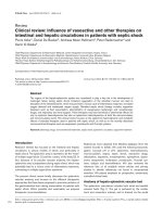
In the following paragraphs, we will examine the main aspects
of these key determinants, following the time-course of a
patient-triggered breath in pressure support [15], as it is the
most commonly used partial ventilatory support mode [16]:
triggering of the ventilator, pressurization slope and
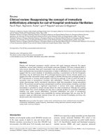
inspiratory flow, level of PS, and cycling (Figure 1). Although
the discussion is centered on intubated patients, aspects
specific to NIV will be outlined as well.
Triggering of the ventilator
Triggering refers to the mechanism by which a patient’s
inspiratory effort initiates a response from the ventilator,
through either a decrease in the ventilator circuit pressure
(pressure trigger), or the presence of an inspiratory flow
(flow trigger). The main determinants affecting inspiratory
muscle workload associated with triggering are the
magnitude of change required and the delay between the
onset of inspiratory effort and ventilator response. Initial
studies comparing flow triggering and pressure triggering
showed that the former entailed a lower work of breathing
[17]. However, the difference is probably of little clinical
relevance [18]. Furthermore, the latest generation intensive
care unit (ICU) ventilators are equipped with very sensitive
mechanisms, in terms of both the inspiratory effort required
and initial delay, triggering thereby adding little to the overall
work of breathing [19]. Nonetheless, on most machines the
trigger sensitivity can be adjusted, and care should be taken
to ensure that the highest possible sensitivity is set, without
auto-triggering [20]. Of note, during NIV, leaks can be
erroneously detected by the ventilator as an inspiratory
effort, thereby triggering the ventilator. The frequency of this
auto-triggering has been shown to correlate with the
magnitude of the leak [7]. Of interest, many recent
ventilators provide an ‘NIV mode’ designed to take leaks into
account and adjust ventilator parameters, including
triggering, accordingly.
Despite the technical progress made, at least one major issue
regarding triggering still persists; that of trigger asynchrony
(TA) [21], which refers to the presence of inspiratory efforts
that do not succeed in triggering the ventilator. The most
common cause of such ineffective inspiratory attempts is the
presence of PEEPi in COPD patients [21,22]. Indeed, for
inspiratory flow to occur and trigger the ventilator, alveolar
pressure (P
alv
) must decrease below the pressure present at
the airway opening (P
ao
). In the presence of PEEPi, however,
P
alv
must decrease by the additional amount of PEEPi. This
added inspiratory threshold load often cannot be offset with
each breath, the patient’s respiratory rate therefore being
higher than that reported by the ventilator. The combination of
ineffective inspiratory efforts and added load even for
triggering breaths can markedly increase the work of
breathing (WOB) [22]. In this situation, even if the trigger
sensitivity is set at its maximum, little improvement is
obtained, since the offsetting of PEEPi is still necessary for
the trigger to react [21]. Two options are available to reduce
TA. One is to add external PEEP (PEEPe), which reduces the
pressure difference between P
alv
and P
ao
, and, therefore, the
magnitude of P
alv
reduction required to generate an
inspiratory flow. Applying PEEPe has been shown to
decrease both the number of ineffective breaths and the
WOB [22]. There is no validated approach to determine the
optimal level of PEEPe. The pragmatic approach used in our
ICU is to start at zero end-expiratory pressure (ZEEP), and to
titrate PEEPe upwards by 1 to 2 cmH
2
O increments, until
ineffective inspiratory attempts markedly decrease or
disappear.
The other option is to reduce the level of pressure support.
Indeed, excessive levels of PS can result in the insufflation of
a high tidal volume, which in turn can increase dynamic
Figure 1
Schematic tracing of a pressure support (PS) cycle, highlighting its
four key phases.
Page 3 of 6
(page number not for citation purposes)
hyperinflation and PEEPi, thereby worsening TA. In this
setting, reducing PS has been shown to effectively decrease
TA [13].
In recent years, new developments relying on micro-
processor technology have attempted to further optimize
triggering. One such approach, based on the analysis of the
inspiratory flow waveform was shown to decrease the
triggering effort in a small group of patients without COPD, at
the price of some degree of instability in the form of auto-
triggering [23]. Another interesting technique is neurally
adjusted ventilators assist (NAVA), which relies on the
recording of the electromyographic activity of the diaphragm
by means of a multi-electrode naso-gastric tube, and its
immediate feedback to the ventilator to control the timing and
level of ventilatory assist [24]. At this stage, clinical studies
are needed to evaluate the potential for NAVA to improve
patient-ventilator interaction and impact outcome.
Pressurization slope and inspiratory flow
During PS, the slope of pressurization, that is, the incremental
increase in P
aw
per time unit, can be adjusted on most
ventilators [19]. The steeper the slope, the faster P
aw
will rise
to its target value. Studies performed in patients with
obstructive mechanics have demonstrated that, compared to
a slow pressurization rise time, a steep slope is associated
with less WOB, and the steeper the slope the lower the
WOB [25]. The same observation was made by Chiumello
and colleagues [26], their results also showing that comfort
was at its lowest at both the lowest and highest
pressurization rates. During NIV, a study performed in COPD
patients showed that the diaphragmatic pressure-time
product was lower at the fastest pressurization rate, which
was associated with a significant increase in air leaks and
proved to be the most uncomfortable for the patients [9].
Therefore, it is probably wise not to decrease the PS rise time
to <100 ms, and, if a patient exhibits discomfort, to increase
the time up to 200 ms.
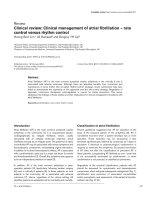
Level of pressure support
As discussed in the first section of this review, in PS mode,
one must take care to avoid both insufficient support leading
to increased respiratory muscle load [27] and excessive
support bearing the risk of worsening dynamic hyperinflation
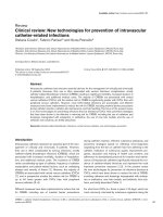
and PEEPi in obstructive patients [13] (Figure 2). Further-
more, recent data show that PS can also disrupt sleep
through episodes of central apneas caused by hypocapnia
[28]. A high level of PS can worsen the delayed cycling
phenomenon as described in the next section. During NIV,
leaks increase in proportion to the pressure generated inside
the mask [10]. This last mechanism probably provides some
safeguard against excessive tidal volume (VT) insufflation and
gastric intake of air, but can lead to delayed cycling.
Furthermore, given that ventilators differ markedly in their
capacity to compensate for leaks [29], any increase in PS
might paradoxically lead to a decrease in delivered VT due to
the increase in leaks. Empirically, PS can be titrated on the
expiratory tidal volume (approximately 8 to 10 ml/kg, the
lowest value being preferred in NIV) and the patient’s
respiratory rate, which should remain below 30/minute.
An innovative approach is to provide automatic titration of the
level of PS, based on the continuous evaluation of respiratory
rate, VT and end-tidal CO
2
[30,31]. Such a system was
shown to improve parameters of patient-ventilator adaptation
in a small group of intubated patients without COPD [31]. In
a recent randomized multicenter study, in which 20% of
patients had COPD, the system led to a decrease in the
duration of weaning, total duration of MV including NIV, and
to a shorter ICU stay compared to standard physician-driven
weaning [32].
Cycling
In PS mode, the transition from inspiration to expiration,
known as cycling, occurs when instantaneous inspiratory flow
(V′
insp
) decreases to a predetermined fraction of peak
inspiratory flow (V′
insp
/V′
peak
), often referred to as an
‘expiratory trigger’ (ET) [15]. In an ideal situation, cycling
coincides with the end of the patient’s inspiratory effort.
Prolonged pressurization by the machine into the patient’s
expiratory phase is known as delayed cycling. Delayed
cycling can lead to expiratory asynchrony and increased
WOB [14,33]. Delayed cycling has been shown to occur
mostly in patients with obstructive airways disease [34-36].
On many ventilators, the cutoff value of ET is pre-determined,
usually at a default setting of 0.25; that is, the ventilator
cycles when V′
insp
has decreased to 25% of V′
peak
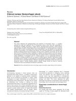
. However,
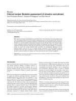
when airway resistance increases, the profile of the
inspiratory flow curve changes, the curve spreading out and
becoming flatter (Figure 3). Hence, the 0.25 point will be
reached later, which in turn increases the likelihood of
delayed cycling. The adverse consequences of delayed
Available online />Figure 2
Conceptual diagram illustrating the adverse effects of both insufficient
and excessive levels of pressure support (PS) on the respiratory
muscle workload. PEEPi, intrinsic positive end-expiratory pressure.
cycling are summarized in Figure 4. Consequently, setting a
higher value of ET should theoretically decrease the
magnitude of delayed cycling [35], and thereby alleviate
some of the adverse consequences outlined above
(Figure 5). This hypothesis was tested in a recent study in
which intubated COPD patients on PS were studied at
various ET settings, ranging from 0.10 to 0.70 [37]. The
study showed that at the higher ET values, the magnitude of
delayed cycling was reduced, entailing as predicted a
reduction in PEEPi, ineffective inspiratory attempts and
inspiratory muscle workload [37].
During NIV, additional factors can contribute to delayed
cycling. Calderini and colleagues [8] showed that leaks
around the mask led to a prolonged pressurization by the
ventilator, in turn leading to an insufficient decrease in V′
insp
to the cycling threshold. Consequently, cycling was
considerably delayed, the patients were attempting to cycle
the ventilator by active expiration [33] and WOB was
increased [8]. The authors convincingly showed that relying
on time rather than on flow cycling, that is, by limiting the
maximum inspiratory time, delayed cycling, the magnitude of
inspiratory efforts and WOB were all markedly reduced [8].
Naturally, reducing leaks can also contribute to alleviate this
problem, but tight-fitting masks are a source of discomfort for
patients, which can lead to overall intolerance to NIV and
reduce its chances of success. Finally, delayed cycling can
also occur as a result of increased leaks caused not by an
insufficient mask seal but by a high pressurization rate [9].
Optimizing patient-ventilator synchrony with
proportional assist ventilation
Proportional assist ventilation (PAV) was developed in the
early 1990s and represents an innovative approach to
respiratory muscle unloading [38,39]. Indeed, with PS, once
the patient triggers the ventilator, P
aw
rises to a preset level,
regardless of patient effort. Thus, PS provides a fixed level of
inspiratory muscle unloading. PAV, on the other hand,
amplifies patient effort without volume or pressure targets, its
basic philosophy being that the more effort the patient
develops to breathe, the more assistance is provided by the
ventilator [38,39]. To achieve this goal, there are no conven-
tional volume or pressure settings, as in other ventilatory
Critical Care Vol 10 No 6 Jolliet and Tassaux
Page 4 of 6
(page number not for citation purposes)
Figure 3
Mathematical modeling of the inspiratory instantaneous flow-time curve
for progressively increasing levels of airway resistance (Rrs), from
normal (5) to severe (20). The cross represents the point at which the
inspiratory flow has decreased to 25% of its peak value, and
corresponds to the default expiratory trigger (ET) on many ventilators.
Figure 4
Consequences of delayed cycling. PEEPi, intrinsic positive
end-expiratory pressure.
Figure 5
Airway and flow-time tracings illustrating the concept of delayed
cycling. (a) Normal mechanics. The expiratory trigger (ET) setting is
0.25. Cycling is ideal, that is, the inspiratory flow (V′) decreases to the
0.25 cycling level at the end of the patient’s neural inspiration (ti
n
).
(b) Obstructive mechanics. The change in inspiratory flow curve
derived from Figure 3 leads to the 0.25 level being reached later, well
after the end of ti
n
. The magnitude of delayed cycling (ti
excess
) is
illustrated by the double arrow. Increasing the level of ET to 0.6 of
peak inspiratory flow corrects this problem, and cycling occurs once
more at the end of ti
n
. Exp., expiration; Insp., inspiration; V′
peak
, peak
inspiratory flow.
modes. Rather, the physician sets a pressure gain applied by
the machine on the patient’s measured or estimated
elastance and resistance. This allows for a compensation by
the ventilator of an increase in either one or both of these
components. Initial studies of PAV, centered on its patho-
physiologcal effects, proved encouraging. PAV decreased
WOB, increased VT and decreased peak P
aw
in intubated
patients without COPD during weaning from mechanical
ventilation [40,41]. In intubated COPD patients, PAV
improved minute ventilation, decreased dyspnea, and
reduced WOB [42], while preserving the physiological
breath-by-breath VT variability better than PS [43]. Further-
more, in case of a sudden increase in mechanical load, PAV
can maintain minute-volume and VT better than PS, and with
less respiratory muscle load [44]. Despite these favorable
effects, PAV can prove difficult to use in the clinical setting.
Indeed, knowledge of the patients’ elastance and resistance,
a prerequisite for the correct titration of their compensation
by PAV, is most often unavailable to the clinician [45].
Therefore, arbitrary levels of compensation are often used,
which, if inappropriately chosen, can increase inspiratory
effort in some patients [46]. Furthermore, although theoreti-
cally PAV should provide optimal cycling characteristics
[38,39], some doubts have recently been cast on this
assumption [47]. In summary, although PAV has been
developed more than a decade ago, its routine clinical
implementation has so far not been achieved on a
widespread scale, most likely because of its relative
complexity and instability, presently positioning it more as a
valuable tool to explore the regulation of ventilation than as an
everyday ventilatory mode [45,48].
Conclusion
Over the past 15 years, considerable knowledge has been
gained in our understanding of the extremely complex issue of
patient-ventilator interaction in COPD patients. Given the
increasing use of assisted modes, this heightened under-
standing has become crucial in the everyday clinical
management of mechanically ventilated patients. The various
key phases and pitfalls of a ventilator-assisted breath should
be understood by ICU physicians and caregivers to reduce
unnecessary respiratory muscle workload and improve
patient comfort. Special attention should be paid to patients
undergoing NIV in whom leaks compound those problems
encountered in intubated patients. New approaches such as
closed-loop modes are beginning to prove their efficacy
during key periods, such as weaning, as they have the
potential for making numerous on-line adjustments to the
patient’s ventilatory demand.
Competing interests
PJ has received financial support for research projects from
ResMed and Draeger but received no financial support for
the present paper.
References
1. Gladwin M, Pierson D: Mechanical ventilation of the patient
with severe chronic obstructive pulmonary disease. Intensive
Care Med 1998, 24:898-910.
2. Sethi J, Siegel M: Mechanical ventilation in chronic obstructive
lung disease. Clin Chest Med 2000, 21:799-818.
3. Rossi A, Polese G, Brandi G, Conti G: Intrinsic positive end-
expiratory pressure. Intensive Care Med 1995, 21:522-536.
4. Pepe, P Marini J: Occult positive end-expiratory pressure in
mechanically ventilated patients with airflow obstruction. Am
Rev Respir Dis 1982, 126:166-170.
5. Tobin MJ, Jubran A, Laghi F: Patient-ventilator interaction. Am J
Respir Crit Care Med 2001, 163:1059-1063.
6. Kondili E, Prinianakis G, Georgopoulos D: Patient-ventilator
interaction. Br J Anaesth 2003, 91:106-119.
7. Bernstein G, Knodel E, Heldt G: Airway leak size in neonates
and autocycling of three flow-triggered ventilators. Crit Care
Med 1995, 23:1739-1744.
8. Calderini E, Confalonieri M, Puccio P, Francavilla N, Stella L, Gre-
goretti C: Patient-ventilator asynchrony during noninvasive
ventilation: the role of expiratory trigger. Intensive Care Med
1999, 25:662-667.
9. Prinianakis G, Delmastro M, Carlucci A, Ceriana P, Nava S: Effect
of varying the pressurisation rate during noninvasive pressure
support ventilation. Eur Respir J 2004, 23:314-320.
10. Schettino GP, Tucci MR, Sousa R, Valente Barbas CS, Passos
Amato MB, Carvalho CR: Mask mechanics and leak dynamics
during noninvasive pressure support ventilation: a bench
study. Intensive Care Med 2001, 27:1887-1891.
11. Brochard L, Harf A, Lorino H, Lemaire F: Inspiratory pressure
support prevents diaphragmatic fatigue during weaning from
mechanical ventilation. Am Rev Respir Dis 1989, 139:513-521.
12. Georgopoulos D, Roussos C: Control of breathing in mechani-
cally ventilated patients. Eur Respir J 1996, 9:2151-2160.
13. Leung P, Jubran A, Tobin M: Comparison of assisted ventilator
modes on triggering, patient effort, and dyspnea. Am J Respir
Crit Care Med 1997, 155:1940-1948.
14. Jubran A, Van de Graaf W, Tobin M: Variability of patient-venti-
lator interactions with pressure support ventilation in patients
with chronic obstructive pulmonary disease. Am J Respir Crit
Care Med 1995, 152:129-136.
15. Brochard L: Inspiratory pressure support. Eur J Anesthesiol
1994, 11:29-36.
16. Esteban A, Anzueto A, Alia I, Gordo F, Apezteguia C, Palizas F,
Cide D, Goldwaser R, Soto L, Bugedo G, et al.: How is mechan-
ical ventilation employed in the intensive care unit? An inter-
national utilization review. Am J Respir Crit Care Med 2000,
161:1450-1458.
17. Aslanian P, El Atrous S, Isabey D, Valente E, Corsi D, Harf A,
Lemaire F, Brochard L: Effects of flow triggering on breathing
effort during partial ventilatory support. Am J Respir Crit Care
Med 1998, 157:135-143.
18. Tütüncu A, Cakar N, Camci E, Esen F, Telci L, Akpir K: Compari-
son of pressure- and flow-triggered pressure-support ventila-
tion on weaning parameters in patients recovering from acute
respiratory failure. Crit Care Med 1997, 25:756-760.
19. Richard JC, Carlucci A, Breton L, Langlais N, Jaber S, Maggiore
S, Fougere S, Harf A, Brochard L: Bench testing of pressure
support ventilation with three different generations of ventila-
tors. Intensive Care Med 2002, 28:1049-1057.
20. Hill LL, Pearl R: Flow triggering, pressure triggering, and
autotriggering during mechanical ventilation. Crit Care Med
2000, 28:579-581.
21. Chao D, Scheinhorn D, Stearn-Hassenpflug M: Patient-ventilator
trigger asynchrony in prolonged mechanical ventilation. Chest
1997, 112:1592-1599.
22. Nava S, Bruschi C, Rubini F, Palo A, Iotti G, Braschi A: Respira-
tory response and inspiratory effort during pressure support
ventilation in COPD patients. Intensive Care Med 1995, 21:
871-879.
23. Prinianakis G, Kondili E, Georgopoulos D: Effects of the flow
waveform method of triggering and cycling on patient-ventila-
tor interaction during pressure support. Intensive Care Med
2003, 29:1950-1959.
24. Sinderby C, Navalesi P, Beck J, Skrobik Y, Comtois N, Friberg S,
Gottfried SB, Lindstrom L: Neural control of mechanical ventila-
tion in respiratory failure. Nat Med 1999, 5:1433-1436.
Available online />Page 5 of 6
(page number not for citation purposes)
25. Bonmarchand G, Chevron V, Chopin C, Jusserand D, Girault C,
Moritz F, Leroy J, Pasquis P: Increased initial flow rate
reduces inspiratory work of breathing during pressure
support ventilation in patients with exacerbation of chronic
obstructive pulmonary disease. Intensive Care Med 1996, 22:
1147-1154.
26. Chiumello D, Pelosi P, Croci M, Bigatello L, Gattinoni L: The
effects of pressurization rate on breathing pattern, work of
breathing, gas exchange and patient comfort in pressure
support ventilation. Eur Repsir J 2001, 18:107-114.
27. Appendini L, Purro A, Patessio A, Zanaboni S, Carone M, Spada
E, Donner C, Rossi A: Partitioning of inspiratory muscle work-
load and pressure assistance in ventilator-dependent COPD
patients. Am J Respir Crit Care Med 1996, 154:1301-1309.
28. Parthasarathy S, Tobin MJ: Effect of ventilator mode on sleep
quality in critically ill patients. Am J Respir Crit Care Med 2002,
166:1423-1429.
29. Mehta S, McCool F, Hill NS: Leak compensation in positive
pressure ventilators: a lung model study. Eur Respir J 2001,
17:259-267.
30. Dojat M, Harf A, Touchard D, Laforest M, Lemaire F, Brochard L:
Evaluation of a knowledge-based system providing ventila-
tory management and decision for extubation. Am J Respir
Crit Care Med 1996, 153:997-1004.
31. Dojat M, Harf A, Touchard D, Lemaire F, Brochard L: Clinical
evaluation of a computer-controlled pressure support mode.
Am J Respir Crit Care Med 2000, 161:1161-1166.
32. Lellouche F, Mancebo J, Jolliet P, Roeseler J, Schortgen F, Dojat
M, Cabello M, Bouadma L, Rodriguez P, Maggiore S, Reynaert M,
Merssmann S, Brochard L: A multicenter randomized trial of
computer-driven protocolized weaning from mechanical ven-
tilation. Am J Crit Care Respir Med 2000, 174:894-900.
33. Parthasarathy S, Jubran A, Tobin M: Cycling of inspiratory and
expiratory muscle groups with the ventilator in airflow limita-
tion. Am J Respir Crit Care Med 1998, 158:1471-1478.
34. Nava S, Bruschi C, Fracchia C, Braschi A, Rubini F: Patient-ven-
tilator interaction and inspiratory effort during pressure
support ventilation in patients with different pathologies. Eur
Respir J 1997, 10:177-183.
35. Tassaux D, Michotte J, Gainnier M, Gratadour P, Fonseca S, Jolliet
P: Expiratory trigger setting in pressure support ventilation:
from mathematical model to bedside. Crit Care Med 2004, 32:
1844-1850.
36. Tokioka H, Tanaka T, Ishizu T, Fukushima T, Iwaki T, Nakamura Y,
Kosogabe Y: The effect of breath termination criterion on
breathing patterns and the work of breathing during pressure
support ventilation. Anesth Analg 2001, 92:161-165.
37. Tassaux D, Gainnier M, Battisti A, Jolliet P: Impact of expiratory
trigger setting on delayed cycling and inspiratory muscle
workload. Am J Respir Crit Care Med 2005, 172:1283-1289.
38. Younes M: Proportional assist ventilation, a new approach to
ventilatory support. Part 1: Theory. Am Rev Respir Dis 1992,
145:114-120.
39. Younes M: Proportional assist ventilation. In Principles and
Practice of Mechanical Ventilation. Edited by Tobin M. New York:
McGraw Hill; 1994:349-369.
40. Younes M, Puddy A, Roberts D, Light RB, Quesada A, Taylor K,
Oppenheimer L, Cramp H: Proportional assist ventilation.
Results of an initial clinical trial. Am Rev Respir Dis 1992, 145:
121-129.
41. Navalesi P, Hernandez P, Wongsa A, Laporta D, Goldberg P, Got-
tfried SB: Proportional assist ventilation in acute respiratory
failure: effects on breathing pattern and inspiratory effort. Am
J Respir Crit Care Med 1996, 154:1330-1338.
42. Ranieri VM, Grasso S, Mascia L, Martino S, Fiore T, Brienza A,
Giuliani R: Effects of proportional assist ventilation on inspira-
tory muscle effort in patients with chronic obstructive pul-
monary disease and acute respiratory failure. Anesthesiology
1997, 86:79-91.
43. Wrigge H, Golisch W, Zinserling J, Sydow M, Almeling G, Bur-
chardi H: Proportional assist versus presure support venila-
tion: effects on breathing pattern and respiratory work of
patients with chronic obstructive pulmonary disease. Intensive
Care Med 1999, 25:790-798.
44. Grasso S, Puntillo F, Mascia L, Ancona G, Fiore T, Bruno F,
Slutsky AS, Ranieri VM: Compensation for increase in respira-
tory workload during mechanical ventilation. Pressure-
support versus proportional-assist ventilation. Am J Respir
Crit Care Med 2000, 161:819-826.
45. Ambrosino N, Rossi A: Proportional assist ventilation (PAV): a
significant advance or a futile struggle between logic and
practice? Thorax 2002, 57:272-276.
46. Delaere S, Roeseler J, D'Hoore W, Matte P, Reynaert M, Jolliet P,
Sottiaux T, Liistro G: Respiratory muscle workload in intubated,
spontaneously breathing patients without COPD: pressure
support vs. proportional assist ventilation. Intensive Care Med
2003, 29:949-954.
47. Du H, Ohtsuji M, Shigeta M, Chao D, Sasaki K, Usuda Y, Yamada
Y: Expiratory asynchrony in proportional assist ventilation. Am
J Respir Crit Care Med 2002, 165:972-977.
48. Vitacca M: New things are not always better: proportional
assist ventilation vs. pressure support ventilation. Intensive
Care Med 2003, 29:1038-1040.
Critical Care Vol 10 No 6 Jolliet and Tassaux
Page 6 of 6
(page number not for citation purposes)