Báo cáo khoa học: "Blood glucose measurements in the critically ill: more than just a blood draw" doc
Bạn đang xem bản rút gọn của tài liệu. Xem và tải ngay bản đầy đủ của tài liệu tại đây (165.88 KB, 2 trang )
Page 1 of 2
(page number not for citation purposes)
Available online />Abstract
A crucial determinant for the success of intensive insulin therapy in
critically ill patients is the frequent and accurate measurement of
blood glucose values with immediate feedback of results. In
general, therefore, this is achieved by point-of-care testing, raising
the question of the best way of monitoring blood glucose.
Corstjens and coworkers, in the previous issue of Critical Care,
demonstrate that, in spite of good correlation to “conventional”
laboratory glucose assessment, absolute glucose levels may differ
systematically. This commentary reviews the problems of glucose
measurements arising from matrix effects, interferences and the
use of different assays.
Intensive insulin therapy in critically ill
patients
In the preceding issue of Critical Care Corstjens and
coworkers [1] investigated three different methods of glucose
measurements. While data about the beneficial effects of
normoglycemia in critically ill patients are conflicting and
inconsistent [2,3], there is no doubt about the importance of
accurate glucose measurements to achieve glycemic control
without increased risk of hypoglycemia. Similar to the results
from the Diabetes Control and Complications Trial, which
showed increasing frequency of hypoglycemia after tight
glycemic control to reduce long-term complications [4], there
is an increase of hypoglycemic episodes in critically ill
patients when strict glycemic control is established [5,6].
Whole blood glucose: what are we actually
measuring?
Although the measurement of glucose is one of the oldest
established tests in the clinical chemistry laboratory, it is
extremely complex and sometimes rather approximate due to
the different fractions of the blood sample used [7,8].
Glucose measurement can be performed in whole blood,
plasma and serum and these may be native or deproteinized,
or hemolyzed in the case of capillary whole blood. Further-
more, blood may be arterial, capillary or venous in origin. Do
all samples give the same result? The simple answer is no,
and moreover, the difference may depend on nutritional state,
perfusion, hematocrit or albumin blood concentrations.
Glucose is dissolved only in the aqueous part of the drawn
sample and not in its entire volume (which contains other
dissolved solids such as proteins). This is the major reason
for differing glucose concentrations in plasma and whole
blood samples. The protein content differs in blood cells
(mostly red blood cells) and plasma (or serum, or
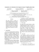
hemolysate). The water content (Figure 1) of red blood cells
is lower (because of a high concentration of hemoglobin)
than that of an equal volume of plasma (which has a lower
concentration of albumin and other plasma proteins). Even
with the glucose concentration being the same in plasma
water and red blood cell water, the concentration of glucose
per unit volume of red blood cells is lower than that per unit
volume of plasma. The concentration of glucose per unit
volume of whole blood is in between that for plasma and red
blood cells. As the water content of whole blood is the sum of
plasma water and red blood cell water, glucose concentration
will strongly depend on the hematocrit of the sample (Figure 1).
With no changes in the protein concentration of plasma or
red blood cells, a change in hematocrit from 0.4 to 0.7 will
change the plasma/whole blood ratio for glucose from 1.10
to 1.38, an error of 26%.
Blood glucose strips retain red blood cells through a filtering
process and measure glucose content in plasma in their
reaction zone. This is yet another way by which hematocrit
can influence the results: whole blood samples with differing
amounts of red blood cells alter flow and volume of plasma
entering the reaction zone. Even the Yellow Springs
Instruments’ Blood Glucose Analyzer, which is often used as
a reference method, such as in the study by Corstjens and
colleagues [1], yields glucose results dependent on hemato-
Commentary
Blood glucose measurements in the critically ill:
more than just a blood draw
Frank M Brunkhorst
1
and Hans G Wahl
2
1
Department of Anesthesiology and Intensive Care Medicine, Friedrich-Schiller University, Erlanger Allee, 07747 Jena, Germany
2
Institute for Clinical Chemistry and Laboratory Medicine, Klinikum Lüdenscheid, Paulmannshöher Str., 58515 Lüdenscheid, Germany
Corresponding author: Frank M Brunkhorst,
Published: 7 December 2006 Critical Care 2006, 10:178 (doi:10.1186/cc5110)
This article is online at />© 2006 BioMed Central Ltd
See related research by Corstjens et al., />Page 2 of 2
(page number not for citation purposes)
Critical Care Vol 10 No 6 Brunkhorst and Wahl
crit when whole blood samples are used. This is due to the
fact that the instrument performs a 25-fold sample dilution
before analysis. In theory, the only systems that should not be
affected by hematocrit are instruments using direct-reading
electrodes without sample dilution, such as those used in
blood gas analysis [9].
Calibration
Next to the composition of the blood sample, calibration of
the instrument is equally important and may often add to
unknown errors. Depending on the glucose standard and
reference method used for calibration, the same instrument
will give varying results, the most obvious being the
calibration for whole blood and plasma. And just what is the
standard reference method for glucose [10], and should
there not be a reference method for each sample type - whole
blood, plasma and hemolysate?
Evaluation of instruments
The methodology of glucose analysis in routine clinical use is
nowadays based on either chromogenic or electrochemical
reactions of the three enzymes glucose oxidase, dehydro-
genase and hexokinase. This gives rise to method-based
specific interferences, such as the blood oxygen tension
dependency of glucose oxidase, which is a major issue in
intensive care unit patients. Critically ill patients may have
very low hematocrits, high or low arterial of venous oxygen
tensions and may present extreme acid-base abnormalities,
all of which have to be evaluated for every glucose analyzer
under consideration [11,12]. Special attention must be paid
to specific interference from intensive care unit typical
medication [13]. Precision and accuracy have to be
determined using standardized protocols and care has to be
taken to choose the suitable reference method [10], which
may not be the one available in the central laboratory where
the study is being performed. Instruments such as the
Continuous Glucose Monitoring System (CGMS System
Gold, Medtronic Minimed) used in the study of Corstjens and
colleagues seem to find their place in the monitoring of
diabetic patients [14] but still need further evaluation of their
clinical utility in critically ill patients.
Conclusion
At the end of their discussion Corstjens and colleagues state
the following: “our study has too few patients and therefore
too little data points under extreme conditions of pH,
temperature, electrolyte disturbances and hypoglycaemia to
make statements about reliability of the specific analyzers
under these circumstances.” But this is exactly what is
needed to be done - otherwise we might never get an
evidence-based answer about benefit versus potential harm
of intensive insulin therapy in critically ill patients.
Competing interests
The authors declare that they have no competing interests.
References
1. Corstjens AM, Ligtenberg JJM, Van der horst ICC, Spanjersberg
R, Lind JSW, Tulleken JE, Meertens JHJM, Zijlstra JG: Accuracy
and feasibility of point-of-care and continuous blood glucose
analyzing in critically ill ICU patients. Crit Care 2006, 10:R135.
2. van den Berghe G, Wouters P, Weekers F, Verwaest C, Bruyn-
inckx F, Schetz M, Vlasselaers D, Ferdinande P, Lauwers P, Bouil-
lon R: Intensive insulin therapy in the critically ill patients. N
Engl J Med 2001, 345:1359-1367.
3. Van den Berghe G, Wilmer A, Hermans G, Meersseman W,
Wouters PJ, Milants I, Van Wijngaerden E, Bobbaers H, Bouillon
R: Intensive insulin therapy in the medical ICU. N Engl J Med
2006, 354:449-461.
4. The Diabetes Control and Complications Trial Research Group:
Hypoglycemia in the Diabetes Control and Complications
Trial. Diabetes 1997, 46:271-286.
5. Van den Berghe G, Wilmer A, Milants I, Wouters PJ, Bouckaert B,
Bruyninckx F, Bouillon R, Schetz M: Intensive insulin therapy in
mixed medical/surgical intensive care units: benefit versus
harm. Diabetes 2006, 55:3151-3159.
6. Kanji S, Singh A, Tierney M, Meggison H, McIntyre L, Hebert PC:
Standardization of intravenous insulin therapy improves the
efficiency and safety of blood glucose control in critically ill
adults. Intensive Care Med 2004, 30:804-810.
7. Wiener K: Whole blood glucose: what are we actually measur-
ing? Ann Clin Biochem 1995, 32:1-8.
8. Burnett RW, D’Orazio P, Fogh-Andersen N, Kuwa K, Kulpmann
WR, Larsson L, Lewnstam A, Maas AH, Mager G, Spichiger-Keller
U; Scientific Division, Working Group on Selective Electrodes:
IFCC recommendation on reporting results for blood glucose.
Clin Chim Acta 2001, 307:205-209.
9. Fogh-Andersen N, D’Orazio P: Proposal for standardizing
direct-reading biosensors for blood glucose. Clin Chem 1998,
44:655-659.
10. Hannestad U, Lundblad A: Accurate and precise isotope dilu-
tion mass spectrometry method for determining glucose in
whole blood. Clin Chem 1997, 43:794-800.
11. Tang Z, Louie RF, Lee JH, Lee DM, Miller EE, Kost GJ: Oxygen
effects on glucose meter measurements with glucose dehy-
drogenase- and oxidase-based test strips for point-of-care
testing. Crit Care Med 2001, 29:1062-1070.
12. Tang Z, Du X, Louie RF, Kost GJ: Effects of pH on glucose mea-
surements with handheld glucose meters and a portable
glucose analyzer for point-of-care testing. Arch Pathol Lab
Med 2000, 124:577-582.
13. Tang Z, Du X, Louie RF, Kost GJ: Effects of drugs on glucose
measurements with handheld glucose meters and a portable
glucose analyzer. Am J Clin Pathol 2000, 113:75-86.
14. Lodwig V, Heinemann L; Glucose Monitoring Study Group: Contin-
uous glucose monitoring with glucose sensors: calibration and
assessment criteria. Diabetes Technol Ther 2003, 5:572-586.
Figure 1
Glucose concentration: water content expressed as percent volume.
With a hematocrit of 0.4 and a water content for the fraction ‘Cells’ of
approximately 70%, the total water content will be 28% of total volume
(whole blood). From the 60% plasma volume, 90% will be water, thus
giving a water content of 54% for the plasma portion of whole blood.
The total water content of whole blood then is 82% (54% + 28%). For
a hematocrit of 0.4 the plasma/whole blood ratio of water content is
0.9/0.82 = 1.10, which reflects the 10% higher glucose values in
plasma compared to whole blood.