Báo cáo khoa học: "Simplified electrophysiological evaluation of peripheral nerves in critically ill patients: the Italian multi-centre CRIMYNE study" potx
Bạn đang xem bản rút gọn của tài liệu. Xem và tải ngay bản đầy đủ của tài liệu tại đây (331.3 KB, 11 trang )
Open Access
Available online />Page 1 of 11
(page number not for citation purposes)
Vol 11 No 1
Research
Simplified electrophysiological evaluation of peripheral nerves in
critically ill patients: the Italian multi-centre CRIMYNE study
Nicola Latronico
1,2
, Guido Bertolini
3,4
, Bruno Guarneri
5
, Marco Botteri
1
, Elena Peli
1
,
Serena Andreoletti
1
, Paola Bera
1
, Davide Luciani
3
, Anna Nardella
1
, Elena Vittorielli
1
, Bruno Simini
4
and Andrea Candiani
1
1
Department of Anesthesiology-Intensive Care, University of Brescia, Spedali Civili, Piazzale Ospedali Civili, 1 – 25123 Brescia, Italy
2
GiViTI, Gruppo Italiano per la Valutazione degli Interventi in Terapia Intensiva Steering Committee, Aldo e Cele Daccò Clinical Research Centre Mario
Negri Institute, Villa Camozzi – 24020 Ranica (BG), Italy
3
Laboratory of Clinical Epidemiology, Aldo e Cele Daccò Clinical Research Centre Mario Negri Institute, Villa Camozzi – 24020 Ranica (BG), Italy
4
GiViTI, Gruppo Italiano per la Valutazione degli Interventi in Terapia Intensiva Steering Committee, Villa Camozzi – 24020 Ranica (BG), Italy
5
Department of Clinical Neurophysiology, University of Brescia, Spedali Civili, Piazzale Ospedali Civili, 1 – 25123 Brescia, Italy
Corresponding author: Nicola Latronico,
Received: 11 Sep 2006 Revisions requested: 9 Nov 2006 Revisions received: 17 Dec 2006 Accepted: 25 Jan 2007 Published: 25 Jan 2007
Critical Care 2007, 11:R11 (doi:10.1186/cc5671)
This article is online at: />© 2007 Latronico et al.; licensee BioMed Central Ltd.
This is an open access article distributed under the terms of the Creative Commons Attribution License ( />),
which permits unrestricted use, distribution, and reproduction in any medium, provided the original work is properly cited.
Abstract
Introduction Critical illness myopathy and/or neuropathy
(CRIMYNE) is frequent in intensive care unit (ICU) patients.
Although complete electrophysiological tests of peripheral
nerves and muscles are essential to diagnose it, they are time-
consuming, precluding extensive use in daily ICU practice. We
evaluated whether a simplified electrophysiological investigation
of only two nerves could be used as an alternative to complete
electrophysiological tests.
Methods In this prospective, multi-centre study, 92 ICU patients
were subjected to unilateral daily measurements of the action
potential amplitude of the sural and peroneal nerves (compound
muscle action potential [CMAP]). After the first ten days,
complete electrophysiological investigations were carried out
weekly until ICU discharge or death. At hospital discharge,
complete neurological and electrophysiological investigations
were performed.
Results Electrophysiological signs of CRIMYNE occurred in 28
patients (30.4%, 95% confidence interval [CI] 21.9% to
40.4%). A unilateral peroneal CMAP reduction of more than two
standard deviations of normal value showed the best
combination of sensitivity (100%) and specificity (67%) in
diagnosing CRIMYNE. All patients developed the
electrophysiological signs of CRIMYNE within 13 days of ICU
admission. Median time from ICU admission to CRIMYNE was
six days (95% CI five to nine days). In 10 patients, the amplitude
of the nerve action potential dropped progressively over a
median of 3.0 days, and in 18 patients it dropped abruptly within
24 hours. Multi-organ failure occurred in 21 patients (22.8%,
95% CI 15.4% to 32.4%) and was strongly associated with
CRIMYNE (odds ratio 4.58, 95% CI 1.64 to 12.81). Six patients
with CRIMYNE died: three in the ICU and three after ICU
discharge. Hospital mortality was similar in patients with and
without CRIMYNE (21.4% and 17.2%; p = 0.771). At ICU
discharge, electrophysiological signs of CRIMYNE persisted in
18 (64.3%) of 28 patients. At hospital discharge, diagnoses in
the 15 survivors were critical illness myopathy (CIM) in six
cases, critical illness polyneuropathy (CIP) in four, combined
CIP and CIM in three, and undetermined in two.
Conclusion A peroneal CMAP reduction below two standard
deviations of normal value accurately identifies patients with
CRIMYNE. These should have full neurological and
neurophysiological evaluations before discharge from the acute
hospital.
CI = confidence interval; CIM = critical illness myopathy; CIP = critical illness polyneuropathy; CMAP = compound muscle action potential; CRIMYNE
= critical illness myopathy and/or neuropathy; EMG = electromyography; ICU = intensive care unit; IQR = interquartile range; MOF = multi-organ
failure; OR = odds ratio; SAPS II = simplified acute physiology score II; SD = standard deviation; SIRS = systemic inflammatory response syndrome;
SOFA = sequential organ failure assessment; SNAP = sensory nerve action potential.
Critical Care Vol 11 No 1 Latronico et al.
Page 2 of 11
(page number not for citation purposes)
Introduction
Critical illness polyneuropathy (CIP) is the commonest and the
best-defined neuromuscular alteration seen in the intensive
care unit (ICU) [1], affecting 58% of patients with prolonged
ICU stay, 70% to 80% of patients with sepsis, septic shock,
or multi-organ failure (MOF), and 100% of patients with sepsis
and coma [2]. CIP is an axonal polyneuropathy and is a com-
mon consequence of systemic inflammatory response
syndrome (SIRS) and MOF [3]. In its classic presentation, CIP
is a sensory-motor axonal polyneuropathy [1]; however, pure
motor and pure sensory forms have also been described [4,5].
CIP is usually suspected in ICU patients who, after a period of
days or weeks, cannot be weaned from the ventilator despite
the absence of pulmonary or cardiac causes of respiratory fail-
ure or because they have various degrees of limb weakness
[3]. Neurological signs of CIP may or may not be present at
this stage [1]. In addition, neurological examination is often
unreliable because of encephalopathy, sedation, or the critical
condition of the patient [6]; therefore, comprehensive electro-
physiological studies of peripheral nerves are necessary to
establish the diagnosis. These should include motor and sen-
sory nerve conduction studies as well as needle electromyog-
raphy (EMG) in upper and lower limbs [7]. A reduced
amplitude of the compound muscle action potential (CMAP)
and sensory nerve action potential (SNAP) is the predominant
finding; latency and nerve conduction velocity remain normal
or are only slightly decreased [7]. Although several studies
have prospectively assessed the evolution of CIP [3-5,8-11],
they did not start at the time of ICU admission and did not
investigate baseline electrophysiological status of peripheral
nerves before the onset of CIP. Only two small case series
have performed electrophysiological investigations in the first
ICU days [12,13]. In one study [12], nine patients with SIRS
had their initial electrophysiological investigations within a
median of five days (range 2 to 25 days) after ICU admission
[12]. All showed a CMAP reduction, whereas most SNAPs
were normal. In the other study [13], nine patients with moder-
ate to severe multi-organ dysfunction syndrome and SIRS or
sepsis had their initial electrophysiological investigations
within two to five days after ICU admission. All had a reduction
in CMAP (SNAPs were not reported), confirming it as the ear-
liest electrophysiological sign of CIP.
Critical illness myopathy (CIM) is a primary muscle disorder
that has been characterised only in recent years [4]. Data on
its incidence are lacking, but evidence is mounting that CIM is
at least as frequent as CIP [4,14-23]. There is currently sub-
stantial consensus about considering CIM as a syndrome with
a continuum of myopathic findings [2,24-27]. Differential diag-
nosis between CIP and CIM is difficult because conventional
conduction studies and needle EMG often provide non-spe-
cific findings that fail to distinguish between CIM and CIP [28].
Both conditions are characterised by low-amplitude CMAPs
and frequently show abnormal spontaneous activity [20,22].
Assessment of recruitment and interference of voluntary EMG
pattern is often problematic because of severe weakness or
poor voluntary effort in most patients. The differentiating fea-
ture may become the SNAP, which may be blunted or masked
by the local oedema in critically ill patients, so that these meas-
ures are often unreliable [20]. Previous studies have shown
that if the patient fails to volitionally activate his/her muscles,
electrophysiological diagnosis is invariably CIP even if CIM is
ongoing [4,29]. Furthermore, CIM and CIP are frequently
associated [4]. We therefore coined the acronym CRIMYNE
(critical illness myopathy and/or neuropathy) to define the neu-
romuscular alterations acquired during the ICU stay. This acro-
nym also identified the current study among the participating
centres.
Early diagnosis of CRIMYNE is important for several reasons.
Knowing CRIMYNE is present aids managing the ventilator
and means the patient has a neuromuscular problem, which is
likely to prolong the patient's ventilator dependency and ICU
stay [30,31]. In critically ill comatose patients developing tetra-
paresis or tetraplegia, knowing that CRIMYNE is present may
prevent an unreasonably pessimistic prognosis and allows the
diagnostician to ascribe paralysis to CRIMYNE rather than to
central nervous system deterioration [4]. Early diagnosis com-
bined with serial electrophysiological studies may also be val-
uable in determining the ultimate prognosis of patients with
CRIMYNE and in gauging the rate of recovery, as well as in
assessing the effects of treatments such as intensive insulin
therapy [32]. However, electrophysiological study is time-con-
suming, requiring 45 to 90 minutes for its completion [6].
We report a multi-centre, prospective study in a mixed cohort
of medical and surgical critically ill adult patients with no evi-
dence of CRIMYNE or MOF at ICU admission who underwent
serial clinical and simplified electrophysiological investigations
during their entire ICU stay.
The main objective of this study was to evaluate whether a sim-
plified electrophysiological test could accurately diagnose
CRIMYNE. Other objectives were to evaluate the onset time of
CRIMYNE in relation to ICU admission and to MOF onset, the
transition from normal electrophysiology to CRIMYNE, and the
evolution of CRIMYNE during the ICU stay.
Materials and methods
This multi-centre prospective cohort study was performed
between January 1998 and March 2001 in nine Italian ICUs
belonging to the GiViTI (Gruppo Italiano per la Valutazione
degli Interventi in Terapia Intensiva). Local ethics committee
approval was obtained beforehand. Written consent was
obtained from the patient whenever possible; otherwise, writ-
ten information was given to their next of kin. Written consent
was obtained from all surviving patients as soon as they
regained mental competency.
Available online />Page 3 of 11
(page number not for citation purposes)
Inclusion and exclusion criteria
Patients more than 15 years of age whose Simplified Acute
Physiology Score II (SAPS II) [33] was between 35 and 70
were eligible for inclusion. This range predicts a risk of devel-
oping MOF of more than 30% (unpublished observation by N.
Latronico and G. Bertolini derived from intensive care medi-
cine data provided by Rui Moreno, Lisbon, Portugal, and from
sepsis study data provided by Martin Langer, Milan, Italy) and
a risk of hospital mortality of between 15% and 85% [33].
Exclusion criteria were (a) CRIMYNE or MOF diagnosed
within 24 hours of ICU admission, (b) previous neuromuscular
disorders, (c) elective surgery, (d) obesity (body mass index of
more than 30 kg/m
2
), (e) lower limb disorders precluding
nerve conduction study and EMG (for example, oedema, frac-
tures, amputation, plaster casts), and (f) brain death. Centres
were allowed to exclude patients if another patient in the same
ICU was being concomitantly studied.
Initial electrophysiological investigations
Twenty-four hours after admission, the SAPS II and Sequential
Organ Failure Assessment (SOFA) [34,35] scores were cal-
culated and complete electrophysiological tests performed.
These consisted of conventional motor (median and common
peroneal nerves) and sensory nerve (median and sural nerves)
conduction studies. SNAPs were recorded from the median
and sural nerves. For the median nerve, the ring recording
electrodes were placed around the proximal (-) and distal (+)
interphalangeal joints of the second or third digit; the nerve
was stimulated at the wrist, on the volar surface, 2 to 3 cm
proximal to the distal crease. For the sural nerve, the surface
recording electrodes were placed above (-) and below (+) the
lateral malleolus as the nerve passes around it or immediately
posteroinferior to the lateral malleolus (-) and 2 to 3 cm distally
along the lateral dorsum of the foot (+); the nerve was stimu-
lated along the posterior surface of the leg (calf), slightly lateral
to the midline and approximately 10 to 12 cm from the active
electrode (-). CMAPs were recorded from the median (abduc-
tor pollicis brevis muscle) and common peroneal (extensor
digitorum brevis muscle) nerves. For the median nerve, surface
recording electrodes were placed over the belly (-) and tendon
(+) of the abductor pollicis brevis; the nerve was stimulated at
the wrist on the volar surface, 2 to 3 cm proximal to the distal
crease and at the elbow over the brachial pulse with the cath-
ode at the volar crease. For the common peroneal nerve, sur-
face recording electrodes were placed over the belly and
tendon of the extensor digitorum brevis; the nerve was stimu-
lated over the dorsum of the foot, near the ankle, 7 to 8 cm
from the recording electrodes, above (at the lateral popliteal
fossa) and below the head of the fibula (below the knee). Incre-
mental electrical stimulation of the nerves was used until the
best SNAP or CMAP amplitudes were obtained. If the clinical
history and physical examination suggested a median nerve
entrapment at the wrist or the median sensory nerve conduc-
tion study was abnormal, the median nerve was substituted by
the ulnar nerve [36]. The ulnar nerve was stimulated above and
below the elbow and the peroneal nerve above and below the
head of the fibula to rule out entrapment neuropathies. EMG
was recorded using a coaxial needle electrode in the tibialis
anterior, quadriceps femori, abductor pollicis brevis, and del-
toid muscles; additional muscles were studied in some
patients. Impaired neuromuscular transmission due to neu-
romuscular blocking agents was excluded by 3-Hz stimulation
of the distal ulnar nerve. Before electrophysiological tests,
heat packs were applied to the skin if its temperature was
below 33°C.
A differential diagnosis between CIP, CIM, or combined CIP
and CIM was not sought during the ICU stay. Electrophysio-
logical diagnosis of CRIMYNE was achieved if the CMAP or
SNAP amplitude of at least two nerves of two limbs was
reduced below two standard deviations (SDs) of the lower
limit of normality with or without abnormal spontaneous mus-
cle activity [7,12]. Normal values were established in normal
control subjects tested in the same laboratory [37] (see Addi-
tional file 1). Organ dysfunction was defined according to the
SOFA score [34,35]. MOF was defined as the failure of two or
more organs in addition to the organ whose failure prompted
ICU admission; CIP was not considered as an organ failure for
the purpose of defining MOF. SIRS and sepsis were defined
according to current standards [38].
Serial clinical and electrophysiological investigations
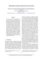
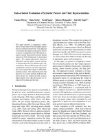
Daily simplified and weekly complete electrophysiological
tests were performed (Figure 1). Simplified electrophysiologi-
cal tests recorded conduction velocity and amplitude of the
sural SNAP and peroneal CMAP in one leg, using surface
stimulation and recording electrodes. We arbitrarily defined a
25% decrease from baseline SNAP and CMAP measured at
ICU admission as the minimum consistently detectable reduc-
tion. If SNAP or CMAP decreased by more than 25% on two
consecutive days, a complete electrophysiological test was
performed. If the latter was consistent with CRIMYNE, com-
plete weekly electrophysiological tests replaced daily tests
until ICU discharge. Otherwise, daily simplified electrophysio-
logical tests were resumed (Figure 1). To minimise artifacts,
the same electrode site and size were used for each patient
[39].
Patient treatment, including control of blood glucose, con-
formed to accepted standards. Intravenous insulin (Actrapid
HM; Novo Nordisk A/S, Bagsvaerd, Denmark), preferably with
the use of a pump, was started if the blood glucose level
exceeded 180 mg/dl. The target was a blood glucose level of
less than 160 mg/dl. Data on blood glucose level were not
collected.
Intensivists and clinical neurophysiologists were unaware of
each other's diagnoses. All electrophysiological recordings
were re-examined by one author (BG) for quality assessment.
Critical Care Vol 11 No 1 Latronico et al.
Page 4 of 11
(page number not for citation purposes)
Follow-up
Patients discharged from the ICU with an electrophysiological
diagnosis of CRIMYNE and who were able to cooperate had
complete electrophysiological investigations, including sen-
sory and motor nerve conduction studies and EMG of upper
and lower limb muscles, before acute hospital discharge. At
this stage, a differential diagnosis between CIM, CIP, and
combined CIM and CIP was sought.
Data presentation and statistical analysis
We expressed continuous variables as means (SD) or as
medians (interquartile range [IQR]) and discrete variables as
counts (percentage) unless otherwise stated. Differences in
the study population were analysed by means of a Student's t
test, Mann-Whitney U test, or χ
2
test (or Fisher exact test) as
appropriate. Ninety-five percent confidence intervals (CIs)
were computed for each estimate of interest. The odds ratio
(OR) was used to quantify the association between electro-
physiological changes and MOF. The times of onset of CIP
and MOF, expressed in terms of cumulative incidence, were
analysed with Kaplan-Meier curves [40]; comparison was
made using the log-rank test. All tests were two-tailed, and a p
value of less than 0.05 was used to define a statistically signif-
icant difference.
Results
Ninety-two patients were enrolled with a mean monthly enrol-
ment rate of 1.2 patients per ICU. One centre (Brescia, Italy)
enrolled 30 patients during the entire study period; the other 8
centres enrolled 4 to 13 patients during 4 to 12 months.
Patient characteristics are shown in Table 1.
The electrophysiological signs of CRIMYNE occurred in 28
patients (30.4%, 95% CI 21.9% to 40.4%) (Table 2), 6 of
whom died (3 in the ICU, 3 after ICU discharge). Thirteen of
the 92 patients died in the ICU (14.1%) and 4 more died in the
hospital after ICU discharge (total of 17 patients [18.5%]).
Hospital mortality was similar in patients with and without
CRIMYNE (6 patients [21.4%] and 11 patients [17.2%],
respectively; Fisher exact test, p = 0.771).
Figure 1
Flow chart of electrophysiological investigationsFlow chart of electrophysiological investigations. CMAP, compound muscle action potential; CRIMYNE, critical illness myopathy and/or neuropathy;
ICU, intensive care unit; SD, standard deviation; SNAP, sensory nerve action potential.
Available online />Page 5 of 11
(page number not for citation purposes)
Time course of CRIMYNE during the ICU stay
An electrophysiological diagnosis of CRIMYNE was preceded
by a 25% peroneal CMAP reduction (compared to the base-
line value at ICU admission) in all 28 patients (sensitivity
100%); however, the specificity of this abnormality was low
(48%) (Table 3). A peroneal CMAP reduction below two SDs
of normal values (according to the single centre) had the same
sensitivity but better specificity (67%) (Table 3). The more
severe the peroneal CMAP reduction, the lower the sensitivity
and the higher the specificity (Table 3).
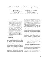
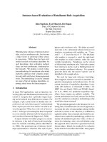
All 28 patients developed the electrophysiological signs of
CRIMYNE within 13 days of ICU admission, 25 (89.3%) within
11 days of ICU admission (Figure 2). The median interval from
ICU admission to CRIMYNE was 6 days (95% CI 5 to 9 days,
IQR 4 to 10 days).
In 18 patients (64.3%), the amplitude of the nerve action
potential amplitude decreased abruptly within 24 hours, and in
10 patients (35.7%) the amplitude dropped progressively over
a median of 3.0 days (IQR 2 to 5 days). In 29 patients (31.5%),
EMG revealed fibrillation potentials and positive sharp waves,
which were evenly distributed among explored muscles. Nerve
conduction velocity was normal in all cases. There were no
complications specifically attributed to serial electrophysiolog-
ical measurements.
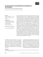
Relationship between MOF and CRIMYNE
MOF occurred in 21 patients (22.8%, 95% CI 15.4% to
32.4%), six of whom died during ICU stay (28.6%). The
median interval from ICU admission to MOF was three days
(95% CI two to five days, IQR two to five days). Respiratory
(17 patients) and cardiovascular (17 patients) failure prevailed
and their combination was responsible for the diagnosis of
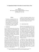
MOF in 12 of the 21 patients (57.1%). There was no
difference between the onset times of CRIMYNE and MOF
(log-rank test 1.03, p = 0.311) (Figure 3).
MOF was strongly associated with CRIMYNE (OR 4.6, 95%
CI 1.6 to 12.8): all but two patients with CRIMYNE had single
(14 patients) or multiple (12 patients) organ failures. If
CRIMYNE were considered an extra organ failure, it would be
the most common organ failure in patients with MOF.
Furthermore, a diagnosis of MOF would be made in ten (48%)
other patients.
Follow-up
Recovery from CRIMYNE and MOF differed. At ICU dis-
charge, MOF had resolved in all survivors (15 patients),
whereas CRIMYNE had resolved in 10 of 28 patients but was
still persisting in 18 (64.3%) (Table 2). Of these 18 patients,
3 died after ICU discharge and 2 were unable to volitionally
activate their muscles in order to have a complete EMG eval-
uation. A precise pathological diagnosis was achieved in the
13 remaining patients, which was CIM in six cases, CIP in four,
and combined CIM and CIP in three.
Table 1
Baseline characteristics of the patients
Characteristic
Total number of patients 92
Age in years
Median 49.5
Interquartile range 31–67
Absolute range 18–85
Female gender, number (percentage) 29 (31.5)
Simplified Acute Physiology Score II
Median 42
Interquartile range 38–49
Sequential Organ Failure Assessment score
Median 7
Interquartile range 6–9
Number of patients artificially ventilated on admission
(percentage)
88 (95.7)
Reason for admission, number (percentage)
Medical 41 (44.6)
Pneumonia 9 (9.8)
Pulmonary oedema 7 (7.6)
Metabolic encephalopathy 6 (6.5)
Post-anoxic encephalopathy 5 (5.4)
Intracranial haemorrhage 5 (5.4)
COPD exacerbation 2 (2.2)
Congestive heart failure 2 (2.2)
Other 5 (5.4)
Emergency surgery 15 (16.3)
Neurosurgery 9 (9.8)
Abdominal surgery 3 (3.3)
Other surgery 3 (3.3)
Trauma 36 (39.1)
Intensive care unit stay in days
Median 13
Mode (bimodal) 2 (11)
Interquartile range 8–22
Absolute range 1–90
COPD, chronic obstructive pulmonary disease.
Critical Care Vol 11 No 1 Latronico et al.
Page 6 of 11
(page number not for citation purposes)
Discussion
CIP and CIM are frequent complications in ICU patients [2]
and are responsible for prolonged disability after ICU dis-
charge [41]. Clinical diagnosis is often unreliable in the ICU
[1,3,6,7], and therefore electrophysiological studies must be
used. Complete electrophysiological investigations are, how-
ever, time-consuming [6], and therefore CIP and CIM are
rarely systematically investigated in the ICU, except for
research purposes. In the present study, we found that a sim-
plified electrophysiological investigation assessment is accu-
rate and can be started early after ICU admission and used in
daily routine. The simplified electrophysiological test we used
consisted of conduction velocity and amplitude of the sural
SNAP and peroneal CMAP in one leg; however, unilateral test-
ing of peroneal CMAP had the best combination of sensitivity
and specificity. This is an important finding because the SNAP
amplitude is 1,000 times lower than CMAP amplitude and is
therefore more difficult to measure accurately, particularly if
oedema is present, and is more prone to misinterpretation.
Although not formally assessed, the time needed to measure
a peroneal CMAP in one leg can be estimated to be 5 to 10
minutes, which is substantially lower than the 45 to 90 minutes
needed for a complete electrophysiological investigation [6].
A 25% reduction of the peroneal CMAP was as sensitive as a
reduction of more than two SDs in diagnosing CRIMYNE. This
first test, however, had a lower specificity (the true-negative
rate) and in order to be calculated needed a baseline evalua-
tion of the peroneal CMAP amplitude at ICU admission. The
second test proved to be not only more accurate but also more
efficient, needing to be compared with normal values and not
with baseline peroneal CMAP. According to Marciniak and
coworkers [37], the possible sources of normal values of elec-
trodiagnostic studies which will permit a report of an abnormal
result to be considered reliable include (a) values obtained in
a normal group (according to the reference standard) enrolled
Table 2
Electrophysiological alterations in the study population
Time of evaluation
At diagnosis of CRIMYNE At ICU discharge
Persisting Resolved
Bilateral peroneal CMAP
reduction
a
16 (57%) 13 3
Only bilateral peroneal CMAP 9 7 2
+ unilateral sural SNAP 1 0 1
+ bilateral sural SNAP 2 2 0
+ bilateral sural SNAP + unilateral
median CMAP
110
+ unilateral median SNAP 1 1 0
+ unilateral median CMAP 1 1 0
+ unilateral median SNAP +
unilateral median CMAP
110
Unilateral peroneal CMAP
reduction
a
12 (43%) 5 7
+ unilateral sural SNAP 1 1 0
+ unilateral sural SNAP+ unilateral
median SNAP
101
+ bilateral sural SNAP 2 0 2
+ bilateral sural SNAP + unilateral
median CMAP + unilateral median
SNAP
211
+ unilateral median SNAP 3 2 1
+ bilateral median SNAP 1 1 0
+ unilateral median CMAP 2 0 2
a
Reduction of the CMAP or SNAP amplitude by more than two standard deviations of its normal value. CMAP, compound muscle action potential;
CRIMYNE, critical illness myopathy and/or neuropathy; ICU, intensive care unit; SNAP, sensory nerve action potential.
Available online />Page 7 of 11
(page number not for citation purposes)
specifically for the article, (b) normal values established in nor-
mal control subjects tested in the same laboratory, and (c) nor-
mal values established in normal control subjects using the
same electrodiagnostic techniques, even if obtained in
another laboratory.
High-sensitivity diagnostic tests have a high negative predic-
tive value and are particularly useful when normal. The test can
therefore be proposed as a screening test before a patient's
discharge from the ICU or the acute hospital: patients with
bilaterally normal peroneal CMAP need no further evaluation;
patients with a peroneal CMAP reduction of more than two
SDs of normal values, either unilateral or bilateral, are referred
to the neurologist for further investigation. The total number of
patients to be investigated would vary according to the defini-
tion of 'high-risk' critically ill patients – possible definitions are
patients with mechanical ventilation longer than three or seven
days, patients with sepsis and/or MOF, or patients with a
SAPS II of between 35 and 70 [3-5,8-11] – but based on the
recruitment rates of this study, it should be in the order of one
to two patients per month per ICU.
The fact that primarily the peroneal nerve, a long lower limb
motor nerve, was affected has implications for the so-called
theory of bioenergetic failure, which is thought to be a relevant
pathophysiological mechanism explaining MOF [42] and CIP
[3,4,43-45]. In fact, nerve action potential generation and ter-
minal axon structural integrity are critically dependent on
axonal transport of proteins and other molecules [46]. Despite
their length, axons are devoid of the machinery for biosynthetic
processes, and all axonal components are synthesised in the
cell body, translocated from the cell body into the axonal proc-
ess, and then transported to their final destination within the
axon [46]. This anterograde transport, particularly the fast
transport, requires considerable energy expenditure because
material is moved rapidly with rates up to 3 µm/second [46]. If
the nerve cell does not receive adequate nourishment due to
microcirculatory alterations [47] or the cell cannot use the
energy due to cellular dysoxia, the axonal transport fails and
distal axonopathy ensues. Bioenergetic failure might explain
the extremely rapid decrease of peroneal CMAP observed
within 24 hours of normal CMAP in 18 (64.3%) of our patients,
which represents a substantial divergence from the traditional
observation that at least one week is needed for axonal neu-
ropathy to become apparent. Although these CMAP changes
could be due to a combination of dysfunction of both periph-
eral nerves and muscles, the important message is that func-
tional derangement happened very early, confirming a
hypothesis we proposed 11 years ago [4]. This early func-
tional derangement may be an important biological sign in crit-
ically ill patients and, as Bolton noted [48], could be used in
Table 3
Sensitivity and specificity of peroneal CMAP reduction to diagnose critical illness myopathy and/or neuropathy
Time of development Sensitivity Specificity
ICU day (True-positive rate) (True-negative rate)
Number (%) Median (IQR)
1. One peroneal CMAP reduced according to criterion A 64 (69.6) 3 (2–5) 28/28 = 100% 28/64 = 44%
2. One peroneal CMAP reduced according to criterion B 49 (53.3) 4 (2–7) 28/28 = 100% 43/64 = 67%
3. Both peroneal CMAPs reduced according to criterion A 26 (28.3) 6 (3–10) 21/28 = 75% 59/64 = 92%
4. One peroneal CMAP reduced according to criterion A plus
the
contralateral peroneal CMAP reduced according to criterion B
23 (25.0) 6 (3–10) 21/28 = 75% 62/64 = 97%
5. Both peroneal CMAPs reduced according to criterion B 16 (17.4) 6 (3.5–10) 16/28 = 57% 64/64 = 100%
Criterion A = CMAP amplitude reduced by more than 25% of its initial value (at ICU admission) but less than two standard deviations (SDs) of its
normal value. Criterion B = CMAP reduced by more than 2 SDs of its normal value. Note that the five categories are not mutually exclusive (for
example, the 16 patients in category 5 are also included in category 2). CMAP, compound muscle action potential; ICU, intensive care unit; IQR,
interquartile range.
Figure 2
Onset time of critical illness myopathy and/or neuropathy during inten-sive care unit (ICU) stayOnset time of critical illness myopathy and/or neuropathy during inten-
sive care unit (ICU) stay.
Critical Care Vol 11 No 1 Latronico et al.
Page 8 of 11
(page number not for citation purposes)
research aiming at interrupting pathological mechanisms at
their onset.
We did not find an association between CIP and SIRS, sepsis,
drugs, or nutrition. Because blood glucose data were not col-
lected, association with hyperglycaemia could not be
confirmed. Conversely, the risk of having CIP was almost five
times greater in patients with MOF than in patients without, a
result in agreement with a recent systematic review [49] and a
prospective multi-centre cohort study [21]. Several previous
studies reported an association between CIP and sepsis or
MOF, although they selectively included patients with sepsis
[4,9,10,50] or with sepsis and MOF [5], used non-validated
MOF-scoring systems [3,5,8,9], or did not provide details of
criteria used to diagnose MOF [3,11,13]. Zochodne and col-
leagues [3] first observed that CIP developed during the
course of MOF and improved in some patients as the critical
illness subsided, and they suggested that the pathogenesis of
failing systemic organs and peripheral nerve damage might be
the same. Indeed, the strong association between CIP and
MOF and the similarity of their onset times suggest that CIP
itself could be considered an organ failure: that of the periph-
eral nervous system.
In our study, hospital mortality was not different in patients with
and without CIP, a result in contrast with two previous studies
[9,11]. In the study by Leijten and colleagues [9] of critically ill
patients mechanically ventilated more than seven days, the
hospital mortality was more than double in patients with CIP
(48%) than in patients without (19%; p = 0.03); however, mor-
tality was no longer significantly different at 1 year (52% and
43% in patients with and without CIP, respectively; p = 0.18).
Garnacho-Montero and colleagues [11] studied a very select
population of patients with sepsis, MOF, and a duration of
mechanical ventilation of more than nine days. A significant
proportion of patients had extremely severe derangement of
physiological variables and 40% had septic shock [41]. Hos-
pital mortality was higher in patients with CIP than in patients
without (84% versus 56.5%, respectively; p = 0.01). These
figures are much higher than ours and suggest that differ-
ences in patients' case mix may have accounted for the differ-
ence. However, we cannot exclude the possibility that the
small number of events in our study population precluded a
thorough statistical evaluation.
The simplified electrophysiological test used in our study
could not and cannot distinguish CIM from CIP [20,22-24,28].
We were able to achieve a precise pathological diagnosis in
only 13 of 28 (46%) patients after ICU discharge. Nine (69%)
of them were found to have CIM alone or in combination with
CIP, confirming that CIM is an often-overlooked diagnosis. We
cannot exclude the fact that a higher number of patients would
have been diagnosed with CIM if we had used muscle biopsy
[4], myosin/actin ratio [51], or specialised electrophysiological
investigations such as direct muscle stimulation [20,22-24].
Recently, a diagnostic algorithm for differentiating CIM from
CIP which combines direct muscle stimulation and conven-
tional techniques was proposed [23]; however, differential
diagnosis between CIP and CIM during ICU stay is of
unproven relevance.
Potential pitfalls of the simplified electrophysiological
test
Acute peroneal palsy, tissue oedema, and advanced age (par-
ticularly more than 70 years) may cause true or artifactual per-
oneal CMAP reduction. Acute peroneal nerve palsy is most
commonly caused by trauma, surgery, or compression of the
nerve trunk at the fibular head [52]. Isolated non-traumatic
Figure 3
Kaplan-Meier curves comparing the times of onset of critical illness myopathy and/or neuropathy (CRIMYNE) and multi-organ failure (MOF)Kaplan-Meier curves comparing the times of onset of critical illness myopathy and/or neuropathy (CRIMYNE) and multi-organ failure (MOF). No dif-
ference between the onset times of CRIMYNE and MOF was observed (log-rank test 1.03, p = 0.311). ICU, intensive care unit.
Available online />Page 9 of 11
(page number not for citation purposes)
lesions are rare. In many patients, however, the cause remains
undetermined and in the absence of other signs is often
assumed to be due to transient compression. Motor conduc-
tion across the segment of fibula head is particularly important
in distinguishing patients with peroneal neuropathy at this level
from patients with other lower-extremity neurological disorders
(class III and class IV evidence) [37]. Inadequate considera-
tion of these potential pitfalls may substantially increase the
number of false-positive cases of CRIMYNE; however, acute
peroneal entrapment neuropathies are a cause of disability
which deserves medical attention.
Conclusion
Assessment of the peroneal nerve CMAP amplitude before
discharge from the ICU is feasible and can be implemented in
clinical routine. A peroneal CMAP reduction of more than two
SDs of normal value accurately identifies patients with
CRIMYNE. These patients should have full neurological and
neurophysiological evaluations before discharge from the
acute hospital. Future availability of low-cost simplified EMG
machines would be desirable for promoting the widespread
use of this important non-invasive diagnostic test in the ICU.
Competing interests
NL, GB, and BS are part of the Steering Committee of the
GiViTI (Gruppo Italiano per la Valutazione degli Interventi in
Terapia Intensiva), which is the recipient of an unconditional
grant from AstraZeneca Italia S.p.A. (Basiglio, Italy), Sanofi-
Aventis (Paris, France), and Draeger Italia (Corsico, Italy). The
other authors declare that they have no competing interests.
Authors' contributions
All authors made a substantial contribution to the study design
and methods. NL conceived the idea of the study. NL, GB, and
BG designed the protocol. GB and DL performed the statisti-
cal analyses. BG was responsible for neurophysiological
investigations of the study. NL, MB, EP, SA, PB, AN, and EV
were responsible for the clinical investigations of the study. NL
drafted the manuscript and all other authors critically revised it
for important intellectual content. All authors read and
approved the final manuscript.
Additional files
Acknowledgements
We are greatly indebted to Rui Moreno (Lisbon, Portugal) and Martin
Langer (Milan, Italy) for providing data to inform the choice of inclusion
criteria.
Centres participating in the study (all in Italy)
Nicola Latronico, Istituto di Anestesia e Rianimazione; Bruno Guarneri,
Servizio di Neurofisiopatologia, Università di Brescia, Spedali Civili,
Brescia; Alessandra Tanfani and Luigi Targa, Unità Operativa di Anes-
tesia e Rianimazione; Chiara Minardi and Fabrizio Rasi, Divisione di Neu-
rologia Ospedale Maurizio Bufalini, Cesena; Diletta Guarducci and
Simona Cardona, Unità Operativa di Anestesia e Rianimazione; Lucia
Toscani and Tiziana Furlan, Servizio di Neurofisiopatologia, Ospedale
SS Annunziata – USL 10/H, Firenze; Anna Piccioli and Sante Ferrarello,
Unità Operativa di Anestesia e Rianimazione I; Aldo Amantini and
Antonello Grippo, Servizio di Neurofisiopatologia, Università di Firenze,
Azienda Ospedaliera Careggi, Firenze; Renata Pinzani and Dorino
Salami, Unità Operativa di Anestesia e Rianimazione; Gian Andrea
Ottonello and Gianna Zocchi, Ospedale San Martino, Genova; Martin
Langer and Francesca Ricciardi, II Unità Operativa di Anestesia e Rian-
imazione; Tullio Mille, Clinica Neurochirurgica, Policlinico S. Matteo,
Pavia; Vincenzo Emmi and Giuseppe Rodi, I Unità Operativa di Anes-
tesia e Rianimazione; Tullio Mille, Clinica Neurochirurgica, Policlinico S.
Matteo, Pavia; Walter Bottari and Roberto Martini, Unità Operativa di
Anestesia e Rianimazione; Rossella Sabadini and Luisa Motti, Clinica
Neurologica, Arcispedale Santa Maria Nuova, Reggio Emilia; Anselmo
Caricato and Francesco Della Corte, Istituto di Anestesia e Rianimazi-
one; Francesca Odoardi and Mauro Lomonaco, Istituto di Neurologia,
Università Cattolica Sacro Cuore, Policlinico Gemelli, Roma.
References
1. Bolton CF: Neuromuscular manifestations of critical illness.
Muscle Nerve 2005, 32:140-163.
2. Latronico N, Peli E, Botteri M: Critical illness myopathy and
neuropathy. Curr Opin Crit Care 2005, 11:126-132.
3. Zochodne DW, Bolton CF, Wells GA, Gilbert JJ, Hahn AF, Brown
JD, Sibbald WA: Critical illness polyneuropathy. A complication
of sepsis and multiple organ failure. Brain 1987, 110(Pt
4):819-841.
4. Latronico N, Fenzi F, Recupero D, Guarneri B, Tomelleri G, Tonin
P, De Maria G, Antonini L, Rizzuto N, Candiani A: Critical illness
myopathy and neuropathy. Lancet 1996, 347:1579-1582.
5. Coakley JH, Nagendran K, Yarwood GD, Honavar M, Hinds CJ:
Patterns of neurophysiological abnormality in prolonged criti-
cal illness. Intensive Care Med 1998, 24:801-807.
Key messages
• A peroneal CMAP reduction of more than two SDs of
normal value accurately identifies patients with
CRIMYNE.
• Transition from normal peripheral nerve electrophysiol-
ogy to CRIMYNE can be extremely rapid (24 hours).
• CRIMYNE, once diagnosed, persists in the majority of
patients at ICU discharge.
• CRIMYNE is associated with MOF, not with SIRS or
sepsis.
• CRIMYNE is not associated with increased hospital
mortality.
The following Additional files are available online:
Additional file 1
A table showing the normal mean value and lower limit of
normality of motor and sensory nerve conduction studies
in the nine participating centres.
See />supplementary/cc5671-S1.doc
Critical Care Vol 11 No 1 Latronico et al.
Page 10 of 11
(page number not for citation purposes)
6. Leijten FSS, Poortvliet DCJ, de Weerd AW: The neurological
examination in the assessment of polyneuropathy in mechan-
ically ventilated patients. Eur Neurol 1997, 4:124-129.
7. Bolton CF, Laverty DA, Brown JD, Witt NJ, Hahn AF, Sibbald WJ:
Critically ill polyneuropathy: electrophysiological studies and
differentiation from Guillain-Barre syndrome. J Neurol Neuro-
surg Psychiatry 1986, 49:563-573.
8. Witt NJ, Zochodne DW, Bolton CF, Grand'Maison F, Wells G,
Young GB, Sibbald WJ: Peripheral nerve function in sepsis and
multiple organ failure. Chest 1991, 99:176-184.
9. Leijten FS, Harinck-de Weerd JE, Poortvliet DC, de Weerd AW:
The role of polyneuropathy in motor convalescence after pro-
longed mechanical ventilation. JAMA 1995, 274:1221-1225.
10. Berek K, Margreiter J, Willeit J, Berek A, Schmutzhard E, Mutz NJ:
Polyneuropathies in critically ill patients: a prospective
evaluation. Intensive Care Med 1996, 22:849-855.
11. Garnacho-Montero J, Madrazo-Osuna J, Garcia-Garmendia JL,
Ortiz-Leyba C, Jimenez-Jimenez FJ, Barrero-Almodovar A, Garna-
cho-Montero MC, Moyano-Del-Estad MR: Critical illness
polyneuropathy: risk factors and clinical consequences. A
cohort study in septic patients. Intensive Care Med 2001,
27:1288-1296.
12. Schwarz J, Planck J, Briegel J, Straube A: Single-fiber electromy-
ography, nerve conduction studies, and conventional electro-
myography in patients with critical-illness polyneuropathy:
evidence for a lesion of terminal motor axons. Muscle Nerve
1997, 20:696-701.
13. Tennila A, Salmi T, Pettila V, Roine RO, Varpula T, Takkunen O:
Early signs of critical illness polyneuropathy in ICU patients
with systemic inflammatory response syndrome or sepsis.
Intensive Care Med 2000, 26:1360-1363.
14. Helliwell TR, Coakley JH, Wagenmakers AJ, Griffiths RD, Camp-
bell IT, Green CJ, McClelland P, Bone JM: Necrotizing myopathy
in critically-ill patients. J Pathol 1991, 164:307-314.
15. Op de Coul AA, Verheul GA, Leyten AC, Schellens RL, Teepen JL:
Critical illness polyneuromyopathy after artificial respiration.
Clin Neurol Neurosurg 1991, 93:27-33.
16. Latronico N, Fenzi F, Guarneri B, Tomelleri G, Tonin P, Rizzuto N,
Candiani A: Critical illness polyneuropathy. Intensive Care Med
1992, 18:204.
17. Rich MM, Bird SJ, Raps EC, McCluskey LF, Teener JW: Direct
muscle stimulation in acute quadriplegic myopathy. Muscle
Nerve 1997, 20:665-673.
18. Lacomis D, Petrella JT, Giuliani MJ: Causes of neuromuscular
weakness in the intensive care unit: a study of ninety-two
patients. Muscle Nerve 1998, 21:610-617.
19. De Letter MA, van Doorn PA, Savelkoul HF, Laman JD, Schmitz PI,
Op de Coul AA, Visser LH, Kros JM, Teepen JL, van der Meche FG:
Critical illness polyneuropathy and myopathy (CIPNM): evi-
dence for local immune activation by cytokine-expression in
the muscle tissue. J Neuroimmunol 2000, 106:206-213.
20. Trojaborg W, Weimer LH, Hays AP: Electrophysiologic studies
in critical illness associated weakness: myopathy or neuropa-
thy – a reappraisal. Clin Neurophysiol 2001, 112:1586-1593.
21. De Jonghe B, Sharshar T, Lefaucheur JP, Authier FJ, Durand-Zale-
ski I, Boussarsar M, Cerf C, Renaud E, Mesrati F, Carlet J, et al.:
Paresis acquired in the intensive care unit: a prospective mul-
ticenter study. JAMA 2002, 288:2859-2867.
22. Bednarik J, Lukas Z, Vondracek P: Critical illness polyneuromy-
opathy: the electrophysiological components of a complex
entity. Intensive Care Med 2003, 29:1505-1514.
23. Lefaucheur JP, Nordine T, Rodriguez P, Brochard L: Origin of ICU
acquired paresis determined by direct muscle stimulation. J
Neurol Neurosurg Psychiatry 2006, 77:500-506.
24. Rich MM, Teener JW, Raps EC, Schotland DL, Bird SJ: Muscle is
electrically inexcitable in acute quadriplegic myopathy. Neu-
rology 1996, 46:731-736.
25. Latronico N, Candiani A: Muscular wasting as a consequence of
sepsis. In Anaesthesia, Pain, Intensive Care and Emergency
Medicine, APICE 13th edition. Edited by: Gullo A. Milan: Springer-
Verlag; 1998:517-522.
26. Lacomis D, Zochodne DW, Bird SJ: Critical illness myopathy.
Muscle Nerve 2000, 23:1785-1788.
27. Friedrich O: Critical illness myopathy: what is happening? Curr
Opin Clin Nutr Metab Care 2006, 9:403-409.
28. Latronico N: Neuromuscular alterations in the critically ill
patient: critical illness myopathy, critical illness neuropathy, or
both? Intensive Care Med 2003, 29:1411-1413.
29. Latronico N, Fenzi F, Boniotti C, Guarneri B, Tonin P, Tomelleri G,
De Maria G, Antonini L, Rizzuto N, Candiani A: Acute reversible
paralysis in critically ill patients. Acta Anaesthesiol Ital 1993,
44:157-171.
30. De Jonghe B, Bastuji-Garin S, Sharshar T, Outin H, Brochard L:
Does ICU-acquired paresis lengthen weaning from mechani-
cal ventilation? Intensive Care Med 2004, 30:1117-1121.
31. Garnacho-Montero J, Amaya-Villar R, Garcia-Garmendia JL,
Madrazo-Osuna J, Ortiz-Leyba C: Effect of critical illness
polyneuropathy on the withdrawal from mechanical ventilation
and the length of stay in septic patients. Crit Care Med 2005,
33:349-354.
32. Van den Berghe G, Schoonheydt K, Becx P, Bruyninckx F, Wout-
ers PJ: Insulin therapy protects the central and peripheral nerv-
ous system of intensive care patients. Neurology 2005,
64:1348-1353.
33. Le Gall JR, Lemeshow S, Saulnier F: A new Simplified Acute
Physiology Score (SAPS II) based on a European/North Amer-
ican multicenter study. JAMA 1993, 270:2957-2963.
34. Vincent JL, Moreno R, Takala J, Willatts S, De Mendonca A, Bruin-
ing H, Reinhart CK, Suter PM, Thijs LG: The SOFA (Sepsis-
related Organ Failure Assessment) score to describe organ
dysfunction/failure. On behalf of the Working Group on Sep-
sis-Related Problems of the European Society of Intensive
Care Medicine. Intensive Care Med 1996, 22:707-710.
35. Ferreira FL, Bota DP, Bross A, Melot C, Vincent JL: Serial evalu-
ation of the SOFA score to predict outcome in critically ill
patients. JAMA 2001, 286:1754-1758.
36. Practice parameter for electrodiagnostic studies in carpal tun-
nel syndrome: summary statement. Muscle Nerve 2002,
25:918-922.
37. Marciniak C, Armon C, Wilson J, Miller R: Practice parameter:
utility of electrodiagnostic techniques in evaluating patients
with suspected peroneal neuropathy: an evidence-based
review. Muscle Nerve 2005, 31:520-527.
38. Levy MM, Fink MP, Marshall JC, Abraham E, Angus D, Cook D,
Cohen J, Opal SM, Vincent JL, Ramsay G: 2001 SCCM/ESICM/
ACCP/ATS/SIS International Sepsis Definitions Conference.
Intensive Care Med 2003, 29:530-538.
39. van Dijk JG, van Benten I, Kramer CG, Stegeman DF: CMAP
amplitude cartography of muscles innervated by the median,
ulnar, peroneal, and tibial nerves. Muscle Nerve 1999,
22:378-389.
40. Kleinbaum D, Kupper L, Morgenstern H: Epidemiologic Research
New York, NY: Van Nostrand Reynhold; 1982.
41. Latronico N, Shehu I, Seghelini E: Neuromuscular sequelae of
critical illness. Curr Opin Crit Care 2005, 11:381-390.
42. Hotchkiss RS, Swanson PE, Freeman BD, Tinsley KW, Cobb JP,
Matuschak GM, Buchman TG, Karl IE: Apoptotic cell death in
patients with sepsis, shock, and multiple organ dysfunction.
Crit Care Med 1999, 27:1230-1251.
43. Bolton CF, Young BG, Zochodne DW: Neurological changes
during severe sepsis. In Current Topics in Intensive Care Vol-
ume 1. Edited by: Dobb GJ, Burehardi H, Dellinger RP. London:
Saunders; 1994:180-217.
44. Latronico N: Monitoring of peripheral nerves and muscle func-
tion in patients with multiple organ dysfunction syndrome. Crit
Care Med 2000, 28:3375.
45. Z'Graggen WJ, Lin CS, Howard RS, Beale RJ, Bostock H: Nerve
excitability changes in critical illness polyneuropathy. Brain
2006, 129:2461-2470.
46. Brown A: Axonal transport of membranous and nonmembra-
nous cargoes: a unified perspective. J Cell Biol 2003,
160:817-821.
47. Bolton CF: Neuromuscular conditions in the intensive care
unit. Intensive Care Med 1996, 22:841-843.
48. Bolton CF: Evidence of neuromuscular dysfunction in the early
stages of the systemic inflammatory response syndrome.
Intensive Care Med 2000, 26:1179-1180.
49. De Jonghe B, Cook D, Sharshar T, Lefaucheur JP, Carlet J, Outin
H: Acquired neuromuscular disorders in critically ill patients: a
systematic review. Groupe de Reflexion et d'Etude sur les
Neuromyopathies En Reanimation. Intensive Care Med 1998,
24:1242-1250.
Available online />Page 11 of 11
(page number not for citation purposes)
50. Coakley JH, Nagendran K, Honavar M, Hinds CJ: Preliminary
observations on the neuromuscular abnormalities in patients
with organ failure and sepsis. Intensive Care Med 1993,
19:323-328.
51. Stibler H, Edstrom L, Ahlbeck K, Remahl S, Ansved T: Electro-
phoretic determination of the myosin/actin ratio in the diagno-
sis of critical illness myopathy. Intensive Care Med 2003,
29:1515-1527.
52. Van Langenhove M, Pollefliet A, Vanderstraeten G: A retrospec-
tive electrodiagnostic evaluation of footdrop in 303 patients.
Electromyogr Clin Neurophysiol 1989, 29:145-152.