Báo cáo khoa học: " The impact of admission diagnosis on gastric emptying in critically ill patients" pot
Bạn đang xem bản rút gọn của tài liệu. Xem và tải ngay bản đầy đủ của tài liệu tại đây (357.07 KB, 10 trang )
Open Access
Available online />Page 1 of 10
(page number not for citation purposes)
Vol 11 No 1
Research
The impact of admission diagnosis on gastric emptying in critically
ill patients
Nam Q Nguyen
1,2
, Mei P Ng
1
, Marianne Chapman
3
, Robert J Fraser
2
and Richard H Holloway
1,2
1
Department of Gastroenterology, Royal Adelaide Hospital, North Terrace, Adelaide, 5000, Australia
2
Department of Medicine, University of Adelaide, Frome Road, Adelaide, 5000, Australia
3
Department of Intensive Care, Royal Adelaide Hospital, North Terrace, Adelaide, 5000, Australia
Corresponding author: Nam Q Nguyen,
Received: 11 Nov 2006 Revisions requested: 11 Jan 2007 Revisions received: 15 Jan 2007 Accepted: 8 Feb 2007 Published: 8 Feb 2007
Critical Care 2007, 11:R16 (doi:10.1186/cc5685)
This article is online at: />© 2007 Nguyen et al.; licensee BioMed Central Ltd.
This is an open access article distributed under the terms of the Creative Commons Attribution License ( />),
which permits unrestricted use, distribution, and reproduction in any medium, provided the original work is properly cited.
Abstract
Introduction Disturbed gastric emptying (GE) occurs
commonly in critically ill patients. Admission diagnoses are
believed to influence the incidence of delayed GE and
subsequent feed intolerance. Although patients with burns and
head injury are considered to be at greater risk, the true
incidence has not been determined by examination of patient
groups of sufficient number. This study aimed to evaluate the
impact of admission diagnosis on GE in critically ill patients.
Methods A retrospective review of patient demographics,
diagnosis, intensive care unit (ICU) admission details, GE, and
enteral feeding was performed on an unselected cohort of 132
mechanically ventilated patients (94 males, 38 females; age 54
± 1.2 years; admission Acute Physiology and Chronic Health
Evaluation II [APACHE II] score of 22 ± 1) who had undergone
GE assessment by
13
C-octanoic acid breath test. Delayed GE
was defined as GE coefficient (GEC) of less than 3.20 and/or
gastric half-emptying time (t50) of more than 140 minutes.
Results Overall, 60% of the patients had delayed GE and a
mean GEC of 2.9 ± 0.1 and t50 of 163 ± 7 minutes. On
univariate analysis, GE correlated significantly with older age,
higher admission APACHE II scores, longer length of stay in ICU
prior to GE measurement, higher respiratory rate, higher FiO
2
(fraction of inspired oxygen), and higher serum creatinine. After
these factors were controlled for, there was a modest
relationship between admission diagnosis and GE (r = 0.48; P
= 0.02). The highest occurrence of delayed GE was observed
in patients with head injuries, burns, multi-system trauma, and
sepsis. Delayed GE was least common in patients with
myocardial injury and non-gastrointestinal post-operative
respiratory failure. Patients with delayed GE received fewer
feeds and stayed longer in ICU and hospital compared to those
with normal GE.
Conclusion Admission diagnosis has a modest impact on GE in
critically ill patients, even after controlling for factors such as
age, illness severity, and medication, which are known to
influence this function.
Introduction
Enteral feeding via the nasogastric route is the preferred
method of nutrition in critically ill patients [1-3]. Continuous
infusion of liquid nutrient into the stomach is convenient, mini-
mally invasive, and cost-effective. However, impaired tolerance
to gastric feeding is common [4,5] and leads to patient dis-
comfort, an increased risk of pulmonary aspiration, and delay
in achieving nutritional goals with the need for prokinetic
agents, post-pyloric feeding, or parenteral nutrition [3-5].
These complications of feed intolerance can adversely affect
patient morbidity and mortality [6-9].
Feed intolerance is an indirect marker of disturbed gastric
motility and delayed gastric emptying (GE) [3-5,9]. Slow GE in
critically ill patients results from disturbed motor function of
both proximal and distal stomach [10-12], but the precise
mechanisms underlying these disturbances remain unclear.
Several factors related to critical illness have been reported to
be associated with gastric dysmotility and feed intolerance,
including hyperglycaemia, the nature of acute illness, mechan-
ical ventilation, sedatives, cytokine release, and splanchnic
hypoperfusion due to shock and sepsis [2-5,13]. Critically ill
patients admitted with traumatic brain injury and burns are
APACHE II = Acute Physiology and Chronic Health Evaluation II; DM = diabetes mellitus; GE = gastric emptying; GEC = gastric emptying coefficient;
ICU = intensive care unit; SIMV = synchronised intermittent mandatory ventilation; SIRS = systemic inflammatory response syndrome; t50 = gastric
half-emptying time.
Critical Care Vol 11 No 1 Nguyen et al.
Page 2 of 10
(page number not for citation purposes)
believed to be at the highest risk of delayed GE and feed intol-
erance [3,4,14-18] (prevalence of up to 80%). However, data
on the incidence of delayed GE in patients with other diag-
noses such as sepsis and multi-trauma are limited and the
techniques used to measure GE in some studies suboptimal.
The phenol red test and gastric residual volume [14,18] have
not been validated in humans. The paracetamol absorption
test [16,17], used in many studies for indirect assessment of
GE, may be less sensitive than other approaches because the
first-pass metabolism of paracetamol is frequently altered in
critically ill patients as a consequence of liver dysfunction or
drug interactions [19]. In addition, this technique has not been
validated against scintigraphy for measurement of GE in criti-
cally ill patients. In contrast,
13
C-octanoic acid breath test
[20,21] has also been compared with the 'gold standard' tech-
nique, gastric scintigraphy, and a strong correlation between
the two techniques has been demonstrated in critically ill
patients [22]. The aims of this study were to examine (a) the
impact of admission diagnosis on delayed GE and (b) factors
associated with delayed GE in critical illness by means of a val-
idated and reliable technique for the measurement of GE, the
13
C-octanoic acid breath test [20,21].
Materials and methods
Subjects
Data from an unselected cohort of critically ill patients, who
were admitted to a mixed surgical and medical intensive care
unit (ICU) from 1999 to 2005, were pooled from six previous
clinical studies that involved measurement of GE by
13
C-octa-
noic acid breath tests. Four of the studies examined the impact
of critical illness on GE and motility in an unselected cohort of
critically ill patients. The specific aims of each study were as
follows: study 1, to evaluate the prevalence of delayed GE
function (n = 45; data of 30 patients from this group were pub-
lished in a study by Ritz and colleagues [21]); study 2, to exam-
ine the relationship between GE and antro-pyloro-duodenal
motility (n = 18; published in a study by Chapman and col-
leagues [12]); study 3, to assess the impact of morphine and
midazolam versus propofol on GE (n = 14; unpublished data);
and study 4, to examine the impact of early versus delayed
feeding on GE in critically ill patients (n = 24; unpublished
data). The other two studies were randomised, placebo-con-
trolled trials that assessed the effects of a single dose of
cephalosporin (50 mg; n = 14; published in a study by Chap-
man and colleagues [23]) and erythromycin (200 mg; n = 30;
data of 20 patients from this group were published in a study
by Chapman and colleagues [24]) on GE. From the latter two
trials, only the results of GE assessment during administration
of placebo therapy (20 ml of normal saline) were included in
this audit.
Although the outcome measures in each trial varied, the inclu-
sion and exclusion criteria as well as GE technique (
13
C-octa-
noic acid breath test) were identical. Patients who participated
in the trials were critically ill, required mechanical ventilation,
and were able to receive nasogastric feeding. The exclusion
criteria were recent major abdominal surgery, any contraindi-
cation to the passage of an enteral tube, previous gastric,
oesophageal, or intestinal surgery, suspected bowel obstruc-
tion or perforation, and use of prokinetic therapy within the pre-
vious 24 hours. In all critically ill patients, written informed
consent was obtained from the next of kin prior to the study.
Both the audit and the assessment of GE were approved by
the Research Ethics Committee of the Royal Adelaide Hospi-
tal (Adelaide, Australia).
Data collection and analysis
All relevant details related to the patients and the ICU admis-
sion were obtained from case notes and intensive care charts.
Patient demographics, admission diagnosis, Acute Physiology
and Chronic Health Evaluation II (APACHE II) score, medica-
tion (prior and during admission), past medical history, blood
glucose concentration, blood biochemistry, ventilation details,
length of stay prior to the assessment of GE, and length of
hospital stay were recorded. The APACHE II score was deter-
mined in all patients by means of a previously published
method [25]. The mean rate of nasogastric feed delivery,
before and after the assessment of GE, was also documented.
The patients were categorised into six major admission diag-
noses: (a) intra-cerebral injury, (b) burns, (c) multi-system
trauma resulting from either motor vehicle accident or fall, (d)
sepsis, (e) respiratory failure after a non-abdominal surgery
requiring ventilation in ICU, and (f) ischaemic myocardial injury
with cardiogenic shock and significant pulmonary oedema.
The 'intra-cerebral injury' category encompassed open or
closed head injury related to mechanical trauma, sub-dural,
sub-arachnoid, or intra-cerebral haemorrhage, and major
ischaemic cerebral events. A patient was deemed to have
'sepsis' if there were clinical signs of systemic inflammatory
response syndrome (SIRS) with documented evidence of
infection on bacteriological assessment [26]. SIRS was rec-
ognised by the presence of two or more of the following: tem-
perature above 38°C or below 36°C, heart rate above 90
beats per minute, respiratory rate above 20 breaths per minute
or PaCO
2
(arterial partial pressure of carbon dioxide) below 32
mm Hg, white cell count greater than 12,000 cells per cubic
millimetre or less than 4,000 cells per cubic millimetre, or a
blood picture showing a proportion of immature white cells of
more than 10% [26].
Technique of GE assessment:
13
C-octanoic acid breath
test
In all patients, the breath test was performed using an identical
standardised technique [20-22]. Studies were performed in
the morning with subjects supine in the 30-degree head-up
position. In patients, enteral feeding was ceased four hours
before the study. After verifying the correct position of the
nasogastric tube (12-French Flexiflo [Ross Products, a divi-
sion of Abbott Laboratories, Abbott Park, IL, USA] or 14-
Available online />Page 3 of 10
(page number not for citation purposes)
French Levin tube [Maersk Medical, Lynge, Denmark]) by rou-
tine clinical radiography or air insufflation and measurement of
the gastric fluid pH, all gastric contents were aspirated and
discarded. In both patients and healthy subjects, 100 μl of
13
C-octanoate (100 mg/ml; Cambridge Isotope Laboratories,
Inc., Andover, MA, USA) was added to 100 ml of Ensure™
(Abbott Australasia Pty. Ltd., Botany, NSW, Australia), a liquid
meal that contains 106 kcal/100 ml. The labelled Ensure™ was
shaken for one minute to distribute the marker in the meal
before it was infused into the stomach over five minutes.
Breath samples were obtained before meal instillation, every 5
minutes for 1 hour and every 15 minutes for a further 3 hours.
Collection of breath samples
In patients, end-expiratory breath samples were collected from
the ventilation tube by means of a T-adapter (Datex-Engstrom,
a division of Instrumentarium Corporation Helsinki, Finland)
and holder for vacutainers (blood needle holder; Reko PTY
Ltd, Lisarow, NSW, Australia), containing a needle (Ven-
oJect
®
; Terumo Corporation, Tokyo, Japan). Previous data
suggested that equilibration of CO
2
concentration between
the ventilation tube and evacuated 10-ml tubes (Exetainer
®
;
Labco Limited, High Wycombe, Buckinghamshire, UK) took a
fraction of a second and was a reliable technique of breath
sampling [21]. To avoid sampling other than end-expiratory air,
samples were timed to the end-expiratory phase by observa-
tion of the patients and the time-flow curve on the ventilation
monitor. In healthy subjects, end-expiratory breath samples
were collected by asking them to fully breathe out into sample
tubes through a straw.
Analysis of breath samples and GE
The concentration of CO
2
and the percentage of
13
CO
2
were
measured in each sample by means of an isotope ratio mass
spectrometer (ABCA model 20\20; Europa Scientific, Crewe,
Cheshire, UK). Samples containing less than 1% CO
2
were
regarded as being non-end-expiratory and were excluded from
further analysis. The
13
CO
2
concentration over time was plot-
ted and the resultant curves were used to calculate a GE coef-
ficient (GEC), a global index for the GE rate, which accounts
for the rates of both appearance and disappearance of the
label in breath [20]. In addition, the gastric half-emptying time
(t50) was calculated [21]. Delayed GE was defined as t50 of
more than 140 minutes and/or GEC of less than 3.2 [21].
Statistical analysis
Data are expressed as mean ± standard error of mean. Differ-
ences in demographic data and GE variables between the
patients groups were compared using χ
2
test with Yates cor-
rection (for categorical data) and Student t test (for continuous
data). Risk factors associated with delayed GE, such as
APACHE II score, age, serum creatinine, length of ICU stay
prior to the breath testing, ventilation parameters, and blood
glucose concentration, were assessed using Pearson's linear
regression analysis. The linear relationship between these risk
factors and GE variables were confirmed on histogram and Q-
Q plot. After these risk factors were controlled for, the influ-
ence of admission diagnosis on GE variables was assessed
using linear and hierarchical regression model analyses. Rela-
tive risk of delayed GE, as compared to that of patients with
cardiac injury, was also calculated with 95% confidence inter-
val. The statistical software used in this study was SAS/STAT
version 9.1 (SAS Institute Inc., Cary, NC, USA). A P value of
less than 0.05 was considered as statistically significant in all
analyses.
Results
A total of 145 patients had completed breath test data availa-
ble. Thirteen patients were excluded from the analysis
because their case notes and/or ICU charts were unable to be
retrieved. Thus, the final analysis was performed on 132 criti-
cally ill patients. Overall patient characteristics and details
related to their ICU admission are summarised in Table 1. The
three most common admission diagnoses were sepsis (n =
44), head injury (n = 30), and multi-system trauma (n = 29).
Seven of 29 multi-system trauma patients also sustained head
injury. Within 24 hours of GE measurement, all but 4 patients
were sedated using morphine and/or midazolam alone (n =
48), propofol alone (n = 18), or a combination of these drugs
(n = 62).
Overall, 60% of the patients had delayed GE with a mean t50
of 163 ± 7 minutes and GEC of 2.9 ± 0.1. The mean interval
from admission to the day of GE measurement was eight days.
Critical illness factors associated with delayed GE
Table 2 summarises the characteristics of patients who had
delayed GE. Slow GE was more common in patients who
were older, had higher admission APACHE II scores, admis-
sion blood glucose, and bilirubin concentrations, and were
ventilated with synchronised intermittent mandatory ventilation
(SIMV) mode. On linear regression analysis, GE (both t50 and
GEC) correlated with older age, higher admission APACHE II
scores, longer length of stay in ICU prior to GE, higher FiO
2
(fraction of inspired oxygen) requirement, and higher serum
creatinine (Table 3). There was no relationship between GE
and patient gender, body mass index, ventilatory pressure,
APACHE II score on study day, the type of sedation, or
requirements for inotropic support.
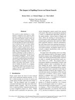
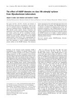
Impact of admission diagnosis on GE
The impact of various admission diagnoses on GE in critical ill-
ness is summarised in Table 4 and Figures 1 and 2. After other
factors that influence GE were controlled for, there was a mod-
est effect of admission diagnosis on GE (r = 0.48; P = 0.02)
with linear and hierarchical regression analyses. GE was
delayed most in patients with burns. Apart from intra-cerebral
and burn injuries, delayed GE was also common in patients
who were admitted with multi-system trauma and sepsis. In
Critical Care Vol 11 No 1 Nguyen et al.
Page 4 of 10
(page number not for citation purposes)
Table 1
Demographics of studied subjects
Critically ill patients (n = 132)
Age (years) 54.4 ± 1.5
Gender (male/female) 95:37
Body mass index (kg/m
2
) 27.4 ± 0.6
Days in ICU prior to study 8.0 ± 0.6
APACHE II score
Admission 23.9 ± 0.5
Study day 17.6 ± 0.6
Enteral feeding rate (ml/hour)
Prior to breath testing 51.1 ± 2.9
After breath testing 56.6 ± 2.8
Diagnosis, % (n)
Sepsis 33% (44)
Head injury
a
23% (30)
Multi-trauma
a
22% (29)
Burns 7% (9)
Non-GI post-operative respiratory compromise 9% (12)
Cardiac injury (ischaemia and failure) 11% (15)
Blood glucose level (mmol/l)
Admission 9.7 ± 0.9
Study day 8.0 ± 0.3
Biochemistry
Albumin (g/l) 23.6 ± 0.5
Bilirubin (μmol/l) 19.5 ± 2.5
White cell count (× 10
9
/l) 12.6 ± 0.5
Serum creatinine (mmol/l) 0.134 ± 0.01
Medications, % (n)
Opioid ± benzodiazepine 83% (110)
Propofol 60% (80)
Inotropes 69% (91)
Mechanical ventilation
SIMV/pressure support ventilation
b
(n) 74:58
Fraction of inspired oxygen 0.5 ± 0.01
Positive end-expiratory pressure (cm H
2
O) 6.5 ± 0.3
Peak inspiratory pressure (cm H
2
O) 24.5 ± 0.8
a
Seven patients had head injury due to multi-trauma.
b
Pressure support ventilation self-triggered mode. APACHE II, Acute Physiology and Chronic
Health Evaluation II; GI, gastrointestinal; ICU, intensive care unit; SIMV, synchronised intermittent mandatory ventilation.
Available online />Page 5 of 10
(page number not for citation purposes)
contrast, patients with myocardial injury and non-gastrointesti-
nal post-operative respiratory failure had the lowest incidence
of delayed GE.
Impact of pre-existing illness on GE
GE was delayed in 58% of patients with no known co-morbid-
ity prior to their ICU admission (t50 = 167 ± 11 minutes and
GEC = 2.9 ± 0.1). When age and admission APACHE II
scores were controlled for, there was a trend for slow GE to
be more common in patients who had pre-existing alcoholic
liver disease (80%; P = 0.04) and chronic renal failure (75%;
P = 0.06) and to be less common in patients with known dia-
betes mellitus (DM) (38%; P = 0.05).
Outcome of delayed GE in critical illness
Patients who had delayed GE received feeds at a lower rate,
both before and after the GE assessment (Table 2). The
lengths of stay in ICU and in hospital were significantly longer
Table 2
Characteristics of patients who had normal or delayed gastric emptying on
13
C-octanoic acid breath test
Delayed gastric emptying (n = 79) Normal gastric emptying (n = 53)
Age (years) 57.8 ± 2.2
a
52.2 ± 2.0
Gender (male/female) 58:21 37:16
Body mass index (kg/m
2
) 27.1 ± 0.8 27.9 ± 0.9
Days in ICU prior to study 7.3 ± 0.6 7.4 ± 0.7
APACHE II score
Admission 24.6 ± 0.6 22.9 ± 0.7
Study day 17.4 ± 0.4 17.8 ± 0.9
Enteral feeding rate (ml/hour)
Prior to breath testing 44 ± 4
a
61 ± 5
After breath testing 55 ± 4
a
60 ± 4
Blood glucose level (mmol/l)
Admission 9.7 ± 0.9
b
7.8 ± 0.2
Study day 8.5 ± 0.5 7.7 ± 0.3
Biochemistry
Albumin (g/l) 23.8 ± 0.6 23.3 ± 0.9
Bilirubin (μmol/l) 24.2 ± 4.0
a
12.0 ± 1.5
White cell count (× 10
9
/l) 12.3 ± 0.6 13.1 ± 0.7
Serum creatinine (mmol/l) 0.148 ± 0.02 0.113 ± 0.01
Medications, % (n)
Opioid ± benzodiazepine 87% (67) 81% (43)
Propofol 63% (50) 57% (30)
Inotropes 66% (52) 73% (39)
Mechanical ventilation
SIMV/pressure support (n) 49:30
a
25:28
Fraction of inspired oxygen 0.49 ± 0.02 0.46 ± 0.02
Positive end-expiratory pressure (cm H
2
O) 6.4 ± 0.4 6.8 ± 0.4
Peak inspiratory pressure (cm H
2
O) 24.6 ± 1.0 24.2 ± 1.2
Length of stay (days)
In ICU 21.0 ± 1.6
b
13.8 ± 1.2
In hospital 37 ± 2
b
28 ± 3
a
P < 0.05, versus normal gastric emptying.
b
P < 0.01, versus normal gastric emptying. APACHE II, Acute Physiology and Chronic Health
Evaluation II; ICU, intensive care unit; SIMV, synchronised intermittent mandatory ventilation.
Critical Care Vol 11 No 1 Nguyen et al.
Page 6 of 10
(page number not for citation purposes)
for patients with delayed GE than for those who had normal
GE (Table 2). Patients with the most delayed emptying (burns
and multi-system trauma) had the longest stays in both ICU
and hospital (Table 4). On the other hand, patients with more
normal GE (that is, cardiac injury group) had a significantly
shorter length of stay in hospital.
Discussion
The current study is the first to examine the impact of admis-
sion diagnosis on GE in critically ill patients by means of the
13
C-octanoic acid breath test technique for the measurement
Table 3
Factors correlated with delayed gastric emptying in critical illness, derived from univariate analyses
P value r
Age < 0.01 0.32
Admission APACHE II score < 0.01 0.27
Fraction of inspiratory oxygen 0.02 0.27
Serum creatinine 0.04 0.17
Length of stay in ICU prior to the study 0.02 0.14
APACHE II, Acute Physiology and Chronic Health Evaluation II; ICU, intensive care unit.
Table 4
Gastric emptying measurements in various groups of diagnosis
Intra-cerebral injury
a
(n = 30)
Burns
(n = 9)
Multi-trauma
(n = 29)
Sepsis
(n = 44)
Non-GI post-operative respiratory failure
(n = 12)
Cardiac injury
(n = 15)
Delayed gastric emptying 67% 77% 72% 61% 33%
b
27%
c
Relative risk of delayed GE
d
(CI)
1.8 (1.1–2.8) 4.2 (1.1–15.0) 2.0 (1.2–3.5) 1.5 (1.02–2.0) 1.1(0.5–2.8)
Age (years) 51.5 ± 1.5 37.2 ± 0.9 47.5 ± 1.8 65.8 ± 0.9 59.3 ± 1.2 58.8 ± 1.3
Days in ICU prior to study 9.8 ± 0.6 9.5 ± 1.5 7.0 ± 0.5 7.7 ± 0.5 6.3 ± 0.4 7.5 ± 2.1
APACHE II score
Admission 23.7 ± 0.4 24.1 ± 0.5 23.2 ± 0.6 25.9 ± 0.5 22.3 ± 0.6 21.9 ± 0.6
Study day 17.5 ± 0.5 14.3 ± 0.6 16.4 ± 0.6 18.5 ± 0.6 17.8 ± 0.5 17.5 ± 0.5
Blood glucose concentrations
on study day (mmol/l)
7.9 ± 0.2 8.9 ± 0.3 7.5 ± 0.2 8.5 ± 0.3 7.3 ± 0.4 8.7 ± 0.3
Medications, % (n)
Opioid ± benzodiazepine 80% (24) 89% (8) 89% (26) 82% (36) 83% (10) 73% (11)
Propofol 83% (25) 33% (3) 69% (20) 57% (25) 58% (7) 40% (6)
Inotropes 63% (19) 89% (8) 52% (15) 91% (40) 58% (7) 53% (8)
Enteral feeding rate (ml/hour)
Prior to breath testing 28 ± 2 47 ± 3 29 ± 3 52 ± 3 53 ± 3 76 ± 3
After breath testing 58 ± 3 67 ± 2 40 ± 2 64 ± 3 55 ± 2 76 ± 2
Length of stay (days)
In ICU 20 ± 3 34 ± 9 19 ± 2 20 ± 2 20 ± 2 16 ± 3
In hospital 46 ± 6 70 ± 5 52 ± 6 37 ± 3 37 ± 2 29 ± 2
Prokinetic therapy for feeding
during ICU admission
e
, % (n)
40% (12) 33% (3) 35% (10) 32% (14) 0% (0) 20% (3)
a
Seven patients had head injury due to multi-trauma.
b
P < 0.05, versus the other four groups on hierarchical regression analysis after controlling
for age, admission APACHE II scores, and serum creatinine.
c
P < 0.01, versus the other four groups on hierarchical regression analysis after
controlling for age, admission APACHE II scores, and serum creatinine.
d
Compared with patients with cardiac injury.
e
In all patients, prokinetic
agents were ceased more than or equal to 24 hours prior to the assessment of GE. APACHE II, Acute Physiology and Chronic Health Evaluation
II; CI, 95% confidence interval; GE, gastric emptying; GI, gastrointestinal; ICU, intensive care unit.
Available online />Page 7 of 10
(page number not for citation purposes)
of GE. Our findings show that admission diagnosis has a sig-
nificant but modest impact on GE in critically ill patients after
controlling for other variables that may influence GE. Consist-
ent with previous reports [2-5,15,17], APACHE II scores, age,
length of ICU stay, blood glucose concentrations on admis-
sion, renal function, and SIMV mode of mechanical ventilation
were associated with delayed GE. Of these, APACHE II
scores correlated best with GE, suggesting that illness sever-
ity is an important determinant of GE in these patients.
The high prevalence of delayed GE in patients with burns and
head injury in the current study is consistent with previous
reports [3,4,14-18]. Apart from factors known to slow GE,
such as high APACHE II score and the use of opioid and ino-
tropic agents, the precise mechanism underlying the media-
tion of disturbed emptying in patients with burns and head
injuries is unknown. Several neuro-humoral abnormalities
related to these injuries, however, may contribute to the gastric
dysmotility [27-29]. Thermal injury has been shown to relax the
fundus, reduce antral motility, and slow GE due to increases in
both sympathetic and opiatergic neural activity and due to
release of systemic inflammatory cytokines [2-6,28,29]. In
patients with a head injury, raised intra-cranial pressure is
thought to be the main mediator of impaired gastric motility
and emptying [27].
Previous data relating the impact of sepsis and trauma on GE
in critical illness have been limited [30-32]. The observation
that GE was delayed in 61% of septic patients has significant
clinical implications given that up to 50% of admissions to the
ICU are due to sepsis and its complications [2-4]. Early detec-
tion and management of feed intolerance in these at-risk
patients are important to prevent subsequent complications. In
animals, release of inflammatory mediators and cytokines dur-
ing sepsis has been shown to impair gastric and small intesti-
nal motor activity and is likely to be the main mechanism
underlying the impact of sepsis on gut function [33,34].
Our results are also consistent with previous reports on GE in
patients admitted following multi-system trauma [31]. The
mechanisms underlying the impact of trauma on gastric func-
tion are unknown but could also be related to the inhibitory
effects that inflammatory mediators and cytokines release dur-
ing the injuries [32]. This speculation is supported by the
observation that GE rates after limb fracture are related to the
severity of pain, swelling, and shock associated with the inju-
ries [32]. However, it is also likely that the analgesia require-
ment in these patients would contribute to the impaired GE.
Both opioids and benzodiazepines impair gastric motility and
reduce GE [35,36].
In contrast, less than a third of the patients with acute ischae-
mic myocardial injury had delayed GE. The reason for the low
prevalence of delayed GE in this group of patients remains
unclear but is unlikely to be related to the lesser degree of ill-
ness severity or the lesser use of opioids and inotropes, as
these factors were similar to those of other diagnostic groups.
In rats, acute myocardial injury delays both GE and intestinal
transit of liquids [35]. The hypotension and the release of atrial
natriuretic peptide induced by the myocardial infarction are
thought to be the main mechanisms that mediate the inhibition
of gut motility, possibly via the non-adrenergic and non-cholin-
ergic pathways [37,38].
In the current study, pre-existing co-morbidities had a moder-
ate effect on GE during critical illness. Diseases previously
reported to be associated with slow GE, such as liver disease
[39] and chronic renal failure [40], increase the likelihood of
delayed GE during critical illness. These findings suggest that
critical illness may have an 'additive' adverse effect on the dis-
turbed GE in these patients. The exception of DM is intriguing
because slow GE is common in patients with both type 1 and
2 DM. However, the lower frequency of delayed GE in our dia-
betic patients is consistent with a previous report that
suggests that GE of liquid is relatively normal in critically ill
patients with type 2 DM [41]. The reason for this difference
may be related to the different proximal gastric motor
responses during critical illness in these patients [42]. In addi-
tion, hyperglycaemia is associated with slowing of GE in DM
and it is possible that the routinely tight control of blood glu-
cose concentrations may reduce the incidence of delayed GE
[43].
The current study has also confirmed that delayed GE in criti-
cally ill patients is associated with suboptimal delivery of
enteral feeds and longer lengths of stay in ICU and hospital
[4,5,7,8]. The adverse effect on successful enteral feeding
was most pronounced prior to the assessment of GE (that is,
in the first weeks of critical illness), with a mean feeding rate
Figure 1
Summary of admission diagnoses in critically ill patients with normal or delayed gastric emptying (GE)Summary of admission diagnoses in critically ill patients with normal or
delayed gastric emptying (GE). *P < 0.05, versus normal GE; **P <
0.01, versus normal GE.
Critical Care Vol 11 No 1 Nguyen et al.
Page 8 of 10
(page number not for citation purposes)
as low as 28 ± 3 ml. Given that the current study is retrospec-
tive, the relationship between delayed GE, illness, and its
impact on admission outcomes such as lengths of stay in ICU
and hospital is unclear, and this requires further study. How-
ever, feed intolerance, an indirect marker of delayed GE, has
been associated with prolonged ICU stay, which may be
related to the increased risk of gastro-oesophageal reflux and
subsequent aspiration pneumonia [3,4,10].
There are some limitations to the current study. Due to the ret-
rospective nature of the study, selection bias cannot be totally
excluded. However, such bias is likely to be minimal given that
the studies into which patients were recruited had similar
inclusion and exclusion criteria and that data for more than
90% of the patients were available for analysis. GE was
assessed only once, approximately 1 week after admission.
The variation in severity of illness and prescribed medications
throughout the ICU course may lead to fluctuation in GE in crit-
ically ill patients over time. The relatively wide time window
between admission and measurement of GE could have
potentially weakened the strength of association between
admission diagnosis and GE. However, because the associa-
tion remained at day eight with other factors known to alter GE
in critical illness having been controlled for, the relationship
between admission diagnosis and GE may have been even
stronger if GE had been assessed within the first three days of
admission. Currently, there are no data on the temporal rela-
tionship between the impact of admission diagnosis and GE in
critically ill patients. Finally, the use of the
13
C-octanoic acid
breath test to measure GE is also a potential source of error.
Figure 2
The impact of various admission diagnoses on gastric emptying variables, (a) t50 and (b) GEC, in critically ill patientsThe impact of various admission diagnoses on gastric emptying variables, (a) t50 and (b) GEC, in critically ill patients. The bars represent median
values of the gastric emptying variables.
#
P < 0.05, versus cardiac injury and non-gastrointestinal (GI) post-operative respiratory failure;
##
P < 0.01,
versus cardiac injury and non-GI post-operative respiratory failure. GEC, gastric emptying coefficient; t50, gastric half-emptying time.
Available online />Page 9 of 10
(page number not for citation purposes)
However, a number of studies, in both healthy humans and
critically ill patients, have demonstrated a strong correlation
between this technique and the current gold standard tech-
nique, gastric scintigraphy [19,20,22].
Conclusion
Admission diagnosis has a modest impact on GE in critically
ill patients, even after controlling for factors such as age, ill-
ness severity, and medication, which are known to influence
this function. Apart from burns and head injury, patients with
sepsis and multi-system trauma are also at high risk of delayed
GE. Patients with these admission diagnoses should be mon-
itored carefully for signs of feed intolerance during enteral
feeding so that treatment for feed intolerance can be instituted
early to prevent reflux complications and optimise nutritional
support.
Competing interests
The authors declare that they have no competing interests.
Authors' contributions
NQN was the main contributor to the conception and design
of the study, the analysis and interpretation of the data, and the
drafting of the manuscript. MPN contributed to the acquisition
and analysis of the data. MC, RJF, and RHH contributed to the
conception and design of the study and the revision of the
manuscript. All authors read and approved the final
manuscript.
Acknowledgements
This study was supported by a grant from the National Health & Medical
Research Council (NH&MRC) of Australia. NQN is an NH&MRC Clini-
cal Research Fellow. The authors would like to thank Ross Butler and
Geoff Davidson, from the Women's and Children's Hospital (Adelaide,
Australia), for assistance in the analysis of the
13
C-octanoic acid breath
test and Nancy Briggs, from Department of Public Health, for assistance
in statistical analysis.
References
1. Napolitano LM, Bochicchio G: Enteral feeding of the critically ill.
Curr Opin Crit Care 2000, 6:136-142.
2. ASPEN Board of Directors and the Clinical Guidelines Taskforce:
Guidelines for the use of parenteral and enteral nutrition in
adult and pediatric patients. JPEN J Parenter Enteral Nutr 2002,
26(1 Suppl):1SA-138SA.
3. Heyland DK, Dhaliwal R, Drover JW, Gramlich L, Dodek P, Cana-
dian Critical Care Clinical Practice Guidelines Committee: Cana-
dian clinical practice guidelines for nutrition support in
mechanically ventilated, critically ill adult patients. JPEN J
Parenter Enteral Nutr 2003, 27:355-373.
4. Multu G, Multu E, Factor P: Gastrointestinal complications in
patients receiving mechanical ventilation. Chest 2001,
119:1222-1241.
5. Mentec H, Dupont H, Bocchetti M, Cani P, Ponche F, Bleichner G:
Upper digestive intolerance during enteral nutrition in critically
ill patients: frequency, risk factors, and complications. Crit
Care Med 2001, 29:1955-1961.
6. Mullen JL, Buzby GP, Matthews D, Smale BF, Rosato EF: Reduc-
tion of operative morbidity and mortality by combined preop-
erative and postoperative nutritional support. Ann Surg 1980,
192:604-613.
7. Dempsey DT, Mullen JL, Buzby GP: The link between nutritional
status and clinical outcome: can nutritional intervention mod-
ify it? Am J Clin Nutr 1988, 47:352-356.
8. Schroeder D, Gillanders L, Mahr K, Hill GL: Effects of immediate
postoperative enteral nutrition on body composition, muscle
function, and wound healing. JPEN J Parenter Enteral Nutr
1991, 15:376-383.
9. Ritz MA, Fraser R, Tam W, Dent J: Impacts and patterns of dis-
turbed gastrointestinal function in critically ill patients. Am J
Gastroenterol 2000, 95:3044-3052.
10. Nguyen NQ, Fraser RJ, Chapman M, Bryant LK, Holloway RH,
Vozzo R, Feinle-Bisset C: Proximal gastric response to small
intestinal nutrients is abnormal in mechanically ventilated crit-
ically ill patients. World J Gastroenterol 2006, 12:4383-4388.
11. Chapman M, Fraser R, Vozzo R, Bryant L, Tam W, Nguyen N, Zach-
arakis B, Butler R, Davidson G, Horowitz M: Antro-pyloro-duode-
nal motor responses to gastric and duodenal nutrient in the
critically ill patients. Gut 2005, 54:1384-1390.
12. Dive A, Moulart M, Jonard P, Jamart J, Mahieu P: Gastroduodenal
motility in mechanically ventilated critically ill patients: a man-
ometric study. Crit Care Med 1994, 22:441-447.
13. Glatzle J, Leutenegger CM, Mueller MH, Kreis ME, Raybould HE,
Zittel TT: Mesenteric lymph collected during peritonitis or sep-
sis potently inhibits gastric motility in rats. J Gastrointest Surg
2004, 8:645-652.
14. Ott L, Young B, Phillips R, McClain C, Adams L, Dempsey R, Tibbs
P, Ryo U: Altered gastric emptying in the head-injured patient:
relationship to feeding intolerance. J Neurosurg 1991,
74:738-742.
15. Kao CH, ChangLai SP, Chieng PU, Yen TC: Gastric emptying in
head-injured patients. Am J Gastroenterol 1998,
93:1108-1112.
16. McArthur C, Gin T, McLaren I, Critchley J, Oh TE: Gastric empty-
ing following brain injury: effects of choice of sedation and
intracranial pressure. Intensive Care Med 1995, 21:573-576.
17. Hu OY, Ho S, Wang J, Ho W, Wang H, Lin C: Evaluation of gas-
tric emptying in severe, burn-injured patients. Crit Care Med
1993, 21:527-531.
18. Weekes E, Elia M: Observations on the patterns of 24-hour
energy expenditure changes in body composition and gastric
emptying in head-injured patients receiving nasogastric tube
feeding. JPEN J Parenter Enteral Nutr 1996, 20:31-37.
19. Kim DY, Myung SJ, Camilleri M: Novel testing of human gastric
motor and sensory functions: rationale, methods and potential
applications in clinical practice. Am J Gastroenterol 2000,
95:3365-3373.
20. Zahn A, Langhans CD, Hoffner S, Haberkorn U, Rating D, Haass
M, Enck P, Stremmel W, Ruhl A: Measurement of gastric emp-
tying by 13C-octanoic acid breath test versus scintigraphy in
diabetics. Z Gastroenterol 2003, 41:383-390.
21. Ritz MA, Fraser R, Edwards N, Di Matteo AC, Chapman M, Butler
R, Cmielewski P, Tournadre JP, Davidson G, Dent J: Delayed gas-
tric emptying in ventilated critically ill patients: measurement
Key messages
• Delayed GE is common in critically ill patients (60%)
and is associated with suboptimal delivery of enteral
feeds and longer lengths of stay in ICU and hospital.
• Admission diagnosis has a modest impact on GE in crit-
ically ill patients, even after controlling for factors that
are known to influence this function: age, illness sever-
ity, and medication.
• Apart from burns and head injury, patients with sepsis
and multi-system trauma are also at high risk of delayed
GE.
• Patients with 'high-risk' admission diagnoses should be
monitored carefully for signs of feed intolerance during
enteral feeding so that treatment for feed intolerance
can be instituted early to prevent reflux complications
and optimise nutritional support.
Critical Care Vol 11 No 1 Nguyen et al.
Page 10 of 10
(page number not for citation purposes)
by 13 C-octanoic acid breath test. Crit Care Med 2001,
29:1744-1749.
22. Nguyen NQ, Chapman M, Bellon M, Fraser R, Butler R, Bryant L,
Holloway RH: The role of
13
C-octanoic acid breath testing in the
assessment of gastric emptying in critically ill patients. Gas-
troenterology 2006, 130:A247.
23. Chapman M, Fraser R, de Beaux I, Creed S, Finis M, Butler R,
Cmielewski P, Zackarkis B, Davidson G: Cefazolin does not
accelerate gastric emptying in the critically ill. Intensive Care
Med 2003, 29:1169-1172.
24. Chapman M, Fraser R, Kluger M, Buist M, De Nichilo D: Erythro-
mycin improves gastric emptying in critically ill patients intol-
erant of nasogastric feeding. Crit Care Med 2000,
28:2334-2337.
25. Castella X, Artigas A, Bion J, Kari A: A comparison of severity of
illness scoring systems for intensive care unit patients: results
of a multicenter, multinational study. Crit Care Med 1995,
23:1327-1335.
26. Balk RA: Severe sepsis and septic shock. Definitions, epidemi-
ology, and clinical manifestations. Crit Care Clin 2000,
16:179-192.
27. McArthur CJ, Gin T, McLaren IM, Critchley J, Oh TE: Gastric emp-
tying following brain injury: effects of choice of sedation and
intracranial pressure. Intensive Care Med 1995, 21:573-576.
28. Alican I, Coskun T, Yegen C, Aktan AO, Yalin R, Yegen BC: The
effect of thermal injury on gastric emptying in rats. Burns
1995, 21:171-174.
29. Takakura K, Hasegawa K, Goto Y, Muramatsu I: Nitric oxide pro-
duced by inducible nitric oxide synthase delays gastric empty-
ing in lipopolysaccharide-treated rats. Anesthesiology 1997,
87:652-657.
30. Kimura F, Suwa T, Sugiura T, Shinoda T, Miyazaki M, Itoh H: Sep-
sis delays gastric emptying following pylorus-preserving pan-
creaticoduodenectomy. Hepatogastroenterology 2002,
49:585-588.
31. Carlin B, Scanlon P, Wagner D, Borghesi L, Geiger J, Long C:
Gastric emptying in trauma patients. Dig Surg 1999,
16:192-196.
32. Zaricznyj B, Rockwood CA Jr, O'Donoughue DH, Ridings GR:
Relationship between trauma to the extremities and stomach
motility. J Trauma 1977, 17:920-930.
33. de Jonge WJ, van den Wijngaard RM, The FO, ter Beek ML, Ben-
nink RJ, Tytgat GN, Buijs RM, Reitsma PH, van Deventer SJ,
Boeckxstaens GE: Postoperative ileus is maintained by intesti-
nal immune infiltrates that activate inhibitory neural pathways
in mice. Gastroenterology 2003, 125:1137-1147.
34. Montuschi P, Tringali G, Curro D, Ciabattoni G, Parente L, Preziosi
P, Navarra P: Evidence that interleukin-1 beta and tumor necro-
sis factor inhibit gastric fundus motility via the 5-lipoxygenase
pathway. Eur J Pharmacol 1994, 252:253-260.
35. Nimmo WS, Heading RC, Wilson J, Tothill J, Prescott LF: Inhibi-
tion of gastric emptying and drug absorption by narcotic
analgesia. Br J Clin Pharmacol 1975, 2:509-513.
36. Steyn PF, Twedt D, Toombs W: The effect of intravenous
diazepam on solid phase gastric emptying in normal cats. Vet
Radiol Ultrasound 1997, 38:469-473.
37. Camurca DF, De Queiroz D, Leal P, Rodrigues C, Aquinogondim
F, Da Graca J, Rola F, Souza M, Santos A: Gastric emptying and
gastrointestinal transit of liquid in awake rats is delayed after
acute myocardial infraction. Dig Dis Sci 2004, 49:757-762.
38. Gondim F, Oliveira G, Graca J, Camurca DF, Gondim R, Santos A,
Rola F: Neural mechanisms involved in the delay of gastric
emptying of liquid elicited by acute blood volume expansion in
awake rats. Neurogastroenterol Motil 1999, 11:93-99.
39. Usami A, Mizukami Y, Onji M: Abnormal gastric motility in liver
cirrhosis: roles of secretin. Dig Dis Sci 1998, 43:2392-2397.
40. Wang X, Luo S, Liu Y: Effects of changes of plasma motilin level
on the motility of gallbladder in patients with chronic renal
failure. Zhonghua Nei Ke Za Zhi 1996, 35:86-88.
41. Nguyen N, Chapman M, Fraser R, Bryant L, Davidson G, Butler R,
Holloway RH: Longstanding type II diabetes mellitus is not a
risk factor for slow gastric emptying in critically ill patients.
Intensive Care Med 2006, 32:1365-1370.
42. Nguyen NQ, Fraser R, Bryant L, Chapman M, Holloway RH: Prox-
imal gastric motility in critically ill patients with type 2 diabetes
mellitus. World J Gastroenterol 2007, 13:270-275.
43. Fraser R, Horowitz M, Maddox A, Chatterton B, Harding P, Dent J:
Hyperglycaemia slows gastric emptying in type 1 DM.
Diabet-
ologia 1990, 30:675-680.