Báo cáo khoa học: "Glutamine induces heat-shock protein and protects against Escherichia coli lipopolysaccharide-induced vascular hyporeactivity in rats" pdf
Bạn đang xem bản rút gọn của tài liệu. Xem và tải ngay bản đầy đủ của tài liệu tại đây (412.23 KB, 7 trang )
Open Access
Available online />Page 1 of 7
(page number not for citation purposes)
Vol 11 No 2
Research
Glutamine induces heat-shock protein and protects against
Escherichia coli lipopolysaccharide-induced vascular
hyporeactivity in rats
Liang Jing, Qiong Wu and Fuzhou Wang
Department of Anesthesiology, Zhongda Hospital and School of Clinical Medicine, Southeast University, 87# Ding Jia Qiao Rd., Nanjing, Jiangsu,
China
Corresponding author: Liang Jing,
Received: 6 Dec 2006 Revisions requested: 16 Jan 2007 Revisions received: 3 Feb 2007 Accepted: 9 Mar 2007 Published: 9 Mar 2007
Critical Care 2007, 11:R34 (doi:10.1186/cc5717)
This article is online at: />© 2007 Jing et al.; licensee BioMed Central Ltd.
This is an open access article distributed under the terms of the Creative Commons Attribution License ( />),
which permits unrestricted use, distribution, and reproduction in any medium, provided the original work is properly cited.
Abstract
Introduction Vascular hyporeactivity is an important problem
associated with sepsis. Although the mechanism involves
inflammatory pathway activation, specific therapeutic
approaches have not been defined. Glutamine (Gln) has been
shown to provide some anti-inflammatory effects and improve
outcomes in sepsis. Here, we tested the hypothesis that Gln
could reduce Escherichia coli lipopolysaccharide (LPS)-
induced vascular hyporeactivity and evaluated the role of heat-
shock protein 70 (HSP70) induction in this process.
Methods Twenty-four male Sprague-Dawley rats were divided
into control, LPS shock, and alanyl-Gln dipeptide+LPS shock
(Ala-Gln+LPS) groups. Six hours after administration of LPS,
phenylephrine (PE) (0.5 to approximately 2.5 μg/kg) was
applied intravenously to all groups, and the percentage increase
in mean arterial pressure (MAP) was detected in the respective
groups. The concentration-response curve of PE was obtained
in tension experiments, and the average values of PE maximum
efficacy (E
max
) and median effective dose (EC
50
) were
calculated. The plasma concentrations of malondialdehyde
(MDA), tumor necrosis factor-alpha (TNF-α), and interleukin-6
(IL-6) were detected in all groups. The expressions of HSP70
from heart, liver, lung, and aorta were also assayed in all groups.
Results The maximal percentage increase in MAP induced by
PE was significantly reduced to 12.7% in the LPS shock group
(P < 0.05) and was restored to 15.6% in the Ala-Gln+LPS
group (P < 0.05), whereas the control group was 24.7%. The
average values of PE E
max
and EC
50
were significantly impaired
in the LPS shock group (P < 0.05) but partially restored in the
Ala-Gln+LPS group (P < 0.05). The expressions of HSP70 from
the heart, aorta, lung, and liver were much higher in the Ala-
Gln+LPS group than those in the LPS shock group (P < 0.05).
The plasma concentrations of TNF-α, IL-6, and MDA were much
lower in the Ala-Gln+LPS group than those in the LPS shock
group.
Conclusion Gln effectively improves vascular reactivity by
inducing the expression of HSP70, reducing inflammatory
cytokine release and peroxide biosynthesis in LPS shock rats.
These results suggest that Gln has a potentially beneficial
therapeutic effect for septic shock patients.
Introduction
Septic shock is a complex pathophysiological state, and
despite considerable therapeutic advances, it remains a major
therapeutic challenge with a high incidence of mortality [1].
Vascular hyporeactivity to catecholamine vasoconstrictors is a
characteristic feature of septic shock, plays a key role in this
pathological process, and results in arterial hypotension, mul-
tiple organ dysfunction, and death. The underlying mechanism
of impaired vasopressor responsiveness in septic shock is not
completely understood but likely involves the activation of
inflammatory pathways [2].
The therapeutic approaches for the treatment of vascular
hyporeactivity in septic shock have included using high-dose
vasoactive agents, nitric oxide synthases inhibitors [3], guan-
ylate cyclase inhibitor [4], low-dose corticosteroids [5], and
Ala-Gln+LPS = alanyl-glutamine dipeptide + lipopolysaccharide shock; EC
50
= median effective dose; E
max
= maximum efficacy; Gln = glutamine;
HSP = heat-shock protein; HSP70 = heat-shock protein 70; IL-6 = interleukin-6; LPS = lipopolysaccharide; LR = lactated Ringer's solution; MAP =
mean arterial pressure; MDA = malondialdehyde; PE = phenylephrine; TNF-α = tumor necrosis factor-alpha.
Critical Care Vol 11 No 2 Jing et al.
Page 2 of 7
(page number not for citation purposes)
antioxidant therapy [6]. These have been experimentally used
in clinical and animal studies, but their value in therapeutics is
not proven. Thus, the precise mechanisms of cardiovascular
dysfunction during sepsis warrant further study and the new
therapeutic approaches should be explored.
Heat-shock proteins (HSPs) are self-protective proteins that
maintain cell homeostasis against various forms of stress as an
adaptive response [7]. These proteins are induced by a wide
variety of stressors and have broad cytoprotective functions.
The 70-kDa family of HSP (HSP70), in particular, plays a vital
role in cellular protection and has been detected in various tis-
sues subject to stress [8,9]. Heat stress, gene transfer, and
some small-molecule agents have been reported to induce
HSP70 expression [10-12], but the potential clinical value of
these approaches has not been defined.
Glutamine (Gln), a non-essential amino acid, has been demon-
strated to attenuate pro-inflammatory cytokine release [13]
and lung metabolic dysfunction in animal models of endotoxin
shock through enhanced HSP expression [14]. No previous
studies have evaluated the impact of Gln administration on
sepsis-related vascular hyporeactivity. In this study, we exam-
ined the hypothesis that pretreatment of Gln could induce
HSP70 expression and improve vascular reactivity in a rele-
vant rat model of lipopolysaccharide (LPS)-induced sepsis.
Materials and methods
Animals
The study was approved by the Ethical Committee of Animal
Research at the College of Medicine, Southeast University,
Nanjing, China. Twenty-four healthy male Sprague-Dawley
rats weighing 250 to approximately 300 g were randomly
divided into three groups: a control group, which received an
intravenous infusion of 5 to 7 ml of lactated Ringer's solution
(LR) (n = 8); an LPS shock group, which received an intrave-
nous infusion of 5 to 7 ml of LR until one hour before intrave-
nous administration of LPS (Sigma-Aldrich, St. Louis, MO,
USA) 10 mg/kg (n = 8); and an alanyl-Gln dipeptide+LPS
shock (Ala-Gln+LPS) group, which received an intravenous
infusion of 5 to 7 ml of 4% Ala-Gln until one hour before intra-
venous administration of LPS (n = 8). All fluids were infused
by micropump at a rate of 5 to 7 ml/hour.
Glutamine administration
Gln was administered as 20% Ala-Gln (Fresenius Kabi Austria
GmbH, Graz, Austria), which was diluted into 4% solution with
LR for intravenous infusion because Ala-Gln must be diluted
five times for intravenous administration in clinical application.
Five to seven milliliters of 4% Ala-Gln was administered to
yield 0.75 g/kg per dose of Gln. Ala-Gln solution or LR vehicle
was administered via femoral vein injection.
Measurement of mean arterial pressure
All rats were anesthetized with sodium pentobarbital (40 mg/
kg intraperitoneally), and a supplemental dose (20 mg/kg) was
added if necessary. The rats were allowed to keep breathing
spontaneously. One catheter was placed in the femoral artery
and connected to the pressure transducers for recording
mean arterial pressure (MAP), and another one was placed in
the femoral vein as a route for drug administration. MAP values
of the rats were decreased after administration of LPS in the
LPS group and the Ala-Gln+LPS group, and an MAP
decrease of 25% to 30% of baseline level was regarded as
endotoxin shock [15]. Six hours after administration of LPS,
phenylephrine (PE) (Shanghai Harvest Pharmaceutical Co.,
Ltd., Shanghai, China) in doses of 0.5, 1.0, 2.0, and 2.5 μg/kg
was applied every 20 minutes via the femoral vein; the per-
centage increase in MAP was recorded in each group.
Isolated vascular function
All rats were anesthetized by sodium pentobarbital and killed
by decapitation after levels of MAP were measured. The tho-
racic aorta was rapidly isolated and prepared with the
endothelium intact. Vascular segments (3 to 4 mm) were sus-
pended by stainless steel hooks in 10-ml tissue baths contain-
ing Krebs buffer at 37°C, oxygenated by constant bubbling of
a 95%/5% O
2
/CO
2
mixture, and incubated for 90 minutes.
Tension data were collected with the Biologic Signal Collect-
ing System (Nanjing Medical University, Nanjing, China). After
five washes, concentration effect data were obtained by
cumulative addition of PE (1 × 10
-9
to 1 × 10
-4
M; Sigma-
Aldrich).
HSP70 protein expression detection with Western
blotting analysis
Six hours after injection of LPS, the heart, liver, lung, and aorta
were harvested, immediately frozen in liquid nitrogen, and
stored at -80°C until analysis. Tissues were homogenized in
buffer (10 mM Tris, 5 mM EDTA [ethylenediaminetetraacetic
acid], 2% Triton X-100, 0.2 mM Na
3
VO
4
, 1 mM phenylmethyl-
sulfonyl fluoride, and 10 μg/ml leupeptin and aprotinin) and
mechanically disrupted. Samples were analyzed by SDS-
PAGE by means of a transfer buffer (25 mM/l Tris, 192 mM/l
glycine, and 20% methanol) in a wet-transfer apparatus. Blots
were blocked with 5% non-fat dry milk in phosphate-buffered
saline with 0.1% Tweens-20 and then incubated with mouse
anti-rat monoclonal HSP70 antibody (Sigma-Aldrich). After
repeated washing, rabbit anti-mouse secondary antibody
(horseradish peroxidase-conjugated) incubation was per-
formed, developed with a chemiluminescence system, and fol-
lowed with film exposure and relative intensity analysis.
Detection of plasma tumor necrosis factor-alpha,
interleukin-6, and malondialdehyde
The arterial blood sample (1.5 ml) was collected from all
groups at 90 minutes (for tumor necrosis factor-alpha [TNF-α]
detection, plasma peak was thought to be achieved within two
Available online />Page 3 of 7
(page number not for citation purposes)
hours after LPS injection [16]) and six hours (for plasma inter-
leukin-6 [IL-6] and malondialdehyde [MDA] detection) after
administration of LPS. Blood was then centrifuged for eight
minutes at 3,500 g and 4°C, and the supernatant was col-
lected. The plasma IL-6 and TNF-α were analyzed using
enzyme-linked immunosorbent assay kits (Shanghai Hua Sen
Science & Technology CO. Ltd., Shanghai, China). Results
were then obtained using a microplate reader (Model 680;
Bio-Rad Laboratories, Inc, Tokyo, Japan). The content of
plasma MDA levels was measured by means of a thibabituric
acid reaction.
Statistical analysis
Concentration-response data were fitted to a sigmoidal maxi-
mum efficacy (E
max
) model by means of GraphPad Prism soft-
ware (GraphPad Software, Inc., San Diego, CA, USA). The
values of PE E
max
and median effective dose (EC
50
) were
determined for each treatment group. Data are presented as
mean ± standard deviation. Statistical analyses were per-
formed using one-way analyses of variance. All analysis was
performed using the Statistical Software Package (SPSS ver-
sion 11.5; SPSS Inc., Chicago, IL, USA). Statistical signifi-
cance was assigned at a P value of less than 0.05.
Results
Changes in mean arterial pressure in response to
phenylephrine
There were no significant differences in baseline levels of MAP
among the groups, but MAP decreased to 75% to 70% of the
baseline level six hours after administration of LPS. PE pro-
duced a dose-dependent increase in MAP in all groups, but
the maximal percentage increase in MAP significantly
decreased to 12.7% in the LPS shock group (P < 0.05) and
was restored to 15.6% in the Ala-Gln+LPS group whereas the
maximal percentage increase in the control group was 24.7%
(P < 0.05) (Figure 1).
Isolated vascular response to phenylephrine
In the vascular tension experiments, each tissue developed
tension to PE in a concentration-dependent way. The average
values of PE EC
50
and E
max
in the control group were 8.55 ±
0.08 nmol/l and 1.86 ± 0.05 g, respectively (Table 1). PE E
max
significantly decreased to 51% (0.95 ± 0.01 g) in the LPS
shock group and was restored to 68% (1.27 ± 0.03 g) in the
Ala-Gln+LPS group, whereas PE E
max
in the control group was
taken as 100% (P < 0.05) (Figure 2, Table 1). Likewise, fitted
PE EC
50
significantly increased to 13.49 ± 0.06 nmol/l in the
LPS shock group and was reversed to 10.15 ± 0.04 nmol/l in
the Ala-Gln+LPS group (P < 0.05) (Table 1).
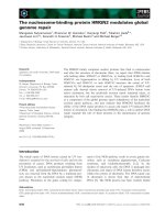
Glutamine enhances HSP70 expression
The analyses from Western blotting showed that HSP70
expressions in heart tissue (Figure 3a), aorta tissue (Figure
3b), lung tissue (Figure 3c), and liver tissue (Figure 3d) were
weak in the control group but markedly stronger in the LPS
shock group (P < 0.05). The expressions of HSP70 were
much higher than those in the LPS shock group from four tis-
sues in the Ala-Gln+LPS group (P < 0.05) (Figure 3a–d).
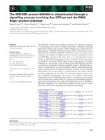
Figure 1
The percentage increase in mean arterial pressure (MAP) induced by phenylephrine in different groups of ratsThe percentage increase in mean arterial pressure (MAP) induced by
phenylephrine in different groups of rats. The maximal percentage
increase in MAP significantly decreased to 12.7% in the LPS shock
group (P < 0.05) and was restored to 15.6% in the Ala-Gln+LPS
group, whereas the maximum percentage increase in the control group
was 24.7% (n = 8, mean ± standard deviation). *P < 0.05 versus the
Ala-Gln+LPS group;
#
P < 0.05 versus the control group. Ala-Gln+LPS,
alanyl-glutamine dipeptide + lipopolysaccharide shock; LPS shock,
lipopolysaccharide shock.
Table 1
The values of PE E
max
and EC
50
on aortic rings in the different groups of rats
Group E
max
(g) EC
50
(nmol/l)
Control 1.86 ± 0.04 8.55 ± 0.08
LPS shock 0.95 ± 0.01
a
13.49 ± 0.06
a
Ala-Gln+LPS 1.27 ± 0.02
a,b
10.15 ± 0.04
a,b
Data are shown as mean ± standard deviation (n = 8 in each group).
a
P < 0.05 versus the control group;
b
P < 0.05 versus the LPS shock group.
Ala-Gln+LPS, alanyl-glutamine dipeptide + lipopolysaccharide shock; E
max
, maximum efficacy; EC
50
, median effective dose; LPS,
lipopolysaccharide.
Critical Care Vol 11 No 2 Jing et al.
Page 4 of 7
(page number not for citation purposes)
Glutamine decreases plasma concentrations of TNF-α,
IL-6, and MDA
The plasma TNF-α, IL-6, and MDA levels were low in the con-
trol group (49.7 ± 12.2 pg/ml, 23.5 ± 9.2 pg/ml, and 4.66 ±
0.55 mol/ml, respectively). They significantly increased in the
LPS shock group (293.1 ± 52.2 pg/ml, 296.2 ± 60.2 pg/ml,
and 9.71 ± 0.87 mol/ml, respectively; P < 0.01) (Table 2). Pre-
treatment with Ala-Gln significantly decreased plasma con-
centrations of TNF-α, IL-6, and MDA compared with the LPS
shock group (131.8 ± 27.7 pg/ml, 204.1 ± 42.2 pg/ml, and
5.89 ± 0.58 mol/ml, respectively; P < 0.05) (Table 2).
Discussion
The results of this study demonstrate that pretreatment of Ala-
Gln significantly improved vascular response to catecho-
lamine vasoconstrictors and that the effect of Ala-Gln is asso-
ciated with its capacity to induce HSP70 expression and
attenuate release of pro-inflammatory cytokines and oxidizing
species production after septic shock. HSP70 is the most
important protein in HSP family to generate a protective effect
against injuries in the presence of various stresses [17]. Previ-
ous studies in rat models of HSP induction to protect against
septic shock have used sodium arsenite or heat as an inducer
for the stress response. Sodium arsenite is known to be quite
toxic; a previous experiment showed a 20% mortality rate from
the arsenite alone [18]. HSP expression has also been
induced by measures that increase core body temperature
[19]. However, these measures are clinically impractical
because they would be poorly tolerated by patients and would
have detrimental effects on many cellular functions [20]. Gln
may have therapeutic value in safely and effectively enhancing
the expression of HSP and may increase the survival from sep-
tic shock [21]. Therefore, we chose Ala-Gln as an HSP
expression inducer in this study.
In the present study, we designed pretreatment of 0.75 g/kg
Gln to be one hour before LPS injection and performed a vas-
cular functional test six hours after administration of LPS. The
dose of Gln dipeptide used in this study was based on our pre-
vious data, which indicate that the maximal HSP70 mRNA
expression in rats occurs at 6 to approximately 12 hours after
an intravenous dose of 0.75 g/kg Gln. This dose has also been
demonstrated to safely induce HSP70 expression in sepsis
rats [22]. One limitation of this study seems to be that we
chose the pretreatment of Ala-Gln rather than to treat at the
onset or after a septic injury since the latter is more likely to get
close to the clinical utility. This was done because from our
previous experimental data, we know that the maximal HSP70
expression occurs at six hours after Ala-Gln is used. However,
administration of LPS results in hypotension at the third to
fourth hour in rats [23]. If Gln injection time is same with or
post LPS application, maybe could not exactly evaluate the
protective effect of Gln on vascular reactivity because Gln-
induced maximal HSP expression dose not obtained.
A potential limitation of this study is that we chose to use a
vehicle-based control rather than an iso-nitrogenous amino
acid control. Previous studies have shown that alanine does
not lead to significant enhancement of HSP70 either in ani-
mals [24] or patients [25], suggesting that the pharmacologi-
cal effects we have observed are related to the Gln treatment.
Ala-Gln must be diluted five times for intravenous administra-
tion based on its description; therefore, there is an excessive
amount of fluid infusion in the present study. This acute volume
overloading results in slight increases in blood pressure and
heart rate but does not lead to death in rats. The effects of vol-
ume overloading on the measurement of MAP should be con-
sidered. However, the volume of infusion in every
administration was strictly controlled according to body
weight to be the same in the three groups, and the MAP still
decreased after administration of LPS. This indicates that,
although volume overloading could temporarily change the
hemodynamics in rats, the change of MAP could still be con-
sidered a sensitive parameter that reflected vascular response
to agonists in the present study. Furthermore, a total of 3 ml of
blood was drawn at 90 minutes and 6 hours before the start
of the experiment and this may have partially reduced the influ-
ence of hypervolemia.
Endotoxin shock is characterized by a marked oxidant stress
[26] and a rapid production of different cytokines [27].
Nuclear factor-kappa-B is a transcription factor that plays a
central role in the modulation of the inflammatory and immune
responses and induces the expression of many genes of
inducible nitric oxide synthase, cytokine tissue factor, and
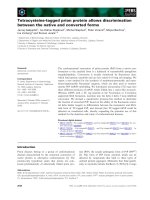
Figure 2
The concentration-response curves of phenylephrine (PE) in aortic rings from different groups of rats (mean ± standard deviation)The concentration-response curves of phenylephrine (PE) in aortic
rings from different groups of rats (mean ± standard deviation). PE max-
imum efficacy (E
max
) significantly decreased to 51% in the LPS shock
group and was restored to 68% in the Ala-Gln+LPS group (P < 0.05),
whereas PE E
max
in the control group was taken as 100%. *P < 0.05
versus the Ala-Gln+LPS group. #P < 0.05 versus the control group.
Ala-Gln+LPS, alanyl-glutamine dipeptide + lipopolysaccharide shock;
LPS shock, lipopolysaccharide shock.
Available online />Page 5 of 7
(page number not for citation purposes)
adhesion molecules involved in the pathogenesis of endotoxin
shock [28]. The results of our study showed that the levels of
plasma TNF-α, IL-6, and MDA in the Ala-Gln+LPS group were
lower than those in the LPS shock group, indicating that the
inhibited pro-inflammatory cytokine release and peroxide pro-
duction may also be attributed to the protective effects of Gln
on LPS-induced vascular hyporeactivity. The mechanism
involved may be that Gln inhibits the expression of the inflam-
matory cytokines directly [22] or that Gln-induced HSP70
expression further enhanced this effect [29].
The data provided in this study demonstrate two discrete
mechanistic effects produced by Ala-Gln, namely the reduced
LPS-induced cytokine presence in plasma and the increased
HSP70 in multiple tissues. The actual upstream mechanisms
responsible for these changes are not clear, but they may be
separately regulated and influenced by Ala-Gln. We
hypothesize that this dual effect of decreased cytokine-
induced vascular injury/action and increased vascular cell sur-
vival may be critical for the improved outcomes we observed.
Further studies to define the molecular pathways responsible
for these discrete actions are clearly warranted, as is further
investigation of Gln as a modulator of sepsis-related cardio-
vascular outcomes.
Figure 3
Effects of alanyl-glutamine on heat-shock protein 70 (HSP70) expression in heart, aorta, lung, and liver in endotoxin ratsEffects of alanyl-glutamine on heat-shock protein 70 (HSP70) expression in heart, aorta, lung, and liver in endotoxin rats. HSP70 expressions were
analyzed by Western blotting analysis. Relative density refers to the ratio of HSP70 to GAPDH. The expression of HSP70 was significantly
increased after lipopolysaccharide (LPS) injection compared with the control group in heart (a), aorta (b), lung (c), and liver (d) tissue. (*P < 0.05; n
= 5). The expressions of HSP70 were much higher than those in the LPS shock group from four tissues in the Ala-Gln+LPS group (#P < 0.05; n =
5). Ala-Gln+LPS, alanyl-glutamine dipeptide + lipopolysaccharide shock; Ala+LPS, alanyl-glutamine dipeptide + lipopolysaccharide shock; GAPDH,
glyceraldehyde-3-phosphate dehydrogenase; LPS+Gln, alanyl-glutamine dipeptide + lipopolysaccharide shock.
Critical Care Vol 11 No 2 Jing et al.
Page 6 of 7
(page number not for citation purposes)
Conclusion
Gln has been well demonstrated to protect against organ dys-
function in animal experiments and in critically ill patients by
inducing HSP70 expression, attenuating sepsis-induced met-
abolic dysfunction, and reducing inflammatory cytokine
release and peroxide production. Here, we further demon-
strated that administration of a dose of 0.75 g/kg Gln could
protect against vascular hyporeactivity in endotoxic shock rats.
Thus, Ala-Gln could be used to induce the protective stress
response and prevent end-organ injury and possibly decrease
mortality from sepsis and improve outcomes in critically ill
patients.
Competing interests
The authors declare that they have no competing interests.
Authors' contributions
LJ carried out the design of the study, established the experi-
mental setup, drafted the manuscript, and participated in part
of the animal experiments. QW carried out the in vivo and in
vitro animal experiments and blood analysis and performed the
statistical analysis. FW carried out the HSP70 protein expres-
sion detection. All authors read and approved the final
manuscript.
Acknowledgements
The authors thank John Anthony Bauer (associate professor at the Divi-
sion of Pharmacology and director of the Center for Cardiothoracic
Research Columbus Children's Research Institute, Columbus, OH,
USA) for his critical reading of and comments on the manuscript. This
work was supported in part by a Jiangsu Province Key Institution of
Anesthesiology Open Foundation grant from the Health Department of
Jiangsu Province (Nanjing, China) (no. WK200502) to LJ. This work was
presented at the 2006 annual meeting of the American Society of
Anesthesiologists, held in Chicago, IL, USA.
References
1. Glauser MP: Pathophysiologic basis of sepsis: considerations
for future strategies of intervention. Crit Care Med 2000, 28(9
Suppl):S4-S8.
2. Sheehan M, Wong HR, Hake PW, Zingarelli B: Protective effects
of isohelenin, an inhibitor of nuclear factor kappaB, in endo-
toxic shock in rats. J Endotoxin Res 2002, 8:99-107.
3. Kilbourn R: Nitric oxide synthase inhibitors – a mechanism-
based treatment of septic shock. Crit Care Med 1999,
27:857-858.
4. Zacharowski K, Berkels R, Olbrich A, Chatterjee PK, Cuzzocrea S,
Foster SJ, Thiemermann C: The selective guanylate cyclase
inhibitor ODQ reduces multiple organ injury in rodent models
of Gram-positive and Gram-negative shock. Crit Care Med
2001, 29:1599-1608.
5. Annane D, Sebille V, Charpentier C, Bollaert PE, Francois B,
Korach JM, Capellier G, Cohen Y, Azoulay E, Troche G, et al.:
Effect of treatment with low doses of hydrocortisone and
fludrocortisone on mortality in patients with septic shock.
JAMA 2002, 288:862-871.
6. d'Emmanuele di Villa Bianca R, Marzocco S, Di Paola R, Autore G,
Pinto A, Cuzzocrea S, Sorrentino R: Melatonin prevents lipopol-
ysaccharide-induced hyporeactivity in rat. J Pineal Res 2004,
36:146-154.
7. Lindquist S, Craig EA: The heat-shock proteins. Annu Rev
Genet 1988, 22:631-677.
8. Ang D, Liberek K, Skowyra D, Zylicz M, Georgopoulos C: Biolog-
ical role and regulation of the universally conserved heat
shock proteins. J Biol Chem 1991, 266:24233-24236.
9. Benjamin IJ, McMillan DR: Stress (heat shock) proteins: molec-
ular chaperones in cardiovascular biology and disease. Circ
Res 1998, 83:117-132.
10. Huber SA: Heat-shock protein induction in adriamycin and
picornavirus-infected cardiocytes. Lab Invest 1992,
67:218-224.
11. Toba T, Shidoji Y, Fujii J, Moriwaki H, Muto Y, Suzuki T, Ohishi N,
Yagi K: Growth suppression and induction of heat-shock pro-
tein-70 by 9-cis beta-carotene in cervical dysplasia-derived
cells. Life Sci 1997, 61:
839-845.
12. Suzuki K, Sawa Y, Kaneda Y, Ichikawa H, Shirakura R, Matsuda H:
In vivo gene transfection with heat shock protein 70 enhances
myocardial tolerance to ischemia-reperfusion injury in rat. J
Clin Invest 1997, 99:1645-1650.
13. Wischmeyer PE, Kahana M, Wolfson R, Ren H, Musch MM, Chang
EB: Glutamine induces heat shock protein and protects
against endotoxin shock in the rat. J Appl Physiol 2001,
90:2403-2410.
14. Singleton KD, Serkova N, Banerjee A, Meng X, Gamboni-Robert-
son F, Wischmeyer PE: Glutamine attenuates endotoxin-
induced lung metabolic dysfunction: potential role of
enhanced heat shock protein 70. Nutrition 2005, 21:214-223.
15. Fink MP, Heard SO: Laboratory model of sepsis and septic
shock. J Surg Res 1990, 49:186-196.
16. Beutler BA, Milsark IW, Cerami A: Cachectin/tumor necrosis
factor: production, distribution, and metabolic fate in vivo. J
Immunol 1985, 135:3972-3977.
Table 2
Plasma concentrations of TNF-α, IL-6, and MDA in the different groups of rats
Group TNF-α (pg/ml) IL-6 (pg/ml) MDA (mol/ml)
Control 49.7 ± 12.2 23.5 ± 9.2 4.66 ± 0.55
LPS shock 293.1 ± 52.2
a
296.2 ± 60.2
a
9.71 ± 0.87
a
Ala-Gln+LPS 131.8 ± 27.7
a,b
204.1 ± 42.2
a,b
5.89 ± 0.58
a,b
Data are shown as mean ± standard deviation (n = 8 in each group).
a
P < 0.05 versus the Ala-Gln+LPS group;
b
P < 0.05 versus the control
group. Ala-Gln+LPS, alanyl-glutamine dipeptide + lipopolysaccharide shock; IL-6, interleukin-6; LPS, lipopolysaccharide; MDA, malondialdehyde;
TNF-α, tumor necrosis factor-alpha.
Key messages
• Alanyl-glutamine improves vascular hyporeactivity in
endotoxic shock rats.
• The protective role of alanyl-glutamine on vascular reac-
tivity comes from inducing HSP70 expression and
reducing inflammatory cytokine release and peroxide
biosynthesis.
• These results suggest that alanyl-glutamine has poten-
tially beneficial therapeutic effects in sepsis.
Available online />Page 7 of 7
(page number not for citation purposes)
17. Jaattela M: Heat shock proteins as cellular lifeguards. Ann Med
1999, 31:261-271.
18. Ribeiro SP, Villar J, Downey GP, Edelson JD, Slutsky AS: Sodium
arsenite induces heat shock protein-72 kilodalton expression
in the lungs and protects rats against sepsis. Crit Care Med
1994, 22:922-929.
19. Chu EK, Ribeiro SP, Slutsky AS: Heat stress increases survival
rates in lipopolysaccharide-stimulated rats. Crit Care Med
1997, 25:1727-1732.
20. De Maio A: Heat shock proteins: facts, thoughts, and dreams.
Shock 1999, 11:1-12.
21. Singleton KD, Serkova N, Beckey VE, Wischmeyer PE: Glutamine
attenuates lung injury and improves survival after sepsis: role
of enhanced heat shock protein expression. Crit Care Med
2005, 33:1206-1213.
22. Singleton KD, Beckey VE, Wischmeyer PE: Glutamine prevents
activation of NF-kappaB and stress kinase pathways, attenu-
ates inflammatory cytokine release, and prevents acute respi-
ratory distress syndrome (ARDS) following sepsis. Shock
2005, 24:583-589.
23. Szabo C: Alterations in nitric oxide production in various forms
of circulatory shock. New Horiz 1995, 3:2-32.
24. Lindemann G, Grohs M, Stange EF, Fellermann K: Limited heat-
shock protein 72 induction in Caco-2 cells by l-glutamine.
Digestion 2001, 64:81-86.
25. Ziegler TR, Ogden LG, Singleton KD, Luo M, Fernandez-Estivariz
C, Griffith DP, Galloway JR, Wischmeyer PE: Parenteral
glutamine increases serum heat shock protein 70 in critically
ill patients. Intensive Care Med 2005, 31:1079-1786.
26. Altavilla D, Squadrito F, Campo GM, Squadrito G, Arlotta M, Urna
G, Sardella A, Quartarone C, Saitta A, Caputi AP: The lazaroid,
U74389G, inhibits inducible nitric oxide synthase activity,
reverses vascular failure and protects against endotoxin
shock. Eur J Pharmacol 1999, 369:49-55.
27. Zuckerman SH, Bryan-Poole N, Evans GF, Short L, Glasebrook
AL: In vivo modulation of murine serum tumour necrosis factor
and interleukin-6 levels during endotoxemia by oestrogen
agonists and antagonists. Immunology 1995,
86:18-24.
28. Barnes PJ, Karin M: Nuclear factor-kappaB: a pivotal transcrip-
tion factor in chronic inflammatory diseases. N Engl J Med
1997, 336:1066-1071.
29. Schell MT, Spitzer AL, Johnson JA, Lee D, Harris HW: Heat shock
inhibits NF-kB activation in a dose- and time-dependent
manner. J Surg Res 2005, 129:90-93.