Báo cáo khoa học: "Nucleated red blood cells in the blood of medical intensive care patients indicate increased mortality risk: a prospective cohort study" doc
Bạn đang xem bản rút gọn của tài liệu. Xem và tải ngay bản đầy đủ của tài liệu tại đây (207.54 KB, 8 trang )
Open Access
Available online />Page 1 of 8
(page number not for citation purposes)
Vol 11 No 3
Research
Nucleated red blood cells in the blood of medical intensive care
patients indicate increased mortality risk: a prospective cohort
study
Axel Stachon
1
, Elmar Segbers
1
, Tim Holland-Letz
2
, Reiner Kempf
1
, Steffen Hering
3
and
Michael Krieg
1
1
Institute of Clinical Chemistry, Transfusion and Laboratory Medicine, BG-University Hospital Bergmannsheil, Buerkle de la Camp-Platz 1, Ruhr-
University Bochum, 44789 Bochum, Germany
2
Department of Medical Informatics, Biometry, and Epidemiology, Overbergstrasse 17, Ruhr-University Bochum, 44801 Bochum, Germany
3
Department of Internal Medicine, BG-University Hospital Bergmannsheil, Buerkle de la Camp-Platz 1, Ruhr-University Bochum, 44789 Bochum,
Germany
Corresponding author: Axel Stachon,
Received: 23 Mar 2007 Revisions requested: 21 Apr 2007 Revisions received: 3 May 2007 Accepted: 5 Jun 2007 Published: 5 Jun 2007
Critical Care 2007, 11:R62 (doi:10.1186/cc5932)
This article is online at: />© 2007 Stachon et al.; licensee BioMed Central Ltd.
This is an open access article distributed under the terms of the Creative Commons Attribution License ( />),
which permits unrestricted use, distribution, and reproduction in any medium, provided the original work is properly cited.
Abstract
Introduction In critically ill patients, the appearance of
nucleated red blood cells (NRBCs) in blood is associated with
a variety of severe diseases. Generally, when NRBCs are
detected in the patients' blood, the prognosis is poor.
Methods In a prospective study, the detection of NRBCs was
used for a daily monitoring of 383 medical intensive care
patients.
Results The incidence of NRBCs in medical intensive care
patients was 17.5% (67/383). The mortality of NRBC-positive
patients was 50.7% (34/67); this was significantly higher (p <
0.001) than the mortality of NRBC-negative patients (9.8%, 31/
316). Mortality increased with increasing NRBC concentration.
Seventy-eight point six percent of the patients with NRBCs of
more than 200/μl died. The detection of NRBCs is highly
predictive of death, the odds ratio after adjustment for other
laboratory and clinical prognostic indicators being 1.987 (p <
0.01) for each increase in the NRBC category (0/μl, 1 to 100/
μl, 101 to 200/μl, and more than 200/μl). Each step-up in the
NRBC category increased the mortality risk as much as an
increase in APACHE II (Acute Physiology and Chronic Health
Evaluation II) score of approximately 4 points. The mortality of
patients who were NRBC-positive on the day of relocation from
the intensive care unit to a peripheral ward was 27.6% (8/27).
This was significantly higher than the mortality of patients who
were NRBC-negative on the relocation day (8.6%, 28/325; p <
0.01). On average, NRBCs were detected for the first time 14
days (median, 3 days) before death.
Conclusion The routine analysis of NRBCs in blood is of high
prognostic power with regard to mortality of critically ill patients.
Therefore, this parameter may serve as a daily indicator of
patients at high mortality risk. Furthermore, NRBC-positive
intensive care patients should not be relocated to a normal ward
but should obtain ongoing intensive care treatment.
Introduction
Under normal conditions, the peripheral blood of healthy
adults is generally free of nucleated red blood cells (NRBCs),
which tend to be found in patients with severe diseases [1-5]
who have a relatively poor prognosis [3,4,6-9]. In most of the
earlier studies on NRBCs, the concentration was determined
microscopically by a stained peripheral blood smear. With
such a technique, it is difficult to detect NRBC concentrations
of less than 200/μl [10]. For several years, a more convenient
and sensitive technique has been available in the form of
mechanized blood analyzers. With such an analyzer, one can
routinely determine NRBC concentrations of less than 100/μl
[11-13] The results of our recent studies with this new tech-
nique indicate that the detection of NRBCs may serve as an
early indicator in patients at increased risk of mortality: on aver-
age, the presence of NRBCs was detected 1 to 3 weeks
before death [14,15]. Furthermore, the analysis of the cytokine
profile in the blood of NRBC-positive patients (without
APACHE II = Acute Physiology and Chronic Health Evaluation II; NRBC = nucleated red blood cell; SAPS II = Simplified Acute Physiology Score II.
Critical Care Vol 11 No 3 Stachon et al.
Page 2 of 8
(page number not for citation purposes)
hematologic diseases) suggests that NRBCs may be consid-
ered a parameter that sums hypoxic and inflammatory injuries.
This may be the reason why the appearance of NRBCs is a
strong predictor of increased mortality [15-17].
Recently, we reported on the poor prognosis of surgical inten-
sive care patients when NRBCs are found in the peripheral
blood [18]. In that study, for the first time, a systematic day-to-
day screening for NRBCs in the blood of surgical intensive
care patients was performed. Our study revealed that 32% of
the surgical intensive care patients were NRBC-positive at
least once. The detection of NRBCs was associated with a
greatly increased mortality of 44% (versus 4% of NRBC-neg-
ative patients). The area under the curve amounted to 0.86.
On average, NRBCs were detected nine days before death.
Therefore, in the present study, we set out to establish
whether the daily screening for NRBCs in medical intensive
care patients could serve as an early indicator of medical inten-
sive care patients at extremely high risk.
Our study revealed that 18% of the medical intensive care
patients were NRBC-positive. The detection of NRBCs was
associated with a greatly increased in-hospital mortality. More
than 50% of the NRBC-positive patients died. Furthermore,
the mortality was three times higher in patients who were
NRBC-positive on the day of relocation from the intensive care
unit to a peripheral ward compared to patients who were
NRBC-negative on the relocation day. On average, NRBCs
were detected 14 days before death. These results suggest
that the routine daily measurement of NRBCs could aid in a
daily risk assessment of medical intensive care patients.
Materials and methods
Subjects and protocol
All intensive care patients treated between April 2003 and
January 2004 in the intensive care unit of the Department of
Internal Medicine of Berufsgenossenschaftliche Universitaet-
sklinik Bergmannsheil GmbH (University Hospital, Ruhr-Uni-
versity Bochum, Germany) (n = 383) were included in this
study. Patients younger than 18 years and patients after sur-
gery were excluded from this study. To evaluate the prognostic
significance of NRBCs in the peripheral blood of medical
intensive care patients, we screened one blood sample of
each patient each day by means of a Sysmex XE-2100 (Sys-
mex Europe GmbH, Norderstedt, Germany). Blood samples
were routinely drawn in the morning. For statistical analysis, a
patient was defined as NRBC-positive when NRBCs were
detected in the blood at least once. Outcome was considered
as in-hospital mortality. Ethical approval to conduct this study
was granted by the Ethical Committee of Ruhr-University
Bochum (reference no. 1982).
Laboratory tests
Blood count parameters (NRBCs, leukocytes, hemoglobin,
and thrombocytes) were measured using a Sysmex XE-2100
blood analyzer in line with the manufacturer's recommenda-
tions. According to the manufacturer, the NRBC detection
limit was greater than 19/μl. Stringent internal quality control
measurements were performed, and the criteria of acceptance
were fulfilled throughout.
Creatinine, alanine aminotransferase, and C-reactive protein
were measured with an LX 20 analyzer (Beckman Coulter
GmbH, Krefeld, Germany), and prothrombine time ratio was
assayed with a BCS (Behring Coagulation System) (Dade
Behring, Schwalbach, Germany), all in accordance with the
recommendations of the manufacturers. The quality assurance
of quantitative determinations was strictly performed accord-
ing to the German Norm: Quality Assurance in Medical Labo-
ratories (DIN [Deutsches Institut für Normung] 58936, 2000).
The criteria of acceptance were fulfilled throughout. Retro-
spective analysis of the laboratory data revealed 0.3% missing
values.
Statistical analysis
Data are presented as the mean ± standard error of mean.
When samples were normally distributed, the differences
between the data for survivors and deceased were analyzed
using the t test procedure. When samples were not normally
distributed, the Mann-Whitney test was used because this test
does not require a normal distribution of data. In the case of
categorical data, the Fisher exact test was used. Correlations
were analyzed by Pearson or non-parametric Spearman corre-
lation. A p value of less than 0.05 was considered statistically
significant.
A receiver operating characteristic curve was obtained by plot-
ting the true-positive proportion (sensitivity) against the false-
negative proportion (1 – specificity). The area under the curve
(C-statistics) was calculated by non-linear regression.
The prognostic significance of NRBCs and other risk indica-
tors was assessed using multiple logistic regression. In this
study, the logistic regression tries to estimate the relative
effect that parameters have on the patients' outcome. This is
facilitated by assuming a functional relationship (the 'logistic
model') between variables and probability of outcome. Then,
for all possible settings, every variable is given a relative impor-
tance that makes the actual observed event 'most likely', taking
into account the effects of all other variables. This is called the
'maximum likelihood' estimate of the variables' influence.
These coefficients provide a relative weighting for each varia-
ble. Moreover, they can be used to derive odds ratios for the
variables. If the odds ratio differs significantly from 1, a signifi-
cant prognostic power that is independent of the other varia-
bles considered may be assumed.
In a first step, laboratory data were analyzed with regard to
mortality. If reasonable for calculation of the odds ratios, the
data were categorized in up to four categories. That is, NRBCs
Available online />Page 3 of 8
(page number not for citation purposes)
were subdivided into four categories: 0/μl, 1 to 100/μl, 101 to
200/μl, and more than 200/μl. A backward selection multiple
logistic regression analysis was performed by first including all
parameters in a multivariate model and subsequently leaving
out the parameters with the largest p values until no parameter
with a p value greater than 0.25 was included. The calcula-
tions were carried out using SAS version 8.02 (SAS Institute
Inc., Cary, NC, USA). The intention of this study was to evalu-
ate the prognostic power of the presence of NRBCs in the
blood with regard to the patients' in-hospital mortality risk.
Nucleated red blood cells and established risk models
for intensive care patients
The prognostic significance of NRBCs was evaluated under
consideration of established risk models: the Acute Physiol-
ogy and Chronic Health Evaluation II (APACHE II) (first evalu-
ated in 1985 [19]) and the Simplified Acute Physiology Score
II (SAPS II) (first evaluated in 1993 [20]). The APACHE II
severity index includes the following risk factors: body temper-
ature, mean arterial pressure, heart rate, respiratory rate, blood
oxygenation, arterial pH, sodium, potassium, creatinine, hema-
tocrit, white blood cell count, Glasgow coma scale, age, and
anamnestic data concerning severe organ insufficiency or
immunocompromised states of health. The SAPS II considers
the following risk factors: age, heart rate, systolic blood pres-
sure, body temperature, blood oxygenation, urinary output,
urea, white blood cell count, potassium, sodium, bicarbonate,
bilirubin, Glasgow coma scale, chronic diseases (that is,
malignancies and acquired immunodeficiency syndrome), and
type of admission (that is, medical and unscheduled surgical).
Both the APACHE II score and the SAPS II are determined
from the most deranged (worst) physiologic value (for exam-
ple, the lowest blood pressure or the highest white blood cell
count) during the initial 24 hours after intensive care unit
admission.
Results
Patient characteristics
We included 383 medical intensive care patients. The mean
age was 66.3 ± 0.8 years (range, 20 to 94 years). Two hun-
dred twenty-five male (58.7%) and 158 female (41.3%)
patients were included in this study. On average, patients
were treated for 4.1 ± 0.3 days (n = 383) in the intensive care
unit. Total mortality was 17.0% (65/383). The APACHE II
score and the SAPS II amounted to 16.0 ± 0.5 and 35.2 ± 0.9,
respectively.
The incidence of NRBCs in the blood was 17.5% (67/383).
No significant difference was found between the incidences in
male (16.4%, 37/225) and female (19.0%, 30/158) patients.
On the day of admission, 7.8% (30/383) of the patients were
NRBC-positive. On average, NRBCs were detected for the
first time on the third day of intensive care treatment (3.1 ±
0.4; Figure 1), but in 44.8% (30/67) of the NRBC-positive
patients, NRBCs were already detected on the admission day.
On average, the highest NRBC concentration of each individ-
ual NRBC-positive patient was 189 ± 41/μl (range, 20 to
1,760/μl; median, 80/μl; n = 67). Some of the basal clinical
characteristics of NRBC-positive and NRBC-negative patients
are summarized in Table 1.
Prognostic significance of nucleated red blood cells
The mortality of NRBC-positive patients was 50.7% (34/67).
The predictive value for death increased with higher NRBC
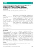
concentrations (Figure 2). The mortality was 46.7% (14/30) in
patients who were NRBC-positive on the day of admission to
the intensive care unit. In contrast, the mortality of NRBC-neg-
ative patients was 9.8% (31/316; p < 0.001).
Furthermore, the mortality of patients who were NRBC-posi-
tive on the day of relocation from the intensive care unit to a
peripheral ward was 27.6% (8/27). This was significantly
higher than the mortality of patients who were NRBC-negative
on the relocation day (8.6%, 28/325; p < 0.01).
Overall, with regard to in-hospital mortality, NRBCs in blood
showed sensitivity and specificity of 52.3% and 89.6%,
respectively. The area under the curve was 0.72.
NRBCs were an early indicator of patients at increased mor-
tality risk. On average, in NRBC-positive patients who died,
NRBCs were detected for the first time 13.6 ± 3.8 days
(median, 3 days; n = 34) before death.
After the first detection of NRBCs in blood and during the fur-
ther course of intensive care treatment, when the NRBCs have
disappeared from the circulation, the mortality again
decreased. That is, when former NRBC-positive patients were
again NRBC-negative for more than 4 days after the final
detection of NRBCs in blood, the mortality decreased to
16.7% (1/6).
As shown in Figure 3, the appearance of NRBCs in blood
seems not to be associated with one particular cause of death.
However, patients who have died from infections or sepsis, in
Figure 1
Intensive care days on which nucleated red blood cells were detected for the first time in the blood of medical intensive care patientsIntensive care days on which nucleated red blood cells were detected
for the first time in the blood of medical intensive care patients.
Critical Care Vol 11 No 3 Stachon et al.
Page 4 of 8
(page number not for citation purposes)
particular, had significantly higher NRBC concentrations than
patients who have died from cerebral or pulmonary complica-
tions. None of the other defined causes of death was associ-
ated with an NRBC concentration that was significantly higher
than the others.
Nucleated red blood cells in relation to other clinical and
laboratory risk indicators
The incidence of NRBCs increased with higher APACHE II
and SAPS II scores (Table 2). Accordingly, the Spearman cor-
relation revealed a significant correlation between the NRBC
concentration and the APACHE II (r = 0.292, p < 0.001) and
the SAPS II scores (r = 0.320, p < 0.001).
The Spearman correlation of the NRBCs with other laboratory
parameters is displayed in Table 3. When correlation was cal-
culated with values measured on the day of the first appear-
ance of NRBCs in blood, NRBCs significantly increased with
the leukocytes (r = 0.373, p < 0.01) and the creatinine con-
centration (r = 0.284, p < 0.05). Moreover, NRBCs increased
with a decreasing prothrombin time ratio (r = -0.408, p <
0.001). The concentrations of hemoglobin, thrombocytes, and
C-reactive protein as well as the alanine aminotransferase
activity were not significantly correlated with the NRBC
concentration.
The detection of NRBCs was an independent risk indicator of
poor outcome. In terms of mortality, the odds ratio for each
stepwise increase in the NRBC categories was calculated in
relation to other clinical and laboratory risk indicators by
means of multiple logistic regression (Table 4). Because the
correlation coefficient calculated by linear regression between
APACHE II score and SAPS II was r = 0.91, only the APACHE
II score was considered for the multiple logistic regression.
The detection of NRBCs is highly predictive of death, the odds
ratio after adjustment for other clinical and laboratory prognos-
tic indicators being 1.987 (p < 0.01) for each increase in the
NRBC category (0/μl, 1 to 100/μl, 101 to 200/μl, and more
than 200/μl). That is, patients with more than 200/μl NRBCs
had a more than seven-fold higher risk of dying than patients
without NRBCs.
Furthermore, under consideration of the APACHE II score, the
'maximum likelihood estimate' for each increase in the NRBC
category was 0.687 ± 0.253 and therefore approximately four
times (exactly 4.40 times) higher than the 'maximum likelihood
estimate' for each one-point increase in the APACHE II score
(0.156 ± 0.025). Therefore, each step-up in the NRBC cate-
gory is equivalent to approximately 4 APACHE II score points.
Consequently, an adjustment of the APACHE II score could
be performed by adding those 4, 8, or 12 points dependent on
Table 1
Clinical data and main diagnosis of treatment of NRBC-positive (n = 67) and NRBC-negative (n = 316) patients
Parameter NRBC-positive NRBC-negative P
Age, years 68 ± 2 66 ± 1 n.s.
Gender, male/female 55%/45% 59%/41% n.s.
Intensive care treatment, days 8.7 ± 1.2 3.2 ± 0.2 < 0.001
Body mass index 27.8 ± 0.9 26.5 ± 0.3 n.s.
Mortality 50.7% 9.8% < 0.001
APACHE II score points 21.5 14.9 < 0.001
SAPS II points 47.5 32.6 < 0.001
Acute coronary syndrome/AMI 16.4% (n = 11) 38.0% (n = 120) < 0.001
Cardiac arrhythmia 10.4% (n = 7) 14.2% (n = 45) n.s.
Cardiac insufficiency 11.9% (n = 8) 6.6% (n = 21) n.s.
Pulmonary diseases 23.9% (n = 16) 8.9% (n = 28) < 0.01
Gastrointestinal diseases 16.4% (n = 11) 7.9% (n = 25) < 0.05
Cerebral diseases 6.0% (n = 4) 10.1% (n = 32) n.s.
Toxication 4.5% (n = 3) 4.1% (n = 13) n.s.
Metabolic diseases 1.5% (n = 1) 4.4% (n = 14) n.s.
Other diagnosis 9.0% (n = 6) 5.7% (n = 18) n.s.
AMI, acute myocardial infarction; APACHE II, Acute Physiology and Chronic Health Evaluation II; NRBC, nucleated red blood cell; n.s., not
significant; SAPS II, Simplified Acute Physiology Score II.
Available online />Page 5 of 8
(page number not for citation purposes)
the patient's NRBC category (1 to 100, 101 to 200, and more
than 200/μl) to the individual APACHE II score of NRBC-pos-
itive patients. In practice, this modification would be a reas-
sessment of the patient's prognosis. The area under the curve
for this modified APACHE II score was 0.91 compared to 0.87
and 0.72 for the APACHE II score and the NRBCs alone,
respectively (Table 5).
Discussion
To our knowledge, this is the first study in which the detection
of NRBCs in the peripheral blood was investigated with regard
to its prognostic significance for the intensive care mortality of
medical intensive care patients. In earlier studies, we and oth-
ers have shown that the detection of NRBCs is associated
with a relatively poor prognosis [3,4,6-8,10,14,21,22]. In most
of those studies, the NRBC detection and quantification were
based on the microscopic analysis of stained blood smears.
This technique is time-consuming and only partly suitable for
the detection and quantification of NRBC concentrations of
less than 200/μl [10].
In this study, the NRBC concentration was screened with a
mechanized blood analyzer of high sensitivity [11,12,23]. The
present study revealed that approximately 18% of all medical
intensive care patients were NRBC-positive at least once.
Interestingly, in nearly half of the NRBC-positive patients,
NRBCs were detected already on the admission day.
Our data confirmed the high prognostic power of the mecha-
nized detection of NRBCs in blood in terms of mortality. The
total in-hospital mortality of NRBC-positive patients of this
study was 50.7%. Furthermore, as shown in earlier studies,
the present data showed that the mortality increased with an
increasing NRBC concentration [10,24,25] Approximately
80% of the patients with NRBC concentrations higher than
200/μl died.
Our study revealed that the daily screening for NRBCs can be
used to estimate the patients' mortality risk. Not only did the
predictive value for death increase with the concentration of
NRBCs in the blood, but the prognosis improved when the
NRBC concentration decreased. In particular, after the first
detection of NRBCs in blood and during the further course of
intensive care treatment, when the NRBCs have disappeared
from the circulation for more than four days, the mortality again
significantly decreased nearly to values of NRBC-negative
patients [18].
In the present study, increased creatinine and leukocyte con-
centrations and a lower prothrombin time ratio were signifi-
cantly correlated with increased NRBC concentrations.
Although these findings suggest that NRBC-positive patients
are more severely burdened than NRBC-negative patients, the
detection of NRBCs is an independent predictor of poor out-
come. To evaluate the independent attributable risk factor, a
logistic regression considering NRBCs, age, gender, body
mass index, APACHE II score, creatinine, hemoglobin, throm-
bocytes, leukocyte, alanine aminotransferase, C-reactive pro-
tein, and the prothrombin time ratio was performed. As a
result, the independent prognostic power of NRBCs is under-
Figure 2
In-hospital mortality of medical intensive care patients in relation to the concentration of nucleated red blood cells (NRBCs) in the bloodIn-hospital mortality of medical intensive care patients in relation to the
concentration of nucleated red blood cells (NRBCs) in the blood. Num-
bers in parenthesis denote the ratio of deceased patients to all patients
with the respective NRBC concentration.
Figure 3
Concentration of nucleated red blood cells (NRBCs) in the blood of medical intensive care patients who have died from various causesConcentration of nucleated red blood cells (NRBCs) in the blood of
medical intensive care patients who have died from various causes. ᭜
indicate the NRBC concentration of each individual deceased patient.
The average concentration is indicated by horizontal bars. * denote the
significance of the difference.
Critical Care Vol 11 No 3 Stachon et al.
Page 6 of 8
(page number not for citation purposes)
lined by an odds ratio of 1.987 for each stepwise increase in
the NRBC category. That is, patients with NRBCs of more
than 200/μl have a more than seven-fold higher risk to die than
NRBC-negative patients. In recent studies, we have already
demonstrated that the detection of NRBCs is a risk indicator
that is independent of several other established risk indicators
[10,14,16,24].
Among the general severity of illness scoring systems for
intensive care patients, APACHE II and SAPS II have become
two of the most accepted and used [26-30]. However, the
present data suggest that the APACHE II score could be sig-
nificantly improved by adding up to 12 score points, consider-
ing the presence of NRBCs as an independent variable in this
score, as suggested for the abbreviated burn severity index in
patients with burns [25].
The analysis of the lifespan of the patients who died indicates
that NRBCs in blood were found not just immediately before
death. Moreover, our present study showed that the detection
of NRBCs is often a relatively early phenomenon prior to
death. In deceased patients, NRBCs were detected 14 days
before death. Therefore, NRBCs would seem to be an early
indicator of increased risk.
Finally, the underlying pathophysiology of NRBCs in blood is
not fully understood. In our study, no association with only one
of the various causes of patient death was found. However,
some authors have claimed that hypoxemia [31,32], acute and
chronic anemia [33,34], or severe infections [35,36] are linked
to the appearance of NRBCs in critically ill patients. In this
context, we recently reported on the cytokine profile and the
erythropoietin concentrations in NRBC-positive patients [17].
Our data suggested an important role of inflammation and/or
decreased tissue oxygenation (caused by local or systemic cir-
culatory disorders) for the appearance of NRBCs in blood.
NRBCs may thus be considered a marker that sums up
hypoxic and inflammatory injuries. It seems obvious that these
complications have an impact on patient prognosis. Therefore,
this could be the reason why the appearance of NRBCs is a
strong predictor of increased mortality.
However, concerning such an association between inflamma-
tion (with or without hypoxia) and NRBCs, it is attractive to
speculate what kind of therapy could improve the poor prog-
nosis of NRBC-positive patients. Currently, studies are under
way in our university hospital to show whether intensifying the
treatment of patients with NRBCs (that is, an earlier adminis-
tration of antibiotics or an anti-inflammatory therapy) can
reduce their mortality rate.
Nonetheless, we observed that the mortality was three times
higher in patients who were NRBC-positive on the day of relo-
cation from the intensive care unit to a normal ward compared
to patients who were NRBC-negative on the relocation day.
Consequently, it seems obvious that NRBC-positive patients
should obtain ongoing intensive care treatment.
Conclusion
This is the first study in which the daily screening for NRBCs
in the peripheral blood of patients in the medical intensive care
unit was investigated with regard to its prognostic power for
in-hospital mortality. The incidence of NRBC-positive patients
was 18%. NRBC detection in critically ill patients was associ-
ated with significantly increased in-hospital mortality (50.7%
versus 9.8%). The predictive value for death increased with
the NRBC concentration and seems to decline again when the
NRBCs have disappeared from the circulation. The prognostic
significance of NRBCs was independent of other laboratory
and clinical risk parameters. An improvement of established
risk models like APACHE II seems feasible. Furthermore, the
detection of NRBCs in blood is a relatively early phenomenon
prior to death, so screening for NRBCs may aid in the early
Table 2
Incidence of NRBCs in blood in medical intensive care patients
in relation to the APACHE II and the SAPS II
Risk model Score range of risk model Incidence of NRBCs
APACHE II < 11 4.7% (6/127)
11–20 18.4% (28/152)
21–30 30.6% (22/72)
> 30 34.4% (11/32)
SAPS II < 21 5.6% (4/71)
21–40 13.3% (27/203)
41–60 34.3% (23/67)
> 60 30.9% (13/42)
APACHE II, Acute Physiology and Chronic Health Evaluation II;
NRBC, nucleated red blood cell; SAPS II, Simplified Acute
Physiology Score II.
Table 3
Spearman correlation of the nucleated red blood cell
concentration with other laboratory parameters (n = 67)
Parameter rP
Hemoglobin -0.113 n.s.
Leucocytes 0.373 < 0.01
Thrombocytes -0.152 n.s.
Creatinine 0.284 < 0.05
Prothrombin time ratio -0.408 < 0.001
Alanine aminotransferase 0.172 n.s.
C-reactive protein 0.169 n.s.
Correlation was calculated with values measured on the day of the
first appearance of nucleated red blood cells in blood. n.s., not
significant.
Available online />Page 7 of 8
(page number not for citation purposes)
identification of patients at high risk. Further studies are
needed to clarify whether the detection of NRBCs could help
to decide on a change of patient management, but our present
data suggest that NRBC-positive patients should obtain ongo-
ing intensive care treatment.
Competing interests
AS and RK obtained a grant from Sysmex Europe GmbH (Nor-
derstedt, Germany) to perform this study. The other authors
declare that they have no competing interests.
Authors' contributions
All authors made substantive intellectual contributions to the
design and conception of this study. AS was responsible for
data acquisition and data presentation, performed the analysis
and interpretation of data, and was responsible for the writing
of the manuscript. RK and ES were responsible for data acqui-
sition and data presentation and performed the analysis and
interpretation of data. SH was responsible for data acquisition
and data presentation. TH-L performed the analysis and inter-
pretation of data. MK was responsible for the writing of the
manuscript. All authors read and approved the final
manuscript.
Table 4
Multivariate odds ratio estimates of clinical and laboratory risk indicators for in-hospital mortality calculated by logistic regression
(n = 383)
Parameter Point estimate 95% confidence limits P
NRBCs
a
1.987 1.211–3.261 < 0.01
Leukocytes (> 10/nl) 0.480 0.178–1.294 0.147
Prothrombin time ratio (< 60%) 3.968 1.733–9.090 < 0.01
Alanine aminotransferase
b
1.223 0.932–1.620 0.162
C-reactive protein
c
1.214 0.901–1.635 0.202
APACHE II 1.168 1.112–1.227 < 0.001
Age, gender, body mass index, APACHE II score, the highest nucleated red blood cell (NRBC) concentration, the highest creatinine
concentration, the lowest hemoglobin concentration, the lowest thrombocytes concentration, the highest leukocyte concentration, the highest
alanine aminotransferase activity, the highest C-reactive protein concentration, and the lowest prothrombin time ratio were considered for the
calculation. All risk indicators with p values greater than 0.25 were removed from the model.
a
Subdivided into four categories (0/μl, 1 to 100/μl,
101 to 200/μl, and more than 200/μl); odds ratio was calculated for each stepwise increase in the category.
b
After log transformation.
c
Subdivided into four categories (0 to 5.0 mg/dl, 5.1 to 10.0 mg/dl, 10.1 to 15.0 mg/dl, and more than 15 mg/dl); odds ratio was calculated for
each stepwise increase in the category. APACHE II, Acute Physiology and Chronic Health Evaluation II.
Table 5
C-statistics for several risk indicators for in-hospital mortality of medical intensive care patients
Parameter Area under curve
NRBC, highest value 0.72
Leukocytes, highest value 0.61
Prothrombin time ratio, lowest value 0.73
Alanine aminotransferase, highest value 0.73
C-reactive protein, highest value 0.72
SAPS II, on admission 0.88
APACHE II, on admission 0.87
APACHE II, on admission + NRBC, highest value
a
0.91
Only data from the intensive care unit were considered.
a
The APACHE II score was incremented under consideration of the NRBC concentration
as follows: NRBC 0/μl: +0, NRBCs 1 to 100/μl: +4, NRBCs 101 to 200/μl: +8, NRBCs more than 200/μl: +12). APACHE II, Acute Physiology
and Chronic Health Evaluation II; NRBC, nucleated red blood cell; SAPS II, Simplified Acute Physiology Score II.
Key messages
• The detection of NRBCs in the blood of medical inten-
sive care patients is associated with significantly
increased in-hospital mortality (50.7% versus 9.8%).
The prognostic significance of NRBCs was independ-
ent of other laboratory and clinical risk parameters. The
predictive value for death increased with the NRBC
concentration and seems to decline again when the
NRBCs have disappeared from the circulation.
• Our present data suggest that NRBC-positive patients
should obtain ongoing intensive care treatment.
Critical Care Vol 11 No 3 Stachon et al.
Page 8 of 8
(page number not for citation purposes)
References
1. Budmiger H, Graf C, Streuli RA: [The leukoerythroblastic blood
picture. Incidence and clinical significance]. Schweiz Rundsch
Med Prax 1984, 73:1489-1493.
2. Mettier SR: Hematologic aspects of space consuming lesions
of the bone marrow (myelophtisic anemia). Ann Intern Med
1940, 14:436-448.
3. Schwartz SO, Stansbury F: Significance of nucleated red blood
cells in peripheral blood; analysis of 1,496 cases. J Am Med
Assoc 1954, 154:1339-1340.
4. Delsol G, Guiu-Godfrin B, Guiu M, Pris J, Corberand J, Fabre J:
Leukoerythroblastosis and cancer frequency, prognosis, and
physiopathologic significance. Cancer 1979, 44:1009-1013.
5. Weick JK, Hagedorn AB, Linman JW: Leukoerythroblastosis.
Mayo Clin Proc 1974, 49:110-113.
6. Tavassoli M: Erythroblastemia. West J Med 1975, 122:194-198.
7. Andres WA: Normoblastemia after thermal injury. Am J Surg
1976, 131:725-726.
8. Böning A, Stachon A, Weisser H, Krismann M, Skipka G, Laczko-
vics A, Krieg M: Postoperative cholesterol and erythroblasts as
a parameter of perioperative mortality after cardiothoracic
surgery. Z Herz-Thorax-Gefäâchir 2001, 15:242-248.
9. Otsubo H, Kaito K, Asai O, Usui N, Kobayashi M, Hoshi Y: Persist-
ent nucleated red blood cells in peripheral blood is a poor
prognostic factor in patients undergoing stem cell
transplantation. Clin Lab Haematol 2005, 27:242-246.
10. Stachon A, Böning A, Krismann M, Weisser H, Laczkovics A,
Skipka G, Krieg M: Prognostic significance of erythroblasts in
blood after cardiothoracic surgery. Clin Chem Lab Med 2001,
39:239-243.
11. Briggs C, Harrison P, Grant D, Staves J, Machin SJ: New quanti-
tative parameters on a recently introduced automated blood
cell counter – the XE 2100. Clin Lab Haematol 2000,
22:345-350.
12. Walters J, Garrity P: Performance evaluation of the Sysmex XE-
2100 hematology analyser.
Lab Hematol 2000, 6:83-92.
13. Stachon A, Sondermann N, Krieg M: Incidence of nucleated red
blood cells in the blood of hospitalised patients. Infusion Ther
Transfus Med 2001, 28:263-266.
14. Stachon A, Sondermann N, Imöhl M, Krieg M: Nucleated red
blood cells indicate high risk for in-hospital mortality. J Lab
Clin Med 2002, 140:407-412.
15. Stachon A, Holland-Letz T, Kempf R, Becker A, Friese J, Krieg M:
Poor prognosis indicated by nucleated red blood cells in
peripheral blood is not associated with organ failure of the
liver or kidney. Clin Chem Lab Med 2006, 44:955-961.
16. Stachon A, Eisenblätter K, Köller M, Holland-Letz T, Krieg M:
Cytokines and erythropoietin in the blood of patients with
erythroblastemia. Acta Haematol 2003, 110:204-206.
17. Stachon A, Bolulu O, Holland-Letz T, Krieg M: Association
between nucleated red blood cells in blood and the levels of
erythropoietin, interleukin-3, interleukin-6, and interleukin-
12p70. Shock 2005, 24:34-39.
18. Stachon A, Kempf R, Holland-Letz T, Friese J, Becker A, Krieg M:
Daily monitoring of nucleated red blood cells in the blood of
surgical intensive care patients. Clin Chim Acta 2006,
366:329-335.
19. Knaus WA, Draper EA, Wagner DP, Zimmerman JE: APACHE II: a
severity of disease classification system. Crit Care Med 1985,
13:818-829.
20. Le Gall JR, Lemeshow S, Saulnier F: A new simplified acute
physiology score (SAPS II) based on a European/North Amer-
ican multicenter study. JAMA 1993, 270:2957-2963.
21. Lehnhardt M, Katzy Y, Langer S, Druecke D, Homann HH, Stein-
straesser L, Steinau HU, Krieg M: Prognostic significance of
erythroblasts in burns. Plast Reconstr Surg 2005, 115:120-127.
22. West CD, Ley AB, Pearson OH: Myelophthisic anemia in cancer
of the breast. Am J Med 1955, 18:923-931.
23. Ruzicka K, Veitl M, Thalhammer-Scherrer R, Schwarzinger I: The
new hematology analyzer Sysmex XE-2100: performance
evaluation of a novel white blood cell differential technology.
Arch Pathol Lab Med 2001, 125:391-396.
24. Stachon A, Holland-Letz T, Krieg M:
High in-hospital mortality of
intensive care patients with nucleated red blood cells in blood.
Clin Chem Lab Med 2004, 42:933-938.
25. Stachon A, Lehnhardt M, Katzy Y, Holland-Letz T, Steinau HU,
Krieg M: Making the case for adapting the abbreviated burn
severity index to include erythroblast count. J Wound Care
2005, 14:97-100.
26. Rosenberg AL: Recent innovations in intensive care unit risk-
prediction models. Curr Opin Crit Care 2002, 8:321-330.
27. Morgenthaler NG, Struck J, Christ-Crain M, Bergmann A, Muller B:
Pro-atrial natriuretic peptide is a prognostic marker in sepsis,
similar to the APACHE II score: an observational study. Crit
Care 2005, 9:R37-R45.
28. Schetz MR, Van den Berghe G: Do we have reliable biochemical
markers to predict the outcome of critical illness? Int J Artif
Organs 2005, 28:1197-1210.
29. Ratanarat R, Thanakittiwirun M, Vilaichone W, Thongyoo S, Perm-
pikul C: Prediction of mortality by using the standard score
systems in a medical intensive care unit in thailand. J Med
Assoc Thai 2005, 88:949-955.
30. Herbert PC, Wells G, Blajchman MA, Marshall J, Marin C,
Pagliarello G, Tweeddale M, Schweitzer I, Yetisir E: A multicenter,
randomized, controlled clinical trial of transfusion require-
ments in critical care. Transfusion Requirements in Critical
Care Investigators, Canadian Critical Care Trials Group. N
Engl J Med 1999, 340:409-417.
31. Ward HP, Holman J: The association of nucleated red cells in
the peripheral smear with hypoxemia. Ann Intern Med 1967,
67:1190-1194.
32. Burkett LL, Cox ML, Fields ML: Leukoerythroblastosis in the
adult. Am J Clin Pathol 1965, 44:494-498.
33. Clifford GO: The clinical significance of leukoerythroblastic
anemia. Med Clin North Am 1966, 50:779-790.
34. Vaughan JM, Oxon DM: Leuco-erythroblastic anaemia. J Pathol
1936, 42:541-563.
35. Okpara RA: The prognostic significance of leukoerythroblastic
anaemia in Nigerians. East Afr Med J 1985, 62:185-188.
36. Retief FP: Leuco-erythroblastosis in the adult. Lancet 1964,
1:639-642.