Radiology for Anaesthesia and Intensive Care - Part 2 pps
Bạn đang xem bản rút gọn của tài liệu. Xem và tải ngay bản đầy đủ của tài liệu tại đây (897.46 KB, 36 trang )
Imaging the chest
1
16
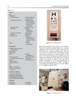
Fig. 1.16 The penetrated film
demonstrates tooth fragments
in the right upper lobe
bronchus and also the left
lower lobe bronchus.
Fig. 1.15 Aspirated
and swallowed teeth
following facial
trauma. Right upper
lobe collapse and
partial collapse of
left lower lobe.
Opacities in the
stomach.
Chap-01.qxd 10/15/02 6:05 PM Page 16
Case illustrations: plain films and CT
1
17
Question 5
Table 1.1 Causes of lobar collapse
Luminal mass
Neoplasm (carcinoma, carcinoid)
Foreign body (peanut) (see Figs 1.15 and 1.16)
Mucus plug/inflammatory exudate
Endoluminal metastasis
Misplaced endotracheal tube (ITU ventilated patients)
Bronchial wall
Inflammation (TB, sarcoid)
Extrinsic compression
Lymph nodes
aneurysm
Fig. 1.17 Quiz case.
Answer
Left lower lobe collapse
Lobar or segmental collapse occurs in large airway obstruction and
subsequent absorbtion of air from the affected lung. Causes are listed
below. Bronchogenic malignancy is one of the commonest causes and the
case study illustrates the subtle signs on plain X-ray. Subsequent CT imaging
of this patient demonstrated a malignant neoplasm originating in the
left lower lobe bronchus.
72-year-old smoker.
Haemoptysis and cough.
What does the chest X-ray
(Fig. 1.17) show?
Chap-01.qxd 10/15/02 6:05 PM Page 17
Imaging the chest
1
18
Fig. 1.18 CT left lower lobe
collapse. Note how the left
lower lobe collapses tight
against the descending aorta.
Fig. 1.19 Right lower lobe
collapse. Loss of volume
in the right lung,
the right hemithorax is
hypertranslucent.
In children, bronchial malignancy is rare and the causes of lobar collapse
differ from those in adults. Inflammatory exudate in pneumonia or mucus
plugging (in patients with cystic fibrosis and asthma) are much more
common causes (Table 1.1).
The five lobes collapse in different directions to produce different
patterns although there are some common features (see below). If the
vessels within the collapsed lobe remain perfused, then a wedge-shaped
opacity is more clearly identified. In lower lobe collapse (both right and
left lower lobe), the lung collapses posteriorly and medially. This is well
illustrated by the CT scan (see Fig. 1.18). In left lower lobe collapse, the
silhouette of the medial aspect of the hemidiaphragm and the descending
Chap-01.qxd 10/15/02 6:05 PM Page 18
Case illustrations: plain films and CT
1
19
aorta is lost because it is no longer outlined by adjacent aerated lung.
A triangular opacity is seen projected through the cardiac outline. In right
lower lobe collapse (Fig. 1.19), the hemidiaphragm silhouette remains
clearly seen as the middle lobe is in contact with it. On a lateral projection
the collapsed lower lobe may be identified as a triangle of increased
density in the posterior costophrenic recess.
X-ray signs of lobar collapse
Volume loss (hilar shift, mediastinal shift, hemidiaphragm elevation,
rib crowding).
Compensatory hyperinflation (translucency) of other lobes.
Movement of fissures.
Wedge-shaped opacity caused by collapsed lobe-specific pattern for
each lobe.
Right upper lobe collapse
The right upper lobe collapses against the mediastinum and thoracic
apex with a broad-based opacity radiating from the hilum. If there is an
outward bulge at the right hilum, this is good evidence that a hilar mass
is responsible for the collapse (see Fig. 1.20). The lower lobe pulmonary
artery is pulled upwards and outwards.
Fig. 1.20 Right upper lobe
collapse. There is a mass
at the right hilum which
merges with the triangular
opacity from the collapsed
right upper lobe –
‘Golden sign’. This
‘S’-shaped appearance is
typical of a neoplastic hilar
mass responsible for the
upper lobe collapse.
Chap-01.qxd 10/15/02 6:05 PM Page 19
Left upper lobe collapse
This does not mirror right upper lobe collapse due to the absence of a middle
lobe. The left upper lobe collapses forward against the anterior chest wall.
The lower lobe expands behind it. The chest X-ray appearance is of a hazy
density in the mid- and upper zones which fades away laterally and inferiorly
(see Fig. 1.21). The collapsed lobe is adjacent to the left cardiac and
mediastinal border, so this silhouette is completely lost. The aortic knuckle is
lost (see Fig. 1.22) unless the lobar collapse is accompanied by overexpansion
of the lower lobe with its superior segment occupying the apex.
Imaging the chest
1
20
Fig. 1.21 Left upper lobe collapse.
Fig. 1.22 Left upper lobe collapse CT. Note how the lobe collapses anteriorly
against the chest wall.
Chap-01.qxd 10/15/02 6:05 PM Page 20
Case illustrations: plain films and CT
1
21
Middle lobe collapse
This is easily missed on the frontal film and is often more obvious on a
lateral projection. On the frontal projection, there is a vague increase in
density seen in the right lower zone and the normally sharp right heart
border is blurred. On a lateral projection the collapsed middle lobe forms
a triangular opacity with its apex at the hilum and base projecting towards
the sternum. (see Figs 1.23 and 1.24).
Fig. 1.24 Lateral projection middle
lobe collapse. The triangular opacity
is the collapsed middle lobe.
Fig. 1.23 Middle lobe collapse. Note
the loss of outline of the right
heart border.
Chap-01.qxd 10/15/02 6:05 PM Page 21
Answer
Pectus excavatum
This chest wall deformity can simulate disease. The rib pairs are heart shaped
and downward pointing in their lateral and anterior aspect. The heart is
shifted to the left and superimposed soft tissue shadows at the right heart
border create the impression of middle lobe disease. A lateral film will
demonstrate the sternal depression and also confirm normality of the
middle lobe. The CT (Fig. 1.26) demonstrates the thoracic cage deformity.
Other normal variants include cervical ribs, which are usually
asymptomatic, but in a minority of cases individuals can be symptomatic
with Raynauld’s phenomenon. Distinction is made from normal transverse
processes by the direction of slope. Cervical ribs slope downwards.
Imaging the chest
1
22
Fig. 1.26 CT pectus
excavatum.
Note the sternal
depression and
the movement of the
heart to the left side.
Question 6
Fig. 1.25 Quiz case.
33-year-old man.
Chest X-ray (Fig. 1.25)
taken for purposes of
obtaining a travel visa.
What are the
findings?
Chap-01.qxd 10/15/02 6:05 PM Page 22
Question 7
Case illustrations: plain films and CT
1
23
Fig. 1.27 Quiz case.
Answer
Diaphragmatic eventration and colonic interposition
This history is designed to mislead as no pathology is demonstrated here.
Interposition of colon between the liver and the diaphragm
(chilaiditis syndrome) can simulate pneumoperitoneum. Haustration of
the bowel can usually be identified but if there is doubt, then a left
lateral decubitus film should be performed.
In diaphragmatic eventration the normal muscular part of a
hemidiaphragm is deficient and replaced by connective tissue. There is
normally a dome-like bulging of the anterior aspect of the diaphragm.
It is common in the elderly when the bulging may be focal. A lateral
projection will show the posterior costophrenic recess to be in the normal
position and if screened under fluoroscopy some diaphragmatic movement
can be identified.
58-year-old man.
Abdominal pain.
Patient has a drug history
of corticosteroids use for
polymyalgia rheumatica.
What does the X-ray
(Fig. 1.27) show?
Does the patient need
laparotomy?
Chap-01.qxd 10/15/02 6:05 PM Page 23
Question 8
Imaging the chest
1
24
Fig. 1.28 Quiz case.
Answer
The tip of the nasogastric tube is in the right main bronchus and the
more proximal part of the tube is coiled in the left main bronchus.
Checking the position of all tubes and lines is crucial for films taken on
intensive care units. This should be done meticulously for each line by
tracing it with the eye throughout its course.
46-year-old patient on
intensive care unit.
What does the chest
X-ray (Fig. 1.28) show?
Chap-01.qxd 10/15/02 6:05 PM Page 24
Case illustrations: plain films and CT
1
25
Fig. 1.29 Quiz case.
Question 9
Chest X-ray (Fig. 1.29) taken prior to varicose vein surgery.
What is the diagnosis?
Are there any precautions necessary prior to anaesthesia?
Chap-01.qxd 10/15/02 6:05 PM Page 25
Answer
Retrosternal thyroid
This is the commonest cause of a superior mediastinal mass. It can
displace and narrow the trachea and it frequently calcifies.
The patient should be euthyroid before elective surgery. Careful
questioning, examination and thyroid function tests should be performed.
If the mediastinal mass is compressing the trachea, then the patient may be
symptomatic. If the tracheal diameter is reduced by more than 50%, then
the patient will develop stridor, which may be positional or occur only on
exercise. CT or MRI should be used to gain more information about the
degree of tracheal compression. If significant, removal of the thyroid
should take priority over elective varicose vein surgery. If there is no, or
minimal, compression, then surgery may proceed. Regional block is an
anaesthetic option.
Mediastinal masses
Mediastinal masses are classified according to their location in
the mediastinum: anterior, middle and posterior mediastinum.
It is possible to localise these with reasonable accuracy on plain films by
assessing which silhouette has been lost. If, for example, part of the
silhouette of the ascending thoracic aorta or heart border is blurred,
then the mass must be anterior. If the lung hilum is seen projected
through the mass and the hilum is of normal appearance, then the
abnormality cannot be in the middle mediastinum. Posterior
mediastinal masses are identified by the loss of the thoracic spine
contour or the descending thoracic aorta outline. Other clues of a
posteriorly sited mass are abnormalities of the adjacent ribs or
bony involvement of the spine (see Fig. 1.30).
Lateral plain films will confirm the position in the mediastinum.
Cross-sectional imaging (CT or MRI) are routinely used to give further
anatomical detail of mediastinal masses (Table 1.2).
Imaging the chest
1
26
Fig. 1.30 Posterior mediastinal
mass, a neurogenic tumour. It could
be difficult to locate this mass in
the posterior mediastinal on frontal
chest X-ray – the clue to look for
is the expansion of the neural
foramina.
Chap-01.qxd 10/15/02 6:05 PM Page 26
Case illustrations: plain films and CT
1
27
Table 1.2 Mediastinal masses
Anterior mediastinal masses – anatomical space anterior to trachea and
major bronchi
Thyroid
Lymph nodes
Thymic tumour (see Figs 1.31 and 1.32)
Teratoma
Ascending aortic aneurysm
Middle mediastinum – space between anterior and posterior mediastinum
Lymph nodes (Fig. 1. 33)
Aortic arch aneurysm
Bronchogenic cyst
Posterior mediastinal mass – anatomical space posterior to anterior aspect
of thoracic spine
Hiatus hernia
Lymph nodes
Descending aortic aneurysm
Neurogenic mass (Fig. 1.30)
Fig. 1.31 Anterior mediastinal mass-thymoma. The mass blends into the aortic arch.
Chap-01.qxd 10/15/02 6:05 PM Page 27
Imaging the chest
1
28
Fig. 1.32 CT thymoma. Cross-sectional imaging demonstrates the position of
the mass in the anterior mediastinum.
Fig. 1.33 Mass in the
middle mediastinum.
Bilateral hilar
lymphadenopathy due to
sarcoidosis.
Chap-01.qxd 10/15/02 6:06 PM Page 28
Question 10
Case illustrations: plain films and CT
1
29
Fig. 1.34 Quiz case.
Answer
Hiatal hernia
A hiatal hernia is herniation of the stomach through the oesophageal
diaphragmatic hiatus into the thorax. The majority of hiatal hernias are
sliding, i.e. the gastro-oesophageal junction as well as part of the stomach
move up into the chest. The gastro-oesophageal junction remains correctly
located in a rolling hiatal hernia and part of the stomach herniates past it
up into the chest. Hiatal hernias are described as incarcerated when they
are irreducible. Hiatal hernias can be diagnosed on plain chest radiographs
where a gas shadow is seen projected through the cardiac silhouette.
If doubt exists, then a lateral projection can be performed (Fig. 1.35).
Hiatal hernias are a frequent finding on barium examinations of the upper
gastrointestinal tract.
64-year-old patient
(Fig. 1.34).
What is the condition?
What anaesthetic
precautions are
necessary?
Chap-01.qxd 10/15/02 6:06 PM Page 29
The appearance following oesophagectomy can also produce a large
mediastinal gas shadow (sometimes with a fluid level) (see Fig. 1.36).
Aspiration is a potential risk of general anaesthesia or sedation.
Regional anaesthesia should be considered if appropriate for the surgery.
The patient should be given an H
2
antagonist or proton pump inhibitor as
pre-medication. If general anaesthesia is undertaken, a rapid sequence
induction with cricoid pressure should be performed and the trachea
intubated with a cuffed tube.
Imaging the chest
1
30
Fig. 1.35 Lateral projection of
hiatus hernia. Note the air
fluid levels in the stomach.
Fig. 1.36 Gastric pull-up. There
has been an oesophagectomy
and gastric pull-up procedure.
The air fluid level is within
the stomach. Note the
right-sided rib changes
from thoracotomy.
Chap-01.qxd 10/15/02 6:06 PM Page 30
Case illustrations: plain films and CT
1
31
Question 11
Fig. 1.37 Quiz case.
Report the chest
X-ray (Fig. 1.37).
Answer
Aortic stenosis
There is cardiomegaly with a prominent ascending thoracic aorta and
calcification in the region of the aortic valve. There is upper lobe blood
diversion consistent with mild left ventricular failure.
Causes of aortic stenosis include congentital stenosis, a bicuspid aortic
valve, rheumatic heart disease and senile calcific valve degeneration.
The gradient across the heart valve produces left ventricular hypertrophy
with increased muscle mass which can cause relative myocardial ischaemia
and the risk of cardiac arrhythmias. If untreated, left ventricular
decompensation leads to left ventricular dilatation and pulmonary venous
congestion.
The X-ray findings include, post-stenotic dilatation of the aorta,
calcification of the aortic valve and a left ventricular configuration seen
at the left heart border with concavity along the mid-lateral border and
increased convexity along the lower left heart border (Table 1.3).
Chap-01.qxd 10/15/02 6:06 PM Page 31
Imaging the chest
1
32
Table 1.3 Causes of cardiomegaly
Ischaemic heart disease
Valvular heart disease
Congenital heart disease
Dilated cardiomyopathy
Viral
Alcohol (Fig. 1.38)
Metabolic
Pericardial effusions
In left ventricular hypertrophy from hypertension or aortic stenosis, the cardiac
silhouette does not dilate until late in the natural history.
Fig. 1.38 Enlarged heart.
Cardiomyopathy – note
the globular appearance.
The differential diagnosis
included pericardial effusion.
Chap-01.qxd 10/15/02 6:06 PM Page 32
Case illustrations: plain films and CT
1
33
Question 12
Fig. 1.39 Quiz case.
Man admitted with acute
chest pain. Report this film
(Fig. 1.39).
What is the most
common cause?
Answer
Pulmonary oedema from left ventricular failure
The heart is enlarged, there is upper lobe blood diversion, septal lines (due
to interstitial oedema) are present at both costophrenic angles and there is
air space shadowing bilaterally in a perihilar distribution. The appearances
are of pulmonary oedema from elevated pulmonary venous pressure.
Ischaemic heart disease is the commonest cause.
Comment
Although in hospital practice the commonest cause of pulmonary
oedema is left ventricular failure, the other causes (see Table 1.4) should
also be considered if only to exclude them. Distinction can be difficult
on the chest X-ray alone. In cases of elevated pulmonary venous pressure,
different X-ray signs develop progressively as it rises. Clinical history,
signs and further investigations all help to limit the diagnosis. Response
to treatment, e.g. diuretics is helpful.
Signs of pulmonary venous hypertension
The various X-ray signs appear as progressive elevation of the venous
pressure occurs (see Table 1.5). The radiological changes correlate with
pulmonary capillary wedge pressure (PCWP). The X-ray changes in the
lungs of patients with chronic left ventricular failure develop at PCWPs
which are about 5 mmHg higher than patients with acute disease and
vascular redistribution is not seen until later.
Chap-01.qxd 10/15/02 6:06 PM Page 33
Imaging the chest
1
34
Table 1.4 Causes of pulmonary oedema
Left ventricular failure
Ischaemic
Cardiomyopathy
Valvular heart disease
Aortic/mitral
Near drowning
Aspiration
ARDS (refer to Fig. 1.49)
Altitude
Raised intracranial pressure
Renal failure and fluid overload
Table 1.5 Signs of pulmonary venous hypertension
1. Vascular redistribution (PCWP 11–19 mmHg)
Upper lobe vessels (arteries and veins)
Increased in diameter (greater than 3 mm in first intercostal space)
Lower lobe vessels reduced in size
2. Interstitial oedema (PCWP
Ͼ
19 mmHg)
Kerley’s B lines
Bronchial cuffing
3. Alveolar oedema (PCWP
Ͼ
25 mmHg)
Perihilar-bat wing distribution
Symmetrical
Clears rapidly with diuretics
4. Pleural effusions
Chap-01.qxd 10/15/02 6:06 PM Page 34
Case illustrations: plain films and CT
1
35
Question 13
Fig. 1.40 Quiz case.
53-year-old female.
Atrial fibrillation (Fig. 1.40).
What is the diagnosis?
Answer
Mitral valve disease
The radiological features are
1. left atrial enlargement (double right heart border),
2. right ventricular enlargement,
3. posterior displacement of the oesophagus,
4. mitral valve calcification,
5. Kerley’s B lines,
6. pulmonary microlithiasis (if chronic).
Chap-01.qxd 10/15/02 6:06 PM Page 35
Imaging the chest
1
36
Question 14
Fig. 1.41 Quiz case.
Comment (Fig. 1.41) on
1. Cardiac size.
2. Pulmonary vascularity.
3. Suggest a possible cause.
Answer
The heart is enlarged. The pulmonary trunk and the pulmonary arteries are
enlarged. There is pulmonary arterial hypertension. The aortic arch is
relatively small. Atrial septal defect (ASD) is the cause in this case.
Atrial septal defect
ASD causes increased pulmonary arterial flow due to blood being shunted
from the left to the right atrium and, subsequently, through the pulmonary
arteries and veins. This overcirculation causes pulmonary plethora making
the vessels appear distended. The right atrium, right ventricle, pulmonary
artery and veins become enlarged. The vessels appear crisper than
pulmonary venous hypertension (due to lack of oedema) and upper lobe
distension is absent. After time persistent overcirculation through the
pulmonary arteries results in increased pulmonary resistance and rapid
tapering of the pulmonary arteries – pruning. Reversal of the shunt can
occur with time – Eisenmenger reaction. Calcification may develop in the
pulmonary arteries in cases of chronic pulmonary hypertension (Table 1.6).
Chap-01.qxd 10/15/02 6:06 PM Page 36
Case illustrations: plain films and CT
1
37
Table 1.6 Causes of pulmonary hypertension
Chronic pulmonary venous hypertension, e.g. mitral valve disease
Congenital heart disease with left to right shunts
ASD
VSD
PDA (see Figs 1.42 and 1.43)
Pulmonary emboli
Primary pulmonary arterial hypertension
Chronic lung disease
Fig. 1.42 Patent ductus
arteriosus (PDA), is a
cause of pulmonary
hypertension – note the
pulmonary artery
enlargement.
A metallic coil has been
placed to occlude the
ductus arteriosus.
Chap-01.qxd 10/15/02 6:06 PM Page 37
Imaging the chest
1
38
Fig. 1.43 PDA lateral
projection.
Chap-01.qxd 10/15/02 6:06 PM Page 38
Case illustrations: plain films and CT
1
39
Question 15
71-year-old male.
Sudden onset of severe,
tearing chest pain.
What is the
diagnosis (Fig. 1.44)?
Fig. 1.44 Quiz case.
Answer
Aortic dissection
The features demonstrated on the CT scan are an aortic intimal flap and
a false lumen (which on higher slices starts distal to the left subclavian
artery) – the features are typical of a type-B aortic dissection.
Aortic dissection has a peak incidence in the sixth to seventh decade
with a male predominance. The associated risk factors are hypertension
and medial degeneration. A variety of congenital diseases are associated
with dissection and these include Marfan’s and Ehlers–Danlos syndrome.
Pregnancy and cardiac catheterisation are further risk factors.
The aorta is composed of three layers (intima, media and adventitia)
and dissection is characterised by haematoma in the media of the aortic
wall. The separation of the intima from the adventitia finally creates a
false lumen that continues for a variable distance. If the dissection ruptures
through the adventitia haemopericardium or haemothorax can result.
Stanford classification
Type A: involving ascending aorta or arch.
Type B: limited to descending aorta (distal to left sub-clavian).
Clinical manifestations vary depending on the site and extent of the
dissection. ECG changes of myocardial infarction can be present if
the coronary arteries are involved, and neurological signs may indicate
haematoma or intimal flap narrowing the head and neck vessels.
Patients with Sandford type-B dissection are treated medically
with management of hypertension and those with type A are treated
surgically. Diagnostic imaging modalities include transthoracic and
trans-oesophageal echocardiography, angiography, CT and MRI.
Chap-01.qxd 10/15/02 6:06 PM Page 39
Imaging the chest
1
40
Question 16
34-year-old female. Taking oral contraceptive pill.
Immobilised for compound tibial fracture.
Suddenly short of breath.
What is the diagnosis (Fig. 1.45)?
What other methods of diagnostic imaging are available?
Fig. 1.45 Quiz case.
Chap-01.qxd 10/15/02 6:06 PM Page 40