Báo cáo y học: " Expression of parathyroid hormone-related protein during immortalization of human peripheral blood mononuclear cells by HTLV-1: Implications for transformation" pps
Bạn đang xem bản rút gọn của tài liệu. Xem và tải ngay bản đầy đủ của tài liệu tại đây (1.98 MB, 12 trang )
BioMed Central
Page 1 of 12
(page number not for citation purposes)
Retrovirology
Open Access
Research
Expression of parathyroid hormone-related protein during
immortalization of human peripheral blood mononuclear cells by
HTLV-1: Implications for transformation
Murali VP Nadella
1,2
, Sherry T Shu
1,2
, Wessel P Dirksen
1,2
, Nanda K Thudi
1
,
Kiran S Nadella
3
, Soledad A Fernandez
2,4
, Michael D Lairmore
1,2
,
Patrick L Green
1,2
and Thomas J Rosol*
1,2
Address:
1
Department of Veterinary Biosciences, The Ohio State University, Columbus, OH, USA,
2
Center for Retrovirus Research, The Ohio State
University, Columbus, OH, USA,
3
Human Cancer Genetics, The Ohio State University, Columbus, OH, USA and
4
Center for Biostatistics, The
Ohio State University, Columbus, OH, USA
Email: Murali VP Nadella - ; Sherry T Shu - ; Wessel P Dirksen - ;
Nanda K Thudi - ; Kiran S Nadella - ; Soledad A Fernandez - ;
Michael D Lairmore - ; Patrick L Green - ; Thomas J Rosol* -
* Corresponding author
Abstract
Background: Adult T-cell leukemia/lymphoma (ATLL) is initiated by infection with human T-lymphotropic virus
type-1 (HTLV-1); however, additional host factors are also required for T-cell transformation and development
of ATLL. The HTLV-1 Tax protein plays an important role in the transformation of T-cells although the exact
mechanisms remain unclear. Parathyroid hormone-related protein (PTHrP) plays an important role in the
pathogenesis of humoral hypercalcemia of malignancy (HHM) that occurs in the majority of ATLL patients.
However, PTHrP is also up-regulated in HTLV-1-carriers and HTLV-1-associated myelopathy/tropical spastic
paraparesis (HAM/TSP) patients without hypercalcemia, indicating that PTHrP is expressed before transformation
of T-cells. The expression of PTHrP and the PTH/PTHrP receptor during immortalization or transformation of
lymphocytes by HTLV-1 has not been investigated.
Results: We report that PTHrP was up-regulated during immortalization of lymphocytes from peripheral blood
mononuclear cells by HTLV-1 infection in long-term co-culture assays. There was preferential utilization of the
PTHrP-P2 promoter in the immortalized cells compared to the HTLV-1-transformed MT-2 cells. PTHrP
expression did not correlate temporally with expression of HTLV-1 tax. HTLV-1 infection up-regulated the
PTHrP receptor (PTH1R) in lymphocytes indicating a potential autocrine role for PTHrP. Furthermore, co-
transfection of HTLV-1 expression plasmids and PTHrP P2/P3-promoter luciferase reporter plasmids
demonstrated that HTLV-1 up-regulated PTHrP expression only mildly, indicating that other cellular factors and/
or events are required for the very high PTHrP expression observed in ATLL cells. We also report that
macrophage inflammatory protein-1α (MIP-1α), a cellular gene known to play an important role in the
pathogenesis of HHM in ATLL patients, was highly expressed during early HTLV-1 infection indicating that, unlike
PTHrP, its expression was enhanced due to activation of lymphocytes by HTLV-1 infection.
Conclusion: These data demonstrate that PTHrP and its receptor are up-regulated specifically during
immortalization of T-lymphocytes by HTLV-1 infection and may facilitate the transformation process.
Published: 9 June 2008
Retrovirology 2008, 5:46 doi:10.1186/1742-4690-5-46
Received: 10 March 2008
Accepted: 9 June 2008
This article is available from: />© 2008 Nadella et al; licensee BioMed Central Ltd.
This is an Open Access article distributed under the terms of the Creative Commons Attribution License ( />),
which permits unrestricted use, distribution, and reproduction in any medium, provided the original work is properly cited.
Retrovirology 2008, 5:46 />Page 2 of 12
(page number not for citation purposes)
Background
Human T-lymphotropic virus type I (HTLV-I) is the etio-
logical agent of adult T-cell leukemia/lymphoma (ATLL),
HTLV-1-associated myelopathy/tropical spastic parapare-
sis (HAM/TSP) and a variety of other disorders [1,2]. ATLL
is an aggressive malignancy of CD4+ T cells that occurs in
approximately 5% of infected individuals after a long
latency period of 20–40 years. The long latency period
and the relatively low proportion of HTLV-1-infected peo-
ple that develop ATLL reflect the inefficiency of the virus
to transform cells and the need for multiple cooperative
changes in growth control mechanisms to induce leuke-
mogenesis.
HTLV-1 is a complex deltaretrovirus and its genome not
only encodes for the essential viral genes gag, pol, and
env, but also additional HTLV-1-specific regulatory pro-
teins Tax and Rex, several accessory proteins p12, p13,
p30 and a minus-strand encoded protein, HTLV-1 bZIP-
factor (HBZ) [7]. Although the precise mechanisms
underlying transformation are not completely under-
stood, the 40-kDa transcriptional transactivator, Tax, is
thought to be principally responsible for tumorigenesis
[8]. The ability to activate cellular genes, including proto-
oncogenes, is a key mechanism leading to immortaliza-
tion and transformation of HTLV-1-infected cells. Rex reg-
ulates the expression of incompletely spliced viral RNAs
by interacting with the Rex response element in the viral
RNA and cellular proteins used by CRM-dependent
nuclear export [15]. Although Rex is not required for
immortalization of lymphocytes in vitro, it is required for
infectivity and persistence in vivo [16]. The accessory genes
p12, p30, p13 and HBZ contribute to establishing persist-
ent viral infection in vivo but are not required for transfor-
mation of cells in vitro [17,18].
About 80% of ATLL patients develop humoral hypercal-
cemia of malignancy (HHM), a life-threatening paraneo-
plastic syndrome that occurs in a wide variety of cancers
in addition to ATLL [19]. ATLL cells express factors such as
interleukin-1, tumor necrosis factor β, parathyroid hor-
mone-related protein (PTHrP), macrophage inflamma-
tory protein-1α (MIP-1α) and receptor activator of
nuclear factor-κB ligand (RANKL) that directly and/or
indirectly stimulate osteoclast differentiation and activity,
resulting in hypercalcemia [20-24]. PTHrP has been
shown to play a central role in the pathogenesis of HHM
in ATLL patients, but likely has additive or synergistic
effects with other tumor-associated cytokines [25].
Although PTHrP was discovered based on its role in the
pathogenesis of HHM, PTHrP is now known to be a com-
plex factor with a broad range of physiologic and/or
pathophysiologic actions in different tissues [34]. PTHrP
has been shown to be an auto/paracrine cell growth regu-
lator that increases proliferation of several cell types
including chondrocytes and renal epithelial cells [43].
PTHrP stimulates proliferation through the PTH1R by
mechanisms involving both PKA and PKC signaling path-
ways.
Watanabe et al have shown that PTHrP was constitutively
expressed in HTLV-1-carriers and ATLL patients with or
without hypercalcemia which suggests that PTHrP is
expressed before transformation of lymphocytes [26].
ATLL cell adhesion up-regulated PTHrP expression [27]
indicating additional roles for PTHrP besides its central
role in the pathogenesis of HHM. Moreover, PTHrP gene
expression was induced during transformation of normal
rat embryo fibroblasts by co-transfection with an activated
ras gene and a mutated p53 gene [40]. Insogna et al have
shown that PTHrP induced transformation of rat fibrob-
lasts with epidermal growth factor [41]. In addition, co-
transfection of rat embryonic fibroblasts with Tax and ras
transformed the fibroblasts and they were highly tumori-
genic in vivo [42]. Based on these findings, it is possible
that PTHrP functions as a transforming factor in conjunc-
tion with other oncogenes.
The goal of this study was to investigate the expression of
PTHrP, its receptor, and MIP-1α during the early stages of
immortalization of human lymphocytes by HTLV-1.
Using long-term liquid culture immortalization assays,
we showed that PTHrP and PTH1R were markedly up-reg-
ulated during immortalization of T-lymphocytes. PTHrP
expression did not correlate temporally with HTLV-1 tax
expression and IL-2 stimulation. Co-transfection of HTLV-
1 with a PTHrP P2/P3 luciferase reporter showed that
PTHrP was up-regulated by HTLV-1 infection.
Results
HTLV-1-infected PBMCs proliferate beyond six weeks
To investigate the expression of PTHrP early after HTLV-1
infection, we used long-term co-culture assays of PBMCs
from healthy human donors with irradiated HTLV-1 pro-
ducer cells (SLB-1) in the presence or absence of IL-2. Via-
ble cells were counted by trypan blue exclusion and the
results are shown in figure 1. Irradiated SLB-1 cells lived
up to 1 week in culture. As expected, PBMCs grown in the
absence of stimulation with either IL-2 or PHA, progres-
sively decreased in numbers and failed to grow in vitro
[31]. PBMCs supplemented with IL-2 or PHA lived and
proliferated up to 2 weeks in culture, at which time they
enter a "growth crisis" phase and decreased in numbers
and lost viability beyond 6 weeks in culture. In contrast,
HTLV-1-infected PBMCs continued to proliferate beyond
6 weeks for up to at least 13 weeks in culture. Cells that
continued to proliferate beyond 8–9 weeks in culture in
the presence or absence of exogenous IL-2 were referred to
as immortalized cells. High levels of p19 Gag protein were
Retrovirology 2008, 5:46 />Page 3 of 12
(page number not for citation purposes)
detected throughout the co-culture demonstrating virus
production (data not presented).
PTHrP was up-regulated during immortalization of PBMCs
with HTLV-1
To determine the temporal expression of PTHrP during
HTLV-1 immortalization of PBMCs, PTHrP mRNA (Figure
2A) and protein (Figure 2B) expression were analyzed at
various time points during the long-term co-culture
assays. Freshly-isolated PBMCs expressed very little PTHrP
mRNA, which was barely detectable by RT-PCR. There was
no increase in PTHrP mRNA or protein expression in
unstimulated PBMCs during culture in vitro. IL-2 stimula-
tion up-regulated PTHrP mRNA expression in the first
week (3.8 to 12-fold) compared to unstimulated PBMCs.
After one week, there was no further up-regulation of
PTHrP mRNA in the IL-2-stimulated PBMCs. Although
there was an increase in the PTHrP mRNA expression due
to IL-2 stimulation, PTHrP protein (2.6 pM) was detecta-
ble in only one of the samples (PBMC-1 + IL-2). No
increase in PTHrP mRNA or protein occurred with PHA
stimulation of PBMCs. In contrast, HTLV-1 infection
markedly up-regulated PTHrP mRNA expression com-
pared to uninfected PBMCs. In PBMCs infected with
HTLV-1 in the presence of IL-2, PTHrP mRNA was up-reg-
ulated 300- to 500-fold 5–11 weeks post co-culture com-
pared to uninfected PBMCs at day 0. In PBMCs infected
with HTLV-1 in the absence of IL-2, PTHrP mRNA was up-
regulated 1300- to 3800-fold 5–11 weeks post co-culture
compared to uninfected PBMCs at day 0. As shown in fig-
ure 2B PTHrP protein was detectable in the conditioned
medium 1 week following co-culture with HTLV-1 pro-
ducer cells and peak PTHrP protein expression occurred
between weeks 10 and 13 post-infection. Peak PTHrP pro-
tein expression ranged from 133 to 212 pM in condi-
tioned medium from PBMCs infected with HTLV-1 in the
presence of IL-2 and from 130 to 160 pM in conditioned
medium from PBMCs infected with HTLV-1 in the
absence of IL-2.
Up-regulation of PTHrP was mediated by the PTHrP P2
and P3 promoters
PTHrP is regulated by three distinct promoters that are
transactivated by different cellular signal transduction
pathways [32]. To understand the molecular mechanisms
involved in the transcriptional up-regulation of PTHrP
following HTLV-1 infection, we investigated the promoter
usage using real-time RT-PCR to detect specific promoter-
initiated transcripts. As shown in figure 3, PTHrP P2 and
P3 promoters were utilized during immortalization in the
presence or absence of IL-2. However, the ratio of P2 to P3
promoter-initiated transcripts was at least 2-fold higher
during immortalization of PBMCs with HTLV-1 (1:2)
(Figure 3A–B) when compared to transformed MT-2 cells
(1:4) (Figure 3C).
HTLV-1 infection up-regulated PTH1R expression
Many of the biological properties of PTHrP result from its
interaction with the PTH1R, which is coupled to adenylyl
cyclase (AC) and/or phospholipase C (PLC), and down-
stream signaling pathways [33,34]. Therefore, we meas-
ured the expression of PTH1R during immortalization of
PBMCs with HTLV-1. As shown in figure 4A, there was
very low PTH1R expression in PBMCs. Stimulation of
PBMCs with IL-2 or PHA did not up-regulate PTH1R.
However, following infection with HTLV-1 there was a
marked induction of PTH1R in PBMCs. Singal intensities
from the PTH1R were quantitated and averages (PBMC-1,
2, 3 + HTLV-1 + IL-2 and PBMC-1, 2 + HTLV-1 samples
combined) were presented as a bar graph in the bottom
panel (Figure 4A). The PTH1R levels were significantly
greater at weeks 5, 7, 9, and 13 compared to PBMCs alone
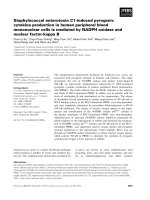
Growth curves and p19 Gag expression in HTLV-1 T-lym-phocyte immortalization assaysFigure 1
Growth curves and p19 Gag expression in HTLV-1 T-
lymphocyte immortalization assays. Human PBMCs (2
× 10
6
) were cultured alone or with irradiated donor cells
(SLB-1) in 24-well plates. Cell viability was measured weekly
by trypan blue exclusion (0–13 weeks after co-cultivation)
and growth curves are shown. PBMCs were infected with
HTLV-1 in the presence of IL-2 (10 U/mL; supplemented
from day 1 following HTLV-1 infection) or in the absence of
IL-2. PBMCs with no stimulation, PBMCs stimulated with
PHA and IL-2 irradiated SLB-1 cells served as controls. The
results showed that only HTLV-1-infected cells continued to
proliferate beyond 6 weeks in culture. Viable cell numbers
were significantly different over time between treatment
groups (p < 0.0001). While the PBMC+HTLV-1+IL-2 group
cell numbers increased slightly over time, the remaining
group cell numbers decreased over time, but the
PBMC+HTLV-1 group cell numbers decreased only slightly.
After using Dunnett's method to adjust for multiple compari-
sons, the HTLV-1-treated groups both had significantly
higher cell numbers than the PBMC (control) group (p <
0.0001).
Retrovirology 2008, 5:46 />Page 4 of 12
(page number not for citation purposes)
PTHrP was markedly up-regulated during immortalization of PBMCs with HTLV-1 infectionFigure 2
PTHrP was markedly up-regulated during immortalization of PBMCs with HTLV-1 infection. (A) PTHrP mRNA
expression during immortalization of PBMCs with HTLV-1. Total RNA was extracted from the co-cultures at various time
points and PTHrP mRNA expression was measured by real-time RT-PCR using the Taqman method. PTHrP expression was
normalized to human β
2
M and the data were represented as fold change over uninfected PBMCs from day 0. After using Dun-
nett's method to adjust for multiple comparisons, the PBMC+HTLV-1 group was shown to have higher PTHrP mRNA level
than the PBMC group (p < 0.0001). The PMBC+HTLV-1+IL-2 group was not different from the PBMC group due to the very
limited data available for the PBMC group. These limited data were caused by low cell viability resulting in low RNA recovery
from the PBMC group. (B) PTHrP protein expression during immortalization of PBMCs with HTLV-1. Secreted PTHrP was
measured in the conditioned medium from the co-culture assays by IRMA. Results showed marked up-regulation of PTHrP
secretion in PBMCs infected with HTLV-1 during the immortalization phase. PTHrP concentrations were significantly different
over time between treatment groups (p < 0.0001). While PTHrP secretion increased in HTLV-1-treated groups over time,
PTHrP secretion in the other 4 groups remained negligible and unchanged. After using Dunnett's method to adjust for multiple
comparisons, both HTLV-1-treated groups had significantly higher protein levels than the PBMC group (p < 0.0001).
Retrovirology 2008, 5:46 />Page 5 of 12
(page number not for citation purposes)
(p < 0.05). We also analyzed the expression of PTH1R in
various HTLV-1-transformed and ATLL cell lines. As
shown in figure 4B, HTLV-1-negative Jurkat cells did not
express PTH1R. High Tax-expressing HTLV-1-positive cells
(MT-2, SLB-1, HT-1RV) expressed moderate levels of
PTH1R. RV-ATL cells expressed low levels of PTH1R while
MET-1 cells did not express the PTH1R. Human β
2
microglobulin (B
2
M) was used as a loading control.
PTHrP expression did not correlate with HTLV-1 tax
expression
HTLV-1 Tax has been shown to transactivate PTHrP; how-
ever, ATLL cells that lack significant Tax expression have
very high levels of PTHrP indicating that PTHrP can be
expressed in a Tax-independent manner [35]. To investi-
gate the basis for up-regulation of PTHrP due to HTLV-1
infection, we analyzed by quantitative real-time RT-PCR
the temporal expression of HTLV-1 viral transcript tax.
The high tax expression during the first week in the co-cul-
tures (data not shown) was contributed by the residual
live irradiated SLB-1 cells. After the first week, the decline
PTHrP was up-regulated by the P2 and P3 promotersFigure 3
PTHrP was up-regulated by the P2 and P3 promoters. Specific PTHrP promoter-initiated transcripts were measured
by real-time quantitative RT-PCR using the SYBR green method. The data was normalized to human β
2
M gene expression. Spe-
cific PTHrP-promoter initiated transcripts are shown for 0, 3, 7 and 13 weeks post co-culture in the presence of IL-2 (A), in
the absence of IL-2 (B) and for MT-2 cells (C). The data showed that PTHrP was up-regulated in PBMCs following HTLV-1
infection by the activation of both the P2 and P3 promoters.
Retrovirology 2008, 5:46 />Page 6 of 12
(page number not for citation purposes)
HTLV-1 infection up-regulated expression of the PTHrP receptor (PTH1R) in PBMCsFigure 4
HTLV-1 infection up-regulated expression of the PTHrP receptor (PTH1R) in PBMCs. PTHrP receptor expres-
sion and human β
2
M were measured by RT-PCR from total RNA at various time points in the co-culture assays. (A) Up-regu-
lation of PTH1R in PBMCs at weeks 1, 3, 5, 7, 9, 11, and 13 following HTLV-1 infection in the presence or absence of IL-2
compared to day 0; controls 1 and 4 are PBMC-1 and PBMC-2; controls 2 and 5 are PBMC-1 and PBMC-2 stimulated with
PHA; controls 3 and 6 are PBMC-1 and PBMC-2 stimulated with IL-2 for one week. ANOVA with Dunnett's tests were used
to analyze the data from PTH1R RT-PCR quantification (bar graph shown at the bottom of the panel). The PTH1R levels were
significantly greater at weeks 5, 7, 9, and 13 (p < 0.05; indicated by asterisks in the figure). (B) PTH1R expression in HTLV-1-
infected T-cells and ATLL cells. Lanes represent: (1) Jurkat (2) MT-2 (3) SLB-1 (4) HUT102 (5) C8166 (6) MET-1 (7) RV-ATL
(8) HT-1RV cells. The data showed that PTH1R expression was very low or absent in the ATLL cells (MET-1 and RV-ATL)
compared to HTLV-1-infected T-cell lines (MT-2, SLB-1 and HT-1RV). Jurkat T-cells were used as a negative control. β
2
M was
used a loading control.
Retrovirology 2008, 5:46 />Page 7 of 12
(page number not for citation purposes)
in tax expression correlated with the death of the irradi-
ated SLB-1 cells and the subsequent tax expression was
from the newly HTLV-1-infected PBMCs. Tax mRNA
expression increased from week 3 to 7 and then decreased
between 9–11 weeks post-infection (Figure 5). As shown
in figure 5, the expression of tax did not correlate tempo-
rally with the expression of PTHrP.
HTLV-1 and HTLV-1 Rex up-regulated PTHrP expression
In order to investigate the direct effect of the HTLV-1 viral
proteins on PTHrP expression, we co-transfected a PTHrP
P2/P3 promoter-driven luciferase plasmid with expres-
sion plasmids for HTLV-1 (ACH), p12, p13, p30, Tax, Rex
and HBZ (Figure 6). Expression of the HTLV-1 ACH pro-
viral clone or Rex up-regulated PTHrP expression (1.6-
fold) 48 h after transfection. The expression of HTLV-1-
p12, p13, p30, HBZ or Tax cDNA vectors did not alter
PTHrP expression (Figure 6).
MIP-1
α
expression correlated with activation of PBMCs
following HTLV-1 infection
Since PTHrP was specifically up-regulated during the
immortalization of PBMCs with HTLV-1, we also meas-
ured the expression of MIP-1α, another chemokine
known to be involved the pathogenesis of HHM in ATLL
patients [22]. As shown in figure 7, MIP-1α expression
was induced by IL-2 (4- to 14-fold) or PHA (3- to 9-fold)
stimulation of PBMCs as expected [36,37], followed by a
return to near-baseline concentrations by week 3 (Figure
7C). However, there was marked up-regulation of MIP-1α
in the first week post co-culture in PBMCs infected with
HTLV-1. The expression of MIP-1α in the immortalization
assays ranged from 10,000 to 46,000 pg/mL. After the
peak induction of MIP-1α at week 1, there was consistent
but lower MIP-1α expression throughout all time points
(Figure 7A & B). PBMCs and irradiated SLB-1 cells
expressed very low levels of MIP-1α (Figure 7D).
Discussion
Although HTLV-1 Tax is known to have pleiotropic effects
that either directly or indirectly contribute to immortali-
zation and transformation of infected T-cells, the exact
mechanisms of transformation are unclear. In this study,
we analyzed the temporal PTHrP gene expression during
virus-mediated immortalization of lymphocytes to char-
acterize its role in the transformation process. We present
data to show that PTHrP is markedly up-regulated during
the immortalization process.
An important step in HTLV-1-induced leukemogenesis is
the induction of abnormal T-cell growth. Long-term
PTHrP expression did not correlate with HTLV-1 tax expressionFigure 5
PTHrP expression did not correlate with HTLV-1 tax
expression. HTLV-1 Tax expression, in co-cultures follow-
ing HTLV-1 Infection, was measured by quantitative real time
RT-PCR using the SYBR green method and the data was nor-
malized to human β
2
M gene expression.
HTLV-1 infection or over-expression of Rex alone up-regu-lated PTHrP expressionFigure 6
HTLV-1 infection or over-expression of Rex alone
up-regulated PTHrP expression. Relative luciferase
activity in 293T cells transfected with either pGL2 or pGL2
PTHrP-P2/P3 Luc constructs alone or with expression plas-
mids for HTLV-1 (ACH), p12, p13, p30, HBZ, Rex and Tax.
The quantity of the expression plasmid is indicated in μg.
Bars represent the mean ± SD of three independent samples.
Relative Luc/Gal units were significantly different across
groups (p = 0.0002). After adjusting for multiple times of
comparison, P2/P3Luc+Rex group and P2/P3Luc+ACH group
had significantly greater relative Luc/Gal units than the P2/
P3Luc group (p = 0.0006, p = 0.0012, respectively; indicated
by asterisks in the figure).
Retrovirology 2008, 5:46 />Page 8 of 12
(page number not for citation purposes)
immortalization assays have been used to study the kinet-
ics of HTLV-1 infection and abnormal T-cell growth that
lead to transformation. The growth curves in our study are
similar to previous reports [31,38]. Human PBMCs that
were cultured in the presence of IL-2, but not exposed to
the virus, survived in vitro only for a few weeks. Following
exposure to HTLV-1, PBMCs initially underwent a prolif-
erative response due to HTLV-1 infection after which the
cells entered a "growth crisis" between weeks 5–7 fol-
lowed by expansion of immortalized cells. The high level
of HTLV-1 p19 antigen expression in the first few weeks of
co-culture was due to the live residual irradiated SLB-1
cells. However, the p19 expression after three weeks in
culture was from the newly infected PBMCs and demon-
strated active HTLV-1 viral infection (data not shown).
Our data showed that PTHrP mRNA expression was grad-
ually up-regulated in PBMCs following HTLV-1 infection;
however, marked expression of PTHrP protein occurred at
the time when the PBMCs were undergoing immortaliza-
tion. This supports an important role for PTHrP during
immortalization and the subsequent transformation
process. The differences between the levels of PTHrP
mRNA and protein expression were likely due to differ-
ences in translation efficiency, processing of the mature
protein, and/or its secretion from the cells. Regulation of
MIP-1α induction due to activation of lymphocytes following HTLV-1 infectionFigure 7
MIP-1α induction due to activation of lymphocytes following HTLV-1 infection. MIP-1α was measured in the condi-
tioned medium from the co-culture assays at various time points (day 0, 1, 3, 5, 7, 9, 11 and 13 weeks of co-culture) by ELISA.
The results showed that MIP-1α expression was up-regulated in PBMCs following stimulation with PHA or IL-2. HTLV-1 infec-
tion markedly up-regulated MIP-1α expression in the first week after infection which demonstrated that HTLV-1 infection acti-
vated the lymphocytes. MIP-1α levels were significantly different across groups and time. Overall, MIP-1α levels significantly
decreased over time (p < 0.0001). After using Dunnett's method to adjust for multiple comparisons, the HTLV-1-treated
groups had significantly higher MIP-1α levels than the PBMC group (p < 0.0001).
Retrovirology 2008, 5:46 />Page 9 of 12
(page number not for citation purposes)
PTHrP secretion is a complex process and it has been
shown that some PTHrP may not be secreted but targeted
directly to the nucleus and function in an intracrine fash-
ion [39]. Abundant expression of PTH1R is normally
found in the target organs that regulate calcium ion home-
ostasis, such as the kidney and bone, with restricted
expression in other tissues. This contrasts with the wide-
spread expression of PTHrP. In our investigation, the
marked induction of both PTHrP and PTH1R by HTLV-1
suggests that PTHrP functioned as an autocrine growth
regulator in the transformation process.
PTHrP is a complex gene that is regulated by three distinct
promoters, P1, P2 and P3, and is transactivated by diverse
cellular signal transduction pathways. We and others have
shown that the P3 promoter in ATLL cells is regulated by
the ETS signaling pathway [45,46] and, recently, we have
shown that the P2 promoter is regulated by the NF-κB
pathway [47]. Our data in this investigation demonstrated
that PTHrP was up-regulated during immortalization
through both the P2 and P3 promoters. The ratio of the
P2/P3 promoter-initiated transcripts during the immor-
talization phase was higher (1:2) than in human HTLV-1-
transformed T-cells (MT-2; 1:4) or ATLL cells (data not
presented) [46]. NF-κB is known to play an important role
during the immortalization process and our data showed
that the P2 promoter was highly expressed during immor-
talization. This suggests that NF-κB activity was responsi-
ble for transactivating the PTHrP P2 promoter during
immortalization.
HTLV-1 tax has been shown to transactivate PTHrP. How-
ever, ATLL cells with no significant Tax expression have
very high levels of PTHrP. Recently, we have shown that
Tax mRNA expression was inversely proportional to
PTHrP mRNA expression and PTHrP can be regulated in a
Tax-independent manner in ATLL cells [46]. To investi-
gate possible mechanisms for up-regulation of PTHrP in
our co-culture assays, we measured the expression of Tax/
Rex mRNA. Our data showed that there was no correla-
tion between PTHrP and Tax/Rex mRNA expression.
Therefore, induction of PTHrP could either be due to an
indirect effect of Tax or possibly a Tax-independent mech-
anism.
Data from the transfection experiments showed that
HTLV-1 infection up-regulated PTHrP expression mildly
and suggested that additional cellular events were
required to induce the high level PTHrP expression seen in
ATLL cells. Alternatively, PTHrP expression might be
dependent on cell-type and require lymphocyte-specific
factors for marked up-regulation. Over-expression of Rex
alone resulted in the up-regulation of PTHrP. Interest-
ingly, Rex and PTHrP have a similar nuclear transport sig-
nal and can bind to CRM1 [39,48]. Therefore, the
increased expression of PTHrP in the presence of Rex may
have been due to increased nuclear export of PTHrP or
alternatively due to increased PTHrP mRNA stability since
Rex increases the mRNA stability of some genes, such as
IL-2Rα [49].
We analyzed the expression of MIP-1α, a second cellular
gene that is known to play an important role in the patho-
genesis of HHM, in the co-cultures. The data showed that
MIP-1α was markedly up-regulated as early as 1 week fol-
lowing HTLV-1 infection of PBMCs. These data are in
agreement with reports that showed MIP-1α was up-regu-
lated during activation of T-lymphocytes [50]. Our data
demonstrated that MIP-1α was up-regulated early in the
co-cultures with HTLV-1 infection due to activation of T-
lymphocytes. In contrast, up-regulation of PTHrP
occurred later during the immortalization, which sup-
ported a specific role for PTHrP in the transformation
process.
Conclusion
Our data demonstrated that PTHrP was dramatically and
specifically up-regulated during the immortalization of
PBMCs with HTLV-1 in a Tax-independent manner.
PTHrP likely functioned in an autocrine manner with the
PTH1R facilitating the transformation process. Although
further investigations are required to understand the role
of PTHrP in the transformation process, it is apparent that
PTHrP is up-regulated not only during HHM but also dur-
ing early HTLV-1 infection implicating an important dual
role for PTHrP in the pathogenesis of ATLL. Novel thera-
pies directed against PTHrP will be an important strategy
to prevent ATLL in HTLV-1-infected patients.
Materials and methods
Cells
293T cells were maintained in Dulbecco's modified eagle
medium (DMEM) supplemented with 10% fetal bovine
serum (FBS), 2 mM glutamine, penicillin (100 U/mL),
and streptomycin (100 μg/mL). PBMCs were cultured in
RPMI 1640 medium supplemented with 20% FBS, 2 mM
glutamine, and antibiotics in the presence or absence of
10 U/mL IL-2 (Boehringer Mannheim, Mannheim, Ger-
many).
Long-term co-culture assays
PBMCs were isolated from the blood of healthy donors by
centrifugation over Ficoll-Paque (Pharmacia, Piscataway,
NJ). Long term co-culture assays were performed as
described previously [51]. Briefly, 2 × 10
6
PBMCs were
cultured alone or co-cultured with 10
6
SLB-1 producer
cells (in approximately 2 mL of culture medium) irradi-
ated with 10,000 rad in 24-well culture plates in the
absence (PBMC-1, 2 + HTLV-1; PBMC-1, 2 represent
PBMCs from two different donors) or presence of 10 U/
Retrovirology 2008, 5:46 />Page 10 of 12
(page number not for citation purposes)
mL human IL-2 (hIL-2) (PBMC-1, 2, 3 + HTLV-1+ IL-2;
PBMC-1, 2, 3 represent PBMCs from three different
donors). Viable cells were counted weekly by trypan blue
exclusion. Cells that continued to produce p19 Gag anti-
gen and proliferate 12 weeks after co-culture were identi-
fied as HTLV-1-immortalized. PBMCs cultured alone
(PBMC-1, PBMC-2) or the in the presence of IL-2 (PBMC-
1 + IL-2, PBMC-2 + IL-2) or phytohemagglutinin (PHA)
(PBMC-1+PHA, PBMC-2 + PHA) without HTLV-1 infec-
tion were used as controls.
Real time RT-PCR
Total RNA was extracted using TRIZOL
®
Reagent (Invitro-
gen, Carlsbad, CA). To measure the total PTHrP mRNA, 1
μg RNA was reverse-transcribed and amplified by real-
time RT-PCR analysis using TaqMan
®
Gene Expression
assays (4331182, Applied Biosystems, CA). β2M
(4333766, Applied Biosystems) was used as a reference
gene. PTHrP P2 and P3 promoter-initiated transcripts,
PTH1R and HTLV-1 Tax mRNAs were measured as
described previously [38,52,53]. The PTH1R gels were
scanned with a Typhoon 9410 Variable Mode Imager (GE
Healthcare Bio-Sciences Corp.) and PTH1R PCR products
were quantified using ImageQuant TL Version 7.0 soft-
ware.
PTHrP Immunoradiometric Assay
PTHrP concentrations were measured in the conditioned
medium using a two-site immunoradiometric assay (DSL,
Webster, TX) specific for the PTHrP N-terminal region
(amino acids 1 to 40) and mid-region (amino acids 57 to
80).
Enzyme Linked Immunosorbant Assays
p19 Gag protein in the culture supernatant was measured
using a commercially available ELISA kit (Zeptometrix,
Buffalo, NY). MIP-1α protein in the conditioned medium
was measured using the Quantikine Human CCL3/MIP-
1α Immunoassay (R&D systems, Minneapolis, MN).
Plasmids and transfections
The PTHrP P2/P3 luciferase construct was made by clon-
ing the PTHrP P2/P3 promoter fragment (-1120 Bam H1
to +1 Hind III) into the pGL2 basic vector. ACH, pcTax,
BCRex, HBZ plasmids were obtained from the laboratory
of Dr. Patrick Green (The Ohio State University). p12, p13
and p30 expression plasmids were obtained from labora-
tory of Dr. Michael Lairmore (The Ohio State University).
293T cells were transfected with either PTHrP P2/P3 PGL2
Luc plasmid alone or with ACH, pcTax, BCRex, HBZ, p12,
p13, p30 vectors. pcDNA-3.1 was used as a "filler" plas-
mid so that the total amount of DNA would be the same
in all transfection groups. The plasmid pβgal-Control Vec-
tor (250 ng) was included in each transfection and served
as an internal control to correct for transfection efficiency.
Luciferase activity was measured with the Luciferase Assay
System (Promega) using 40 μl of lysate. Simultaneously,
β-galactosidase activity was measured with the Lumines-
cent β-Galactosidase Detection Kit II (BD Biosciences).
Statistical analyses
For the co-culture experiments, linear mixed models with
repeated measures (ANOVA with repeated measures)
were used to study the effects of time, treatment and the
interaction between time and treatment. The square-root
transformation was used for cell number and MIP-1α data
to achieve normality and homogeneous variances. Dun-
nett's method was used to adjust for multiple compari-
sons versus the control group. In some treatments (PBMC,
PBMC+IL2, PBMC+PHA), cell numbers and protein level
were zero after 6 weeks. Thus, a non-parametric method
(Wilcoxon sum rank) was used for the comparison among
non-zero groups to the zero groups after week 6. ANOVA
with Dunnett's tests were used to analyze the data from
transfection experiments and PTH1R RT-PCR quantifica-
tion. A multiplicity-adjusted p value less than alpha = 0.05
was considered significant. In the figures, either raw data
or averages were plotted to improve readability and visu-
alization of the data.
Competing interests
The authors declare that they have no competing interests.
Authors' contributions
MVPN, SS, WPD, NKT, NKS, ML, PLG, SAF, MDL and TJR
have all met the definition of author as outlined by the
Retrovirology journal.
Acknowledgements
This work was supported by the National Cancer Institute (CA100730 and
CA77911). MVPN was supported by the Glenn C Barber Fellowship from
the College of Veterinary Medicine, The Ohio State University; TR and SS
were supported by the National Center for Research Resources
(RR00168) and the NCRR T32 (RR07073), respectively.
References
1. Osame M, Arimura K, Nakagawa M, Umehara F, Usuku K, Ijichi S:
HTLV-I associated myelopathy (HAM): review and recent
studies. Leukemia 1997, 11(Suppl 3):63-64.
2. Poiesz BJ, Ruscetti FW, Gazdar AF, Bunn PA, Minna JD, Gallo RC:
Detection and isolation of type C retrovirus particles from
fresh and cultured lymphocytes of a patient with cutaneous
T-cell lymphoma. Proc Natl Acad Sci USA 1980, 77:7415-7419.
3. Edlich RF, Arnette JA, Williams FM: Global epidemic of human T-
cell lymphotropic virus type-I (HTLV-I). J Emerg Med 2000,
18:109-119.
4. Bangham CR: Human T-lymphotropic virus type 1 (HTLV-1):
persistence and immune control. Int J Hematol 2003,
78:297-303.
5. Igakura T, Stinchcombe JC, Goon PK, Taylor GP, Weber JN, Griffiths
GM, et al.: Spread of HTLV-I between lymphocytes by virus-
induced polarization of the cytoskeleton. Science 2003,
299:1713-1716.
6. Arisawa K, Soda M, Endo S, Kurokawa K, Katamine S, Shimokawa I,
et al.: Evaluation of adult T-cell leukemia/lymphoma inci-
dence and its impact on non-Hodgkin lymphoma incidence
in southwestern Japan. Int J Cancer 2000, 85:319-324.
Retrovirology 2008, 5:46 />Page 11 of 12
(page number not for citation purposes)
7. Matsuoka M, Jeang KT: Human T-cell leukaemia virus type 1
(HTLV-1) infectivity and cellular transformation. Nat Rev Can-
cer 2007, 7:270-280.
8. Yasunaga J, Matsuoka M: Human T-cell leukemia virus type I
induces adult T-cell leukemia: from clinical aspects to molec-
ular mechanisms. Cancer Control 2007, 14:133-140.
9. Siekevitz M, Feinberg MB, Holbrook N, Wong-Staal F, Greene WC:
Activation of interleukin 2 and interleukin 2 receptor (Tac)
promoter expression by the trans-activator (tat) gene prod-
uct of human T-cell leukemia virus, type I. Proc Natl Acad Sci
USA 1987, 84:5389-5393.
10. Ballard DW, Bohnlein E, Lowenthal JW, Wano Y, Franza BR, Greene
WC: HTLV-I tax induces cellular proteins that activate the
kappa B element in the IL-2 receptor alpha gene. Science
1988, 241:1652-1655.
11. Ressler S, Morris GF, Marriott SJ: Human T-cell leukemia virus
type 1 Tax transactivates the human proliferating cell
nuclear antigen promoter. J Virol 1997, 71:1181-1190.
12. Fujii M, Sassone-Corsi P, Verma IM: c-fos promoter trans-activa-
tion by the tax1 protein of human T-cell leukemia virus type
I. Proc Natl Acad Sci USA 1988, 85:8526-8530.
13. Ratner L: Regulation of expression of the c-sis proto-onco-
gene. Nucleic Acids Res 1989, 17:4101-4115.
14. Marriott SJ, Semmes OJ: Impact of HTLV-I Tax on cell cycle
progression and the cellular DNA damage repair response.
Oncogene 2005, 24:5986-5995.
15. Younis I, Green PL: The human T-cell leukemia virus Rex pro-
tein. Front Biosci 2005, 10:431-445.
16. Ye J, Silverman L, Lairmore MD, Green PL: HTLV-1 Rex is
required for viral spread and persistence in vivo but is dis-
pensable for cellular immortalization in vitro. Blood 2003,
102:3963-3969.
17. Albrecht B, Lairmore MD: Critical role of human T-lympho-
tropic virus type 1 accessory proteins in viral replication and
pathogenesis. Microbiol Mol Biol Rev 2002, 66:396-406.
18. Arnold J, Yamamoto B, Li M, Phipps AJ, Younis I, Lairmore MD, et al.:
Enhancement of infectivity and persistence in vivo by HBZ,
a natural antisense coded protein of HTLV-1. Blood 2006,
107:3976-3982.
19. Kiyokawa T, Yamaguchi K, Takeya M, Takahashi K, Watanabe T, Mat-
sumoto T, et al.: Hypercalcemia and osteoclast proliferation in
adult T-cell leukemia. Cancer 1987, 59:1187-1191.
20. Niitsu Y, Urushizaki Y, Koshida Y, Terui K, Mahara K, Kohgo Y, et al.:
Expression of TGF-beta gene in adult T cell leukemia. Blood
1988, 71:263-266.
21. Nosaka K, Miyamoto T, Sakai T, Mitsuya H, Suda T, Matsuoka M:
Mechanism of hypercalcemia in adult T-cell leukemia: over-
expression of receptor activator of nuclear factor kappaB lig-
and on adult T-cell leukemia cells. Blood 2002, 99:634-640.
22. Okada Y, Tsukada J, Nakano K, Tonai S, Mine S, Tanaka Y: Macro-
phage inflammatory protein-1alpha induces hypercalcemia
in adult T-cell leukemia. J Bone Miner Res 2004, 19:1105-1111.
23. Senba M, Kawai K: Hypercalcemia and production of parathy-
roid hormone-like protein in adult T-cell leukemia-lym-
phoma. Eur J Haematol 1992, 48:278-279.
24. Wano Y, Hattori T, Matsuoka M, Takatsuki K, Chua AO, Gubler U,
et al.: Interleukin 1 gene expression in adult T cell leukemia.
J Clin Invest 1987, 80:911-916.
25. Prager D, Rosenblatt JD, Ejima E: Hypercalcemia, parathyroid
hormone-related protein expression and human T-cell
leukemia virus infection. Leuk Lymphoma 1994, 14:395-400.
26. Watanabe T, Yamaguchi K, Takatsuki K, Osame M, Yoshida M: Con-
stitutive expression of parathyroid hormone-related protein
gene in human T cell leukemia virus type 1 (HTLV-1) carri-
ers and adult T cell leukemia patients that can be trans-acti-
vated by HTLV-1 tax gene. J Exp Med 1990, 172:759-765.
27. Wake A, Tanaka Y, Nakatsuka K, Misago M, Oda S, Morimoto I, et al.:
Calcium-dependent homotypic adhesion through leukocyte
function-associated antigen-1/intracellular adhesion mole-
cule-1 induces interleukin-1 and parathyroid hormone-
related protein production on adult T-cell leukemia cells in
vitro. Blood 1995, 86:2257-2267.
28. Dunbar ME, Wysolmerski JJ, Broadus AE: Parathyroid hormone-
related protein: from hypercalcemia of malignancy to devel-
opmental regulatory molecule. Am J Med Sci 1996, 312:287-294.
29. Gessi M, Monego G, Calviello G, Lanza P, Giangaspero F, Silvestrini A,
et al.: Human parathyroid hormone-related protein and
human parathyroid hormone receptor type 1 are expressed
in human medulloblastomas and regulate cell proliferation
and apoptosis in medulloblastoma-derived cell lines. Acta
Neuropathol 2007, 114:135-145.
30. Wysolmerski JJ, Stewart AF: The physiology of parathyroid hor-
mone-related protein: an emerging role as a developmental
factor. Annu Rev Physiol 1998, 60:431-460.
31. Franzese O, Balestrieri E, Comandini A, Forte G, Macchi B, Bonmas-
sar E: Telomerase activity of human peripheral blood mono-
nuclear cells in the course of HTLV type 1 infection in vitro.
AIDS Res Hum Retroviruses 2002, 18:249-251.
32. Richard V, Rosol TJ, Foley J: PTHrP gene expression in cancer:
do all paths lead to Ets? Crit Rev Eukaryot Gene Expr 2005,
15:115-132.
33. Martin TJ, Moseley JM, Williams ED: Parathyroid hormone-
related protein: hormone and cytokine. J Endocrinol 1997,
154(Suppl):S23-S37.
34. Philbrick WM, Wysolmerski JJ, Galbraith S, Holt E, Orloff JJ, Yang KH,
et al.: Defining the roles of parathyroid hormone-related pro-
tein in normal physiology. Physiol Rev 1996, 76:127-173.
35. Richard V, Lairmore MD, Green PL, Feuer G, Erbe RS, Albrecht B, et
al.:
Humoral hypercalcemia of malignancy: severe combined
immunodeficient/beige mouse model of adult T-cell lym-
phoma independent of human T-cell lymphotropic virus
type-1 tax expression. Am J Pathol 2001, 158:2219-2228.
36. Menten P, Wuyts A, Van Damme J: Macrophage inflammatory
protein-1. Cytokine Growth Factor Rev 2002, 13:455-481.
37. Sharma V, Lorey SL: Autocrine role of macrophage inflamma-
tory protein-1 beta in human T-cell lymphotropic virus type-
I tax-transfected Jurkat T-cells. Biochem Biophys Res Commun
2001, 287:910-913.
38. Li M, Green PL: Detection and quantitation of HTLV-1 and
HTLV-2 mRNA species by real-time RT-PCR. J Virol Methods
2007, 142:159-168.
39. Lam MH, Thomas RJ, Martin TJ, Gillespie MT, Jans DA: Nuclear and
nucleolar localization of parathyroid hormone-related pro-
tein. Immunol Cell Biol 2000, 78:395-402.
40. Motokura T, Endo K, Kumaki K, Ogata E, Ikeda K: Neoplastic
transformation of normal rat embryo fibroblasts by a
mutated p53 and an activated ras oncogene induces parath-
yroid hormone-related peptide gene expression and causes
hypercalcemia in nude mice. J Biol Chem 1995, 270:30857-30861.
41. Insogna KL, Stewart AF, Morris CA, Hough LM, Milstone LM, Cen-
trella M: Native and a synthetic analogue of the malignancy-
associated parathyroid hormone-like protein have in vitro
transforming growth factor-like properties. J Clin Invest 1989,
83:1057-1060.
42. Pozzatti R, Vogel J, Jay G: The human T-lymphotropic virus type
I tax gene can cooperate with the ras oncogene to induce
neoplastic transformation of cells. Mol Cell Biol 1990,
10:413-417.
43. Clemens TL, Cormier S, Eichinger A, Endlich K, Fiaschi-Taesch N,
Fischer E, et al.: Parathyroid hormone-related protein and its
receptors: nuclear functions and roles in the renal and cardi-
ovascular systems, the placental trophoblasts and the pan-
creatic islets. Br J Pharmacol 2001, 134:1113-1136.
44. Sourbier C, Massfelder T:
Parathyroid hormone-related protein
in human renal cell carcinoma. Cancer Lett 2006, 240:170-182.
45. Dittmer J, Pise-Masison CA, Clemens KE, Choi KS, Brady JN: Inter-
action of human T-cell lymphotropic virus type I Tax, Ets1,
and Sp1 in transactivation of the PTHrP P2 promoter. J Biol
Chem 1997, 272:4953-4958.
46. Richard V, Nadella MV, Green PL, Lairmore MD, Feuer G, Foley JG,
et al.: Transcriptional regulation of parathyroid hormone-
related protein promoter P3 by ETS-1 in adult T-cell leuke-
mia/lymphoma. Leukemia 2005, 19:1175-1183.
47. Nadella MV, Dirksen WP, Nadella KS, Shu S, Cheng AS, Morgenstern
JA, et al.: Transcriptional regulation of parathyroid hormone-
related protein promoter P2 by NF-kappaB in adult T-cell
leukemia/lymphoma. Leukemia 2007, 21:1752-1762.
48. Cingolani G, Bednenko J, Gillespie MT, Gerace L: Molecular basis
for the recognition of a nonclassical nuclear localization sig-
nal by importin beta. Mol Cell 2002, 10:1345-1353.
Publish with BioMed Central and every
scientist can read your work free of charge
"BioMed Central will be the most significant development for
disseminating the results of biomedical researc h in our lifetime."
Sir Paul Nurse, Cancer Research UK
Your research papers will be:
available free of charge to the entire biomedical community
peer reviewed and published immediately upon acceptance
cited in PubMed and archived on PubMed Central
yours — you keep the copyright
Submit your manuscript here:
/>BioMedcentral
Retrovirology 2008, 5:46 />Page 12 of 12
(page number not for citation purposes)
49. Kanamori H, Suzuki N, Siomi H, Nosaka T, Sato A, Sabe H, et al.:
HTLV-1 p27rex stabilizes human interleukin-2 receptor
alpha chain mRNA. EMBO J 1990, 9:4161-4166.
50. Sharma V, May CC: Human T-cell lymphotrophic virus type-I
tax gene induces secretion of human macrophage inflamma-
tory protein-1alpha. Biochem Biophys Res Commun 1999,
262:429-432.
51. Xie L, Yamamoto B, Haoudi A, Semmes OJ, Green PL: PDZ binding
motif of HTLV-1 Tax promotes virus-mediated T-cell prolif-
eration in vitro and persistence in vivo. Blood 2006,
107:1980-1988.
52. Richard V, Luchin A, Brena RM, Plass C, Rosol TJ: Quantitative
evaluation of alternative promoter usage and 3' splice vari-
ants for parathyroid hormone-related protein by real-time
reverse transcription-PCR. Clin Chem 2003, 49:1398-1402.
53. Southby J, O'Keeffe LM, Martin TJ, Gillespie MT: Alternative pro-
moter usage and mRNA splicing pathways for parathyroid
hormone-related protein in normal tissues and tumours. Br
J Cancer 1995, 72:702-707.