Báo cáo y học: "Host proteins interacting with the Moloney murine leukemia virus integrase: Multiple transcriptional regulators and chromatin binding factors" pot
Bạn đang xem bản rút gọn của tài liệu. Xem và tải ngay bản đầy đủ của tài liệu tại đây (1.25 MB, 23 trang )
BioMed Central
Page 1 of 23
(page number not for citation purposes)
Retrovirology
Open Access
Research
Host proteins interacting with the Moloney murine leukemia virus
integrase: Multiple transcriptional regulators and chromatin
binding factors
Barbara Studamire
1,3
and Stephen P Goff*
1,2
Address:
1
Department of Biochemistry and Molecular Biophysics, Columbia University College of Physicians and Surgeons, Hammer Health
Sciences Center, Room 1310c, New York 10032, USA,
2
Howard Hughes Medical Institute Columbia University College of Physicians and
Surgeons, Hammer Health Sciences Center, Room 1310c, New York 10032, USA and
3
Brooklyn College of CUNY, 2900 Bedford Avenue,
Brooklyn, NY 11210, USA
Email: Barbara Studamire - ; Stephen P Goff* -
* Corresponding author
Abstract
Background: A critical step for retroviral replication is the stable integration of the provirus into
the genome of its host. The viral integrase protein is key in this essential step of the retroviral life
cycle. Although the basic mechanism of integration by mammalian retroviruses has been well
characterized, the factors determining how viral integration events are targeted to particular
regions of the genome or to regions of a particular DNA structure remain poorly defined.
Significant questions remain regarding the influence of host proteins on the selection of target sites,
on the repair of integration intermediates, and on the efficiency of integration.
Results: We describe the results of a yeast two-hybrid screen using Moloney murine leukemia
virus integrase as bait to screen murine cDNA libraries for host proteins that interact with the
integrase. We identified 27 proteins that interacted with different integrase fusion proteins. The
identified proteins include chromatin remodeling, DNA repair and transcription factors (13
proteins); translational regulation factors, helicases, splicing factors and other RNA binding
proteins (10 proteins); and transporters or miscellaneous factors (4 proteins). We confirmed the
interaction of these proteins with integrase by testing them in the context of other yeast strains
with GAL4-DNA binding domain-integrase fusions, and by in vitro binding assays between
recombinant proteins. Subsequent analyses revealed that a number of the proteins identified as Mo-
MLV integrase interactors also interact with HIV-1 integrase both in yeast and in vitro.
Conclusion: We identify several proteins interacting directly with both MoMLV and HIV-1
integrases that may be common to the integration reaction pathways of both viruses. Many of the
proteins identified in the screen are logical interaction partners for integrase, and the validity of a
number of the interactions are supported by other studies. In addition, we observe that some of
the proteins have documented interactions with other viruses, raising the intriguing possibility that
there may be common host proteins used by different viruses. We undertook this screen to
identify host factors that might affect integration target site selection, and find that our screens have
generated a wealth of putative interacting proteins that merit further investigation.
Published: 13 June 2008
Retrovirology 2008, 5:48 doi:10.1186/1742-4690-5-48
Received: 20 July 2007
Accepted: 13 June 2008
This article is available from: />© 2008 Studamire and Goff; licensee BioMed Central Ltd.
This is an Open Access article distributed under the terms of the Creative Commons Attribution License ( />),
which permits unrestricted use, distribution, and reproduction in any medium, provided the original work is properly cited.
Retrovirology 2008, 5:48 />Page 2 of 23
(page number not for citation purposes)
Background
A required step for retroviral gene expression and propa-
gation is the stable integration of the double-stranded
DNA viral genome into the genome of their hosts. The
viral integrase protein is key in this essential step of the
retroviral life cycle [1]. The organization of the various
integrase structural domains is conserved from retrotrans-
posons to retroviruses, in that they all possess an N-termi-
nal domain containing a Zinc finger motif, an internal
catalytic domain known as the D,D(35)E motif, and a C-
terminal region that is far less conserved [2,3]. Following
virion entry into the cytoplasm, the viral RNA genome is
reverse transcribed to form a linear double-stranded DNA
molecule. The viral cDNA and integrase enter the nucleus
as a large nucleoprotein complex, termed the preintegra-
tion complex (PIC) [4]. For Moloney murine leukemia
virus (MoMLV), nuclear entry occurs only in mitotic cells,
likely reflecting a requirement for disruption of the
nuclear membrane [5]. However, human immunodefi-
ciency virus type 1 (HIV-1) does not require disruption of
the nuclear membrane to enter the nucleus, and thus non-
dividing cells are equally susceptible to infection [6]. The
viral DNA ends are processed by integrase, producing
recessed 3' OH termini with a free CA dinucleotide at each
end of the long terminal repeat (LTR) [7]. The subsequent
steps of integration have been well characterized in vitro:
the two free 3'-OH viral DNA ends are used, in a nucle-
ophilic attack on the host DNA, to covalently join the viral
and host DNA strands, leaving a gapped intermediate
with free 5'-phosphodiester viral DNA ends which pre-
sumably are repaired by host enzymes [8,9]. Although the
basic mechanism of integration by mammalian retrovi-
ruses has been well characterized, the factors determining
how viral integration events are targeted to particular
regions of the genome or to regions of a particular DNA
structure remain poorly defined. Thus, significant ques-
tions remain regarding the influence of host proteins on
the selection of target sites, on the repair of integration
intermediates, and on the efficiency of integration.
Early reports of mammalian and avian retroviral systems
suggested that the selection of integration sites might be
non-random with respect to the chromatin structure of
the DNA target, and perhaps with respect to the primary
sequence [10-13]. In addition to the early reports, more
recent findings suggest that host cellular proteins are
involved in the integration reaction and may also play a
role in target site selection, as appear to be the case for
yeast retrotransposons Ty1, Ty3 and Ty5. For the gypsy-
like retroelement Ty3, in vivo targeting to within one or
two nucleotides of tRNA gene transcription start sites is
most likely mediated by an interaction with TFIIIB and
TFIIIC [14]. As another example, the copia-like element
Ty1 frequently integrates within 750-bp of the 5'end of
tRNA genes [15], and deletion of the RecQ helicase SGS1
results in increased multimerization of the Ty1 genome
and the transposition of heterogeneous Ty1 multimers
[16]. Mutations in Sir4p that disrupt telomeric silencing
result in a loss of targeting of the copia-like element Ty5
to heterochromatic regions of DNA, indicating that target-
ing is controlled by transcriptional modifiers [17].
Identification and biochemical analysis of host proteins
known to interact with retroviral integrase proteins has
been limited by the difficulty of manipulating the viral
proteins in vitro due to poor solubility and aggregation.
However, laboratories using a variety of methods have
isolated a growing number of HIV integrase-interacting
host factors. Many of these factors have been identified by
analyzing the components of the PIC and by yeast two-
hybrid screening. Among many other applications, yeast
two-hybrid analysis [18] has been used successfully to
identify host proteins that interact with Mo-MLV RT pro-
tein (eRF1) [19]; HIV-1 Gag protein (Cyclophilins A and
B) [20] and HIV-1 IN protein (Ini1). Ini1 was the first
identified integrase interacting protein [21]. In early stud-
ies, HIV-1 integrase was used as the bait to screen an
human cDNA library using the yeast two-hybrid system
[21]. This screen resulted in the identification and isola-
tion of the SNF5 homologue integrase interactor 1 (Ini1).
In the presence of integrase, Ini1 was found to stimulate
the DNA-joining reaction in vitro. More recent reports
suggest that Ini1 is incorporated into virions and is
required for efficient particle production [22].
Human lens epithelium-derived growth factor (LEDGF),
the first host cofactor for HIV-1 integration whose role has
been most clearly elucidated, was identified both in a
yeast two-hybrid screen (S. Emiliani et al., personal com-
munication), and by its association with exogenously
expressed HIV-I IN in cells [23]. Subsequent analysis of
this factor has suggested a unique role for LEDGF/p75 in
nuclear targeting of integrase in HIV-1 infected cells
[23,24] and an essential role for LEDGF/p75 in HIV-1
integration [25] and in viral replication [26]. Thus,
LEDGF/p75 appears to play a major role in HIV-1 integra-
tion and is the first host protein conclusively identified as
having an integral and direct role in targeting integration
[27].
There have been no reported yeast two-hybrid screens
using Mo-MLV integrase as bait, and there are no proteins
known to interact directly with MoMLV IN. In an effort to
identify host proteins that interact with MoMLV integrase,
we performed a series of yeast two-hybrid screens of
murine cDNA libraries. Three primary screens were per-
formed which produced 121 putative interacting proteins.
We chose to further characterize the interactions of 27 of
these factors with MoMLV integrase and to test their inter-
actions with HIV integrase. A subset of the proteins iden-
Retrovirology 2008, 5:48 />Page 3 of 23
(page number not for citation purposes)
tified was found to interact with HIV-1 integrase. As
presented below, we identified three groups of interacting
proteins in the screens: Group I, transcription factors and
chromatin binding proteins; Group II, RNA binding pro-
teins; and Group III, miscellaneous proteins. A subset of
the proteins identified in the screens was tested for bind-
ing to recombinant IN proteins in vitro, and by secondary
analysis of two-hybrid interactions in different yeast
strains. A smaller subset of the proteins identified in the
screens was tested with integrase deletions in yeast-two
hybrid assays to localize the region of interaction with
MoMLV integrase. In this paper, we present the first exam-
ples of proteins interacting directly with both MoMLV and
HIV-1 integrase in vitro and in vivo in yeast cells. These
proteins represent a rich source of candidate interactors
that may impact retroviral integration target site selection.
Results
Analysis of MoMLV integrase-integrase interactions in the
yeast two-hybrid system
Lysates from the CTY10-5d yeast strain bearing lexA MLV
integrase (pSH2-1 and pNlexA) constructs were examined
for protein expression on Western blots probed with an
anti-LexA antibody (Figure 1A). To examine potential
autonomous activation of the DNA binding domain
fusions and to confirm the expected multimerization of
MoMLV IN, plasmids pSH2-mIN, pSH2-mIN 6G, and
mIN-pNlexA were introduced into the reporter strain
CTY10-5d alone, or co-transformed with the GAL4-AD
plasmids pGADNOT, pGADNOT-mIN, plasmid pACT2,
or pACT2-mIN. Colonies were lifted onto nitrocellulose
membranes and stained with X-gal to score for β-galactos-
idase activity. No self-activation was observed with the
two lexA-DB empty vectors, with the lexA-DB-mIN
fusions transformed singly, nor with either of the empty
GAL4 AD vectors pGADNOT or pACT2 (Table 1 and data
not shown). Activation of the β-galactosidase reporter was
observed when mIN was expressed in the following plas-
mid combinations in pair-wise homodimerization tests:
pSH2-mIN/pGADNOT-mIN, pSH2-mIN6G/pGADNOT-
mIN, pSH2-mIN/pACT2-mIN, pNlexA-mIN/pGADNOT-
mIN, and pNlexA-mIN/pACT2-mIN (data not shown).
Thus, we were assured that the proposed full-length inte-
grase bait plasmid constructs to be used for the screens
and retest assays were appropriately capable of multimer-
ization in vivo, and would produce no background activa-
tion of the lexA operator-β-galactosidase reporter fusion.
The MoMLV integrase bait plasmids were also tested for
interactions with GAL4 AD fusions of HIV-RT p51 [28] as
a negative control, and Mus musculus LEDGF (pGADNOT-
mLEDGF): no interactions were observed between pSH2-
mIN with either of these activation domain plasmids in
strain CTY10-5d (Table 1). We did not know if HIV-1 IN
and mLEDGF would exhibit an interaction in yeast, so we
also tested the lexA DB fusions of HIV-1 IN (pSH2-hIN)
with pGADNOT-mLEDGF, and pSH2-mLEDGF with
pGADNOT-hIN. The hIN and mLEDGF lexA transform-
ants were examined in the X-gal colony lift assay, and pro-
tein expression was examined by Western blot (Figure
1A). Positive interactions were observed in CTY10-5d in
both cases (Table 1 and data not shown).
Interactions of cDNA clones with MoMLV IN and with HIV
IN in yeast two-hybrid assays
We examined all of the rescued clones in the context of
both vectors used to isolate them in the screens (C-termi-
nal and N-terminal mIN fusions) in colony lift assays. Not
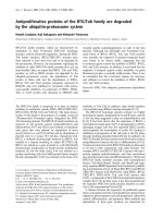
Expression of DNA binding domain-IN plasmids and control plasmids used in the yeast two-hybrid screensFigure 1
Expression of DNA binding domain-IN plasmids and
control plasmids used in the yeast two-hybrid
screens. (A) Lysates from strain CTY10-5d were electro-
phoresed on 10% SDS-PAGE gels, transferred to PVDF
membrane and probed with anti-lexA. Lane 1, pSH2-1 empty
vector; lane 2, pSH2-MoMLV IN; lane 3, pSH2-MoMLV IN
with 5'six-glycine linker; lane 4, pSH2-HIV-1 IN; lane 5, pSH2-
mouse LEDGF; lane 6, pNlexA empty vector; lane 7, MoMLV
IN-pNlexA. (B) Lysates from strain SFY526 were electro-
phoresed on 10% SDS-PAGE gels, transferred to PVDF and
probed with anti-GAL4-DB. Lane 1, strain without vector;
lane 2, pGBKT7 empty vector; lane 3, pGBKT7-MLV Gag;
lane 4, pGBKT7-MoMLV IN; lane 5, pGBKT7-HIV-1 IN; lane
6, pGBKT7-mLEDGF.
75-
50-
37-
pSH2-1
mIN-pNlexA
pSH2-mIN
pSH2-mIN 6gly
pSH2-hIN
pSH2-mLEDGF
pNlexA
1 2 3 4 5 6 7
75-
50-
37-
SFY526
pGBKT7
pGBKT7-mGag
pGBKT7-mIN
pGBKT7-hIN
pGBKT7-mLEDGFG
1 2 3 4 5 6
Retrovirology 2008, 5:48 />Page 4 of 23
(page number not for citation purposes)
all clones interacted with the pSH2-mIN and mIN-pNlexA
constructs equally, suggesting that the conformation of
the integrase fusion has an impact on its ability to bind
the putative interacting protein (Enx-1, ABT1, TIF3, B-
ATF, AF9, Ankrd49, U5snRNP, Znfp15, Znfp38, Ddx p18,
Ddx p68, and Trpc2; see Table 1). A common problem
encountered in yeast two-hybrid assays is that of back-
ground reporter activation. Because we observed some
background binding of Ku70 with both empty vectors
(pSH2-1 and pNlexA; Table 1) we tested the putative
Ku70 clone for interaction with pSH2-CLIP170 (CAP-GLY
domain containing linker protein 1) as a negative control.
There was no interaction between Ku70 and this protein
(data not shown), suggesting that the background activa-
tion we observed between the empty vectors and Ku70
may be due to the intrinsic DNA binding activity of the
acidic domain of the protein. In addition to Ku70, three
other clones, Radixin, Trpc2 and U2AF
26
also exhibited
weak background reporter activation in the CTY10-5d col-
ony lift assay in the context of the empty C-terminal lexA
Table 1: Yeast two-hybrid clone interactions with lexA C-terminal and N-terminal fused MoMLV integrase and with C-terminal fused
HIV-1 integrase
lexA fusions No. isolates in each
library
Total number
isolates
GALAD
fusions
pSH2-1 pSH2-MLV IN pSH2-HIV IN pNlexA MLV IN-
pNlexA
WEHI-3B T-cell
Controls
pGADNOT - na na na
pACT2 - na na na
mLEDGF - - ++ nt nt na na na
HIV-RTp51 +/ nt na na na
HIV IN - - +++ nt nt na na na
Gal4-AD
clones isolated
Fen-1 -+ ++- +from Fv-1 screen na 1
Enx-1 -+ +- - 404
TFIIE-β subunit -+ +- + 314
Ku70 + ++ +++ +/- +++ 011
TBP ABT1 - ++++ + - + 022
PRC - +++ ++ - ++ 213
B-ATF - +++ +/- - + 101
Brd2 - ++++ + - +++ 729
AF9/Mllt3 - ++++ + - ++ 404
Baz2b - ++++ + - +++ 101
Ankrd49 -++ + - - 101
Zn finger p15 - ++ +/- - +/- 101
Zn finger p38 - + +++ - +/- 101
SLU7 -+ ++- + 011
HSL bp -++ + - ++ 033
TIF3/eIFs2/TRIP1 -++ - - - 303
SF3b2 - +++ +++ - +++ 404
SF3a3 - +++ ++ - ++++ 011
U2Af
26
+/- +++ + - ++ 011
U5snRNP -+ +/ - 101
SMN - +++ +++ - +++ 011
Ddx p18 -+/- ++-+++ 505
Ddx p68 -+/- + -+++ 202
Kif3A -+ ++- + 202
Radixin +/- +++ ++ - ++ 011
Ran bp 10 -+ ++- + 011
Trpc2 +/- + + - +++ 011
Interactions between MoMLV IN, HIV-1 IN, and the clones isolated in the yeast two-hybrid screen. The pACT or pGADNOT plasmids containing
the cDNAs isolated from the yeast two-hybrid screens were introduced into strain CTY10-5d bearing either the pSH2-mIN, mIN-pNlexA, or
pSH2-hIN plasmids. Qualitative β-galactosidase colony lift assays were performed. No. of isolates in each library: the number of times a clone
identified as the indicated insert was retrieved, specific to each library screened. Total number of times an insert corresponding to each protein was
retrieved from all screens. Legend: - white; +/- pale blue; + light blue; ++ intermediate blue; +++, ++++ dark blue. Additional controls not shown:
pSH2-mLEDGF/pGADNOT-hIN, +++; pSH2-mIN/pGADNOT-mLEDGF, -; pSH2-mLEDGF/pGADNOT-mIN,
Retrovirology 2008, 5:48 />Page 5 of 23
(page number not for citation purposes)
DNA binding domain plasmid pSH2-1. To address this
issue, we examined these clones in this strain without the
DNA binding domain plasmid. None of these proteins
were able to activate the reporter in this context (data not
shown), suggesting that the background activation
observed may be due to the conformation of bait plasmid
used. We speculate that because we observed no activa-
tion signal with the empty pNlexA plasmid, and each of
these clones were isolated with the mIN-pNlexA fusion,
the conformation of the truncated lexA reporter in the
empty pSH2-1 vector may expose residues not available
for interaction in the full length lexA DB, leading to a spu-
rious interaction peculiar to these clones (Table 1).
The proteins isolated represent novel putative interacting
partners for MoMLV IN. As there have been no proteins
demonstrated conclusively to interact directly with
MoMLV IN, and because relatively few HIV-1 IN interact-
ing proteins have been identified, we examined our puta-
tive MoMLV IN interactors with HIV-1 IN in yeast two-
hybrid assays. Four of the proteins that interacted with
mIN interacted equally strongly with hIN. Those that
exhibited robust interactions with hIN were Ku70,
Znfp38, SF3b2, and SMN, and the interactions between
hIN with Ku70 and hIN with Znfp38 were stronger than
the interactions observed between mIN and these proteins
(Table 1). Intermediate interactions were observed for
hIN and Fen-1, PRC, SLU7, SF3a3, Ddx p18, Kif3A,
Radixin, and Ran bp10. Some of the proteins isolated in
the screen did not interact with hIN at all in these assays
(TIF3), or exhibited relatively moderate interactions
(Table 1).
Yeast two-hybrid cDNA library screens
We performed a pilot yeast two-hybrid screen of a mouse
WEHI-3B cDNA library in the GAL4 activation domain
plasmid pGADNOT using the plasmids pSH2-mIN and
pSH2-mIN 6G as baits in strain CTY10-5d. Our pilot
screen yielded a high percentage of interacting clones (96
putative interacting clones, data not shown). Due to the
large number of interactors isolated in the first screen, we
performed two additional independent screens of a
mouse T-cell cDNA library in the GAL4 AD plasmid
pACT2 in a different isolate of strain CTY10-5d with both
C-terminal and an N-terminal fusions of MoMLV inte-
grase as baits. In the T-cell library screen, we obtained 25
interacting clones (see Table S1 in Additional file 1).
We re-examined the phenotypes of each clone identified
in the WEHI-3B and T-cell library screens in strain CTY10-
5d. We rescued a total of 121 plasmids from yeast and
retested each of these putative interacting plasmids with
pSH2-mIN and mIN-pNlexA in the X-gal colony lift assay
in a minimum of three independent transformations. Of
the 121 plasmids rescued, we chose 27 of the clones that
retested successfully to characterize on the basis of their
phenotypes in the colony lift assay (intensity of activation
based on blue color), the number of times the gene was
isolated, and our interest in their proposed functions.
There are a number of other clones identified in the
screens that remain to be examined in greater detail and
are not included in this report, but the level of analysis
required is extensive and will be included in another
report. The clones presented in this report were placed
into three general categories according to functions attrib-
uted to them after BLAST [29] and database searches. The
proteins identified were categorized as follows and are
presented in Table 2: Group I, transcription factors and
chromatin binding proteins; Group II, RNA binding and
splicing factors; and Group III, miscellaneous and trans-
porter proteins. In cases where we obtained multiple iso-
lates of the same protein, very few of the clones were
siblings, as the isolated inserts represent different frag-
ments of these proteins (Table 2, column 2). Three of the
interacting proteins identified in the WEHI-3B screen
were also identified in the T-cell screen: general transcrip-
tion factor 2E beta subunit [(TFIIE-β), three isolates from
the WEHI-3B library and one from the T-cell library]; per-
oxisome proliferative activated receptor, gamma, coacti-
vator-related 1 [(PRC), two WEHI-3B and one T-cell
isolate]; and bromodomain 2 [(Brd2), alternatively
known as RING3 and female sterile homeotic related -1,
seven WEHI-3B and two T-cell isolates] (Table 2).
Interactions in yeast strain SFY526
In addition to the X-gal colony lift assays in CTY10-5d, we
also examined interactions between the integrases and the
putative interacting clones in the context of a strain utiliz-
ing a GAL4 DNA binding domain-IN fusion protein, and
activating a GAL4-responsive reporter. We wished to
examine interactions between the integrases and the vari-
ous GAL4 AD yeast two-hybrid clones in the context of a
plasmid with a weak promoter and thus lower expression
levels of the fusion bait proteins. Before performing these
tests, we subcloned mIN, hIN, MoMLV Gag and mLEDGF
into the GAL4 DB plasmid pGBKT7, and examined pro-
tein expression in the GAL4 reporter strain SFY526 by
Western blotting using an anti-GAL4 DB antibody (Figure
1B). MoMLV Gag/Gag interactions were used as controls
in these assays and activation of the GAL4 reporter was
observed with cotransformations of pGBKT7-mGag/
pACT2-mGag, pGBKT7-mGag/pGADNOT-mGag [30],
pGBKT7-hIN/pGADNOT-hIN, pGBKT7-hIN/pGADNOT-
mLEDGF, and pGBKT7-mIN/pACT2-mIN (data not
shown and Table 3). This series of control assays assured
us that there was no integrase-mediated self-activation in
this strain. We examined GAL4 DB fusions of mIN and
hIN in S. cerevisiae strain SFY526 and noted that strong
interactions previously observed with both IN proteins
were recapitulated in this context for Ku70, Brd2, AF9,
Retrovirology 2008, 5:48 />Page 6 of 23
(page number not for citation purposes)
Table 2: MoMLV integrase interacting proteins identified in the yeast two-hybrid screens
Insert aliases Complete residues/
peptides retrieved
a
Proposed function/properties
b
GenBank
accession Nos.
c
Reference
Group I, Chromatin binding and transcription factors
Enhancer of zeste homolog 1
(Ezh1/Enx-1/Ezh2)
742/31–292; 31–266; 371–615;
371–641
Polycomb group; chromatin structure
maintenance and transcriptional regulation;
binds ATRX via SET domain
U52951.1
[93]
Transcription factor IIE, beta
subunit (TFIIE-β)
292/18–292; 18–228- gap-249–
290; 18–233-gap-247–290; 50–
292
Subunit of RNA polII holoenzyme; recruits
TFIIH to the PolII-TFIIB-TFIID complex
NM_026584
[94]
Ku70/XRCC6 608/1–608 NHEJ, chromosome maintenance, 70 kD
subunit with Ku80 subunit of DNA-PKcs
AB010282
[95]
Flap endonuclease-1 (Fen1) 381/143–381 Removes 5' initiator tRNA from Okazaki
fragments; DNA repair in NHEJ and V(D)J
AY014962
[96]
Tata binding protein ABT1
(ABT1)
269/20–269 (2) Associates with Tata binding protein and
activates basal transcription of class II
promoters
AB021860
[97]
B-Activating transcription
factor (B-ATF)
120/1–120 AP-1/ATF superfamily; Basic leucine zipper
transcription factor; blocks transformation
by H-Ras and v-Fos
AF017021
[48]
Bromodomain containing
protein 2 (Brd2)/RING3/female
sterile homeotic gene-related 1
(fsrg 1)
798/311–543; 357–541; 530–
798; 558–798; 560–798; 562–
798; 563–798; 594–798; 595–
798
Bromodomain-containing protein; interacts
with Latency-associated nuclear antigen
(LANA-1) of KHSV; mitogen-activated
kinase activity; homolog of Drosophila
female sterile homeotic gene
AF045462
[98]
All1 fused translocated to
Chromosome 9 (AF9)/mixed
lineage-leukemia translocated
to 3 (Mllt3)
568/238–428, 476–560; 238–
428; 182–362
Pc3 interacting protein; Implicated in H3
hypermethylation; YEATS family member
(YNL107w/ENL/'AF-9/and TFIIF small
subunit)
AF333960
[39]
Bromodomain adjacent to zinc
finger domain, 2B (Baz2b)
2123/615–883 Putative member of ISWI containing
chromatin remodeling machinery; DDT,
PHD-type zinc finger and putative histone
acetyltransferase-Methyl-CpG binding
domain (HAT-MBD)
NM_001001182
[47]
Zinc finger p15 (Znfp15) 2192/1526–1808 Binds to Z-box response element between
two Pit-1 elements in the growth hormone
(GH) promoter; activates GH transcription
100 fold above basal levels
AF017806
[99]
Zinc finger p38 (Znfp38) 555/137–540 Transactivation via SCAN domain; granule
cell specification in brain; upregulated in
spermatogenesis
NM_011757
[52]
Peroxisome proliferative
activated receptor, gamma,
coactivator-1 related (PRC)
1644/1181–1644; 1321–1644;
1321–1644
Serum-inducible coactivator of nuclear
respiratory factor 1- dependent
transcription from RNA pol II promoters;
stress response protein
AAH66048
[100]
Ankyrin rep domain 49
(Ankrd49)
238/6–190 Putative transcription factor; contains acidic
activation domain; ankyrin repeat domain is
similar to SWI6
NM_019683.3
[101]
Group II, RNA binding proteins
Translation initiation factor 3
(TIF3/eIFs2/TRIP1)
325/128–325 (4) Translation initiation factor; 5 WD repeats;
dissociates ribosomes, promotes initiator
Met-tRNA and mRNA binding; yeast
homolog TUP12 acts as transcriptional
repressor
NM_018799
[102]
Splicing factor 3b, subunit 2
(SF3b2)
878/389–844; 385–606; 397–
579; 554–781; 397–576
Has putative DNA-binding (bihelical) motif
predicted to be involved in chromosomal
organization; has SAP domain; proline-rich
domain in spliceosome assoc. proteins;
basic domain in HLH proteins of MYOD
family
NM_030109
[103]
Splicing factor 3a, subunit 3
(SF3a3)
501/318–501 Zinc finger, C2H2-type; RNA splicing,
mRNA processing
BC092058
[100]
U2 auxiliary factor 26 (U2AF
26
) 220/53–220 Pre-RNA splicing factor; can replace
U2AF
35
in vitro
AF419339
[104]
Retrovirology 2008, 5:48 />Page 7 of 23
(page number not for citation purposes)
U5 small nuclear
ribonucleoprotein (U5 snRNP)
2136/1939–2136 Transcriptional regulation; SNF2 N-
terminal domain; conserved C-terminal
helicase domain; GTP binding factor;
ortholog of S. cerevisiae splicing factor
Prp8p; mutations in hPRPC8 are autosomal
dominants in retinitis pigmentosum
NP_796188
[105]
Step II Splicing factor SLU7 585/27–585 Pre mRNA splicing, required for 3' splice
site choice
NM_148673
[106]
Survival motor neuron (SMN) 288/12–254 Component of an import snRNP complex
containing GEMIN2, 3, 4, 5, 6 and 7;
contains one Tudor domain; deficiency
leads to apoptosis
Y12835
[70]
Dead box p18 (Ddx18) 660/366–592; 366–610; 366–
660; 366–660; 366–590
RNA-dependent helicase; RNA-dependent
ATPase activity; stimulated by ss-RNA
NM_025860.2
[107]
Dead box p68 (Ddx68/Ddx5) 615/247–490; 247–510 RNA-dependent helicase and ATPase
activity; stimulated by ss-RNA; interacts
with HDAC1
BC129873
[100]
Histone stem loop binding
protein (HSLbp)
275/1–275; 1–204; 1–248 RNA transcription events, required for
histone pre mRNA processing
NM_009193
[108]
Group III, Miscellaneous
and transport proteins
Ran binding protein 10
(Ranbp10)
503/60–387 Interacts with MET (receptor protein
tyrosine kinase) via its SPRY domain; does
not interact with SOS, competes with
Ranbp9 for MET binding; interacts with Ran
in vitro
AY337314
[109]
kinesin super family member
3A (Kif3A)
701/443–701; 443–650 Transport of organelles, protein complexes,
and mRNAs in a microtubule- and ATP-
dependent manner; chromosomal and
spindle movements during meiosis and
mitosis
NM_008443.2
[110]
Radixin 389/13–330 Member of ezrin, radixin, moesin family of
actin binding proteins. Binds directly to
ends of actin filaments at plasma membrane
BC053417
[100]
Transient receptor potential
prot.2 (TrpC2)
313/3–313 Calcium ion entry channel; putative
involvement in DNA damage response
AF111108
[111]
Identities and BLAST search information obtained for MoMLV IN interacting proteins identified in the yeast two-hybrid screens. (
a
) The first
number reflects the length of the full-length protein; the next sets of numbers refer to the residues retrieved for each clone. (
b
) Other functions
may exist. (
c
) Database accession numbers are current as of May 19, 2007
Table 2: MoMLV integrase interacting proteins identified in the yeast two-hybrid screens (Continued)
Znfp38, Ranbp10, and SMN (Table 3). We also observed
that some weaker interactions between hIN and the
inserts were not recapitulated for Baz2b, ABT1, SF3a3, and
Radixin (data not shown and Table 3).
Deletion analysis of mIN and isolated clones
We mapped the region of mIN that interacted with a sub-
set of the clones identified in the yeast two-hybrid screen
by introducing deletions into MoMLV IN. We constructed
lexA-mIN fusions containing the Zinc binding motif
(mIN-Zn), the Zinc binding motif and the catalytic
domain (mIN-ZnDDE), the catalytic domain alone (mIN-
DDE), the catalytic domain and the C-terminus (mIN-
DDECH), and the C-terminus alone (mIN-COOH) (Fig-
ure 2A). First, we examined lysates from the mIN dele-
tions to insure that the proteins were expressed (Figure
2B). We then examined the interactions between these
deletions and various clones in yeast two-hybrid assays.
The most robust interactions were observed between the
B-ATF, AF9, Brd2, Enx-1, and ABT1 clones and the mIN-
DDECH fusion (Table 4). The interaction between TFIIE-
β and the mIN-Zn fusion was stronger than its interaction
with any of the other deletion constructs (Table 4). Ku70
interacted with multiple regions, but the most robust
interaction was observed between Ku70 and the mIN-Zn
fusion (Table 4). These results suggest that there may be
discrete regions of mIN that interact with different groups
of host factors. More detailed mapping experiments are
required to localize the precise residues of mIN responsi-
ble for the interactions observed.
In vitro binding assays
We next examined the interactions between maltose bind-
ing protein (MBP)-fused mIN and hIN with 17 of the
putative interacting proteins in in vitro binding assays. E.
coli strains overproducing the MBP IN fusions or the GST
fused two-hybrid clones were examined for protein
expression (Figure 3A, B). Relative levels of expression
were used to determine the amounts of input protein for
the binding assays. For the assays, the MBP fusion lysates
Retrovirology 2008, 5:48 />Page 8 of 23
(page number not for citation purposes)
Table 3: Yeast two-hybrid tests in strain SFY526
GAL4 DNA binding domain fusions
GAL4 AD fusions pGBKT7 pGBKT7-mIN pGBKT7-hIN
pGADNOT-empty -
pACT2-empty -
pGADNOT-HIV IN - nt ++++
pGADNOT-Gag -nt nt
pGADNOT-mLEDGF - - +++
Fen-1 +
Enx-1 -
TFIIE-β +
Ku70 - + ++++
ABT1 -+ -
B-ATF -
BRD2/RING3 - ++++ +/-
AF9/Mllt3 -+/- +/-
PRC +/-
Baz2b -
Zn finger p15 +/-
Zn finger p38 -+/- +
Ankrd49 +/-
SF3b2 +/-
SF3a3 -
U2AF26 +/- - +/-
U5snRNP +/- - +/-
splicing factor SLU7 -
SMN -+/- +/-
Ran bp 10 +++ ++++ ++++
KIF3A -+/- -
Radixin -+ -
Trpc2 +
Interactions between selected clones isolated in the yeast two-hybrid screens with GAL4-MoMLV IN and GAL4-HIV-IN. The pACT or pGADNOT
plasmids containing the cDNAs isolated in the yeast two-hybrid screen were introduced into SFY526 strains bearing the pGBKT7 integrase fusions.
Qualitative colony lift assays were performed.
were first incubated with amylose resin and washed exten-
sively. Lysates from E. coli strains overproducing the GST
fused two-hybrid subclones were incubated with the
washed MBP-amylose resin-bound integrase proteins. We
performed these binding assays to determine if the GST
proteins could interact specifically with the MBP-integrase
fusions. The MBP-IN/GST-putative interacting protein
complexes were eluted from the amylose resin by compe-
tition with maltose. This was done to resolve bona fide
complexes between the integrases and the putative inter-
acting fusions, rather than non-specific interactions
between the resin and input proteins. There was some C-
terminal proteolytic cleavage of both MLV and HIV inte-
grases in these expression studies, the extent of which var-
ied from preparation to preparation, as can be seen by the
cleavage products visible in both the Coomassie stained
gels and in the Western blots employing these proteins
(Figure 3A, lanes 3 and 4 and Figures 4A, B, C, D, and 4E).
In general, the intensity of the interactions between the
GST subclones and the two retroviral integrases correlated
well with the strength of the interactions observed in the
yeast two-hybrid assays. The MBP-mIN fusion interacted
with the 17 proteins examined as GST fusions: Brd2, AF9,
Ankrd49, Fen-1, Enx-1, TFIIE-β, Ku70, PRC, Baz2b, ABT1,
SF3a3, U5snRNP, Kif3A, Radixin, Znfp38, U2AF
26
, and
Ranbp10 (Figures 4A, B, C, D, and 4E). The MBP-hIN
fusion interacted with 15 of the GST fusions analyzed:
Brd2, AF9, Ankrd49, Fen-1, Enx-1, TFIIE-β, Ku70, Baz2b,
SF3a3, U5snRNP, Kif3A, Radixin, Znfp38, U2AF
26
, and
Ranbp10 (Figures 4A, B, C, D, and 4E). Only weak inter-
actions were observed in vitro between hIN with PRC and
ABT1 (Figure 4C). These data confirm and extend the
yeast two-hybrid results, indicating that the interactions
are likely direct.
Both mIN and hIN proteins interacted to different extents
with Ku70, PRC and ABT1, as was observed in their yeast
two-hybrid interactions, but both integrases interacted
equally with Baz2b in these assays (compare Figure 4C
and Table 1). The mIN and hIN integrases exhibited
apparent equivalent interactions in vitro with SF3a3,
Retrovirology 2008, 5:48 />Page 9 of 23
(page number not for citation purposes)
Construction and expression of MoMLV IN deletion plasmids in CTY10-5dFigure 2
Construction and expression of MoMLV IN deletion plasmids in CTY10-5d. (A)Schematic of pSH2-1 MLV IN
truncation constructs. 1–408, full-length mIN; 1–124, mIN-Zn; 1–296, mIN-ZnDDE; 97–225, mIN-DDE; 107–408, mIN-
DDECOOH; 220–408, mIN-COOH. (B) Lysates from strain CTY10-5d were electrophoresed on 12% SDS-PAGE gels, trans-
ferred to PVDF membranes and probed with anti-LexA. The indicated lysates are shown left to right.
LexA Zinc motif DDE domain C-terminal
pSH2-mIN 1-408
pSH2-mIN-Zn 1-124
pSH2-mIN-ZnDDE 1-296
pSH2-mIN-DDE 97-225
pSH2-mIN-DDECOOH 107-408
pSH2-mIN-COOH 220-408
50 -
37 -
25 -
pSH2-1
pSH2-mIN
pSH2-mIN-Zn
pSH2-mIN-ZnDDE
pSH2-mIN-DDE
pSH2-mIN-DDECOOH
pSH2-mIN-COOH
-Non-specific band
Table 4: Interactions between pSH2-MoMLV IN deletions and selected yeast two-hybrid interacting proteins
Fusions lexADB lexA-p66 lexA-mIN mIN-Zn mIN-ZnDDE mIN-DDE mIN-DDECH mIN-COOH
GAL4 AD - - - - - -
RT p51 - ++++ - nt nt nt nt nt
mIN - ++ + - +++ ++ -
B-ATF -++ +/
AF9 - ++++ - - - +++ -
Brd2 -++ +-
Enx-1 -+ +/
Ku70 +++++++++-+/-
TFIIE-β -++
ABT1 -+++ +/
Analyses of MoMLV IN truncations with selected interacting proteins. pSH2-MoMLV IN deletions were introduced into CTY10-5d with the
indicated clones in pGADNOT. Qualitative colony lift assays were performed.
U5snRNP, and Kif3A, although the intensity of their inter-
actions in vivo was dependent on the LexA fusion (Figure
4D and see Table 1). The in vitro interactions between
mIN and hIN with Radixin also did not mirror their in
vivo interactions, with hIN exhibiting a stronger interac-
tion than mIN with this protein (Figure 4D and see Table
1). Znfp38, U2AF
26
and Ran bp10 interacted equally with
both integrases (Figure 4E).
The observed in vitro binding of pairs of proteins derived
from crude lysates could in principle be facilitated,
enhanced, or even mediated entirely by nucleic acids,
Retrovirology 2008, 5:48 />Page 10 of 23
(page number not for citation purposes)
Expression and binding tests of maltose binding and glutathione-S transferase fusion proteinsFigure 3
Expression and binding tests of maltose binding and glutathione-S transferase fusion proteins. (A) MBP lysates
were bound to amylose resin, eluted with 15 mM maltose, electrophoresed on 10% SDS-PAGE gels, and stained with Coomas-
sie brilliant blue. Lanes 2–4, expression of pmalc2 (empty vector), pmalc2-mIN, and pmalc2-hIN in TB1 cells. For the GST
fusions, the lysates were bound to glutathione sepharose, eluted with 10 mM reduced glutathione, electrophoresed on 10%
SDS-PAGE gels and stained with Coomassie brilliant blue. Lanes 5–13, representative loads of GST-yeast two hybrid clones:
pGEX2TPL, mLEDGF, Fen-1, Enx-1, TFIIE-β, Ku70, ABT1, PRC, and Brd2. (B) Lanes 2–12, GST-yeast-two hybrid clones: AF9,
Baz2b, B-ATF, Ankrd49, Znfp38, SF3a3, U2AF
26
, U5snRNP, KIF3A, Radixin, and Ran bp10. Lane 1 in A and B: Molecular weight
marker.
MBP
mIN
hIN
GST
mLEDGF
Fen-1
Enx-1
TFIIE-1
Ku70
ABT1
PRC
Brd2
100-
75-
50-
37-
25-
1 2 3 4 5 6 7 8 9 10 11 12 13
100-
75-
50-
37-
25-
1 2 3 4 5 6 7 8 9 10 11 12
AF9
Baz2b
B-ATF
Ankr49
Znfp38
SF3a3
U2AF
26
U5SnRNP
KIF3A
Radixin
Ran bp10
Retrovirology 2008, 5:48 />Page 11 of 23
(page number not for citation purposes)
In vitro binding interactions between MoMLV and HIV-1 integrases and selected proteins identified in the yeast two-hybrid screenFigure 4
In vitrobinding interactions between MoMLV and HIV-1 integrases and selected proteins identified in the yeast
two-hybrid screen. In vitro binding assays between the pmalc2 empty vector (MBP), full-length pmalc2-MoMLV IN (mIN) or
full-length pmalc2-HIV-1 IN (hIN) and seventeen of the clones isolated in the screen, plus mLEDGF expressed as GST fusions.
The MBP fusion lysates were incubated with amylose resin, washed extensively, resuspended in equal volumes of buffer, and
then aliquoted to separate tubes. These tubes were incubated with the GST fusion lysates, washed and eluted with 15 mM mal-
tose. 25 μl of each eluate was electrophoresed on 10 or 12% SDS-PSGE gels, transferred to PVDF membranes, and the same
Western was probed with anti-GST, stripped, and then probed with anti-MBP. All Westerns are loaded from left to right: MBP,
mIN, and hIN fusion reactions. All upper panels, anti-MBP. All lower panels, anti-GST. (A) Maltose binding protein fusions with
empty GST vector; MBP fusions with Brd2, AF9, and Ankrd49. (B) MBP fusions with mLEDGF, Fen-1, Enx-1, and TFIIE-β. (C)
MBP fusions with Ku70, PRC, Baz2b, and ABT1. (D) MBP fusions with SF3a3, U5snRNP, KIF3A, and Radixin. (E) MBP fusions
with Znfp38, U2AF
26
, and Ran bp10.
Retrovirology 2008, 5:48 />Page 12 of 23
(page number not for citation purposes)
In vitro binding interactions between MoMLV and HIV-1 integrases and selected proteins after treatment of the lysates with nucleases to eliminate nucleic acid bridges between the proteinsFigure 5
In vitro binding interactions between MoMLV and HIV-1 integrases and selected proteins after treatment of
the lysates with nucleases to eliminate nucleic acid bridges between the proteins. In vitro binding assays between
the empty vector (MBP), full-length pmalc2-MoMLV IN (mIN) or full-length pmalc2-HIV-1 IN (hIN) and a subset of the clones
isolated in the screen. All Westerns are loaded from left to right: MBP, mIN, hIN and the indicated GST fusion reactions.
Upper panels, anti-MBP. Lower panels, anti-GST. (A) Left, maltose binding protein fusions with empty GST vector; right, MBP
fusions with Ku70 and Brd2. (B) MBP fusions with U2AF
26
, TFIIE-β, and Ankr49. (C) MBP fusions with the indicated proteins
AF9, PRC, Fen-1, Baz2b, and Enx-1.
Retrovirology 2008, 5:48 />Page 13 of 23
(page number not for citation purposes)
either RNA or DNA, that bridge the two proteins and
mimic direct protein-protein interactions. To address this
possibility, a subset of the lysates examined in the pull-
down assays were treated with DNase and RNase to elim-
inate potential contaminating nucleic acids, and the in
vitro interaction of the proteins in the lysates was assessed
as before. Examination of the lysates for residual nucleic
acids showed that the nucleases were highly effective (see
Figure S1 in Additional file 2). The binding studies show
that the majority of the protein-protein interactions were
maintained following nuclease treatment (Figure 5). Of
the 18 GST-fusions examined in the in vitro binding
assays shown in Figure 4, we examined 13 GST-fusions in
assays in which each of the MBP-integrase and GST-clone
fusion lysates were treated with DNase and RNase prior to
performing the binding reactions. Of the 13 lysates
treated, five of the interactions with mIN and hIN were
unchanged: Brd2, TFIIE-β, Ankr49, Fen-1 and ABT1 (Fig-
ure 5 and data not shown); four were increased, in some
cases differentially with respect to the integrase used in the
assay: PRC, Ku70, U2AF
26
, and Radixin (Figure 5 and data
not shown); and three were decreased: AF9, Baz2b, and
mLEDGF (Figure 5 and data not shown). Ten of these
binding reactions are shown in Figure 5. No interactions
were observed between any of the MBP fusions and the
GST vector (Figure 5A, lanes MBP, mIN and hIN). There
was some background interaction between Ku70 and
MBP, but much lower than the increased interactions
observed between this protein with mIN and hIN (com-
pare Figure 5A with Figure 4C). This result may be a func-
tion of improved binding between Ku70 and all MBP-
fusions due to removal of residual nucleic acids. Of the 14
pairs, the interaction between mIN and U2AF
26
(compare
Figure 5B with Figure 4E), between AF9 and hIN (com-
pare Figure 5C with Figure 4A), and between PRC and hIN
were enhanced (compare Figure 5C with Figure 4C). The
interaction between MLV IN with AF9, Baz2b and PRC
was decreased in this particular assay, suggesting that
some bridging by nucleic acids could not be ruled out
(Figure 5C). Binding between Moloney and HIV inte-
grases with Radixin was consistently enhanced following
this treatment (data not shown). Although the tests for
residual nucleic acids in the lysates suggest that the nucle-
ase treatments were almost completely effective, it is pos-
sible that undetected traces of nucleic acids remained, and
are still serving as bridges. More extensive testing of the
binding interactions following nuclease treatment is
required to definitively state that there are no residual
nucleic acids remaining in the lysates.
Discussion
In this report, we used Moloney MLV integrase in the con-
text of two different lexA DNA binding domain fusion
vectors as bait to screen two mouse GAL4 activation
domain cDNA libraries. We present 27 proteins that inter-
acted with MoMLV integrase in the yeast two-hybrid
screens. Twenty of the proteins identified in the screens
interact strongly with Mo-MLV IN, and 7 have relatively
weaker interactions. We also show that a subset of 12 of
these interact strongly with HIV-1 IN, that 11 have inter-
mediate interactions, that three have weak interactions,
and that one exhibited no interaction (TIF3). It is of inter-
est to note that the screen has revealed 13 DNA binding
proteins, 10 RNA binding proteins, and four proteins
involved in transport or signaling. Seven of the isolated
clones were examined for their interactions with MLV IN
deletions. We found that B-ATF, AF9, Brd2, Enx-1, and
ABT1 interacted with the truncated fragment containing
both the catalytic and the C-terminal domains. TFIIE-β
interacted with the amino terminus of MLV IN and Ku70
interacted with multiple regions of IN. The IN/Ku70 inter-
action was lost when only the catalytic/C-terminal frag-
ment of IN was expressed. As each of the proteins tested in
the truncation assays were DNA binding proteins or tran-
scription factors, we may have identified domains of inte-
grase that interact with a range of transcription factors and
DNA binding proteins.
We have examined interactions between 18 of these pro-
teins in vitro using binding assays with both MoMLV and
HIV-1 integrases. Of the 18 proteins examined in vitro, we
find that 14 exhibited strong interactions with MLV IN
and 12 exhibited strong interactions with HIV IN. We find
that the intensity of the in vivo interactions in yeast varies
between mIN and hIN, which is not surprising, given that
the two integrases have little sequence identity and the
host protein requirements for their respective integration
reaction pathways are presumed to differ, even though the
structure of the major functional domains are conserved.
Tests for nucleic acid bridging between a subset of the pro-
teins suggest that most of the detected interactions are
likely to be direct protein-protein interactions, as also sup-
ported by the differential binding of the host proteins to
the two integrases.
The results of our assays in yeast and in the in vitro bind-
ing assays suggest that there may be many common host
proteins used by both viruses. Since the cDNA libraries we
screened were murine, we do not presume that all of the
clones isolated will exhibit equal effects on both HIV and
MLV integration or on virus infectivity, but the isolation
of so many putative interacting proteins in our screens
merit further investigation for potential roles in the viral
life cycle. It is of interest to note that a large group of these
proteins, 13 factors, are chromatin binding proteins or
transcription factors. Although these various proteins
have no obvious simple sequence similarity, it is plausible
that the MLV IN protein is recognizing a common feature
present on many of these proteins. For example, IN may
detect and bind to transcriptional activation domains; the
Retrovirology 2008, 5:48 />Page 14 of 23
(page number not for citation purposes)
common thread between such proteins may be as inap-
parent as the acidic protein-protein interaction domains
thought to mediate the tethering of transcriptional activa-
tors to DNA by promoter or enhancer binding proteins.
The significance and consequence of these interactions on
viral infectivity and integration await functional analyses.
In early tests for protein-protein binding in vitro, we
observed an interaction between HIV-1 IN and LEDGF, a
factor widely reported as affecting the efficiency of infec-
tion and the target site selection for viral integration. We
also observed an unexpected in vitro interaction between
mLEDGF and mIN. These proteins did not interact in
yeast and there is no documented evidence of an interac-
tion between MLV IN and hLEDGF [31]. When we treated
the lysates with nucleases, both the mIN- and hIN-LEDGF
interactions disappeared (data not shown), suggesting
that the interactions observed in vitro might have only
been mediated by nucleic acid bridging. Thus, the signifi-
cance of the in vitro interaction between mLEDGF and
MBP-mIN is unclear. We do not know if the interactions
observed between mLEDGF and hIN suggest that
mLEDGF could play a similar role in the integration of
HIV in mouse cells to its role in human cells though
indeed a recent study of HIV-1 integration in wild-type
and mutant mouse cells suggest that it is a significant
player in virus integration [27]. It is interesting to note
that when we aligned the protein sequences of the mouse
and human LEDGF proteins, we observed that the pro-
teins share 92% identity overall and the integrase binding
domain of hLEDGF identified by Cherepanov [32] shares
100% consensus with the corresponding region in
mLEDGF (data not shown).
Chromatin binding and transcriptional activators
One category of proteins isolated in the screens is of par-
ticular interest because it includes DNA binding and chro-
matin modification factors. Enhancer of Zeste homolog 1,
(Enx-1/Ezh2), is a member of Polycomb repressive com-
plex 2 (PRC2). The isolation of a member of this class of
proteins is not without precedence: one of its PRC2 part-
ner proteins, embryonic ectodermal development factor
(EED), has been identified as an interactor with other ret-
roviral proteins. EED was isolated in a yeast two-hybrid
screen with HIV-1 MA as bait and later shown to interact
with HIV-1 IN [33,34]. The interaction with HIV-1 IN led
to an increase in integration in vitro [34]. Another yeast
two-hybrid screen using HIV-1 Nef as bait recovered EED
from a Jurkat cDNA library [35]. Analyses of the interac-
tion between Nef and EED revealed that Nef mimics an
integrin receptor signal and translocates EED from the
nucleus and relocalizes it to the plasma membrane, result-
ing in an increase in Tat mediated HIV transcription [35].
Enhancer of zeste [E(Z)] and extra sex combs (Esc), the
drosophila homologs of mammalian Enx-1 and EED
respectively, are part of the same repressive complex in
both drosophila and mammalian cells. In fact, Enx-1 and
EED interact both in vitro in yeast and in vivo in mouse
cells [36]. Intriguing questions are whether or not Enx-1 is
also translocated to the plasma membrane in a complex
with EED, and whether both proteins play similar roles in
the viral life cycle or have a comparable effect independ-
ently on viral infectivity and integration. Although none
of the studies cited above investigated an interaction
between EED and MoMLV IN, the isolation of Enx-1 in
our screen, and our finding that it also interacts with HIV
IN suggests the intriguing possibility of a role for the
PRC2 chromatin repressor complex in the viral life cycle.
Acute lymphocytic leukemia gene 1 fused from chromo-
some 9 (AF9), also known as mixed lineage leukemia
translocated to chromosome 3 homolog (Mllt3) is fre-
quently found in balanced translocations with the mixed
lineage leukemia gene (MLL), a trithorax homolog, in
acute myeloid leukemia cells. In mice, MLL is required for
normal embryogenesis and likely regulates Hox gene
expression by binding to promoter sequences [37]. The
precise function of AF9 is unknown, but it has been pro-
posed as a transcriptional activator as it contains a serine-
and proline-rich domain, as well as a nuclear localization
signal, consistent with such a role. Null af9 mice exhibit
homeotic transformations and perinatal lethality, suggest-
ing that AF9 may be a master regulator of Hox genes [38].
The C-terminus of AF9 interacts with the mouse and
human homologs of the Drosophila Polycomb group
protein Pc3, and with the BCL6 corepressor BcoR: both
Pc3 and BcoR normally act to repress transcription
[39,40]. In this report, we isolated four clones of AF9 in
our screens and we show that at least one of these clones
interacts with HIV IN and MoMLV IN in yeast and in the
in vitro binding assays. An intriguing question raised is
whether disruption of the opposing activities of Poly-
comb and Trithorax proteins will reveal a role for these
proteins in retroviral integration, given that Trithorax pro-
teins are transcriptional activators and Polycomb proteins
are transcriptional repressors.
In our screens, the largest number of clones isolated cor-
responded to the cDNA for bromodomain containing
protein 2 (Brd2/fsrg1/RING3) (nine isolates). Proteins
that contain bromodomain motifs function in the regula-
tion of chromatin and in epigenetics [41]. The bromodo-
main is found in the majority of histone acetyltransferases
and in transcriptional activators, and derives its name
from the Drosophila brahma protein in which the motif
was initially identified [42]. Brd2 functions as a transcrip-
tional co-activator and as a nuclear-localized kinase [43].
Recent studies have identified a Brd2 complex that con-
tains, among others, E2F (E2 promoter binding factor),
histones, HDAC11, CBP, p300, Cyclin A2, TAF
II
250, and
Retrovirology 2008, 5:48 />Page 15 of 23
(page number not for citation purposes)
Swi/Snf chromatin remodeling complex member Brg-1
[41,44]. In the Denis et al. studies, overproduction of
Brd2 led to elevated Cyclin A transcription and a pre-
sumed destabilization of the cell cycle, as Brd2 was asso-
ciated with the cyclin A promoter at both the G
1
and S
phases [41]. In addition, Brd2 was shown to interact with
the chromatin-binding domain in the Kaposi's sarcoma-
associated Herpes virus (KSHV) latency-associated
nuclear antigen 1 (LANA-1) to modulate transcription
and episomal DNA replication [45]. LANA-1 may interact
with Brd2 to tether the KSHV genome to mitotic chromo-
somes in a manner similar to that observed between the
Bovine papillomavirus (BPV) E2 protein and Brd4 [46].
Although the observed interaction between Brd2 and
HIV-1 IN in yeast was weaker than its interaction with
MLV IN, the finding that the Brd2-HIV IN in vitro interac-
tion is apparently equal in intensity to that observed for
MLV IN suggests that this protein may play a role in the
integration of both retroviruses. Baz2b is another bromo-
domain family member identified in our screen, whose
precise function remains to be elucidated [47]. Baz2b
exhibits the same behavior as that observed for Brd2 in
our assays: it displays a weaker interaction in yeast with
HIV IN than that observed for MLV IN, but an in vitro
binding apparently equivalent to that observed for MLV
IN.
B-ATF is a member of the AP-1/ATF superfamily of tran-
scription factors [48] and its expression in human and
mouse is tissue specific, primarily limited to hematopo-
etic tissues and cells [49]. B-ATF contains a basic Leucine
zipper motif, does not homodimerize, does not contain a
functional transcription activation domain, and does not
dimerize with Fos, but does form heterodimers with the
Jun family proteins (c-Jun, JunD and JunB) to bind Acti-
vator protein-1 (AP-1) consensus DNA sites [49]. B-ATF is
a natural dominant-negative regulator of AP-1 mediated
transcription, acting as a non-activating competitor for c-
Fos in the AP-1 dimer to reduce cell growth [49]. Ectopi-
cally expressed B-ATF reduced transformation by v-fos and
H-ras oncogenes in mouse cells [49]. Rasmussen et al. [50]
identified T-cell lymphoma-specific MoMLV integrations
at the Fos/Jdp2/Batf locus in mouse cells. The B-ATF clone
isolated in our screen did not interact with HIV-IN in
yeast, but a role for this factor in transformation by
MoMLV should be investigated.
Zinc finger p38 is a transcriptional activator that contains
seven Cys
2
His
2
type zinc fingers, a SCAN box (SRE-ZBP,
C
Tfin51, AW-1 (znf174), and Number 18), also known as
the Leucine rich region, and a novel N-terminal domain
[51]. The SCAN domain may be a protein-protein interac-
tion motif, as mammalian two-hybrid studies have iden-
tified this region as capable of transcriptional activation
[52,53]. The finding that our Znfp38 clone interacted with
both MLV IN and HIV-1 IN both in yeast and in vitro, sug-
gests a role for this transcription factor in the life cycle of
both retroviruses.
DNA repair proteins
A surprising find was the isolation of Ku70/XRCC6, the 70
kD subunit of the Ku70/Ku80 thyroid autoantigen, also
known as the Ku heterodimer. Ku70 was initially identi-
fied by the isolation of an abundant antibody found in
patients with autoimmune thyroid disease and lupus ery-
thematosus [54]. The Ku86 heterodimer has ATP-depend-
ent DNA helicase activity [55], is thought to be the first
protein to bind to a DNA double strand break [56], func-
tions as a sliding clamp on DNA and recruits DNA-PK
cs
,
DNA polymerases, and ligases to the site of damage [57]
in a manner similar to the mechanism employed by
PCNA [58]. The Ku heterodimer participates in the non-
homologous DNA end joining (NHEJ) pathway of DNA
repair [59], in V(D)J recombination, and with Telomere
repeat factor 2 (TRF2) to suppress homologous recombi-
nation of telomeres between sister chromatids [60]. Addi-
tional studies have identified a role for the NHEJ complex
in Ty1 retrotransposition [61] and in retroviral integration
[62,63]. The isolation of Ku70 in our screen and the in
vitro binding data suggest that this protein may play a
direct role in integration for both MLV and HIV-1.
Flap endonuclease-1 (Fen1), or RAD two homolog-1
(Rad27 or RTH1) is a structure-specific 5' endo/exonucle-
ase that functions in the maintenance of genome stability,
long-patch base excision repair, NHEJ, and the resolution
of Okazaki fragments in lagging strand DNA synthesis
[64]. Deletions of Fen-1/Rad27 in yeast cells lead to a high
frequency of chromosome loss and an increased rate of
recombination [64]. The C-terminus of Fen-1 interacts
with the transcription coactivator p300, which acetylates
Fen-1 [65], and has been implicated in retroviral integra-
tion [66,67]. Although Fen-1 was identified in a yeast two-
hybrid screen as an interaction partner of Friend virus sus-
ceptibility 1 protein (Fv-1) (Subarna Bhattacharyya,
unpublished data), the report of Rumbaugh et al. [68]
demonstrating the involvement of Fen-1 in the processing
of HIV-1 integration intermediates [68] prompted us to
examine a possible direct interaction between Fen-1 and
the integrases of MoMLV and HIV-1. The in vivo and in
vitro interactions observed in our report support a direct
interaction between Fen-1 and the two integrases, suggest-
ing that experiments designed to delineate the precise role
of Fen-1 in the DNA repair step of integration in vivo
should be pursued.
RNA binding proteins
Spliceosomal small ribonucleoproteins (snRNPs) are
major components of the mRNA splicing machinery and
each snRNP is comprised of one or two small nuclear
Retrovirology 2008, 5:48 />Page 16 of 23
(page number not for citation purposes)
RNAs (snRNAs) bound to a set of RNA-binding proteins,
called Sm proteins (SmB/B', SmD1, SmD2, SmD3, SmDE,
SmF, and SmG) [69]. The Sm proteins bind to a highly
conserved uridine rich sequence on each snRNA called the
Sm site. Sm cores are assembled in vivo onto snRNAs by
the SMN complex [69]. Survival motor neuron (SMN) is the
gene for spinal muscular atrophy (SMA) whose disruption
in mouse embryos leads to massive cell death and early
embryonic lethality [70]. SMN is part of a large complex
with at least six to seven Gemin proteins (Gemins 2–8)
that function to organize snRNPs [69]. SMN interacts
directly with Gemins 2, 3, and 8 [71]. Reduction of SMN
levels by an SMA-causing mutation leads a decrease in the
relative amounts of Gemins as part of the SMN complex
[71]. A recent report identified Gemin2 as an HIV-1 inte-
grase interactor by yeast two-hybrid screening [72]. The
Hamamoto [72] report used siRNA to downregulate
Gemin2 and SMN in cells subsequently infected by HIV-
1, showing that disruption of these proteins blocked HIV-
1 infection, and Gemin2 disruption reduced viral DNA
copy number, 2-LTR circle accumulation, and proviral
integration [72]. Interestingly, SMN also interacts with
snRNPs U1, U2 and U5. The U2snRNP associated factor
U2AF
26
and U5snRNP were also isolated in our screen,
suggesting the possibility of an interaction between the
incoming viral RNA and the spliceosomal network, or that
integrase may co-opt these factors for downstream viral
functions.
The U2 snRNP is an essential component of the spliceo-
some and binds to the pre-mRNA branch site by base-
pairing with the complementary RNA sequence of the U2
snRNA [73]. U2 snRNP interacts with the U1 snRNP
which binds to the 5' splice site, and a complex of U1
snRNP/U2 snRNP/pre-mRNA recruits the U4/U6/U5
snRNPs to form an active spliceosome [73]. The core 12S
U2 snRNP binds splicing factor 3b (SF3b), to form a pre-
mature 15S U2 snRNP [74]. In turn, this complex binds
SF3a to form a mature 17S snRNP, which interacts with
nucleotides upstream of the branch site within the intron
[74]. Splicing factor 3a subunit 3 (also known as SF3a3,
Sf3a60 and Spf3a3) is the mammalian homolog of S. cer-
evisiae PRP9 and is a C2H2- type zinc finger protein
required for the core complex assembly [75]. The SF3a
complex is composed of SF3a60, SF3a66 and SF3a120, of
which we have isolated the 60 kD subunit (SF3a3) in our
screen. In addition, we isolated the SF3b2 subunit of SF3b
in our screen, which interacts directly with SF3a. We also
isolated the factors U2AF
26
, U5 snRNP, and SMN as
described above. Would the incoming virus interact with
these proteins? The isolation of these core spliceosome
components suggests that a new perspective on integrase-
host factor interactions may be required upon further
analysis of these factors.
Other factors
Peroxisome proliferative-activated receptor gamma coac-
tivator-1α, PGC-1α (formerly PGC-1), is a nuclear hor-
mone receptor that coordinates diverse organ- and cell-
specific transcription programs in response to stress stim-
uli [76]. Two additional genes in the family have been
identified, PGC-1-related coactivator (PRC) and PGC-1β
(PERC/ERRL-1) [77,78]. Each of the genes share domain
organization: an N-terminal region containing a nuclear
hormone receptor interacting motif, an LXXLL coactivator
motif, an RS-rich domain, and a C-terminal RNA binding
motif [77,78]. Both PGC-1α and PRC interact via their C-
terminal domains with nuclear respiratory factor 1 (NRF-
1), a transcription factor that activates a number of mito-
chondrion-related genes. In addition, NRF-1 has been
implicated in biosynthetic pathways of two rate-limiting
enzymes in purine nucleotide biosynthesis by the pres-
ence of functional NRF-1 binding sites in their promoters:
the CXCR4 chemokine receptor, and the human poliovi-
rus receptor CD155 [79-81]. PRC enhances NRF-1-
dependent transcription in vitro and in vivo [77]. Unlike
PGC-1α, PRC is ubiquitously expressed in all tissues, but
is cell cycle regulated as cells arrested in G
0
exhibit barely
detectable levels of mRNA or protein, but expression lev-
els return to detectable levels after addition of serum [77].
The PRC clones in our study all contain the C-terminal
RNA recognition motif, and the clone examined in our
assays interacted with MLV IN in vivo and in vitro and
exhibited a moderate interaction with HIV IN in these
studies.
Our screen identified Radixin, a member of the ERM
(Ezrin-Radixin-Moesin) family of proteins, as an interac-
tor with MoMLV IN and HIV-1 IN. This protein family reg-
ulates cortical structure and has a role in Rho and Rac
signaling pathways [82]. The ERM proteins exhibit
approximately 75% amino acid sequence identity
between them and each protein contains a domain
known as the band 4.1 ERM domain (the f
our-point one
e
zrin radixin moesin, or FERM domain), which comprises
about 300 residues of the amino-terminal region in each
protein, and binds the plasma membrane. Each ERM pro-
tein also contains a stretch of approximately 30 residues
in their carboxyl-terminal domains that bind to F-actin.
Expression of these proteins is often cell type- and organ-
specific: it is of interest to note that although some T-cell
lines do not express detectable levels of radixin, the cDNA
corresponding to radixin was isolated from a T-cell library
in our screen. Radixin is activated by the unmasking of
FERM domains by the binding of phosphatidylinositol 4,
5 bisphosphate (PIP
2
) [83]. Growth factor-induced phos-
phorylation at C-terminal threonines by Rho-associated
kinase, protein kinase C (PKC)-α, or PKC-θ stabilizes the
unmasked ERM proteins in an open form, thus regulating
binding to actin [84]. Thus far, none of the ERM proteins
Retrovirology 2008, 5:48 />Page 17 of 23
(page number not for citation purposes)
has been identified as a bona fide tumor suppressor except
Merlin (moesin-ezrin-radixin-like protein), which was
identified as the gene for neurofibromatiosis-2 (NF-2)
[83]. Recently, overexpression of Moesin was found to
inhibit infection of both HIV and MLV viruses at a step
prior to the initiation of reverse transcription [85]. In
addition, endogenous levels of Moesin inhibited viral rep-
lication [85]. Investigation of a possible role for Radixin
in the integration reaction may yield new insights into a
regulatory function for another member the ERM family
of proteins in retroviral infectivity.
Conclusion
There are many steps during retroviral infection that may
afford opportunities for the viral integrase to interact with
host factors: following cytoplasmic entry, during reverse
transcription, at or during nuclear entry, prior to and after
genomic integration, during transcription of viral RNA, or
even during virus gene expression and virion production.
As different retroviruses appear to favor different integra-
tion target sites, a preference for specific host factors as
chromatin tethers or for targeting the viral genome to spe-
cific sites may be influenced by target site preferences spe-
cific to the virus [86,87].
In summary, we used MoMLV integrase as bait in a series
of yeast two-hybrid screens to isolate 27 putative integrase
interacting proteins. These proteins also interacted to var-
ying degrees with HIV-1 IN in two-hybrid assays. Seven-
teen of these proteins were examined in MBP-GST binding
assays with MBP fusions of MLV and HIV integrases and
the clones interacted to varying degrees with MLV IN and
HIV IN in these assays. The isolation of chromatin remod-
eling factors (Enx-1, AF9, Brd2, Baz2b), DNA repair pro-
teins (Ku70 and Fen1), transcriptional activators (B-ATF3,
TFIIE-β, PRC, Ankrd49, Znfp15 and Znfp38) and several
distinct components of the spliceosome (U5 snRNP,
U2AF
26
, SMN, SF3a3, SF3b2, SLU7) suggest new path-
ways to explore in the analysis of integrase host factor
interactions. Many of the proteins identified in the screen
are logical interaction partners for integrase, and the valid-
ity of the interactions are supported by other studies
(Ku70, Fen-1 and SMN). In addition, the finding that
Brd2 interacts with KHSV protein LANA-1 raises the
intriguing possibility that there may be common host pro-
teins used by viruses other than retroviruses. We originally
undertook this screen to obtain potential host factors that
might affect integration target site selection. The yeast
two-hybrid screens described herein have generated a
wealth of putative interacting proteins that merit further
investigation. We make no strong assumptions that each
of the proteins presented in this work will exhibit pro-
found effects on the integration reaction in vitro, nor in
vivo. We present a group of potential interaction partners
for Moloney and HIV-1 integrases that we hope will pro-
vide new avenues to explore in our efforts to understand
interactions between viral integrases and host proteins.
Methods
Yeast strains
The Saccharomyces cerevisiae strain CTY10-5d (MATa ade2
trp1-901 leu2-3,112 his3-200 gal4 gal80 URA3::lexAop-lacZ
ura3-52), a generous gift from Dr. Rolf Sternglanz, State
University of New York at Stonybrook, was the strain used
to screen the cDNA libraries and to examine the interac-
tions between MoMLV IN deletions and the putative inter-
acting proteins identified in the screens. We also used
CTY10-5d to examine interactions between HIV-1 IN and
a subset of clones identified in the screen. SFY526 (MATa
ura3-52 his3-200 ade2-101 lys2-801 trp1-901 leu2-3,112
can
r
gal4-542 gal80-538 URA::GAL1
UAS
-GAL1
TATA
-lacZ), a
generous gift from Dr. Michael Stallcup (University of
Southern California, Los Angeles, CA), was used to exam-
ine weaker interactions of clones obtained in the screen.
Yeast two-hybrid bait shuttle vectors
Moloney murine leukemia virus integrase was subcloned
from the plasmid pNCA, which contains the entire provi-
ral genome of MoMLV. The PCR fragments corresponding
to the MoMLV integrase inserts were subcloned into the
EcoRI and SalI sites of the plasmid pSH2-1 [88], using the
primer pairs listed in Table S2 in additional file 3, result-
ing in the plasmid herein known as pSH2-mIN. This plas-
mid contains a truncated lexA DNA binding domain and
allows fusions to the carboxyl-terminus of lexA. We also
constructed a version of this plasmid containing a six gly-
cine linker at the N-terminus of IN, pSH2-mIN 6G (see
Table S2 in Additional file 3 for oligos used). The full-
length lexA reporter (amino acids 1–202, a derivative of
pEG202) plasmid pNlexA (constructed by M. Sainz and S.
Nottwehr and a gift from Erica Golemis, Fox Chase Cancer
Center, Philadelphia, PA) was used to generate an amino
terminal lexA fusion of MoMLV integrase. The mIN insert
was subcloned into the EcoRI and BamHI sites by PCR
(Expand High Fidelity, Roche) using the primer pairs
listed in Table S2 in Additional file 3, generating plasmid
mIN-pNlexA. MoMLV Integrase was subcloned into the
GAL4 DNA binding domain vector pGBKT7 (Clontech,
USA) by insertion of the EcoRI-SalI integrase fragment
from pSH2-mIN to generate pGBKT7-mIN. The pSH2-
HIV-1 integrase construct (herein called pSH2-hIN) was
described previously [21], and the integrase insert was
subcloned into pGBKT7 using the BamHI-SalI insert from
pSH2-hIN to generate pGBKT7-hIN. The cDNA corre-
sponding to Mus musculus LEDGF was subcloned by PCR
from MGC:57990, IMAGE:6400529, Genbank accession
number BC043079
/BU702373 in pYX-ASC (Invitrogen
Clones, USA) into pSH2-1, pGBKT7 and pGADNOT [20]
using the primers listed on Table S2 in Additional file 3.
The insert from pMA424-MoMLV Gag [89] was subcloned
Retrovirology 2008, 5:48 />Page 18 of 23
(page number not for citation purposes)
into the following vectors for use as controls: pGBKT7,
pGADNOT, and pACT2. All yeast plasmids, including
library plasmids, were sequenced using the following oli-
gonucleotides:5'ADH: 5'-GTTTGCCGCTTTGCTATCAAG-
3' and 3'ADH: 5'-GTTTTAAAACCTAAGAGTCAC-3'. All
constructs were also sequenced with internal oligonucle-
otides.
Yeast protein isolation
Single colonies corresponding to each of the bait and con-
trol plasmids were isolated and grown in 5 ml minimal
media lacking either His or Trp at 30°C until the O.D.
600
reached 0.7. For processing, the pellets were thawed on ice
and resuspended in 200 μl Yeast Extraction Buffer (25 mM
Tris-HCl pH 7.5, 150 mM NaCl, 1 mM EDTA, 1 mM
PMSF, 7 mM β-mercaptoethanol (β-Me), 10% glycerol).
The cell suspensions were lysed using glass beads (425–
600 micron, Sigma) by vortexing 30 seconds, followed by
a 30 second incubation on ice; this procedure was
repeated 5 times, after which the tubes were centrifuged
for 15 minutes at 14,000 rpm, 4°C. The supernatant was
transferred to chilled tubes and the beads were washed
with 100 μl of fresh extraction buffer, followed again by
centrifugation. The resulting supernatant was pooled with
the first and diluted 1:1 with 2X Protein Sample buffer
(0.125 M Tri-HCl pH 6.8, 2% SDS, 20% glycerol, 0.1 mg/
ml Bromophenol blue, 5% β-Me) and loaded to 10 or
12% SDS-PAGE gels for Western blotting, transferred to
PVDF membranes (Immobilon-P, IPVH00010, Milli-
pore), and probed with anti-lexA (R990-25, Invitrogen),
or anti-GAL4 DB (RK5C1, Santa Cruz Biotechnology).
Library plasmids and screens
The WEHI-3B cDNA library, in the GAL4 activation
domain vector pGADNOT, was described previously [30].
The T-cell mouse cDNA library in λ ACT2 was a generous
gift from Dr. Stephen J. Elledge, Harvard University. The
Escherichia coli strain LE392 (F
-
e14
-
(McrA
-
) hsdR514 (r
K
-
m
K
+
) supE44 supF58 lacY1 or •(lacIZY)6 galK2 galT22
metB1 trpR55), a gift from Dr. Max Gottesman, Columbia
University, was used to titer the λACT2 phage library and
the strain BNN132 (JM107/λKC, kan
r
, lambda lysogen
containing the cre gene (F' traD36 lacI
q
• (lacZ)M15 proA
-
B
+
/e14
-
(McrA
-
) •(lac-proAB) thi gyrA96 (Nal
r
) endA1
hsdR17(r
k
-
m
k
+
)relA1 supE44), also a gift from Dr. Elledge,
was used to convert the λ ACT2 library to the plasmid
library in pACT2, using the method described by Durfee
et al. [90]. Clonal expansions of all bait and control plas-
mids were performed in the E. coli strain DH5α prepared
by standard CaCl
2
transformation procedures.
Three independent yeast two-hybrid screens were per-
formed using two cDNA libraries, the pGADNOT-WEHI-
3B cDNA library described above, and the pACT2-T-cell
cDNA library. For all screens, a single CTY5-10d colony
bearing a pre-transformed lexA-integrase fusion plasmid
was transformed with 30 μg library DNA into 500 ml log
phase cultures by the Lithium Acetate method of Schiestl
and Gietz [91]. Transformants were plated on 15-cm syn-
thetic complete media plates lacking Histidine and Leu-
cine and allowed to grow for three days, after which time
the colonies were transferred to nitrocellulose membranes
(Schleicher and Schuell), stored at -80°C for 2 hours to
overnight. The nitrocellulose membranes were thawed at
room temperature and X-gal colony lift assays were per-
formed at 30°C and monitored every hour for six hours to
overnight for the development of blue colonies indicative
of β-galactosidase activity [92]. Blue colonies were iso-
lated and streaked to fresh SC-His-Leu plates and lifted
onto nitrocellulose membranes and assayed again in the
X-gal colony lift assay. One-half of three blue colonies
from each plate were patched to master plates for prepara-
tion of stocks, and the other half was transferred to 5 ml
of SC-Leu media and incubated at 30°C overnight for
plasmid rescue. Yeast DNA was extracted using the Zymo-
prep Yeast Plasmid Minipreparation I Kit (Zymo Research,
Orange, CA) with the following modification: the DNA
pellets were washed three times in 70% ethanol. A com-
bined total of 1.2 × 10
6
transformants were analyzed in
the three screens.
Rescued yeast DNAs were transformed into E. coli strain
KC8 by electroporation using standard procedures. The
transformants were plated on M9-Leu ampicillin selective
plates and a minimum of six colonies from each putative
clone were isolated and amplified. The rescued plasmids
were then retransformed into CTY10-5d, bearing either
the pSH2-mIN or mIN-pNlexA bait plasmid, and the X-gal
colony lift assay repeated. Plasmids DNAs corresponding
to positive clones, as indicated by blue color in the lift
assay, were sequenced. The positive clones identified in
the screen were also transformed into CTY10-5d bearing
pSH2-hIN and tested in the colony lift assay. The rescued,
sequenced, positive clones were also transformed into
SFY526 strains bearing the empty vector pGBKT7,
pGBKT7-mIN or pGBKT7-hIN plasmids and tested in the
colony lift assay.
MoMLV IN deletion constructs
Domain or motif deletions of MoMLV integrase were con-
structed by PCR (Expand High Fidelity, Roche) using the
proviral plasmid pNCA as template. The deletions were
engineered with 5' EcoRI and 3' SalI sites for subcloning
into pSH2-1. The constructs retained the following
regions: Zinc binding motif only (pSH2-mIN-Zn); Zinc
binding motif and catalytic domain (pSH2-mIN-
ZnDDE); catalytic domain (pSH2-mIN-DDE); catalytic
domain and C-terminal domain (pSH2-mIN-DDE-
COOH); and the C-terminal domain only (pSH2-mIN-
COOH).
Retrovirology 2008, 5:48 />Page 19 of 23
(page number not for citation purposes)
Protein expression vectors
The pmalc2-MoMLV integrase plasmid used for protein
expression studies was constructed by subcloning the
EcoRI/SalI insert from pSH2-MLV IN into the maltose
fusion vector pmalc2 (New England Biolabs) to generate
pmalc2-mIN and the HIV-1 IN plasmid was constructed
by subcloning a BamHI-XhoI insert generated by PCR
from pSH2-HIV-1 IN, and ligating it into the BamHI-SalI
site of pmalc2, to generate pmalc2-hIN. The pmalc2-
MoMLV IN and pmalc2-HIV-1 IN constructs were trans-
formed into E. coli strain TB1 or DH5α for expression. The
library inserts were subcloned into the vector pGEX2TPL,
a laboratory modified version of the glutathione-S trans-
ferase fusion vector pGEX2T (Pharmacia/GE healthcare),
into which an extensive polylinker was inserted, using the
following sites for the various WEHI-3B/library inserts: for
AF9, TFIIE-β, Brd2, B-ATF, and PRC: XbaI/BglII; for Zinc
finger p38, Ankyrin repeat domain 49, KIF3A, Baz2b, and
U5 snRNP: SpeI/BglII; and for Enx-1, and Fen-1: AvaI/
BglII. The pACT2 T-cell library inserts for U2AF
26
, Tata
binding protein Activator of Basal Transcription-1, Brd2,
Ran binding protein 10 were subcloned using the XhoI
site. The inserts for Ku70, PRC, and SF3a3 were subcloned
by PCR using oligonucleotides designed with BamHI/
EcoRI sites; or for Radixin and TFIIE-β using BamHI/XhoI
sites (Table S2 in Additional file 3). The resulting GST
fusion plasmids containing the yeast two-hybrid inserts
were transformed into BL21 for expression. Protein
expression for all bacterial strains was induced when the
optical density at 600 nm reached 0.8 by the addition of
100–200 μM or 400 μM isopropyl-β-D-thiogalactoside
(IPTG) for pGEX2T-PL or pmalc2 constructs respectively,
in 50 ml or 100 ml cultures for 3–5 hours at 37°C, or at
28°C for pGEX2TPL-Ku70, -PRC and -Radixin. All
induced cultures were collected by centrifugation at 4,000
rpm for 15 minutes, washed twice in Buffer A (50 mM
Tris-HCl, pH 8, 200 mM NaCl, 5 mM EDTA, 10 mM 2-
mercaptoethanol, 1 mM phenylmethylsulfonyl fluoride
(PMSF), protease inhibitors (Roche), or Buffer C (100
mM Tris-HCl pH 8, 200 mM NaCl, 5 mM EDTA, 10 mM
dithioretol (DTT), protease inhibitors (Roche), 1 mM
PMSF) and the pellets stored at -80°C until processing.
MBP – GST in vitro binding assays
Pellets for pmalc2-MoMLV IN, pmalc2-HIV IN, or the
pGEX2T-PL two-hybrid fusion expression plasmids were
thawed on ice, resuspended in Buffer A or C plus 0.5 mg/
ml lysozyme and incubated one hour at 4°C on a rocking
platform. Pellets were sonicated and the crude lysates
were centrifuged 30 min. at 13,000 rpm, 4°C, the clarified
supernatants collected, glycerol added to 20%, aliquoted
in 100 μl volumes, and flash frozen or used immediately.
Expression of GST fusion proteins was examined follow-
ing manufacturer's instructions (GE Healthcare). For the
amylose resin binding assay, 2–25 μl of each maltose
fusion protein lysate, depending on expression levels, in a
total volume of 200 μl was mixed with 200 μl of pre-equil-
ibrated amylose resin (prepared according to manufactur-
ers instructions, New England Biolabs) that was pre-
equilibrated in Buffer A or Buffer C. The binding reactions
were incubated at 4°C for one hour and washed four
times in Buffer A or Buffer C. For each MBP fusion binding
of library-GST fusion protein lysate, 50 – 100 μl of each
GST fusion lysate was added to the washed MBP fusion
protein binding reaction and incubation was continued
for one hour at 4°C on a rocking platform. The MBP-GST
complexes were then washed four times in Buffer A or
Buffer C containing 0.1–0.3% IGEPAL CA-630 (I-3021,
Sigma), and a total of four elutions were performed as fol-
lows. The washed complexes were incubated overnight at
4°C in 200 μl, 15 mM maltose prepared in Buffer C with-
out detergent, followed by centrifugation for 10 minutes
at 4,000 rpm. This elution step was repeated three times
with 50 μl of 15 mM maltose in Buffer C (instead of 200
μl) and incubated at room temperature for one hour each
time for a total of four elutions. The eluates were pooled
and 20 – 25 μl was electrophoresed on 10 or 12% SDS-
PAGE gels (Nu-Sep, N.A. Austell GA) and transferred to
PVDF membranes (Immobilon-P, IPVH00010, Millipore)
for Western blotting by standard procedures. The mem-
branes were incubated successively with GST antibody at
1:2000 dilution (MMS-112P, Covance) and MBP anti-
body at 1:5000 dilution (93–5100, Zymed), stripping
between each probe. Secondary antibodies were horserad-
ish peroxidase (HRP)-conjugated anti-mouse (NA931, GE
Healthcare) used at 1:10,000 dilution in 6% non-fat dry
milk/TBST and were visualized with chemiluminescent
substrate (Perkin-Elmer Western Lightning).
Nuclease treatment of MBP and GST lysates
Each E. coli lysate from strains expressing MBP- or GST-
fusions were treated independently with 2 μl (4U) of
Turbo DNA-free (Applied Biosystems) and 2 μg of RNase
(Roche) in Turbo DNA-free reaction buffer in a total vol-
ume of 50–100 μl per reaction and incubated at 25°C for
30–60 minutes. Samples of treated and untreated lysates
were removed and electrophoresed in 1.5% agarose gels
and stained with ethidium bromide to determine the pres-
ence or absence of nucleic acids (see Figure S1 in Addi-
tional file 2). Following nuclease treatment, the MBP-
integrase and GST-fusion lysates were mixed, and binding
assays performed as previously described. The nuclease
treated binding reactions were electrophoresed on 10%
SDS-PAGE gels (NuSep) and transferred for Western blot-
ting and probed successively with anti-GST and anti-MBP
antibodies in the same manner as described in this report.
Competing interests
The authors declare that they have no competing interests.
Retrovirology 2008, 5:48 />Page 20 of 23
(page number not for citation purposes)
Authors' contributions
BS designed and performed all of the experiments, drafted
and edited the manuscript, SPG reviewed and edited the
manuscript. Both authors approved the manuscript.
Additional material
Acknowledgements
We thank Andrew Yueh, Juliana Leung, Mariana Orlova, and Gilda Tached-
jian for plasmids and technical advice, and Subarna Bhattacharyya and David
Lim for plasmids. We also thank Kenia de los Santos and Martha De Los
Santos for technical assistance. BS was supported by the Howard Hughes
Medical Institute and a supplement to 5 R01 CA30488-20. SPG was sup-
ported by the Howard Hughes Medical Institute.
References
1. Schwartzberg P Colicelli J, Goff SP: Construction and analysis of
deletion mutations in the pol gene of Moloney murine leuke-
mia virus: a new viral function required for productive infec-
tion. Cell 1984, 37:1043-1052.
2. Brown P: Integration. In Retroviruses Edited by: Coffin J, Hughes SH,
Varmus HE. Cold Spring Harbor, NY , Cold Spring Harbor Labora-
tory Press; 1997:161-204.
3. Hindmarsh P Leis J: Retroviral DNA integration. Micro Molec Biol
Rev 1999, 63(4):836-843.
4. Goff SP: Intracellular trafficking of retroviral genomes during
the early phases of infection: viral exploitation of cellular
pathways. J Gene Medicine 2001, 3:517-528.
5. Roe T Reynolds TC, Yu G, Brown PO: Integration of murine
leukemia virus DNA depends on mitosis. EMBO J 1993,
12(5):2099-2108.
6. Lewis P Hensel M, Emerman M: Human immunodeficiency virus
infection of cells arrested in the cell cycle. EMBO J 1992,
11(8):3053-3058.
7. Roth MJ Schwartzberg PL, Goff SP: Structure of the termini of
DNA intermediates in the integration of retroviral DNA:
dependence on IN function and terminal DNA sequence. Cell
1989, 58(1):47-54.
8. Vink C Yeheskiely E, van der Marel GA, van Boom JH, Plasterk RH:
Site-specific hydrolysis and alcoholysis of human immunode-
ficiency virus DNA termini mediated by the viral integrase
protein. Nucleic Acids Res 1991, 19(24):6691-6698.
9. Aiyar A Hindmarsh P, Skalka AM, Leis J: Concerted integration of
linear retroviral DNA by the avian sarcoma virus integrase
in vitro: dependence on both long terminal repeat termini. J
Virol 1996, 70(6):3571-3580.
10. Pryciak P, Varmus HE: Nucleosomes, DNA-binding, proteins,
and DNA sequence modulate retroviral integration target
site selection. Cell 1992, 69:769-80.
11. Pruss D, Reeves R, Bushman FD, Wolffe AP: The influence of
DNA and nucleosome structure on integration events
directed by HIV integrase. J Biol Chem 1994, 269:
25031-25041.
12. Muller H, Varmus H: DNA bending creates favored sites for
retroviral integration: an explanation for preferred insertion
sites in nucleosomes. EMBO J 1994, 13:4704-4714.
13. Withers-Ward E, Kitamura Y, Barnes JP, Coffin JM: Distribution
of targets for avian retrovirus DNA integration in vivo. Genes
Dev 1994, 8:1473-1487.
14. Chalker D Sandmeyer SB: Ty3 integrates within the region of
RNA polymerase III transcription initiation. Genes Dev 1994,
6:117-128.
15. Ji H Moore DP, Blomberg MA, Braiterman LT, Voytas DF, Natsoulis
G, Boeke JD: Hotspots for unselected Ty1 transposition
events on yeast chromosome III are near tRNA genes and
LTR sequences. Cell 1993, 73(5):1007-1018.
16. Bryk M Banerjee M, Conte D, Curcio MJ: The Sgs1 helicase of Sac-
charomyces cerevisiae inhibits retrotransposition of Ty1
multimeric arrays. Mol Cell Biol 2001, 21(16):5374-5388.
17. Sandmeyer S: Integration by design. Proc Natl Acad Sci 2003,
100(10):5586-5588.
18. Fields S Song OK: A novel genetic system to detect protein-
protein interactions. Nature 1989, 340:245-246.
19. Orlova M Yueh A, Leung J, Goff SP: Reverse transcriptase of
Moloney murine leukemia virus binds to eukaryotic release
factor 1 to modulate suppression of translational termina-
tion. Cell 2003, 115:319-331.
20. Luban J Bossolt KL, Franke EK, Kalpana GV, Goff SP: Human immu-
nodeficiency virus type 1 Gag protein binds to cyclophilins A
and B. Cell 1993, 73:1067-1076.
21. Kalpana GV Marmon S, Wang W, Crabtree GR, Goff SP: Binding
and stimulation of HIV-1 integrase by a human homolog of
yeast transcription factor SNF5. Science 1994, 266:2002-2006.
22. Yung E Sorin M, Pal A, Craig E, Morozov A, Delattre O, Kappes J, Ott
D, Kalpana GV: Inhibition of HIV-1 virion production by a
transdominant mutant of integrase interactor 1. Nature Med
2001, 7:920-926.
23. Cherepanov P Maertens G, Proost P, Devreese B, Van Beeumen J,
Engelborghs Y, De Clercq E, Debyser Z: HIV-1 integrase forms
stable tetramers and associates with LEDGF/p75 in human
cells. J Biol Chem 2003, 278:372-381.
24. Maertens G Cherepanov P, Pluymers W, Busschots K, De Clercq E,
Debyser Z, Engelborghs Y: LEDGF/p75 is essential for nuclear
and chromosomal targeting of HIV-1 integrase in human
cells. J Biol Chem 2003, 278:33528-33539.
25. Llano M Saenz DT, Meehan A, Wongthida P, Peretz M, Walker WH,
Teo W, Poeschla EM: An essential role for LEDGF/p75 in HIV
integration. Science 2006, 314:461-464.
26. De Rijck J Vandekerckhove L, Gijsbers R, Hombrouck A, Hendrix J,
Vercammen J, Engelborghs Y, Christ F, Debyser Z: Overexpression
of the lens epithelium-derived growth factor/p75 integrase
binding domain inhibits human immunodeficiency virus rep-
lication. J Virol 2006, 80(23):11498-11509.
27. Shun M, Raghavendra NK, Vandegraaff N, Daigle JE, Hughes S, Kellam
P, Cherepanov P, Engelman A: LEDGF/p75 functions down-
stream from preintegration complex formation to effect
gene-specific HIV-1 integration. Genes Dev 2008, 21:1767-1778.
28. Tachedjian G Aronson HG, de los Santos M, Seehra J, McCoy JM, Goff
SP: Role of residues in the tryptophan repeat motif for HIV-1
reverse transcriptase dimerization. J Molec Biol 2003,
326:381-396.
29. Altschul S Madden FT, Schaffer AA, Zhang J, Zhang Z, Miller W, Lip-
man DJ: Gapped BLAST and PSI-BLAST: a new generation of
protein database search programs. Nucleic Acids Res 1997,
25(17):3389-3402.
30. Bacharach E Gonsky J, Alin K, Orlova M, Goff SP: The carboxy-ter-
minal fragment of nucleolin interacts with the nucleocapsid
Additional file 1
Table S1. Results of the T-cell library yeast two-hybrid screen.
number of colonies screened in T-cell library.
Table S1. Results of the T-cell library yeast two-hybrid screen. number
of colonies screened in T-cell library.
Click here for file
[ />4690-5-48-S1.pdf]
Additional file 2
Figure S1. Nuclease treatment of MBP and GST lysates. Ethidium bro-
mide-stained agarose gel of nuclease treated lysates.
Click here for file
[ />4690-5-48-S2.pdf]
Additional file 3
Table S2. Oligonucleotides used in this study. Oligonucleotides used to
construct the bait and protein expression constructs.
Click here for file
[ />4690-5-48-S3.pdf]
Retrovirology 2008, 5:48 />Page 21 of 23
(page number not for citation purposes)
domain of retroviral Gag proteins and inhibits virion assem-
bly. J Virol 2000, 74(23):11027-11039.
31. Llano M Vanegas M, Fregoso O, Saenz D, Chung S, Peretz M, Poeschla
EM: LEDGF/p75 determines cellular trafficking of diverse len-
tiviral but not murine oncoretroviral integrase proteins and
is a component of functional lentiviral preintegration com-
plexes. J Virol 2004, 78(17):9524-9537.
32. Cherepanov P Devroe E, Silver PA, Engleman A: Identification of
an evolutionarily conserved domain in human lens epithe-
lium-derived growth factor/transcriptional co-activator p75
(LEDGF) that binds HIV-1 integrase. J Biol Chem 2004,
279(47):48883-48892.
33. Peytavi R Hong SS, Gay B, d'Angeac AD, Selig L, Benichou S, Benarous
R, Boulanger P: HEED, the product of the human homolog of
the murine eed gene, binds to the matrix protein of HIV-1. J
Biol Chem 1999, 274:1635-1645.
34. Violot S Hong SS, Rakatobe D, Petit C, Gay B, Moreau K, Billaud G,
Priet S, Sire J, Schwartz O, Mouscadet JF, Boulanger P: The human
Polycomb group EED protein interacts with the integrase of
human immunodeficiency virus type 1. J Virol 2003,
77:12507-12522.
35. Witte V Laffert B, Rosorius O, Lischka P, Blume K, Galler G, Stilper
A, Willbold D, D'Aloja P, Sixt M, Kolanus J, Ott M, Kolanus W,
Schuler G, Baur AS: HIV-1 Nef mimics an integrin receptor sig-
nal that recruits the Polycomb group protein Eed to the
plasma membrane. Mol Cell 2004, 13:179-190.
36. van Lohuizen M Tijms M, Voncken JW, Schumacher A, Magnuson T,
Wientjens E: Interaction of mouse Polycomb-Group (Pc-G)
proteins Enx1 and Enx2 with Eed: indication for separate Pc-
G complexes. Mol Cell Biol 1998, 18(6):3572-3579.
37. Milne TA Briggs SD, Brock HW, Martin ME, Gibbs D, Allis CD: MLL
targets SET domain methyltransferase activity to Hox gene
promoters. Molec Cell 2002, 10:1107-1117.
38. Collins EC Appert A, Ariza-McNautghton L, Pannell R, Yamada Y,
Rabbitts TH: Mouse Af9 is a controller of embryo patterning,
like Mll, whose human homologue fuses with AF9 after chro-
mosomal translocation in leukemia. Mol Cell Biol 2002,
22(20):7313-7324.
39. Hemenway CS de Erkenez AC, Gould GC: The polycomb protein
MPc3 interacts with AF9, an MLL fusion partner in
t(9;11)(p22;q23) acute leukemias. Oncogene 2001,
20:3798-3805.
40. Srinivasan RS de Erkenez AC, Hememway CS: The mixed lineage
leukemia fusion partner AF9 binds specific isoforms of the
BCL-6 corepressor. Oncogene 2003, 22:3395-3406.
41. Denis GV McComb ME, Faller DV, Sinha A, Romesser PB, Costello
CE: Identification of transcription complexes that contain
the double bromodomain protein Brd2 and chromatin
remodeling machines. J Proteome Res 2006, 5:502-511.
42. Tamkun JW Deuring R, Scott MP, Kissenger M, Pattatucci AM,
Kaufman TC, Kennison JA: brahma: a regulator of Drosophila
homeotic genes structurally related to the yeast transcrip-
tional activator SWI2/SNF2. Cell 1992, 68:561-572.
43. Denis GV Green MR: A novel, mitogen-activated nuclear
kinase is related to a Drosophila developmental regulator.
Genes Dev 1996, 10:261-271.
44. Crowley TE Kaine EM, Yoshida M, Nandi A, Wolgemuth DJ: Repro-
ductive cycle regulation of nuclear import, euchromatic
localization, and association with components of Pol II medi-
ator of a mammalian double-bromodomain protein. Mol
Endocrinol 2002, 16(8):1727-1737.
45. Viejo-Borbolla A Ottinger M, Bruning E, Burger A, Konig R, Kati E,
Sheldon JA, Schulz TF: Brd2/RING3 interacts with a chromatin-
binding domain in the Kaposi’s sarcoma-associated Herpes-
virus latency-associated nuclear antigen 1 (LANA-1) that is
required for multiple functions of LANA-1. J Virol 2005,
79(21):13618-13629.
46. You J Croyle JL, Nishimura A, Ozato K, Howley PM: Interaction of
the bovine papillomavirus E2 protein with Brd4 tethers the
viral DNA to host mitotic chromosomes. Cell 2004,
117:349-360.
47. Jones MH Hamana N, Nezu J, Shimane M: A novel family of bro-
modomain genes. Genomics 2000, 63:40-45.
48. Dorsey MJ Tae HJ, Sollenberger KG, Mascarenhas NT, Johansen LM,
Taparowsky EJ: B-ATF: a novel human bZIP protein that asso-
ciates with members of the AP-1 transcription factor family.
Oncogene 1995, 11:2255-2265.
49. Echlin DR Tae HJ, Mitin N, Taparowsky EJ: B-ATF functions as a
negative regulator of AP-1 mediated transcription and
blocks cellular transformation by Ras and Fos. Oncogene 2000,
19:1752-1763.
50. Rasmussen MH Sorensen AB, Morris DW, Dutra JC, Engelhard EK,
Wang CL, Schmidt J, Pedersen FS: Tumor model-specific proviral
insertional mutagenesis of the Fos/Jdp2/Batf locus. Virology
2005, 337:353-364.
51. Edelstein LC Collins T: The SCAN domain family of zinc finger
transcription factors. Gene 2005, 359:1-17.
52. Chowdhury K Goulding M, Walther C, Imai K, Fickenscher H: The
ubiquitous transactivator Zfp-38 is upregulated during sper-
matogenesis with differential transcription. Mechanisms of
Development 1992, 39:129-142.
53. Williams AJ Blacklow SC, Collins T: The zinc finger-associated
SCAN box is a conserved oligomerization domain. Mol Cell
Biol 1999, 19(12):8526-8535.
54. Reeves WH Sthoeger ZM: Molecular cloning of cDNA encoding
the p70 (ku) lupus autoantigen. J Biol Chem 1989,
264:5047-5052.
55. Tuteja N Tuteja R, Ochem A, Taneja J, Huang NW, Simoncsits A,
Susic S, Rahman K, Marusic L, Chen J, Zhang J, Wang S, Pongor S,
Falaschi A: Human DNA helicase II: a novel DNA unwinding
enzyme identified as the Ku autoantigen. EMBO J 1994,
13:4991-5001.
56. Lieber MR Ma Y, Pannicke U, Schwartz K: Mechanism and regula-
tion of human non-homologous DNA end-joining. Nat Rev Mol
Cell Biol 2003, 4:712-720.
57. Lieber MR Yu K, Raghvan SC: Roles of non-homologous DNA
end joining, V(D)J recombination, and class switch recombi-
nation in chromosomal translocations. DNA Repair 2006,
5:1234-1245.
58. Burgers PM: Eukaryotic DNA polymerases in DNA replication
and repair. Chromosoma 1998, 107:218-227.
59. Hartley KO Gell D, Smith GC, Zhang H, Divecha N, Connelly MA,
Admon A, Lees-Miller SP, Anderson CW, Jackson SP: DNA-
dependent protein kinase catalytic subunit: a relative of
phosphadtidylinositol 3-kinase and the ataxia telangiectasia
gene product. Cell 1995, 82:849-856.
60. Celli GB Denchi EL, de Lange T: Ku70 stimulates fusion of dys-
functional telomeres yet protects chromosome ends from
homologous recombination. Nature Cell Biol 2006, 8(8):885-890.
61. Kiechle M Friedl AA, Manivasakam P, Eckardt-Schupp F, Schiestl RH:
DNA integration by Ty integrase in yku70 mutant Saccharo-
myces cerevisiae. Mol Cell Biol 2000, 20:8836-8844.
62. Li L Olvera JM, Yoder KE, Mitchell RS, Butler SL, Lieber M, Martin SL,
Bushman FD: Role of non-homologous DNA end joining path-
way in the early steps of retroviral infection. EMBO J 2001,
20:3272-3281.
63. Daniel R Greger JG, Katz RA, Taganov DD, Wu X, Kappes JC, Skalka
AM: Evidence that stable retroviral transduction and cell sur-
vival following DNA integration depend on components of
the nonhomologous end joining repair pathway. J Virol 2004,
78(16):8573-8581.
64. Liu Y Kao HI, Bambara RA: Flap endonuclease 1: a critical com-
ponent of DNA metabolism. Ann Rev Biochem 2004, 73:589-615.
65. Hasan S Stucki M, Hassa PO, Imhof R, Gehrig P: Regulation of
human flap endonuclease-1 activity by acetylation through
the transcriptional coactivator p300. Mol Cell 2001,
7:1221-1231.
66. Cereseto A Manganaro L, Gutierrez MI, Terreni M, Fittipladi A, Lusic
M, Marcello A, Giacca M: Acetylation of HIV-1 integrase by
p300 regulates viral integration. EMBO J 2005, 24:3070-3081.
67. Yoder KE and Bushman FD.: Repair of gaps in retroviral DNA
integration intermediates. J Virol 2000, 74:11191-11200.
68. Rumbaugh J Fuentes GM, Bambara RA: Processing of an HIV rep-
lication intermediate by the human DNA replication
enzyme FEN1. J Biol Chem 1998, 273(44):28740-28745.
69. Battle DJ Kasim M, Yong J, Lotti F, Lau CK, Mouaikel J, Zhang Z, Han
K, Wan L, Dreyfuss G: The SMN complex: an assembly
machine for RNPs. Cold Spring Harbor Symposia on Quantitative Biol-
ogy 2006, LXXI:313-320.
70. Schrank B Gotz R, Gunnersen J, Ure JM, Toyka KV, Smith AG, Send-
tner M: Inactivation of the survival motor neuron gene, a can-
Retrovirology 2008, 5:48 />Page 22 of 23
(page number not for citation purposes)
didate gene for human spinal muscular atrophy, leads to
massive cell death in early mouse embryos. Proc Natl Acad Sci
1997, 94:9920-9925.
71. Otter S Grimmler M, Neuenkirchen N, Chari A, Sickmann A, Fischer
U: A comprehensive interaction map of the human survival
of motor neuron (SMN) complex. J Biol Chem 2007,
282(8):5825-5833.
72. Hamamoto S Nishitsuji H, Amagasa T, Kannagi M, Masuda T: Identi-
fication of a novel human immunodeficiency virus type 1
integrase interactor, Gemin2, that facilitates efficient viral
cDNA synthesis in vivo. J Virol 2006, 80(12):5670-5677.
73. Will CL Lührmann R: Spliceosomal UsnRNP biogenesis, struc-
ture and function. Curr Opin Cell Biol 2001, 13:290-301.
74. Kramer A Mulhauser F, Wersig C, Groning K, Bilbe G: Mammalian
splicing factor SF3a120 represents a new member of the
SURP family of proteins and is homologous to the essential
splicing factor PRP21p of Saccharomyces cerevisiae. RNA
1995, 1:260-272.
75. Brosi R Groning K, Behrens SE, Luhrmann R, Kramer A: Interaction
of mammalian splicing factor SF3a with U2 snRNP and rela-
tion of its 60-kD subunit to yeast PRP9. Science 1993,
262:102-105.
76. Wu Z Puigserver P, Andersson U, Zhang C, Adelmant G, Mootha V,
Troy A, Cinti S, Lowell B, Scarpulla RC, Spiegelman BM: Mecha-
nisms controlling mitochondrial biogenesis and function
through the thermogenic coactivator PGC-1. Cell 1999,
98:115-124.
77. Andersson U Scarpulla RC: PGC-1-related coactivator, a novel,
serum-inducible coactivator of nuclear respiratory factor 1-
dependent transcription in mammalian cells. Mol Cell Biol
2001, 21(11):3738-3749.
78. Lelliott C Medina-Gomez G, Petrovic N, Kis A, Feldmann HM,
Bjursell NP, Curtis K, Campbell M, Hu P, Zhang D, Litwin SE, Zaha
VG, Fountain KT, Boudina S, Jimenez-Linan M, Blount M, Lopez M,
Meirhaeghe A, Bohlooly-Y M, Storlien L, Stromstedt M, Snaith M,
Oresic M, Abel ED, Cannon B, Vidal-Puig A: Ablation of PGC-
1beta results in defective mitochondrial activity, thermo-
genesis, hepatic function, and cardiac performance. PLoS Biol-
ogy 2006, 4(11):
2042-2056.
79. Chen S, Nagy PL, Zalkin H: Role of NRF-1 in bidirectional tran-
scription of the human GPAT-AIRC purine biosynthesis
locus. Nucleic Acids Res 1997, 25:1809-1816.
80. Wegner S, Ehrenberg PK, Chang G, Dayhoff DE, Sleeker AL,
Michael NL: Genomic organization and functional characteri-
zation of the chemokine receptor CXCR4, a major entry co-
receptor for human immunodeficiency virus type 1. J Biol
Chem 1998, 273:4754-4760.
81. Solecki D, Bernhardt G, Lipp M, Wimmer E: Identification of a
nuclear respiratory factor-1 binding site within the core pro-
moter of the human polio virus receptor/CD155 Gene. J Biol
Chem 2000, 275:12453-12462.
82. Bretscher A: Regulation of cortical structure by the ezrin-
radixin-moesin protein family. Curr Opin Cell Biol 1999,
11(1):109-116.
83. Hoeflich KP Ikura M: Radixin: cytoskeletal adopter and signal-
ing protein. Int J Biochem Cell Biol 2004, 36:2131-2136.
84. Bretscher A Edwards K, Fehon RG: ERM proteins and merlin:
integrators at the cell cortex. Mol Cell Biol 2002, 3:586-599.
85. Naghavi MH Valente S, Hatziioannou T, de los Santos K, Wen Y, Mott
C, Gundersen GG, Goff SP: Moesin regulates stable microtu-
bule formation and limits retroviral infection in cultured
cells. EMBO J 2007, 26:41-52.
86. Schröder RW Shinn P, Chen H, Berry C, Ecker JR, Bushman F: HIV
integration in the Human genome favors active genes and
local hotspots. Cell 2002, 110:521-529.
87. Wu X Li Y, Crise B, Burgess SM: Transcription start regions in
the human genome are favored targets for MLV integration.
Science 2003, 300:1749-1751.
88. Hanes SD Brent R: DNA specificity of the bicoid activator pro-
tein is determined by homeodomain recognition helix resi-
due 9. Cell 1989, 57:1275-1283.
89. Alin K Goff SP: Mutational analysis of interactions between the
gag precursor proteins of murine leukemia viruses.
Virology
1996, 216:418-424.
90. Durfee T Becherer K, Chen PL, Yeh SH, Yang Y, Kilburn AE, Lee WH,
Elledge SJ: The retinoblastoma protein associates with the
protein phosphatase type 1 catalytic subunit. Genes Dev 1993,
7:555-569.
91. Schiestl RH Gietz RD: High efficiency transformation of intact
yeast cells using single stranded nucleic acids as a carrier.
Current Genet 1989, 16(5-6):339-346.
92. Ramer SW Elledge SJ, Davis RW: Dominant genetics using a
yeast genomic library under the control of a strong inducible
promoter. Proc Natl Acad Sci 1992, 89:11589-11593.
93. Hobert O Sures I, Ciossek T, Fuchs M, Ullrich A: Isolation and
developmental expression analysis of Enx-1, a novel mouse
Polycomb group gene. Mechanisms of Development 1996,
55(2):171-184.
94. Suzuki H Fukunishi Y, Kagawa I, Saito R, Oda H, Endo T, Kondo S,
Bono H, Okazaki Y, Hayashizaki Y: Protein-protein interaction
panel using mouse full-length cDNAs. Genome Res 2001,
11(10):1758-1765.
95. Fukumura R Araki R, Fujimori A, Mori M, Saito T, Watanabe F, Sarashi
M, Itsukaichi H, Eguchi-Kasai K, Sato K, Tatsumi K, Abe M: Murine
cell line SX9 bearing a mutation in the dna-pkcs gene exhib-
its aberrant V(D)J recombination not only in the coding joint
but also in the signal joint. J Biol Chem 1998,
273(21):13058-13064.
96. Karanjawala ZE Shi X, Hsieh CL, Lieber MR: The mammalian
FEN-1 locus: structure and conserved sequence features.
Microb Comp Genomics 2000, 5(3):173-177.
97. Oda T Kayukawa K, Hagiwara H, Yudate HT, Masuho Y, Murakami Y,
Tamura TA, Muramatsu MA: A novel TATA-binding protein-
binding protein, ABT1, activates basal transcription and has
a yeast homolog that is essential for growth. Molec Cell Biol
2000, 20(4):1407-1418.
98. Rhee K Brunori, M., Besset, V., Trousdale, R., and Wolgemuth, D.J.:
Expression and potential role of Fsrg1, a murine bromodo-
main-containing homologue of the Drosophila gene female
sterile homeotic. J Cell Sci 1998, 111:3541-3550.
99. VanderHeyden TC Wojtkiewicz PW, Voss TC, Mangin TM, Harrel-
son Z, Ahlers KM, Phelps CJ, Hurley DL: Mouse growth hormone
transcription factor Zn-16: unique bipartite structure con-
taining tandemly repeated zinc finger domains not reported
in rat Zn-15. Molec Cell Endocrin
2000, 159:89-98.
100. Strausberg RL Feingold EA, Grouse LH, Derge JG, Klausner RD, et al.:
Generation and initial analysis of more than 15,000 full-
length human and mouse cDNA sequences. Proc Natl Acad Sci
2002, 99(26):16899-16903.
101. Yang Y Duan Z, Skarpidi E, Li Q, Papayannopoulou T, Stamatoyannop-
oulos G: Cloning and characterization of a potential tran-
scriptional activator of human gamma-globin genes. Blood
Cells Mol Dis 2001, 27(1):1-15.
102. Joseph P Lei YX, Whong WZ, Ong TM: Molecular cloning and
functional analysis of a novel cadmium-responsive proto-
oncogene. Cancer Res 2002, 62(3):703-707.
103. Warner DR Roberts EA, Greene RM, Pisano MM: Identification of
novel Smad binding proteins. Biochem Biophys Res Commun 2003,
312(4):1185-1190.
104. Shepard JB Reick M, McKnight SL: Characterization of U2AF(6),
a splicing factor related to U2AF(35). Mol Cell Biol 2002,
22(1):221-230.
105. Lauber J Fabrizio P, Teigelkamp S, Lane WS, Hartmann E, Luhrmann
R: The HeLa 200 kDa U5 snRNP-specific protein and its
homologue in Saccharomyces cerevisiae are members of the
DEXH-box protein family of putative RNA helicases. EMBO J
1996, 15(15):4001-4015.
106. Chua K Reed R: An upstream AG determines whether a down-
stream AG is selected during catalytic step II of splicing. Mol
Cell Biol 2001, 21(5):1509-1514.
107. Bailey PJ Klos JM, Andersson E, Karlen M, Kallstrom M, Ponjavic J,
Muhr J, Lenhard B, Sandelin A, Ericson J: A global genomic tran-
scriptional code associated with CNS-expressed genes. Exp
Cell Res 2006, 312(16): 3108-3119.
108. Wang ZF Whitfield ML, Ingledue TC, Dominski Z, Marzluff WF: The
protein that binds the 3' end of histone mRNA: a novel RNA-
binding protein required for histone pre-mRNA processing.
Genes Dev 1996, 10(23):3028-3040.
109. Wang D Li Z, Schoen SR, Messing EM, Wu G: A novel MET-inter-
acting protein shares high sequence similarity with RanBPM,
but fails to stimulate MET-induced Ras/Erk signaling. Biochem
Biophys Res Commun 2004, 313(2):
320-326.
Publish with BioMed Central and every
scientist can read your work free of charge
"BioMed Central will be the most significant development for
disseminating the results of biomedical research in our lifetime."
Sir Paul Nurse, Cancer Research UK
Your research papers will be:
available free of charge to the entire biomedical community
peer reviewed and published immediately upon acceptance
cited in PubMed and archived on PubMed Central
yours — you keep the copyright
Submit your manuscript here:
/>BioMedcentral
Retrovirology 2008, 5:48 />Page 23 of 23
(page number not for citation purposes)
110. Kondo S Sato-Yoshitake R, Noda Y, Aizawa H, Nakata T, Matsuura Y,
Hirokawa N: KIF3A is a new microtubule-based anterograde
motor in the nerve axon. J Cell Biol 1994, 125(5):1095-1107.
111. Vannier B Peyton M, Boulay G, Brown D, Qin N, Jiang M, Zhu X, Birn-
baumer L: Mouse trp2, the homologue of the human trpc2
pseudogene, encodes mTrp2, a store depletion-activated
capacitative Ca2+ entry channel. Proc Natl Acad Sci 1999,
96(5):2060-2064.