Báo cáo y học: " Modulation of microtubule assembly by the HIV-1 Tat protein is strongly dependent on zinc binding to Tat" pptx
Bạn đang xem bản rút gọn của tài liệu. Xem và tải ngay bản đầy đủ của tài liệu tại đây (2.23 MB, 13 trang )
BioMed Central
Page 1 of 13
(page number not for citation purposes)
Retrovirology
Open Access
Research
Modulation of microtubule assembly by the HIV-1 Tat protein is
strongly dependent on zinc binding to Tat
Caroline Egelé
1,2
, Pascale Barbier
2
, Pascal Didier
1
, Etienne Piémont
1
,
Diane Allegro
2
, Olivier Chaloin
3
, Sylviane Muller
3
, Vincent Peyrot
2
and
Yves Mély*
1
Address:
1
Université Louis Pasteur, Strasbourg 1, Institut Gilbert Laustriat, CNRS, UMR 7175, Département Photophysique des Interactions
Biomoléculaires, Faculté de Pharmacie, 74, Route du Rhin, 67401, Illkirch, Cedex, France,
2
Aix-Marseille Université, INSERM UMR 911, Centre de
Recherche en Oncologie biologique et en Oncopharmacologie, Faculté de Pharmacie, 27, Boulevard Jean Moulin, 13385, Marseille, Cedex 5,
France and
3
CNRS UPR 9021, Institut de Biologie Moléculaire et Cellulaire, 15 rue René Descartes, Strasbourg, France
Email: Caroline Egelé - ; Pascale Barbier - ;
Pascal Didier - ; Etienne Piémont - ;
Diane Allegro - ; Olivier Chaloin - ; Sylviane Muller - ;
Vincent Peyrot - ; Yves Mély* -
* Corresponding author
Abstract
Background: During HIV-1 infection, the Tat protein plays a key role by transactivating the
transcription of the HIV-1 proviral DNA. In addition, Tat induces apoptosis of non-infected T
lymphocytes, leading to a massive loss of immune competence. This apoptosis is notably mediated
by the interaction of Tat with microtubules, which are dynamic components essential for cell
structure and division. Tat binds two Zn
2+
ions through its conserved cysteine-rich region in vitro,
but the role of zinc in the structure and properties of Tat is still controversial.
Results: To investigate the role of zinc, we first characterized Tat apo- and holo-forms by
fluorescence correlation spectroscopy and time-resolved fluorescence spectroscopy. Both of the
Tat forms are monomeric and poorly folded but differ by local conformational changes in the
vicinity of the cysteine-rich region. The interaction of the two Tat forms with tubulin dimers and
microtubules was monitored by analytical ultracentrifugation, turbidity measurements and electron
microscopy. At 20°C, both of the Tat forms bind tubulin dimers, but only the holo-Tat was found
to form discrete complexes. At 37°C, both forms promoted the nucleation and increased the
elongation rates of tubulin assembly. However, only the holo-Tat increased the amount of
microtubules, decreased the tubulin critical concentration, and stabilized the microtubules. In
contrast, apo-Tat induced a large amount of tubulin aggregates.
Conclusion: Our data suggest that holo-Tat corresponds to the active form, responsible for the
Tat-mediated apoptosis.
Published: 9 July 2008
Retrovirology 2008, 5:62 doi:10.1186/1742-4690-5-62
Received: 25 April 2008
Accepted: 9 July 2008
This article is available from: />© 2008 Egelé et al; licensee BioMed Central Ltd.
This is an Open Access article distributed under the terms of the Creative Commons Attribution License ( />),
which permits unrestricted use, distribution, and reproduction in any medium, provided the original work is properly cited.
Retrovirology 2008, 5:62 />Page 2 of 13
(page number not for citation purposes)
Background
Human Immunodeficiency Virus type 1 (HIV-1) infection
is characterized by a massive depletion of CD4+ T cells
that leads to the loss of immune competence [1,2]. This is
in part mediated by the HIV-1 Tat protein, which is pro-
duced by HIV-infected cells and is efficiently taken up by
the neighboring cells [3-5]. Tat is an 86 to 106-amino
acid-long protein whose primary role is to transactivate
the transcription of the HIV-1 proviral DNA from the long
terminal repeat (LTR) by binding to the nascent TAR
(Trans-Acting Responsive element) RNA sequence [6-8].
In addition, extracellular Tat shows many additional func-
tions, which contribute to the AIDS syndrome. In particu-
lar, Tat induces the apoptosis of macrophages and
cytotoxic T-lymphocytes by several mechanisms [9]. These
different pathways include the up-regulation of Fas ligand
[10], the down-regulation of cellular genes encoding for
superoxide-dismutase [11] and manganese-dependent
superoxide dismutase [12], and the activation of cyclin
dependent kinases [13]. Another mechanism of Tat-medi-
ated apoptosis involves microtubules [14-16], which are
polymers of α- and β-tubulin dimers involved in numer-
ous cellular functions such as mitosis, cell motility, or
intracellular traffic. Tat is thought to interact in the cyto-
plasm with tubulin dimers and microtubules through a
four-amino acid subdomain (amino acids 36 to 39)
within its highly conserved 13-amino acid core region
(amino acids 36 to 48) [15]. These interactions alter the
microtubule dynamics [14-17], inducing the mitochon-
drial pathway of cellular apoptosis [15,18] as well as neu-
ronal cytoskeletal changes leading to the
neurodegenerative diseases associated with AIDS [17].
Tat has been shown to bind two Zn
2+
ions in vitro [19-21]
through its conserved cysteine-rich domain (residues 22–
37), which is well exposed to solvent [22,23]. However,
the role of zinc in the structure and functions of Tat is still
debated. Indeed, while Tat has been proposed to form a
metal-linked dimer with zinc ions bridging the cysteine-
rich regions from each monomer [19], Tat was described
by others to remain monomeric in the presence of zinc
[6,21,24]. Moreover, while the binding of zinc was
reported to be dispensable for the binding of Tat to the
TAR sequence [19] and for the role of Tat in the transacti-
vation step [24], it was shown to be required for the inter-
action with T1 cyclin, essential for the transactivation of
proviral DNA transcription [25]. Interestingly, zinc bind-
ing has also been shown to be critical for Tat-induced
apoptosis [26]. Since apoptosis mediated by Tat partly
relies on the interaction of Tat with tubulin [14-17], we
hypothesized that zinc binding might play a role in the
modulation by Tat of the microtubule dynamics.
Thus, in order to get insight in the role of zinc in the
molecular mechanism of Tat-induced apoptosis, we ana-
lyzed the conformations of the apo-form and zinc-bound
form of Tat, and studied the interaction of the two forms
of Tat with tubulin. The 86-aa-long Tat protein was syn-
thesized by solid-phase chemistry and was shown to be
highly pure and biologically active [27]. Using fluores-
cence correlation spectroscopy (FCS) and time-resolved
fluorescence spectroscopy, the two forms were found to
be monomeric and poorly folded, and to differ by local
conformational changes in the vicinity of the cysteine-rich
region. Moreover, using turbidity measurements and elec-
tron microscopy, both forms were found to promote
tubulin assembly, but only the holo-Tat decreased the
tubulin critical concentration and promoted cold stable
microtubules. These observations were correlated with the
different binding modes of the two Tat forms on tubulin
dimers.
Methods
Chemical synthesis of Tat protein from HIV-1 Lai
The full-length Tat protein from HIV-1 Lai strain
(
1
MEPVDPRLEPWKHPGSQPKTACTTCYCKKCCFHCQV
CFTTKAL
GISYGRKKRRQRRRPPQGSQTHQVSLSKQPTSQPRGDPT
GPKE
86
) was chemically synthesized and purified as
described previously [27]. Tat-RhB was synthesized using
the same strategy. Tat samples were stored lyophilized at -
20°C to prevent oxidation. The thirteen aa-long Tat(36–
48) peptide was synthesized by NeoMPS (France).
Treatments of Tat proteins
Apo-Tat was used four hours after dissolution in the
appropriate buffer. In these conditions, apo-Tat was spon-
taneously oxidized with the formation of essentially
intramolecular disulfide bridges [24]. Reduced apo-Tat
was obtained by adding 1 mM TCEP (Tris (2-carboxye-
thyl) phosphine hydrochloride), which keeps the -SH
groups in a reduced form, to the buffer. Holo-Tat was pre-
pared by addition of two molar equivalents of zinc
(ZnSO
4
). For fluorescence measurements, Tat proteins
were dissolved in 50 mM Hepes buffer, pH7.5. For FCS
measurements, the 50 mM Hepes buffer pH7.5 contained
also 0.05% (v/v) of IGEPAL CA-630 to limit Tat adsorp-
tion to the walls of the Lab-Tek wells. For the other tech-
niques, Tat proteins were dissolved in 20 mM sodium
phosphate (NaPi) buffer, pH6.5 to monitor Tat-tubulin
interactions. Tat concentration was determined on a Cary
400 spectrophotometer (Varian, Australia) by using an
extinction coefficient of 8,300 M
-1
cm
-1
at 280 nm. For Tat-
RhB, we used an extinction coefficient of 65,950 M
-1
cm
-1
at 555 nm.
Retrovirology 2008, 5:62 />Page 3 of 13
(page number not for citation purposes)
Determination of Tat sulfhydryl concentration
The oxidation of Tat was monitored by Ellman's method
[28]. The titration of the sulfhydryl groups was performed
with DTNB (5,5'-dithiobis(2-nitrobenzoic acid), in the
presence of EDTA. The concentration of the free -SH
groups of Tat was monitored by measuring the absorb-
ance at 412 nm with a Cary 4000 spectrophotometer,
using ε
412 nm
= 13,600 M
-1
cm
-1
[29].
FCS setup and data analysis
FCS measurements were performed on a two-photon plat-
form including an Olympus IX70 inverted microscope, as
described previously [30,31]. Two-photon excitation at
850 nm is provided by a mode-locked Tsunami Ti:sap-
phire laser pumped by a Millenia V solid state laser (Spec-
tra Physics, U.S.A.). The measurements were carried out in
an eight-well Lab-Tek II coverglass system, using a 400-μL
volume per well. The focal spot was set about 20 μm
above the coverslip. The normalized autocorrelation func-
tion, G(
τ
) was calculated online by an ALV-5000E correla-
tor (ALV, Germany) from the fluorescence fluctuations,
δ
F(t), by G(τ) = <δF(t)δF(t+τ)>/<F(t)>
2
where <F(t)> is
the mean fluorescence signal, and
τ
is the lag time. Assum-
ing that Tat-Rhodamine B (Tat-RhB) undergoes triplet
blinking and diffuses freely in a Gaussian excitation vol-
ume, the correlation function, G(
τ
), calculated from the
fluorescence fluctuations was fitted according to [32]:
where
τ
d
is the diffusion time, N is the mean number of
molecules within the sample volume, S is the ratio
between the axial and lateral radii of the sample volume,
f
t
is the mean fraction of fluorophores in their triplet state
and
τ
t
is the triplet state lifetime. The excitation volume is
about 0.3 μm
3
and S is about 3 to 4. Using carboxytetram-
ethylrhodamine (TMR) in water as a reference (D
TMR
=
2.8× 10
-6
cm
2
·s
-1
) [33], the diffusion coefficient, D
exp
, of
the labeled peptide was calculated by: D
exp
=D
TMR
×
τ
d(TMR)
/τ
d(Tat)
where
τ
d(TMR)
and
τ
d(Tat)
are the measured
correlation times for TMR and Tat-RhB, respectively. Typ-
ical data recording times were 10 min.
Time-resolved fluorescence measurements
Time-resolved fluorescence measurements were per-
formed with the time-correlated, single-photon counting
technique, as previously described [34,35]. The excitation
and emission wavelengths for Trp residues were set at 295
nm and 350 nm, respectively. For lifetime measurements,
the polarizer in the emission path was set at the magic
angle (54.7°). For time-resolved anisotropy measure-
ments, this polarizer was set at the vertical position. I
⊥
(t)
and I
//
(t) were recorded alternatively every 5 s, by using
the vertical polarization of the excitation beam with and
without the interposition of a quartz crystal that rotates
the beam polarization by 90°. Time-resolved data analy-
sis was performed by the maximum entropy method
using the Pulse5 software [36]. For the analysis of the flu-
orescence decay, a distribution of 200 equally spaced life-
time values on a logarithmic scale between 0.01 and 10 ns
was used. The anisotropy decay parameters were extracted
from both I
⊥
(t) and I
//
(t). The anisotropy at any time t is
given by:
where r
0
is the fundamental anisotropy, and
β
i
corre-
sponds to the fractional amplitude, which decays with the
correlation time
θ
i
.
Tubulin purification
Tubulin was purified from lamb brains by ammonium
sulfate fractionation and ion exchange chromatography.
The protein was stored in liquid nitrogen and prepared as
previously described [37-39]. Protein concentrations were
determined spectrophotometrically with an extinction
coefficient of ε
275nm
= 1.07 L.g
-1
·cm
-1
in 0.5% SDS in neu-
tral aqueous buffer, or with ε
275 nm
= 1.09 L.g
-1
·cm
-1
in 6
M guanidine hydrochloride.
Sedimentation velocity
Experiments were performed in PG buffer (20 mM NaPi,
10 μM GTP, pH6.5), at 20°C (non-assembly conditions).
Experiments were carried out at 40,000 rpm in a Beckman
Optima XL-A analytical ultracentrifuge equipped with
absorbance optics, using an An55Ti rotor and 12 mm alu-
minum double-sector centerpieces. Tubulin solutions (5
μM), in the absence or in the presence of Tat were centri-
fuged and the absorbance was recorded in the continuous
mode at 290 nm to minimize the contribution of Tat
absorption. The apparent sedimentation coefficients were
determined using the SEDFIT program [40] and corrected
to the standard conditions by the SEDNTERP program
(retrieved from the RASMB server).
Microtubule formation
The classical buffer used to measure microtubule assem-
bly is the PEMG buffer: 20 mM NaPi, 1 mM EGTA (ethyl-
ene glycol tetraacetic acid), 10 mM MgCl
2
, 0.1 mM GTP,
and 3.4 M glycerol, pH 6.5 [41]. We performed our exper-
iments in PMG buffer without EGTA, to avoid chelating
zinc from Tat. Various concentrations of Tat were mixed
with 15 μM tubulin (assembly conditions above the criti-
cal concentration Cr to obtain tubulin polymerization) or
6 μM tubulin (assembly conditions under the Cr) at 4°C
on ice. The assembly reactions were started by warming
the samples to 37°C in a 0.2 × 1 cm cell, and the polymer
G
N
d
s
d
f
t
f
t
t
() exp
τ
τ
τ
τ
τ
ττ
=+
⎛
⎝
⎜
⎞
⎠
⎟
+
⎛
⎝
⎜
⎞
⎠
⎟
+
−
⎛
⎝
⎜
⎞
⎠
⎟
−
−−
1
11
1
1
2
1
1
1
2
(()
⎛
⎝
⎜
⎜
⎞
⎠
⎟
⎟
(1)
rt r e
i
i
t
i
()
=
∑
−
0
β
θ
/
(2)
Retrovirology 2008, 5:62 />Page 4 of 13
(page number not for citation purposes)
formation was monitored by turbidimetry at 350 nm
using a thermostated Beckman DU7400 spectrophotome-
ter.
Critical concentration determination
Holo-Tat (8 μM) was added to tubulin samples (concen-
trations ranging from 0.3 to 25 μM tubulin) in PMG
buffer. The samples were incubated for 40 min at 37°C
and centrifuged for 30 min at 50,000 rpm with a TL100
Beckman ultracentrifuge in a prewarmed TLA 100.2 rotor.
Supernatants were carefully removed by aspiration. The
tubulin concentration in the supernatant, which corre-
sponds to Cr, was measured spectrofluorometrically, by
comparison with a calibration curve of the fluorescence
emission as a function of known tubulin concentrations.
Fluorescence emission spectra were recorded on a Fluoro-
Max spectrofluorometer (Jobin Yvon) with an excitation
wavelength of 295 nm. A control with holo-Tat alone (8
μM) was done in parallel following the same procedure in
order to subtract holo-Tat fluorescence from the samples.
Electron Microscopy
Samples were adsorbed onto 200 meshes, Formvar car-
bon-coated copper grids, stained with 2% (w/v) uranyl
acetate, and blotted to dryness. Grids were observed using
a JEOL JEM-1220 electron microscope operated at 80 kV.
For assembly assays at 37°C, to ensure that the polymers
do not disassemble, grids were prepared in a thermostated
room at 37°C.
Results
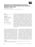
Zinc binding prevents Tat oxidation
As a first step, we measured the effect of zinc binding on
Tat oxidation. To this end, we monitored with time the
number of free -SH groups per molecule of Tat. At pH7.5
in the absence of zinc, oxidation occurs rapidly, as well
documented [21]. Five out of the seven -SH groups were
oxidized within three hours (Fig. 1). Since Tat-tubulin
interaction was investigated at pH6.5, we also measured
the oxidation of Tat at this pH. Oxidation was slower than
that at pH7.5, but nevertheless three out of the seven -SH
groups were oxidized after four hours. In contrast, two
equivalents of zinc preserved Tat from oxidation since five
out of seven -SH groups remained in their reduced form,
even after more than 24 hours (data not shown). There
was no difference with five equivalents of zinc, suggesting
that Tat is saturated with two equivalents of zinc. This is
in agreement with mass spectrometry data, which showed
the disappearance of apo-Tat when two zinc equivalents
were added (data not shown).
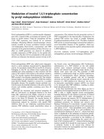
Zinc binding induces a local folding of Tat
In a next step, we characterized the effect of zinc on the
structure of Tat. To this end, we first performed fluores-
cence correlation spectroscopy (FCS) using Tat labeled at
its N-terminus by rhodamine B (Tat-RhB). The autocorre-
lation curves of apo-Tat-RhB and holo-Tat-RhB were
indistinguishable (Fig. 2). Their diffusion constants were
1.46(± 0.05) × 10
-6
cm
2
s
-1
and 1.38(± 0.08) × 10
-6
cm
2
s
-1
,
respectively, in excellent agreement with the theoretical
diffusion constant (D
th
= 1.44 × 10
-6
cm
2
s
-1
) calculated
from the Stokes-Einstein equation for the diffusion of a
sphere with the molecular mass of the Tat protein and
30% hydration. This suggests that both protein forms are
monomeric with a nearly spherical shape. Moreover, the
identical brightness (5.1 ± 0.1 kHz/molecule) of the two
Tat forms confirmed that they exhibit the same oligomeric
state. Interestingly, the monomeric state of both Tat forms
was further substantiated by mass spectrometry (data not
shown).
Then, we performed steady-state and time-resolved fluo-
rescence measurements, by monitoring the signal of
Trp
11
, which is a strictly conserved residue among Tat var-
iants [22,23]. Steady-state fluorescence results (data not
shown) showed that apo-Tat and holo-Tat displayed their
maximum emission wavelength at 346 nm, consistent
with a well exposed Trp residue [42]. The fluorescence
intensity decay of apo-Tat was characterized by four life-
times ranging from 0.21 ns to 4.5 ns, with comparable
populations (Table 1). Addition of two equivalents of zinc
resulted in a significant increase of the long-lived lifetime
from 4.5 ns to 5.1 ns. In contrast, the other lifetimes as
well as the amplitudes associated with the various life-
times were only marginally affected by the binding of
Effect of zinc binding on Tat oxidationFigure 1
Effect of zinc binding on Tat oxidation. The number or
free -SH groups per Tat molecule was measured according
to the Ellman reaction. Tat in NaPi 20 mM buffer, pH6.5 (●),
or in Hepes buffer 50 mM, pH7.5, in the absence (᭝), or in
the presence of 2 (■) or 5 (ᮀ) zinc equivalents.
Retrovirology 2008, 5:62 />Page 5 of 13
(page number not for citation purposes)
zinc. This suggests that the environment of Trp
11
is only
moderately modified by the binding of zinc ions.
Fluorescence anisotropy decays showed that both forms
were characterized by two correlation times (Table 2). The
short correlation time was about 0.25 ns for both forms
and can be assigned to the local motion of the Trp residue
[42]. The long correlation time was 2 ns for apo-Tat and
was thus markedly lower than the 4.1 ns theoretical value
expected for the tumbling motion of a sphere with the
molecular mass of Tat and 30% hydration [42]. The long
correlation time likely describes the segmental motion of
a domain, which includes the Trp residue. A significant
increase of this long correlation time (from 2 ns to 2.8 ns)
was observed with addition of zinc, indicating a signifi-
cant slowing down of the motion of the Trp-containing
domain. This slowing down is likely related to a zinc-
induced folding of the Cys-rich sequence (residues 22–
37), which is close to the Trp
11
residue.
Noticeably, no significant changes in the steady-state and
time-resolved fluorescence parameters of the apo-Tat were
observed in the presence of TCEP that keeps the -SH
groups in a reduced form. This indicates that the intramo-
lecular disulfide bridges in the oxidized form of apo-Tat
do not significantly affect the environment and the local
motion of Trp
11
as well as the segmental motion of the
Trp-containing domain.
Zinc binding to Tat promotes discrete Tat-tubulin
complexes under non-assembly conditions
We first investigated the interaction of Tat with tubulin
dimers at 20°C in 20 mM NaPi, 10 μM GTP, pH6.5 (PG
buffer). This buffer normally allows neither the associa-
tion of tubulin nor microtubule assembly at a tubulin
concentration ≤ 5 μM [43]. Analytical ultracentrifugation
(AUC) was used to characterize the binding of both apo-
Tat and holo-Tat to tubulin dimers. Control tubulin (5
μM) was found to sediment as a single species, as indi-
cated by the single Gaussian distribution of the continu-
ous sedimentation coefficient, C(S) (Fig. 3A) centered at
5.64 ± 0.01 S, in line with the standard value of 5.8
S [39]. Control experiments with zinc sulfate at concentra-
tions up to 20 μM, corresponding to the total concentra-
tion of zinc used in the holo-Tat samples, did not change
the apparent sedimentation coefficient (S
apparent
) of tubu-
lin and its corresponding area (data not shown). In con-
trast, the S
apparent
of tubulin in the presence of 10 μM holo-
Tat increased to 6.12 ± 0.01 S, suggesting a direct interac-
tion of the holo-Tat with tubulin dimers. In the presence
of apo-Tat at the same concentration (10 μM), the S
apparent
value of tubulin also increased and reached a value of 6.29
± 0.02 S. However, the area of the corresponding peak
drastically decreased in favor of a distribution of S
apparent
S
W20
0
,
Effect of zinc on Tat-RhB diffusion, as monitored by FCSFigure 2
Effect of zinc on Tat-RhB diffusion, as monitored by
FCS. The normalized autocorrelation curves were recorded
with 1 μM apo-Tat-RhB (❍) or holo-Tat-RhB (■) in Hepes
buffer 50 mM, 0.05% IGEPAL CA-230, pH7.5, at 20°C. The
continuous lines are fits to the experimental points with
Equation 1.
Table 1: Fluorescence intensity decay parameters of apo-Tat and holo-Tat
a
τ
1
(ns)
α
1
(%)
τ
2
(ns)
α
2
(%)
τ
3
(ns)
α
3
(%)
τ
4
(ns)
α
4
(%) <τ> (ns)
Apo-Tat 0.21 ± 0.03 25 ± 2 1.35 ± 0.01 35 ± 3 2.60 ± 0.20 19 ± 1 4.5 ± 0.2 21 ± 5 1.96 ± 0.08
Holo-Tat 0.22 ± 0.05 18 ± 4 1.30 ± 0.20 37 ± 3 2.79 ± 0.09 25 ± 3 5.1 ± 0.2 20 ± 3 2.24 ± 0.07
a
Experiments were performed with 1.5 μM Tat proteins in 50 mM Hepes buffer, pH7.5, at 20°C. The lifetimes, τ
i
, and relative amplitudes, α
i
, are
expressed as means for at least three independent experiments. The mean lifetimes were calculated with: Ότ = ∑α
i
τ
i
. The excitation and emission
wavelengths for Trp were set at 295 nm and 350 nm, respectively.
Table 2: Fluorescence anisotropy decay parameters of apo-Tat
and holo-Tat
a
θ
1
(ns)
β
1
(%)
θ
2
(ns)
β
2
(%)
Apo-Tat 0.28 ± 0.03 42 ± 3 2.0 ± 0.2 58 ± 3
Holo-Tat 0.24 ± 0.07 43 ± 6 2.8 ± 0.4 57 ± 6
a
Experimental conditions were as in Table 1. The correlation times,
θ
i
, and relative amplitudes,
β
i
, are expressed as means for at least
three experiments.
Retrovirology 2008, 5:62 />Page 6 of 13
(page number not for citation purposes)
values ranging from 20 to 90 S (Fig. 3A inset), suggesting
the formation of tubulin oligomers. Electron microscopy
of the tubulin/apo-Tat samples (Fig. 3B) showed the pres-
ence of small particles, consistent with the formation of
oligomers, which are absent in the control and the tubu-
lin- holo-Tat samples (data not shown).
Holo-Tat promotes and stabilizes microtubules under
assembly-conditions
Having shown some differences between apo-Tat and
holo-Tat with respect to their interaction with tubulin
dimers in PG buffer at 20°C, we measured the effects of
various concentrations of apo-Tat and holo-Tat on micro-
tubule formation in PMG buffer (20 mM NaPi, 10 mM
MgCl
2
, 0.1 mM GTP, 3.4 M glycerol, pH6.5) (Fig. 4). The
reactions with 15 μM tubulin were started by warming the
samples to 37°C. For the control in the absence of Tat,
after a lag time of several minutes, the turbidity increased
and reached a plateau (Fig. 4A). Lowering the temperature
to 10°C induced a drop in turbidity to its initial values,
indicating a total reversibility of the reaction. In the pres-
ence of apo-Tat (Fig. 4A) and holo-Tat (Fig. 4B) added at
concentrations that have been shown to interact effi-
ciently with microtubules and promote apoptosis in cells
[15,16], we observed a shortening of the lag time as well
as a strong increase in the rate of assembly and final pla-
teau value. The Tat-induced changes on tubulin assembly
were strongly dependent on the protein concentration for
both of the Tat forms. At the highest Tat concentration (4
μM), the turbidity plateau was increased by 1.6- and 2.1-
fold for apo-Tat and holo-Tat, respectively, as compared
with the control plateau value obtained with tubulin
alone. Our data obtained with Tat Lai are in line with
those previously obtained with Tat HxB2, suggesting that
the Tat proteins from both strains exhibit similar activities
on tubulin assembly [16].
However, the Tat proteins from the two strains were found
to differ in the disassembly step. Indeed, in contrast to Tat
HxB2 (Fig. 1A in [16]), when the temperature of the sam-
ples was decreased to 10°C, we did not observe a com-
plete disassembly of the microtubules in the presence of
both apo-Tat and holo-Tat Lai species. This indicated the
presence of cold stable aggregates or polymers with the Tat
Lai variant.
To compare further the tubulin assembly induced by the
apo- and holo-forms of Tat Lai, the samples were exam-
ined by electron microscopy at 37°C at the turbidity pla-
teau and at 10°C, after cold depolymerisation (Fig. 4C).
At 37°C, the electron micrographs confirmed the forma-
tion of microtubules in the presence of both Tat forms,
similar in shape to the controls. However, in addition to
microtubules, numerous tubulin aggregates were
observed in the presence of apo-Tat. At 10°C, in all condi-
tions (with and without Tat) we observed large rings (out-
side diameter ≈ 50 nm), likely due to the lack of EGTA in
our experiments. Indeed, rings are favored by divalent cat-
ions such as Ca
2+
[44,45] that are chelated by the EGTA
added in the classical buffer used to study microtubule
formation [41]. These rings are the main if not, the only
observable form in the control. In contrast, we also
observed cold stable microtubules in the presence of the
holo-Tat (Fig. 4C). With apo-Tat, amorphous tubulin
aggregates were observed but microtubules were absent.
As a consequence, though the turbidity traces of apo- and
holo-Tat forms were similar (Fig. 4A and Fig. 4B), signifi-
cant differences appear in the nature of the tubulin poly-
mers induced by the two forms of Tat.
Zinc binding to Tat promotes discrete Tat-tubulin complexes under non-assembly conditionsFigure 3
Zinc binding to Tat promotes discrete Tat-tubulin
complexes under non-assembly conditions. A. Charac-
terization of Tat-tubulin interaction by analytical ultracentrif-
ugation, in PG buffer. Continuous sedimentation coefficient
distribution C(S) of tubulin (5 μM) in the absence (black solid
line), or in the presence of 10 μM holo-Tat (blue dashed line)
or 10 μM apo-Tat (red dotted line). Inset: Full range C(S) of
tubulin (5 μM) in the presence of 10 μM apo-Tat (red dotted
line). Tat contributed to less than 10% of the signal. B. Elec-
tron micrograph of 5 μM tubulin in the presence of 10 μM
apo-Tat, in PG buffer.
Retrovirology 2008, 5:62 />Page 7 of 13
(page number not for citation purposes)
Effect of Tat on tubulin assembly above the critical concentration (Cr) of tubulinFigure 4
Effect of Tat on tubulin assembly above the critical concentration (Cr) of tubulin. A and B. Effect of Tat on tubulin
(15 μM) assembly, as measured by turbidimetry at 350 nm. Measurements were performed in the absence (black solid line), or
in the presence of 2 μM (blue dashed line), 3 μM (red dotted line), or 4 μM (green dashed-dotted line) of A) apo-Tat or B)
holo-Tat, in PMG buffer at 37°C. At the time indicated by the arrow, samples were cooled to 10°C. C. Electron micrographs
of 15 μM tubulin in the absence or the presence of 4 μM apo-Tat, or 4 μM holo-Tat at 37°C and after cold depolymerisation at
10°C, in PMG buffer.
Retrovirology 2008, 5:62 />Page 8 of 13
(page number not for citation purposes)
In the next step, the interaction between the different
forms of Tat Lai and tubulin were characterized at a tubu-
lin concentration below the critical concentration (Cr),
where no tubulin assembly occurs at 37°C (for a review,
see [46]). In the absence of Tat, the tubulin Cr value was
found to be 9 ± 1 μM, in line with the 8 μM value deter-
mined in the presence of EGTA [47]. To be below the Cr,
we investigated Tat-tubulin interaction at a 6 μM concen-
tration of tubulin. As for the control (black solid line in
Fig. 5A), no significant increase in turbidity was observed
when apo-Tat at 8 μM was added at 37°C. In contrast, the
same concentration of holo-Tat (8 μM) resulted in a
strong increase in turbidity (red dashed-dotted line). This
effect was dependent on the holo-Tat concentration, as
seen by the different turbidity traces with 4 μM and 8 μM
holo-Tat. When the samples were cooled to 10°C, the tur-
bidity slightly decreased but did not fall to zero even after
several hours (data not shown). This indicates that a large
fraction of the tubulin polymers induced by holo-Tat was
stable at 10°C. Further incubation at 4°C during one hour
induced a drop of turbidity.
In line with the turbidity data, electron microscopy
showed no polymers with tubulin alone or when apo-Tat
(See additional file 1) was added to tubulin at 37°C. In
contrast, the polymers induced by holo-Tat corresponded
to normal microtubules (Fig. 5B). At 10°C, we also
observed microtubules. A few stable microtubules were
still present after one hour of incubation at 4°C, and were
thus responsible for the residual turbidity (Fig. 5A).
The temperature-induced reversibility of tubulin assembly
in the presence of holo-Tat indicates that holo-Tat -
induced tubulin polymers and tubulin dimers are in equi-
librium. This allowed us to calculate a Cr value of 4 ± 1 μM
of tubulin in the presence of 8 μM holo-Tat. This Cr value
is about two-fold less than the Cr value for tubulin assem-
bly in the absence of holo-Tat.
Since zinc is known to induce tubulin sheets [48-50], we
also monitored the effect of zinc on tubulin assembly (Fig.
5A). Only, at the highest zinc concentration (16 μM) that
would correspond to a total release of Zn from 8 μM holo-
Tat, a strong increase in turbidity was observed (green
dashed-dotted-dotted line in Fig. 5A). However, the lag
time of this turbidity increase was much longer than the
one observed with 8 μM holo-Tat. Moreover, at 8 μM con-
centration of zinc, which would correspond to a total
release of Zn from 4 μM holo-Tat, the effect on turbidity
was much weaker than that with 4 μM holo-Tat. In con-
trast to the microtubules observed in the presence of holo-
Tat at all temperatures, tubulin sheets were observed at
37°C and 10°C in the presence of ZnS0
4
(Fig. 5B). These
sheets were no more present at 4°C, in line with the
strong drop in turbidity (Fig. 5A).
Thus, the effect of holo-Tat on tubulin assembly can not
be attributed to the release of free zinc from holo-Tat.
Moreover, these data confirm that in our experimental
conditions, two equivalents of zinc are mainly bound to
Tat.
Since the 36–48 region of Tat has been previously shown
to be necessary and sufficient for the Tat-tubulin interac-
tion [15], we checked whether a Tat(36–48) peptide was
able to induce tubulin assembly. Both above and below
the Cr, the turbidity traces were indistinguishable from
the control ones, even at peptide concentration up to 60
μM (data not shown). This indicates that the 36–48
region is not sufficient to promote microtubule forma-
tion.
Discussion
HIV-1 Tat protein is involved in the weakening of
immune defense in AIDS, notably by interacting with
microtubules. Several studies showed that Tat from differ-
ent HIV isolates, and specifically residues 38–72, was able
to enhance tubulin assembly in vitro, and induce apopto-
sis via the mitochondrial pathway [14-16]. The efficiency
of different Tat variants to promote tubulin assembly was
correlated with their efficiency to induce apoptosis and
the progression to AIDS [14,16]. However, in these stud-
ies, the zinc binding status of Tat was not checked, despite
the evidence that Tat is able to bind zinc ions through its
cysteine-rich domain in vitro [19-21] and that the Tat
transactivation function and apoptosis induction seem to
depend upon zinc [25,26]. Moreover, mutations of the
Cys residues (except Cys
31
) have been shown to impair
Tat functions [51], confirming further the relevance of
zinc binding in the biological functions of Tat. In addi-
tion, the Tat-Oyi variant from highly exposed but persist-
ently seronegative patients has been shown to differ from
other Tat variants by a Cys
22
→Ser substitution, which has
the consequences of a decrease in the transactivation
activity of Tat [52] and Tat-microtubules interaction [16].
In this study, to further understand the importance of zinc
in Tat functions, its role on the conformation and the
interaction of Tat Lai with tubulin was investigated. Tat
Lai was selected since this variant is representative of the
subtype B HIV-1 virus, commonly found in infected indi-
viduals in Europe and North America [53]. First, we com-
pared the conformations of the apo-form and zinc-bound
form of Tat Lai. For the apo-form, an excellent agreement
between the diffusion constant measured by FCS and the
theoretical diffusion constant of a sphere with the mass of
the hydrated Tat protein suggested that the protein was
monomeric and poorly folded, in line with the data
obtained earlier with other Tat variants [54]. The poor
folding of the apo-Tat form was substantiated by the
important segmental motion of the Trp
11
-containing
Retrovirology 2008, 5:62 />Page 9 of 13
(page number not for citation purposes)
Holo-Tat promotes and stabilizes microtubules under assembly-conditions, at a tubulin concentration below the critical con-centration (Cr)Figure 5
Holo-Tat promotes and stabilizes microtubules under assembly-conditions, at a tubulin concentration below
the critical concentration (Cr). A. Effect of Tat on tubulin (6 μM) assembly, as measured by turbidimetry at 350 nm. Meas-
urements were performed in the absence (black solid line), or in the presence of 4 μM holo-Tat (blue dashed line), 8 μM holo-
Tat (red dashed-dotted line), 8 μM zinc sulfate (purple dotted line), or 16 μM zinc sulfate (green dashed-dotted-dotted line), in
PMG buffer at 37°C. At the time indicated by the first arrow, samples were cooled to 10°C. The second arrow represents one
hour of incubation at 4°C. The trace with 8 μM apo-Tat was indistinguishable from the control and was thus not represented.
B. Electron micrographs of 6 μM tubulin in the presence of 8 μM holo-Tat, or 16 μM zinc sulfate, in PMG buffer at 37°C and
after cold depolymerisation at 10°C or 4°C.
Retrovirology 2008, 5:62 />Page 10 of 13
(page number not for citation purposes)
domain that prevented the observation of the protein
tumbling motion (Table 2). Moreover, the high maxi-
mum emission wavelength and complex fluorescence
intensity decay of Trp
11
suggested that it was well exposed
to the solvent and explored a large number of conforma-
tions, in agreement with a flexible and poorly folded
structure of Tat. This large exposure of Trp
11
to the solvent
differs, however, from the inclusion of Trp
11
in a hydro-
phobic pocket suggested by the NMR-derived structure of
Tat Lai/Bru at pH4.5 [55]. This difference could not be
attributed to the oxidation state of Tat since addition of
the reducing agent TCEP that prevented oxidation of the -
SH groups did not significantly affect any of the measured
fluorescence parameters (data not shown). Though a pH-
dependent folding involving Trp
11
can not be excluded,
our data support also recent reports showing that the Trp-
containing region is not folded [53,54].
The holo-form of Tat Lai was found to bind two zinc ions
through five of its seven cysteine residues, in full agree-
ment with previous results with the Tat(21–38) peptide
[21]. The diffusion constant and the mass spectrum of the
zinc-bound form strongly suggested that it remains mon-
omeric, in line with most previously published data
[6,21,24]. Interestingly, the large solvent-exposure and
the complex intensity decay of the Trp
11
residue, as well as
the absence of a rotational correlation time corresponding
to the protein tumbling suggested that the holo-form
remains poorly folded. Nevertheless, the increase of the
long rotational correlation time (Table 2) suggested a
local folding, most likely at the level of the cysteine-rich
sequence close to the Trp
11
residue. This partial folding is
in line with previous observations made with a different
variant of Tat [19], suggesting that it may be a general fea-
ture in holo-Tat proteins.
Both apo- and holo-Tat were found to promote tubulin
assembly at concentrations above the Cr value (9 ± 1 μM
in our conditions). Monitoring the assembly by turbidim-
etry, both Tat forms were found to decrease the initial lag
time and increase the rate of assembly. This suggests that
both protein forms can promote the nucleation and elon-
gation phases of microtubule formation [46]. Moreover,
both forms increased the turbidity plateau by about two-
fold over the control (in the absence of Tat). Electron
microscopy data as well as the reversibility of the major
part of the holo-Tat-induced turbidity increase at 10°C
indicate that holo-Tat mainly induces the formation of
microtubules. As a consequence, the increase of the tur-
bidity plateau over the control suggests that Tat promotes
a larger amount of microtubules than in the control and
thus, likely decreases the Cr. This was confirmed by the
measured two-fold decrease in the tubulin Cr value
induced by holo-Tat (from 9 ± 1 μM to 4 ± 1 μM of tubu-
lin), and the observation of holo-Tat -induced microtu-
bules at a 6 μM tubulin concentration (Fig. 5).
Moreover, the significant fraction of cold-stable microtu-
bules at 10°C further suggests that holo-Tat also prevents
microtubule depolymerization. This assumption is
strengthened by the observation of cold-stable microtu-
bules after one hour of incubation at 4°C. In the case of
the apo-Tat, the turbidity traces were associated with the
formation of both microtubules and tubulin aggregates.
Since turbidity is a complex function of the number, size
and the shape of the scattering particles [56-58], the effect
of apo-Tat on the amount of tubulin polymers is difficult
to evaluate. Nevertheless, since in contrast to holo-Tat, no
microtubules were induced by apo-Tat at a concentration
below the Cr, it is likely that apo-Tat marginally affects the
Cr value. In addition, the absence of cold-stable microtu-
bules with apo-Tat further suggests that it does not prevent
microtubule depolymerization. The cold stabilization of
microtubules by only holo-Tat is highly significant, since
this cold stabilization in vitro has been shown to be repre-
sentative of the stabilization of the microtubule network
in cells [59,60].
The differences between apo-Tat and holo-Tat with
respect to tubulin assembly may be partly accounted by
their different binding modes to the tubulin dimers.
Holo-Tat was found to bind tubulin dimers in discrete
complexes while apo-Tat promoted a distribution of tubu-
lin oligomers. In assembly conditions, the discrete com-
plexes with holo-Tat likely nucleate and elongate
microtubules more efficiently than control tubulin dim-
ers. Holo-Tat has the same effect than Paclitaxel [61] and
Taxotere [62] that also stabilize the microtubules, causing
a mitotic block and a subsequent cell death by apoptosis
[60], but it remains to be demonstrated that their mecha-
nisms are similar. The tubulin oligomers observed with
apo-Tat probably contribute to the formation of tubulin
aggregates and microtubules observed in assembly condi-
tions above the Cr. Since oligomers are thought to be pre-
cursors for microtubule nuclei [46], their presence may
explain the observed increase in the rate of nucleation and
elongation in the apo-Tat-promoted assembly of tubulin.
Noticeably, the concentration of Tat in our assays was sub-
stantially larger than the nM range concentration of Tat in
sera of HIV-1-infected patients [63]. However, such Tat
concentrations could be locally achieved in lymphoid tis-
sues, where HIV-1 actively replicates [10,63] or within the
intracellular medium, as a consequence of efficient inter-
nalization of Tat.
Importantly, our data with holo-Tat are fully consistent
with the previously reported prevention by cellular Tat of
microtubule depolymerization and the concurrent reduc-
tion of the level of unpolymerized tubulin in cells [15].
Retrovirology 2008, 5:62 />Page 11 of 13
(page number not for citation purposes)
Consequently, holo-Tat likely constitutes the active form
of Tat in the cell cytoplasm. By altering the microtubule
dynamics, holo-Tat may then lead to a release of the pro-
apoptotic Bim protein, leading to apoptosis through the
mitochondria pathway [15].
The Tat(36–48) peptide was found to be unable to pro-
mote tubulin assembly though this sequence mediates the
binding of Tat to tubulin [15]. Since a Tat(38–72) peptide
has been previously reported to promote microtubule for-
mation as efficiently as the full-length Tat [16], the 49–72
region of Tat is likely to be required for promoting tubulin
polymerization. The basic region of Tat (residues 49–59)
is probably important since basic domains play a key role
in microtubule-associated proteins [64] by neutralizing a
negatively charged region of the tubulin dimer involved in
tubulin assembly. The glutamine rich region of Tat (resi-
dues 60–72) may be important too, since this region was
shown to modulate the binding of Tat to tubulin and the
efficiency of Tat in inducing apoptosis [14].
The differences between apo-Tat and holo-Tat in their
binding to tubulin dimers and their activation of tubulin
assembly are probably a consequence of the limited con-
formational changes between the two forms. The binding
of zinc to the cysteine-rich region and probably to Cys
37
most likely modifies the conformation of the
36
Val-Cys-
Phe-Thr
39
sequence, which is determinant for binding to
tubulin [15]. This conformational change is probably
required for the proper positioning of Tat on its tubulin
binding site(s) in order to change the assembly properties
of tubulin. Large effects on Tat properties resulting from
limited conformational changes are not unprecedented
since the strong differences in apoptosis induction by Tat
proteins from two different strains have also been related
to minor structural modifications of Tat [14].
Conclusion
We demonstrated in this work that the binding of zinc to
the Cys-rich region of Tat Lai modulates the protein con-
formation, most likely by inducing a partial folding. This
probably affects the
36
Val-Cys-Phe-Thr
39
region, critical
for tubulin binding. This allows Tat to bind tubulin dim-
ers in discrete complexes, while apo-Tat induces oligom-
ers of different sizes. Moreover, holo-Tat but not apo-Tat
reduces the Cr and stabilizes the microtubules similarly to
intracellular Tat [15], suggesting that holo-Tat is the intra-
cellular active form involved in apoptosis. Inhibition of
Tat-induced apoptosis in non infected cells is thought to
impair at least in part the loss of immunocompetence pro-
voked by HIV-1 and hopefully convert HIV infection from
a progressively immunosuppressive and ultimately fatal
disease to a chronic manageable infection. Since the
highly conserved cysteine-rich domain of Tat [22,23]
likely induces a structure distinct from the eukaryotic zinc
fingers, interference with zinc binding to Tat or targeting
the binding site of the holo-Tat to tubulin could be prom-
ising as new approaches to design antiviral drugs that
would not affect the host proteins.
List of abbreviations
AIDS: Acquired immunodeficiency syndrome; HIV-1:
Human immunodeficiency virus type 1; TAR: Trans-acting
responsive element; LTR: Long terminal repeat; AUC: Ana-
lytical ultracentrifugation; RhB: Rhodamine B; FCS: Fluo-
rescence correlation spectroscopy; TMR:
Carboxytetramethylrhodamine.
Competing interests
The authors declare that they have no competing interests.
Authors' contributions
CE performed experiments and wrote part of the manu-
script. PB participated in the design of the experiments
and in the interpretation of the results. PD and EP pro-
vided technical support for FCS and fluorescence time-
resolved measurements. DA contributed in the design of
the experiments. OC and SM synthesized Tat protein and
peptides. VP and YM directed the work and finalized the
writing of the manuscript. All authors read and approved
the final manuscript.
Additional material
Acknowledgements
The Agence Nationale de Recherches sur le SIDA (ANRS), the Association
Ensembles Contre le SIDA (SIDACTION), and the Association pour la
Recherche sur le Cancer are gratefully acknowledged for financial support
to this work. CE was a fellow from the French Ministère de la Recherche.
References
1. Fauci AS: Immunopathogenesis of HIV infection. J Acquir
Immune Defic Syndr 1993, 6(6):655-662.
2. Meyaard L, Otto SA, Jonker RR, Mijnster MJ, Keet RP, Miedema F:
Programmed death of T cells in HIV-1 infection. Science 1992,
257(5067):217-219.
3. Chang HC, Samaniego F, Nair BC, Buonaguro L, Ensoli B: HIV-1 Tat
protein exits from cells via a leaderless secretory pathway
and binds to extracellular matrix-associated heparan sulfate
proteoglycans through its basic region. Aids 1997,
11(12):1421-1431.
4. Ensoli B, Buonaguro L, Barillari G, Fiorelli V, Gendelman R, Morgan
RA, Wingfield P, Gallo RC: Release, uptake, and effects of extra-
cellular human immunodeficiency virus type 1 Tat protein
on cell growth and viral transactivation. J Virol 1993,
67(1):277-287.
Additional file 1
Electron micrograph of 6
μ
M tubulin in the presence of 8
μ
M apo-Tat, in
PMG buffer at 37°C.
Click here for file
[ />4690-5-62-S1.pdf]
Retrovirology 2008, 5:62 />Page 12 of 13
(page number not for citation purposes)
5. Frankel AD, Pabo CO: Cellular uptake of the tat protein from
human immunodeficiency virus. Cell 1988, 55(6):1189-1193.
6. Dingwall C, Ernberg I, Gait MJ, Green SM, Heaphy S, Karn J, Lowe AD,
Singh M, Skinner MA: HIV-1 tat protein stimulates transcrip-
tion by binding to a U-rich bulge in the stem of the TAR RNA
structure. Embo J 1990, 9(12):4145-4153.
7. Gatignol A, Jeang KT: Tat as a transcriptional activator and a
potential therapeutic target for HIV-1. Adv Pharmacol 2000,
48:209-227.
8. Karn J: Tackling Tat. J Mol Biol 1999, 293(2):235-254.
9. Muller S, Desgranges C: HIV-1 Tat and apoptotic death (Chap-
ter 9), in Cell death during HIV infection. (A. Badley, Ed) CRC
Taylor and Francis; 2006.
10. Westendorp MO, Frank R, Ochsenbauer C, Stricker K, Dhein J, Wal-
czak H, Debatin KM, Krammer PH: Sensitization of T cells to
CD95-mediated apoptosis by HIV-1 Tat and gp120. Nature
1995, 375(6531):497-500.
11. Flores SC, Marecki JC, Harper KP, Bose SK, Nelson SK, McCord JM:
Tat protein of human immunodeficiency virus type 1
represses expression of manganese superoxide dismutase in
HeLa cells. Proc Natl Acad Sci U S A 1993, 90(16):7632-7636.
12. Westendorp MO, Shatrov VA, Schulze-Osthoff K, Frank R, Kraft M,
Los M, Krammer PH, Droge W, Lehmann V: HIV-1 Tat potenti-
ates TNF-induced NF-kappa B activation and cytotoxicity by
altering the cellular redox state. Embo J 1995, 14(3):546-554.
13. Li CJ, Friedman DJ, Wang C, Metelev V, Pardee AB: Induction of
apoptosis in uninfected lymphocytes by HIV-1 Tat protein.
Science 1995, 268(5209):429-431.
14. Campbell GR, Pasquier E, Watkins J, Bourgarel-Rey V, Peyrot V,
Esquieu D, Barbier P, de Mareuil J, Braguer D, Kaleebu P, Yirrell DL,
Loret EP: The glutamine-rich region of the HIV-1 Tat protein
is involved in T-cell apoptosis. J Biol Chem 2004,
279(46):48197-48204.
15. Chen D, Wang M, Zhou S, Zhou Q: HIV-1 Tat targets microtu-
bules to induce apoptosis, a process promoted by the pro-
apoptotic Bcl-2 relative Bim. Embo J 2002, 21(24):6801-6810.
16. de Mareuil J, Carre M, Barbier P, Campbell GR, Lancelot S, Opi S,
Esquieu D, Watkins JD, Prevot C, Braguer D, Peyrot V, Loret EP:
HIV-1 Tat protein enhances microtubule polymerization.
Retrovirology 2005, 2:5.
17. Battaglia PA, Zito S, Macchini A, Gigliani F: A Drosophila model of
HIV-Tat-related pathogenicity. J Cell Sci 2001, 114(Pt
15):2787-2794.
18. Giacca M: HIV-1 Tat, apoptosis and the mitochondria: a tubu-
lin link? Retrovirology 2005, 2:7.
19. Frankel AD, Bredt DS, Pabo CO: Tat protein from human immu-
nodeficiency virus forms a metal-linked dimer. Science 1988,
240(4848):70-73.
20. Frankel AD, Chen L, Cotter RJ, Pabo CO: Dimerization of the tat
protein from human immunodeficiency virus: a cysteine-rich
peptide mimics the normal metal-linked dimer interface.
Proc Natl Acad Sci U S A 1988, 85(17):6297-6300.
21. Huang HW, Wang KT: Structural characterization of the metal
binding site in the cysteine-rich region of HIV-1 Tat protein.
Biochem Biophys Res Commun 1996, 227(2):615-621.
22. Gregoire C, Peloponese JM Jr., Esquieu D, Opi S, Campbell G, Solo-
miac M, Lebrun E, Lebreton J, Loret EP: Homonuclear (1)H-NMR
assignment and structural characterization of human immu-
nodeficiency virus type 1 Tat Mal protein. Biopolymers 2001,
62(6):324-335.
23. Kuppuswamy M, Subramanian T, Srinivasan A, Chinnadurai G: Multi-
ple functional domains of Tat, the trans-activator of HIV-1,
defined by mutational analysis. Nucleic Acids Res 1989,
17(9):3551-3561.
24. Koken SE, Greijer AE, Verhoef K, van Wamel J, Bukrinskaya AG,
Berkhout B: Intracellular analysis of in vitro modified HIV Tat
protein. J Biol Chem 1994, 269(11):8366-8375.
25. Garber ME, Wei P, KewalRamani VN, Mayall TP, Herrmann CH, Rice
AP, Littman DR, Jones KA: The interaction between HIV-1 Tat
and human cyclin T1 requires zinc and a critical cysteine res-
idue that is not conserved in the murine CycT1 protein.
Genes Dev 1998, 12(22):3512-3527.
26. Misumi S, Takamune N, Ohtsubo Y, Waniguchi K, Shoji S: Zn2+
binding to cysteine-rich domain of extracellular human
immunodeficiency virus type 1 Tat protein is associated with
Tat protein-induced apoptosis. AIDS Res Hum Retroviruses 2004,
20(3):297-304.
27. Chaloin O, Peter JC, Briand JP, Masquida B, Desgranges C, Muller S,
Hoebeke J: The N-terminus of HIV-1 Tat protein is essential
for Tat-TAR RNA interaction. Cell Mol Life Sci 2005,
62(3):355-361.
28. Ellman GL: Tissue sulfhydryl groups. Arch Biochem Biophys 1959,
82(1):70-77.
29. Riddles PW, Blakeley RL, Zerner B: Reassessment of Ellman's
reagent. Methods Enzymol 1983, 91:49-60.
30. Azoulay J, Clamme JP, Darlix JL, Roques BP, Mely Y: Destabilization
of the HIV-1 complementary sequence of TAR by the nucle-
ocapsid protein through activation of conformational fluctu-
ations. J Mol Biol 2003, 326(3):691-700.
31. Clamme JP, Azoulay J, Mely Y: Monitoring of the formation and
dissociation of polyethylenimine/DNA complexes by two
photon fluorescence correlation spectroscopy. Biophys J 2003,
84(3):1960-1968.
32. Thompson NL: Fluorescence correlation spectroscopy, in
Topics in fluorescence spectroscopy . In Volume 1: Plenum
Publishers, NY.; 1991.
33. Egele C, Schaub E, Piemont E, de Rocquigny H, Mely Y: Investigation
by fluorescence correlation spectroscopy of the chaperoning
interactions of HIV-1 nucleocapsid protein with the viral
DNA initiation sequences. C R Biol 2005, 328(12):1041-1051.
34. Bombarda E, Ababou A, Vuilleumier C, Gerard D, Roques BP, Pie-
mont E, Mely Y: Time-resolved fluorescence investigation of
the human immunodeficiency virus type 1 nucleocapsid pro-
tein: influence of the binding of nucleic acids. Biophys J 1999,
76(3):1561-1570.
35. Mely Y, Jullian N, Morellet N, De Rocquigny H, Dong CZ, Piemont E,
Roques BP, Gerard D: Spatial proximity of the HIV-1 nucleo-
capsid protein zinc fingers investigated by time-resolved flu-
orescence and fluorescence resonance energy transfer.
Biochemistry 1994, 33(40):12085-12091.
36. Livesey AK, Brochon JC: Analyzing the distribution of decay
constants in pulse-fluorimetry using the maximum entropy
method. Biophys J 1987, 52:693-706.
37. Andreu JM, Diaz JF, Gil R, de Pereda JM, Garcia de Lacoba M, Peyrot
V, Briand C, Towns-Andrews E, Bordas J: Solution structure of
Taxotere-induced microtubules to 3-nm resolution. The
change in protofilament number is linked to the binding of
the taxol side chain. J Biol Chem 1994, 269(50):31785-31792.
38. Lee JC, Frigon RP, Timasheff SN: The chemical characterization
of calf brain microtubule protein subunits. J Biol Chem 1973,
248(20):7253-7262.
39. Weisenberg RC, Borisy GG, Taylor EW: The colchicine-binding
protein of mammalian brain and its relation to microtu-
bules. Biochemistry 1968, 7(12):4466-4479.
40. Schuck P, Rossmanith P: Determination of the sedimentation
coefficient distribution by least-squares boundary modeling.
Biopolymers 2000, 54(5):328-341.
41. Barbier P, Gregoire C, Devred F, Sarrazin M, Peyrot V: In vitro
effect of cryptophycin 52 on microtubule assembly and tubu-
lin: molecular modeling of the mechanism of action of a new
antimitotic drug. Biochemistry 2001, 40(45):13510-13519.
42. Lakowicz JR: Principles of fluorescence spectroscopy, Second
Edition. Kluwer Academic, Plenum Publishers, NY.; 1999.
43. Devred F, Barbier P, Douillard S, Monasterio O, Andreu JM, Peyrot
V: Tau induces ring and microtubule formation from alpha-
beta-tubulin dimers under nonassembly conditions. Biochem-
istry 2004, 43(32):10520-10531.
44. Howard WD, Timasheff SN: GDP state of tubulin: stabilization
of double rings. Biochemistry 1986, 25(25):8292-8300.
45. Nogales E, Wang HW, Niederstrasser H: Tubulin rings: which
way do they curve? Curr Opin Struct Biol 2003, 13(2):256-261.
46. Valiron O, Caudron N, Job D: Microtubule dynamics. Cell Mol Life
Sci 2001, 58(14):2069-2084.
47. Devred F, Douillard S, Briand C, Peyrot V: First tau repeat domain
binding to growing and taxol-stabilized microtubules, and
serine 262 residue phosphorylation. FEBS Lett 2002, 523(1-
3):247-251.
48. Gaskin F: In vitro microtubule assembly regulation by divalent
cations and nucleotides. Biochemistry 1981, 20(5):1318-1322.
49. Gaskin F, Kress Y: Zinc ion-induced assembly of tubulin. J Biol
Chem 1977, 252(19):6918-6924.
Publish with BioMed Central and every
scientist can read your work free of charge
"BioMed Central will be the most significant development for
disseminating the results of biomedical research in our lifetime."
Sir Paul Nurse, Cancer Research UK
Your research papers will be:
available free of charge to the entire biomedical community
peer reviewed and published immediately upon acceptance
cited in PubMed and archived on PubMed Central
yours — you keep the copyright
Submit your manuscript here:
/>BioMedcentral
Retrovirology 2008, 5:62 />Page 13 of 13
(page number not for citation purposes)
50. Nogales E, Wolf SG, Zhang SX, Downing KH: Preservation of 2-D
crystals of tubulin for electron crystallography. J Struct Biol
1995, 115(2):199-208.
51. Jeang KT, Xiao H, Rich EA: Multifaceted activities of the HIV-1
transactivator of transcription, Tat. J Biol Chem 1999,
274(41):28837-28840.
52. Peloponese JM Jr., Collette Y, Gregoire C, Bailly C, Campese D,
Meurs EF, Olive D, Loret EP: Full peptide synthesis, purification,
and characterization of six Tat variants. Differences
observed between HIV-1 isolates from Africa and other con-
tinents. J Biol Chem 1999, 274(17):11473-11478.
53. Pantano S, Carloni P: Comparative analysis of HIV-1 Tat vari-
ants. Proteins 2005, 58(3):638-643.
54. Shojania S, O'Neil JD: HIV-1 Tat is a natively unfolded protein:
the solution conformation and dynamics of reduced HIV-1
Tat-(1-72) by NMR spectroscopy. J Biol Chem 2006,
281(13):8347-8356.
55. Peloponese JM Jr., Gregoire C, Opi S, Esquieu D, Sturgis J, Lebrun E,
Meurs E, Collette Y, Olive D, Aubertin AM, Witvrow M, Pannecou-
que C, De Clercq E, Bailly C, Lebreton J, Loret EP: 1H-13C nuclear
magnetic resonance assignment and structural characteri-
zation of HIV-1 Tat protein. C R Acad Sci III 2000,
323(10):883-894.
56. Berne BJ: Interpretation of the light scattering from long rods.
J Mol Biol 1974, 89(4):755-758.
57. Gaskin F: Techniques for the study of microtubule assembly in
vitro. Methods Enzymol 1982, 85 Pt B:433-439.
58. Stoylov SP, Vuilleumier C, Stoylova E, De Rocquigny H, Roques BP,
Gerard D, Mely Y: Ordered aggregation of ribonucleic acids by
the human immunodeficiency virus type 1 nucleocapsid pro-
tein. Biopolymers 1997, 41(3):301-312.
59. Horwitz SB: Taxol (paclitaxel): mechanisms of action. Ann
Oncol 1994, 5 Suppl 6:S3-6.
60. Jordan MA: Mechanism of action of antitumor drugs that
interact with microtubules and tubulin. Curr Med Chem Antican-
cer Agents
2002, 2(1):1-17.
61. Kumar N: Taxol-induced polymerization of purified tubulin.
Mechanism of action. J Biol Chem 1981, 256(20):10435-10441.
62. Diaz JF, Andreu JM: Assembly of purified GDP-tubulin into
microtubules induced by taxol and taxotere: reversibility,
ligand stoichiometry, and competition. Biochemistry 1993,
32(11):2747-2755.
63. Xiao H, Neuveut C, Tiffany HL, Benkirane M, Rich EA, Murphy PM,
Jeang KT: Selective CXCR4 antagonism by Tat: implications
for in vivo expansion of coreceptor use by HIV-1. Proc Natl
Acad Sci U S A 2000, 97(21):11466-11471.
64. Hirokawa N: Microtubule organization and dynamics depend-
ent on microtubule-associated proteins. Curr Opin Cell Biol
1994, 6(1):74-81.