Báo cáo y học: "Direct Vpr-Vpr Interaction in Cells monitored by two Photon Fluorescence Correlation Spectroscopy and Fluorescence Lifetime Imaging" pps
Bạn đang xem bản rút gọn của tài liệu. Xem và tải ngay bản đầy đủ của tài liệu tại đây (756.21 KB, 17 trang )
BioMed Central
Page 1 of 17
(page number not for citation purposes)
Retrovirology
Open Access
Research
Direct Vpr-Vpr Interaction in Cells monitored by two Photon
Fluorescence Correlation Spectroscopy and Fluorescence Lifetime
Imaging
Joëlle V Fritz
1
, Pascal Didier
1
, Jean-Pierre Clamme
2
, Emmanuel Schaub
1
,
Delphine Muriaux
3
, Charlotte Cabanne
4
, Nelly Morellet
5
, Serge Bouaziz
5
,
Jean-Luc Darlix
3
, Yves Mély
1
and Hugues de Rocquigny*
1
Address:
1
Département de Pharmacologie et Physico-Chimie des Interactions Cellulaires et Moléculaires, UMR 7175 CNRS, Faculté de Pharmacie,
Université Louis Pasteur, Strasbourg 1, 74, Route du Rhin, 67401 Illkirch Cedex, France,
2
Department of Immunology, The Scripps Research
Institute, 10550 North Torrey Pines Road, La Jolla, CA 92037, USA,
3
LaboRétro Unité de Virologie Humaine INSERM 758, IFR 128 Ecole Normale
Supérieure de Lyon, 46 allée d'Italie, 69364 Lyon, France,
4
Ecole Supérieure de Technologie des Biomolécules de Bordeaux, Université V Ségalen,
Bordeaux 2, 146, rue Léo Saignat, 33076 Bordeaux Cedex, France and
5
Unité de Pharmacologie Chimique et Génétique, Inserm U640 CNRS
UMR8151 UFR des Sciences Pharmaceutiques et Biologiques 4, Avenue de L'observatoire, 75006 Paris, France
Email: Joëlle V Fritz - ; Pascal Didier - ; Jean-
Pierre Clamme - ; Emmanuel Schaub - ; Delphine Muriaux - delphine.muriaux@ens-
lyon.fr; Charlotte Cabanne - ; Nelly Morellet - ;
Serge Bouaziz - ; Jean-Luc Darlix - ; Yves Mély - ;
Hugues de Rocquigny* -
* Corresponding author
Abstract
Background: The human immunodeficiency virus type 1 (HIV-1) encodes several regulatory proteins,
notably Vpr which influences the survival of the infected cells by causing a G2/M arrest and apoptosis. Such
an important role of Vpr in HIV-1 disease progression has fuelled a large number of studies, from its 3D
structure to the characterization of specific cellular partners. However, no direct imaging and
quantification of Vpr-Vpr interaction in living cells has yet been reported. To address this issue, eGFP- and
mCherry proteins were tagged by Vpr, expressed in HeLa cells and their interaction was studied by two
photon fluorescence lifetime imaging microscopy and fluorescence correlation spectroscopy.
Results: Results show that Vpr forms homo-oligomers at or close to the nuclear envelope. Moreover,
Vpr dimers and trimers were found in the cytoplasm and in the nucleus. Point mutations in the three α
helices of Vpr drastically impaired Vpr oligomerization and localization at the nuclear envelope while point
mutations outside the helical regions had no effect. Theoretical structures of Vpr mutants reveal that
mutations within the α-helices could perturb the leucine zipper like motifs. The ΔQ44 mutation has the
most drastic effect since it likely disrupts the second helix. Finally, all Vpr point mutants caused cell
apoptosis suggesting that Vpr-mediated apoptosis functions independently from Vpr oligomerization.
Conclusion: We report that Vpr oligomerization in HeLa cells relies on the hydrophobic core formed
by the three α helices. This oligomerization is required for Vpr localization at the nuclear envelope but
not for Vpr-mediated apoptosis.
Published: 22 September 2008
Retrovirology 2008, 5:87 doi:10.1186/1742-4690-5-87
Received: 16 May 2008
Accepted: 22 September 2008
This article is available from: />© 2008 Fritz et al; licensee BioMed Central Ltd.
This is an Open Access article distributed under the terms of the Creative Commons Attribution License ( />),
which permits unrestricted use, distribution, and reproduction in any medium, provided the original work is properly cited.
Retrovirology 2008, 5:87 />Page 2 of 17
(page number not for citation purposes)
Background
As for any replication competent retrovirus, the human
immunodeficiency virus type 1 (HIV-1) encodes the pre-
cursors to the major structural proteins, enzymes and
envelope glycoproteins of the viral particle. In addition,
HIV-1 codes for essential regulatory factors, notably Tat,
Rev and Vpr. Over the past decade, Vpr has been the sub-
ject of many studies because it was suspected to play a
direct role in the physiopathology of the viral infection. In
fact, Vpr was found to interact with the C-terminus of Gag,
causing its virion incorporation [1-4], and with cellular
proteins in infected cells. Due to these interactions Vpr
promotes the transactivation of HIV-1 long terminal
repeat (LTR) and can cause a G2/M arrest and apoptosis of
cells, but the relationship between these two roles of Vpr
is still a matter of debate (reviewed in [5-7]). Also Vpr
appears to contribute to the nuclear import of the pre-
integration complex (PIC) and thus of the viral DNA
[8,9]. This last function is supported by the nuclear enve-
lope (NE) localization of Vpr, which is mediated by inter-
action with components of the nuclear pore complex
(NPC) [10-12].
Vpr is a 96 amino acid protein with an N- terminal
domain required for virion incorporation, nuclear locali-
zation and oligomerization [13,14]. Its C-terminal
domain is involved in the G2/M cell cycle arrest [15],
apoptosis [16] and interaction with the viral nucleocapsid
protein and nucleic acids [17,18]. Moreover, Vpr-Vpr
interaction was shown to be required for nuclear localiza-
tion but not for cell cycle blockade [19].
The 3D structure of Vpr peptides and of full length Vpr in
hydrophobic solvents or in the presence of micelles was
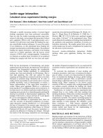
solved by NMR [20,21]. As illustrated in Figure 1, Vpr is
composed of three amphipathic α helices spanning resi-
dues (17–33), (38–50) and (54–77), surrounded by flex-
ible N- and C-terminal sequences [22]. Two loops
spanning residues (34–37) and (51–53) allow a mutual
orientation of these helices, conferring a globular confor-
mation to the protein and promoting the formation of a
hydrophobic core with numerous hydrophobic amino
acids scattered throughout Vpr. The difficulties encoun-
tered to solve the Vpr 3D structure might be explained by
its ability to oligomerize via the formation of leucine zip-
per like motifs [14,23-26].
NMR based structure of VprFigure 1
NMR based structure of Vpr. The NMR-based 3D- structure of Vpr (1–96) is characterised by three α helices in close
vicinity surrounded by flexible N and C termini [22]. Helices are presented in dark blue (17–33), green (38–50) and orange
(54–77). Mutated amino acids Q3R, L23A, ΔQ44, W54G, I60A, L67A, R77Q and R90K are represented in CPK mode. Notice-
ably, the NMR studies were carried out on the Vpr sequence of the HIV-1 pNL43 strain with a Leucine at the position 60
instead of an Isoleucine for the HIV-1
LAI
strain used here. Nevertheless, a predictive study on I60 Vpr showed that the third α
helix was not altered compared to L60 Vpr (data not shown).
Retrovirology 2008, 5:87 />Page 3 of 17
(page number not for citation purposes)
To further characterize the formation of Vpr oligomers
and their intracellular localization, we used eGFP and
mCherry Vpr fusion proteins and studied their interaction
by two photon fluorescence lifetime imaging microscopy
(FLIM) and fluorescence correlation spectroscopy (FCS).
We found that Vpr oligomerization relies on both the N-
and the C- termini and occurs at the nuclear envelope, in
the cytoplasm and in the nucleus. Mutations in the three
α helices elicited a large decrease in Vpr-Vpr interaction
while mutations in the loops or in the N- or C-termini had
little influence on its oligomerization. This study also
shows that Vpr oligomerization determines its subcellular
localization but not its proapoptotic activity. Finally,
molecular modeling of Vpr mutants has been performed
in an attempt to draw a possible correlation between Vpr
structure and activity.
Results
Confocal microscopy visualisation of eGFP or mCherry
fused to Vpr N and C termini
In order to monitor Vpr-Vpr interaction by FRET, eGFP or
mCherry proteins were fused to Vpr at their C- or N- ter-
mini. The eGFP and mCherry were used as a donor/accep-
tor pair for FRET for several reasons. Firstly, eGFP exhibits
a high quantum yield (0.8) and its time resolved fluores-
cence is characterized by a mono-exponential decay (2.5
ns) [27]. This single exponential decay strongly contrasts
with the complex decay of CFP [28], another fluorescent
protein commonly used as a donor for FRET, which makes
eGFP highly suitable for monitoring FRET due to the
decrease of its fluorescence lifetime. Secondly, mCherry
was used as the acceptor since its absorption spectrum
overlaps the fluorescence spectrum of eGFP, giving a large
Förster R
0
distance (where the transfer efficiency is 50%)
of about 54 Å [29]. Moreover, in contrast to the com-
monly used DsRed protein, mCherry is monomeric and
readily matures, which avoids the generation of several
proteins with different lifetimes [30]. Lastly, its spectro-
scopic properties are preserved in mCherry-tagged pro-
teins [31] and its use in association with eGFP to monitor
protein/protein interaction by FRET has been validated
[28,29,31].
Four labelled Vpr proteins were obtained by fusing eGFP
or mCherry to Vpr either to its N- or C-terminus. Since
both eGFP and mCherry are large with respect to Vpr, we
first checked whether the fusion affects the intracellular
localization of Vpr. To this end, we analyzed by confocal
microscopy at 24 h post transfection the expression of
both mCherry- (Figure 2, panels A2-3) and eGFP Vpr
fusions in HeLa cells (Figure 2, panels B 2-3). Both Vpr-
eGFP and Vpr-mCherry showed a nuclear rim staining
coincident with the nuclear envelope (NE) (Figure 2, pan-
els A2 and B2) in agreement with the localization of HA-
Vpr (additional file 1, [12]). This localization of Vpr at the
NE is not driven by the eGFP and mCherry proteins since
both fluorescent proteins were found to be spread all over
the cells when expressed in their free form (Figure 2 A1
and B1). Localization of HA-Vpr (additional file 1) or His-
Vpr [12] confirms that these proteins are predominantly
localized at the nuclear membrane and in the nucleus
with some cytoplasmic localization. Thus, the fusion of
either mCherry or eGFP to the C terminus of Vpr has a
limited effect on Vpr localization in the cell even though
the relative proportion of Vpr in the nucleus, at the
nuclear envelope or in the cytoplasm was modified
[10,12,13,24,32]. The distribution pattern of mCherry-
Vpr was close to that of Vpr-mCherry except that a larger
amount of protein diffused out in the cytoplasm, indicat-
ing a limited alteration of Vpr intracellular distribution by
the mCherry fused to the N-terminus of Vpr. In contrast,
eGFP-Vpr showed a diffuse distribution in both the cyto-
plasm and the nucleus (Figure 2, panel B 3) similar to the
nuclear staining of eYFP-Vpr [10,12]. At least, it should be
mentioned that Vpr distribution was not time dependent
since the same pattern of localization was monitored at 48
and 72 h (data not shown).
Co-localization of Vpr-eGFP and either mCherry-Vpr or
Vpr-mCherry was visualized by confocal microscopy. As a
control, Vpr-eGFP was first co-expressed with mCherry.
Localization of Vpr-eGFP at the nuclear rim (Figure 3,
panel A1) was similar to that in Figure 2 (panel B2), indi-
cating that the expression of mCherry did not affect the
intracellular distribution of Vpr-eGFP. When Vpr-eGFP
was co-expressed with Vpr-mCherry, both green and red
fluorescence emissions were localised at the rim of the
nucleus and to a lesser extent in the cytoplasm and in the
nucleus (Figure 3, panels B1-3). A full co-localization of
the two Vpr fusion proteins in the same cellular compart-
ments was further evidenced by the yellow color in Figure
3 (panel B3), that shows a nice superposition of the green
and red emissions of the two Vpr fusion proteins. Interest-
ingly, expression of Vpr-eGFP with mCherry-Vpr resulted
in a partial redistribution of Vpr-eGFP from the nuclear
rim toward the cytoplasm (compare Figure 3, panel C1
with Figure 2, panel B2). The overlap of their emissions all
over the cell confirmed their similar intracellular distribu-
tion (Figure 3, panel C3).
The re-localization of Vpr-eGFP mediated by mCherry-
Vpr in a human cell line suggests that the mCherry-Vpr
fusion protein interacts with Vpr-eGFP. However, due to
the limited resolution of optic microscopic methods (≈
200 nm), co-localization does not constitute an absolute
proof for direct protein interaction. Direct evidence for the
interaction between the eGFP and mCherry Vpr fusion
proteins and thus Vpr oligomerization, can be provided
by FRET between the two proteins as measured by FLIM.
Retrovirology 2008, 5:87 />Page 4 of 17
(page number not for citation purposes)
Investigating intracellular Vpr-Vpr interaction by FLIM
Due to its exquisite dependence on the inter-chromo-
phore distance, FRET between eGFP- and mCherry tagged
proteins will occur only if they are less than 10 nm apart
[33,34]. This implies that FRET will only be observed
when the tagged proteins directly interact with each other
[35,36]. In cells, the FRET efficiency can be directly meas-
ured by imaging with the FLIM technique the decrease of
the fluorescence lifetime of the donor at each pixel or
group of pixels. Indeed, in contrast to fluorescence inten-
sities, the fluorescence lifetimes are absolute parameters
that do not depend on the instrumentation or the local
concentration of the fluorescent molecules. Thus, changes
of the fluorescence lifetimes of the donor will provide a
direct evidence for a physical interaction between the
labelled proteins with high spatial and temporal resolu-
tion [37].
HeLa cells were transfected and FLIM measurements were
monitored at 24, 48 and 72 hours but since no time
dependant effect was monitored; only measurements at
24 h are presented. Experiments were performed first on
cells expressing eGFP or Vpr eGFP fusion protein as a con-
trol (Figure 4, panels A1-3) and next on cells co-expressing
Vpr-eGFP and mCherry fusion proteins (Figure 4, panels
B1-3 and C1-3). An arbitrary color scale, ranging from
blue to red, illustrates short to long lifetimes. The Vpr-
eGFP fluorescence was mainly localized at the nuclear
envelope and also in other cell compartments, where
FLIM measurements can be carried out. We focused on
three distinct regions, namely the nuclear rim, the cyto-
plasm and the nucleus (Table 1). For the cytoplasm and
the nuclear region, care was taken to exclude pixels with
contribution from the nuclear envelope. Moreover, due to
the thickness of the nuclear envelope, the pixels used to
calculate the lifetime values of the nuclear envelope
involved contributions from cytoplasmic and nuclear Vpr.
Nevertheless, due to the strong accumulation of Vpr at the
nuclear membrane, we assumed that the lifetimes mainly
reflected the behaviour of the Vpr fusion proteins at this
site (see Table 1). FLIM measurements were carried out
The lifetimes (2.4–2.5 ns) of Vpr eGFP fusion proteins
expressed alone (Figure 4, panels A2 and A3) or co-
expressed with mCherry (Figure 4, panels B1 and B2) were
identical to that of eGFP alone (Figure 4, panel A1) [27].
Subcellular localization of eGFP or mCherry tagged Vpr by confocal microscopyFigure 2
Subcellular localization of eGFP or mCherry tagged Vpr by confocal microscopy. HeLa cells were co-transfected
with 0.5 μg of each plasmid and 0.5 μg pcDNA3. Cells were observed by confocal microscopy 24 h post transfection. Each
panel shows the major phenotype. (A) mCherry images with excitation at 568 nm and emission at 580 to 700 nm. (B) eGFP
images with excitation at 488 nm and emission at 500 to 550 nm. Note the intracellular redistribution of eGFP and mCherry
upon fusion with Vpr.
Retrovirology 2008, 5:87 />Page 5 of 17
(page number not for citation purposes)
Visualization of the intracellular co-expression eGFP or mCherry tagged VprFigure 3
Visualization of the intracellular co-expression eGFP or mCherry tagged Vpr. Plasmid DNA (0.5 μg each) express-
ing the Vpr fusion proteins were cotransfected in HeLa cells. One day post transfection, images were recorded with an excita-
tion at 488 nm and emission at 500–550 nm to monitor eGFP expression, and with an excitation at 568 nm and emission at
580–700 nm to monitor mCherry expression, respectively. In the merge images, co-localization of the two proteins is indi-
cated in yellow. Each image is representative of the major phenotype. Note the accumulation of the Vpr fusion proteins at or
close to the nuclear envelope.
Table 1: Lifetime and FRET efficiency of eGFP- and eGFP-tagged Vpr in living cells
Nuclear envelope Cytoplasm Nucleus Whole Cell
E(%) τ(ns) E(%) τ(ns) E(%) τ(ns) E(%) τ(ns)
eGFP - - - 2.50 (± 0.01) - 2.50 (± 0.01) - 2.50 (± 0.01)
Vpr-eGFP - 2.36 (± 0.01) - 2.40 (± 0.01) - 2.41 (± 0.01) - 2.39 (± 0.01)
eGFP-Vpr - 2.47 (± 0.01) - 2.46 (± 0.01) - 2.47 (± 0.01) - 2.47 (± 0.01)
Vpr-eGFP+mCherry - 2.41 (± 0.02) - 2.42 (± 0.01) - 2.42 (± 0.01) - 2.42 (± 0.01)
Vpr-eGFP+Vpr-mCherry 27 1.72 (± 0.02) 23 1.86 (± 0.03) 19 1.95 (± 0.03) 23 1.85 (± 0.03)
Vpr-eGFP+mCherry-Vpr 17 1.95 (± 0.02) 14 2.06 (± 0.02) 13 2.09 (± 0.02) 15 2.02 (± 0.03)
eGFP-Vpr+mCherry - 2.43 (± 0.01) - 2.43 (± 0.01) - 2.43 (± 0.02) - 2.43 (± 0.01)
eGFP-Vpr+Vpr_mCherry 13 2.14 (± 0.03) 9 2.25 (± 0.03) 6 2.32 (± 0.02) 9 2.25 (± 0.03)
eGFP-Vpr+mCherry-Vpr 13 2.14 (± 0.03) 7 2.28 (± 0.03) 6 2.31 (± 0.02) 8 2.28 (± 0.03)
The fluorescence lifetimes (τ) of eGFP alone or linked to the Vpr C-terminus are the average values (+/- standard deviation) for 10 to 35 cells. For
each cell, measurements were performed at the nuclear envelope, in the nucleus and in the cytoplasm. The FRET efficiency (E) is related to the
distance between the two chromophores and is calculated from the lifetime ratio with and without the acceptor using equation (2). The whole cell
E and τ values represent the average values calculated over the entire cell.
Retrovirology 2008, 5:87 />Page 6 of 17
(page number not for citation purposes)
These results show that the eGFP fluorescence was not
altered when fused to Vpr and that no short range interac-
tion occurred between the Vpr eGFP fusion protein and
free mCherry.
In contrast, a strong decrease in the average fluorescence
lifetime of Vpr-eGFP was observed all over the cell when
it was co-expressed with Vpr-mCherry (Figure 4, panel
C1), thus indicating a direct physical interaction between
the two Vpr chimeric proteins. The strongest decrease was
observed at the nuclear rim where the fluorescence life-
time dropped down to 1.72 ns, corresponding to a trans-
fer efficiency of 27% (Table 1). Vpr-Vpr interaction also
occurred in the cytoplasm and the nucleus, as shown by
the 19–23% energy transfer measured at these sites.
As reported in Table 1 and Figure 4, the energy transfer
efficiency is dependent upon the couple of the Vpr fusion
proteins. Indeed, the transfer efficiency dropped by a fac-
tor of about 1.5 when Vpr-eGFP was co-expressed with
mCherry-Vpr (15%; Figure 4, panel C2) and by a factor of
about 2.5 when eGFP-Vpr was co-expressed with either
Direct Vpr-Vpr interaction in HeLa cells visualized by FLIMFigure 4
Direct Vpr-Vpr interaction in HeLa cells visualized by FLIM. Cells were transfected with the DNA construct encoding
eGFP or eGFP-Vpr alone or in combination with mCherry-Vpr. In the FLIM images, the lifetimes are represented using an arbi-
trary color scale ranging from blue to red for short and long lifetimes in nanoseconds (right bottom), respectively. The Vpr-
eGFP or eGFP-Vpr with short lifetime fluorescence symbolized by the blue color were mainly localized at the nuclear envelope
and also in other cell compartments when co transfected with mCherry tagged Vpr. Panels A1 to A3 show the lifetime images
of cells expressing eGFP or eGFP-tagged Vpr alone. Panels B1 and B2 represent cells coexpressing eGFP-tagged Vpr and
mCherry; Panels B3 and C1-C3 show the lifetime images of cells coexpressing eGFP-tagged Vpr and mCherry-tagged Vpr.
Note the accumulation of Vpr fusion proteins at or near the nuclear envelope.
Retrovirology 2008, 5:87 />Page 7 of 17
(page number not for citation purposes)
Vpr-mCherry (9%; Figure 4, panel C3) or mCherry-Vpr
(8%; Figure 4, panel B3). Although Vpr-Vpr interaction
was clearly taking place in all cases, a comparison of the
energy transfer values suggests that fusion of a fluorescent
protein at the Vpr N-terminus is detrimental to Vpr-Vpr
interaction.
Taken together, these data indicate that Vpr-Vpr interac-
tions occur in the cytoplasm, in the nucleus and at the
nuclear rim and are best visualized when the fluorescent
proteins are linked to the C-terminus of Vpr.
Mapping Vpr-Vpr interaction
In an attempt to map the Vpr domains involved in Vpr-
Vpr interaction, site directed mutagenesis was carried out
on Vpr-eGFP and Vpr-mCherry constructs based on struc-
tural criteria [22] (Figure 1). Several amino acids (L23,
Q44, I60 and L67) located in the three α-helices were
changed to F (L23F) or A (I60A, L67A) or deleted (ΔQ44).
Residues I60 and L67 are involved in Vpr dimerisation
through a leucine zipper type motif [21,26]. The L23F and
ΔQ44 Vpr mutants retained their ability to translocate to
the nucleus but were poorly incorporated into virions
[13,24,38].
In parallel, amino acids Q3, and R90 located in the N- and
C-flexible termini and residues W54 and R77 located at
the extremities of the third helix, were changed to R, K, G,
Q respectively (Figure. 1). The Q3R and R77Q mutants
were shown to be impaired in their proapoptotic activity
and to be associated with long-term non-progressive HIV-
1 infection [39,40] while the R90K mutant failed to cause
the G2/M cell arrest [41]. Moreover, the W54G mutant
was shown to be critical for the interaction with cellular
UNG (Uracil DNA glycosilase) and its virion incorpora-
tion [41].
Mutated proteins were expressed in HeLa cells. Immuno-
detection by Western Blots revealed that none of the point
mutations impeded expression of the Vpr fusion proteins
(data not shown). The fluorescence lifetime images were
recorded and compared with those of the two wild type
Vpr fusion proteins. Figure 5 shows the lifetime images of
the Vpr-eGFP mutants expressed in the absence (Column
A) and in the presence of the corresponding Vpr-mCherry
mutant (Column B). The mean values obtained for the
entire cell are reported on the right of the figure. Among
the eight mutants, four of them, namely Q3R, W54G,
R77Q and R90K, showed a staining pattern similar to that
of the wild type fusion proteins with an accumulation at
the nuclear rim (compare with Figure 4, panel A2). Oli-
gomers of these mutant proteins were found in the cyto-
plasm, the nucleus and at the nuclear envelope. The
transfer efficiency in the whole cell for these mutants was
respectively 19%, 16%, 22% and 18%, similar to the value
obtained for the wild type fusion protein (23%). Thus, the
Q3, W54, R77 and R90 residues located outside the α-hel-
ices are probably not critical for the intracellular localiza-
tion and oligomerization of Vpr.
On the contrary, the Vpr L23F, ΔQ44, I60A and L67A
mutants have lost their ability to accumulate at the
nuclear rim. Their intracellular distribution resembled
that of eGFP-Vpr, which was evenly distributed in the cell
with some accumulation in the nucleus. Interestingly, this
different staining pattern of L23F-Vpr-eGFP and ΔQ44-
Vpr-eGFP compared to the wild type was also found with
L23F-Vpr and ΔQ44-Vpr using immunostaining method-
ology, indicating that eGFP does not interfere with Vpr
distribution [13,24].
A very low transfer efficiency was found for L23F, ΔQ44
and L67A, indicating that these Vpr mutants failed to oli-
gomerize even at or near the nuclear envelope. Thus, the
three residues located respectively in the first, second and
third helix seemed to be directly involved in Vpr-Vpr inter-
action and its cellular localization. Furthermore, a small
but significant FRET was observed between I60A-Vpr-
eGFP and its red counterpart (6% in the whole cell; 7%
inside the nucleus- figure 5C) even though the I60A-Vpr-
eGFP mutant lost its ability to accumulate at the nuclear
envelope. Thus, a minor population of Vpr-eGFP/Vpr-
mCherry complex was still observed despite this muta-
tion. In line with this result, transfection of I60A-Vpr-
eGFP with wild type Vpr-mCherry restored up to 100% of
the nuclear rim staining of the I60AVpr-eGFP mutant
(data not shown). Such an important nuclear envelope
localization rescue was not observed with the L23F, ΔQ44
and L67A Vpr-eGFP mutants.
Thus, the mapping of Vpr-Vpr interaction reveals that
amino acids located in the hydrophobic central core are
directly involved in Vpr oligomerization while residues in
non-structured domains are dispensable. These results
also indicate that the localization of Vpr at the rim of the
nucleus probably relies on Vpr-Vpr interaction.
Vpr oligomerization monitored by FCS
To further characterize Vpr-Vpr interaction in cells, Fluo-
rescence Correlation Spectroscopy (FCS) was performed.
This technique characterizes the translational dynamics of
fluorescent molecules (or molecular complexes) in any
liquid environment. By using the intensity fluctuations of
fluorescent species within a femtoliter volume (defined by
the laser excitation), several physical parameters – diffu-
sion time, local concentration, molecular brightness,
related to the hydrodynamic and photophysical proper-
ties of these species – can be monitored [42].
Retrovirology 2008, 5:87 />Page 8 of 17
(page number not for citation purposes)
Mapping of Vpr-Vpr interaction by FLIMFigure 5
Mapping of Vpr-Vpr interaction by FLIM. HeLa cells were co transfected with mutated Vpr-eGFP and its own counter-
part fused to mCherry. FLIM was carried out 24 h posttransfection (see methods). Column A corresponds to the FLIM images
of the Vpr-eGFP mutants alone, column B to the FLIM images of cells co expressing the mutant Vpr-eGFP and the mutant Vpr-
mCherry. FRET efficiency (E) expressed in percentage represents the average value calculated over the entire cell (column C).
The color scale used to create theses images is the same than the one used for figure 4. Note the drastic reduction of Vpr-Vpr
interaction and the loss of Vpr nuclear envelope accumulation upon mutating residues L23, Q44, I60 and L67 (column B and
C).
Retrovirology 2008, 5:87 />Page 9 of 17
(page number not for citation purposes)
Due to the strong eGFP photobleaching, no FCS measure-
ment was possible at the nuclear rim. FCS measurements
were thus carried out in the cytoplasm and in the nucleus.
Figure 6 reports the histograms of τ
A
(diffusion time), α
(anomalous diffusion coefficient) and the count rate per
molecule τ
A
represents the average time needed to cross
the focal volume, which depends on the size of the mole-
cule or the molecular complex. The α value corresponds
to the anomalous diffusion coefficient that accounts for
the concentration, size, mobility and reactivity of the
obstacles encountered by the diffusing species. Anoma-
lous diffusion was preferred over the two-component dif-
fusion since it takes into account the molecular crowding
in the intracellular environment [43]. Moreover, the FCS
parameters were obtained from sequential short-time
measurements at numerous cell locations to avoid prob-
lems due to the non steady-state conditions in cells [42].
Using this protocol, the anomalous diffusion time of
eGFP (Figure 6B) displays a narrow distribution centred
around 0.4 ms [42], compared to 0.2 ms for purified eGFP
in aqueous solution (data not shown). In addition, the α
value peaks around 1 (Figure. 6A), suggesting that eGFP
freely diffuses as monomers in the cell in agreement with
the monomeric structure found by RX [44,45]. A com-
pletely different behaviour was observed for Vpr-eGFP.
Firstly, the distribution of the apparent diffusion time is
shifted to 4 ms (Figure 6E) with dispersion larger than
that obtained with eGFP. Since τ
A
roughly varies as the
cubic root of the molecular mass of the diffusing species,
the tenfold increase of τ
A
implies a thousand fold
increased in the molecular mass, unambiguously showing
that Vpr fusion proteins form large complexes in cells.
Moreover, the anomalous coefficient of Vpr-eGFP
presents a distribution centred around 0.75 showing that
such complexes do not freely diffuse in the cell but inter-
act with cellular components (Figure 6D). To further char-
acterize these complexes, their molecular brightness (i.e.
the number of photons emitted by a particle per second
for a given excitation intensity) was compared with that of
eGFP (Figure. 6C and 6F). The histogram of eGFP displays
a narrow distribution centred around 1 kHz/particle sim-
ilar to purified eGFP in aqueous solution showing that the
photophysical properties of eGFP are not modified by the
cellular environment. Since eGFP does not form oligom-
ers, this value can be taken as a reference for eGFP mono-
mers [44,45]. In contrast, the count rate histogram for
Vpr-eGFP shows a broad distribution with a major popu-
lation centred around 2–3 kHz/particle and a minor pop-
ulation with a rather large distribution of brightness
(Figure. 6F). This confirms that Vpr forms oligomers as
observed by FLIM and suggests that Vpr-eGFP self associ-
ates in the cytoplasm and the nucleus notably in the form
of dimers and trimers, assuming that the eGFP fluores-
cence is not modified by Vpr oligomerization. These small
oligomers do not explain the aforementioned 10
3
-fold
difference between the molar masses of eGFP and Vpr-
eGFP complexes, thus indicating that Vpr oligomers prob-
ably interact with cellular proteins [46].
FLIM analyses showed that the ΔQ44 mutant of Vpr-eGFP
did not interact with Vpr-mCherry (Figure. 5 panel B3).
This prompted us to perform FCS experiments with the
ΔQ44 Vpr-eGFP to confirm its inability to oligomerize. As
shown in Figure 6I, the count rate of ΔQ44 Vpr-eGFP is
centred around 1.2 kHz, close to the value obtained for
eGFP (Figure. 6C), which confirms that the ΔQ44 Vpr-
eGFP does not form oligomers. Interestingly, the diffusion
coefficient τ
A
for the Vpr ΔQ44 mutant is about 2 ms (Fig-
ure. 6H), a value in between that for eGFP (0.4 ms) and
that for Vpr-eGFP (4 ms). Moreover, the distribution of
the anomalous coefficient was similar to that for Vpr-
eGFP with a peak value around 0.75. The five-fold
increase of τ
A
with respect to free eGFP, which corre-
sponds to a 100-fold increase in the molar mass, indicates
that this Vpr mutant probably interacts with host proteins
in a monomeric form.
Vpr oligomerization is not necessary for the induction of
cell apoptosis
Vpr can induce apoptosis of infected cells and probably of
bystander cells [5,6]. In order to evaluate the role of Vpr
oligomerization on its pro-apoptotic activity, FACS analy-
ses were carried out. To this end, annexin V and propid-
ium iodide staining of HeLa cells expressing eGFP, Vpr-
eGFP or Vpr-eGFP mutants were performed 72 hours after
transfection (see methods). Results show that 6% of mock
transfected cells (data not shown) and 16% of cells
expressing eGFP were apoptotic (Figure. 7). The percent-
ages of apoptotic cells expressing either Vpr-eGFP or one
mutant varied from 45 to 70% as compared to the 43%
obtained with wt Vpr (data not shown) [12,47]. Thus, no
significant reduction of apoptosis was monitored for the
Vpr-eGFP mutants examined here. As a consequence there
is no clear correlation between the intracellular oligomer-
ization of Vpr and its pro-apoptotic properties.
Discussion
We report here a study on Vpr oligomerization in the cel-
lular context by confocal microscopy, two photon FCS
and FLIM. Using eGFP or mCherry tagged at their N or C
terminus by Vpr, we confirmed that Vpr oligomerization
occurs in human cells [19], notably at the nuclear enve-
lope (Figure. 3 and 4) in line with the preferential locali-
zation of the wild type Vpr [13,24,32,48]. Moreover, FCS
experiments also show that Vpr could form two popula-
tions of oligomers in the cytoplasm and in the nucleus,
one containing mainly dimers and/or trimers and a sec-
ond composed by a large number of molecules (Figure.
6). This heterogeneity of Vpr oligomers is in agreement
Retrovirology 2008, 5:87 />Page 10 of 17
(page number not for citation purposes)
Distribution histograms of anomalous diffusion coefficients, diffusion times and count rates/species of eGFP, Vpr-eGFP and ΔQ44 Vpr-eGFPFigure 6
Distribution histograms of anomalous diffusion coefficients, diffusion times and count rates/species of eGFP,
Vpr-eGFP and ΔQ44 Vpr-eGFP. The anomalous diffusion coefficient (coefficient that accounts for the obstacles encoun-
tered by the diffusing species), diffusion times (average time needed to cross the focal volume) and brightness (count rates/spe-
cies) determined by FCS are expressed as a function of the number of occurrences. A-C correspond to eGFP; D-F correspond
to Vpr-eGFP; G-I correspond to ΔQ44 Vpr-eGFP.
Retrovirology 2008, 5:87 />Page 11 of 17
(page number not for citation purposes)
with biochemical data showing that the stoichiometry of
Vpr oligomers could vary from two to six [14,23,49].
Moreover, FCS analyses of Vpr-eGFP showed that Vpr
does not freely diffuse in the cell and thus most probably
forms oligomers that interact with cellular proteins
[10,11,50-53] and membranes [54,55]. These Vpr oligom-
ers explain the energy transfer observed between eGFP-
and mCherry-tagged Vpr proteins by FLIM. The maximum
energy transfer was obtained when Vpr was linked to the
N terminus of the two reporter proteins (Table 1), which
further highlights the role of the N-terminal domain in
Vpr oligomerization [14,24].
The 3D structure of Vpr is characterized by three amphip-
athic α-helices with relative orientations displaying two
accessible hydrophobic domains and a hydrophilic one
(Figure 1). To map the Vpr-Vpr interactions, we studied
Vpr mutants harbouring a single mutation in the helical
or flanking regions of the protein [22]. In a first step, we
characterized the L23F, I60A and L67A mutations of the
hydrophobic central core. Predictive structural studies
performed on the mutated protein revealed that these α
helices were not significantly altered by these mutations
and as a consequence the global 3D structure of the
mutant proteins closely resembles that of the wild type
Vpr (data not shown).
The L23F and L67A Vpr mutants were distributed
throughout the cell, indicating that these residues are crit-
ical for addressing Vpr at the nuclear envelope. A similar
intracellular distribution of non-fluorescently labelled
L23F-Vpr has been previously found by immunofluores-
cence [13,24], indicating that eGFP did not interfere with
the cellular distribution of such Vpr mutants. As the α-
helix (17–33) containing the L23 residue is predicted to
adopt a coiled-coil conformation, this mutation might
well cause an interruption of the leucine stretch formed by
residues L20, L22, L23 and L26 located on the same side
of the helix (additional file 2). Thus, this hydrophobic
platform formed by the N terminal alpha helix (17–33) is
a recognition motif for Vpr-Vpr oligomerization in the cel-
lular context.
Mutating residue I60 is less detrimental than mutating
residues L23 and L67 for addressing Vpr at the nuclear
envelope (Figure 5) since a residual energy transfer was
Pro-apoptotic properties of the Vpr-eGFP mutantsFigure 7
Pro-apoptotic properties of the Vpr-eGFP mutants. Cells expressing either the wild type Vpr-eGFP or mutant Vpr-
eGFP were selected by fluorescence cytometry, using the eGFP fluorescence. The percentage of cells undergoing apoptosis
was assessed by the number of cells labeled with cells with Cy5 alone, or with both Cy5 and PI. Statistical analysis was achieved
using the multi-factorial ANOVA test and the Dunnett analysis. Three independent measurements were performed for each
assay.
Retrovirology 2008, 5:87 />Page 12 of 17
(page number not for citation purposes)
observed. Moreover, the peri-nuclear localization of the
I60A-Vpr mutant was recovered upon co-expression with
Vpr-mCherry (data not shown). Residues I60 and L67 are
involved in a hydrophobic stretch constituted by residues
L61, I63, L64, L68, I70 and I74 in the helix (54–77). Since
I60 is the first residue of this stretch (additional file 2),
changing I to Ala should not affect drastically this hydro-
phobic motif, and thus Vpr oligomerization and nuclear
localization. On the opposite, L67 is located in the center
of this hydrophobic motif and changing it to Ala should
cause a significant disorder that likely perturbs Vpr oli-
gomerization and nuclear localization. Mutation of resi-
due 67 and the loss of Vpr-Vpr binding was reported and
explained by the presence of the negative charge of the
glutamic residue placed at this position [19]. Neverthe-
less, since alanine differs only moderately from leucine it
appears that the length and the hydrophobicity of the leu-
cine side chain is critical for maintaining the leucine zip-
per like structure and the hydrophobic core of Vpr.
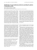
The ΔQ44 mutation drastically impaired the oligomeriza-
tion and localization of Vpr at the nuclear envelope, fur-
ther suggesting a direct correlation between these two
phenomena. Molecular modeling of this mutant shows a
partial unfolding of the second helix, from residues W38
to L42 (Figure 8). These structural modifications reorient
the residue side-chains involved in the hydrophobic inter-
actions within helix (54–77). Thus, the hydrophobic core
formed by hydrophobic stretches of the second and third
helices is disrupted and reorganized, leading to a strong
modification of the overall Vpr structure. This altered
structure might explain why the mutant Vpr has lost its
ability to form oligomers and its localization at the
nuclear envelope.
In contrast to the aforementioned mutants, a wild type
Vpr docking at the nuclear rim was observed for muta-
tions in the loops (W54G, R77Q) and the flexible N- and
C- terminal regions (Q3R, R90K), indicating that these
residues are dispensable for Vpr cellular localization. In
addition, molecular modeling indicates that these muta-
tions should not modify the overall structure of Vpr (data
not shown).
Targeting of Vpr at the nuclear envelope most probably
relies on its interaction with components of the nuclear
pore complex (NPC) [8,11] and especially with the nucle-
oporin hCG1 [10]. The Vpr/hCG1 interaction is mediated
by the hydrophobic core of Vpr independently of its N-
and C-termini [10]. For instance, the L23F mutation that
alters the Vpr-hCG1 complex was recently shown to cause
a lack of Vpr accumulation at the nuclear rim [38]. Thus,
the hydrophobic residues of Vpr core are most probably
required both for Vpr-Vpr and Vpr-hCG1 interactions. It
can thus be speculated that Vpr-hCG1 recognition
depends on Vpr oligomerization.
The role of the nuclear localization and oligomerization
of Vpr on the induction of apoptosis was studied. In fact,
Vpr-eGFP is still able to induce apoptosis, indicating that
eGFP does not impair the Vpr apoptotic activity [12]. Sim-
ilar levels of apoptosis were found for all Vpr mutants. The
apoptotic activity of the Q3R and R77Q Vpr mutants are
in variance with published reports [39,40] but could be
explained by a possible eGFP-mediated gain of function
[12,47]. The apoptotic activity of all Vpr mutants shows
that this activity is not correlated with Vpr oligomeriza-
tion. Meanwhile, Bolton and Lenardo have recently
showed that Vpr oligomerization of Vpr was dispensable
for mediating G2/M arrest [19].
Conclusion
Taken together, our data show that i) Vpr oligomerizes in
the nucleus and the cytoplasm in HeLa cells, ii) Vpr oli-
gomerization is required for Vpr localization at the
nuclear envelope, iii) the structural determinants for Vpr
oligomerization are located in the hydrophobic core
formed by the three α helices and iv) nuclear localization
and oligomerization are neither required nor sufficient for
apoptosis as for G2/M cell cycle arrest [19].
Methods
Plasmid DNA construction
Construction of eGFP-Vpr and Vpr-eGFP was previously
described [32]. To construct mCherry-Vpr, we PCR ampli-
fied the full length coding sequence of Vpr (from HIV-
1
LAI
) using the mammalian HA-tagged Vpr expressing vec-
tor [56]. The reverse primer
5'
GCCCCGCTCGAGCT
AGGATCTACTGGC
3'
used in the PCR amplification was
designed to include an XhoI restriction site (underlined).
The PCR DNA product was digested by EcoRV and XhoI
and cloned into a mCherry expression vector under the
control of the CMV promoter.
The Vpr-mCherry recombinant was constructed by a two
step protocol. Firstly, the full length coding sequence of
Vpr was amplified by PCR from the HA-tagged Vpr expres-
sion vector described above. The forward
5'
CCCAAGCTTG
ATCTACCATGGAACAAGCCCCAGAAG
3'
and reverse
5'
CGCGGATCCCCGGATCTACTGGCTCCATTTC
3'
prim-
ers were designed to include the restriction sites HindIII
and BamHI (underlined). The complementary sequence
corresponding to the Kozak consensus for optimal trans-
lation initiation is shown in bold. The PCR fragment was
digested and cloned into pDsRED-Monomer-N1 (Clon-
tech) to obtain Vpr-DsRED. Secondly, the DsRED coding
sequence was cut out with BamHI and NotI and replaced
by the mCherry coding sequence. The latter was amplified
by PCR from a mCherry expressing plasmid using the fol-
Retrovirology 2008, 5:87 />Page 13 of 17
(page number not for citation purposes)
Comparison of the wild type and ΔQ44 Vpr mutant structuresFigure 8
Comparison of the wild type and ΔQ44 Vpr mutant structures. Stereoview of the three dimensional structure of the
wild type Vpr determined by NMR (A) and theoretical model for the Vpr ΔQ44 mutant (B). Helices (17–33), (38–50) and (54–
77) are represented as ribbon and colored in blue, pink and green, respectively and loops (34–37) and (51–53) are colored in
yellow. For clarity, the two disordered extremities of the molecule have not been represented. Residues showing long range
correlations on NOESY NMR experiments have been displayed in the stick representation and colored according to their
hydrophobicity. Only their side chain atoms have been represented. The network of hydrophobic residues can be observed at
the interface of the three α-helices. Note the impact of the ΔQ44 deletion (B) on the partial unfolding of the second helix and
the rearrangement of the hydrophobic residues at the interface.
Retrovirology 2008, 5:87 />Page 14 of 17
(page number not for citation purposes)
lowing primers:
5'
CGCGGATCCAGGAGGCGGTGGGATG
GTGAGCAAGGGCGAG
3'
and
5'
ATA GTTTAGCGGCCGC
TTACTTGTACAGCTCG TCCATGCC
3'
.
Deletion or substitution mutants were carried out by PCR
based site-directed mutagenesis on the Vpr-eGFP and Vpr-
mCherry expressing vector using a protocol from Strata-
gene.
Cell culture and DNA transfection
HeLa cells (10
5
) were cultured on 35 mm glass coverslips
(μ-Dish IBIDI, Biovalley, France) in Dulbecco's modified
eagle medium supplemented with 10% fetal calf serum
(Invitrogen Corporation, Cergy Pontoise, France) at 37°C
in a 5% CO
2
atmosphere. Transfection of HeLa cells with
0.5 μg of each plasmid was achieved with FuGENE™ 6
transfection agent (Roche) or jetPEI™ (PolyPlus transfec-
tion, Illkirch, France) according to supplier's recommen-
dations. To keep a constant amount of transfected DNA,
each transfection assay was supplemented with the neces-
sary amount of pcDNA3 (Invitrogen Corporation, Cergy
Pontoise, France) up to 1 μg of total DNA.
Immunodetection of Vpr and Vpr derivatives by Western
blotting
HeLa cells (2 × 10
5
), transfected with 3 μg of plasmids
expressing either eGFP, Vpr-eGFP or Vpr-eGFP mutants,
were treated with trypsin and resuspended in ice cold lysis
buffer (1% Triton X-100, 100 mM NaF, 10 mM NaPPi, 1
mM NA
3
VO
4
in PBS supplemented with a complete anti-
protease cocktail from Roche, Meylan, France). After son-
ication and centrifugation, total protein concentrations
were assessed by a Bradford assay (Bio-Rad). 25 μg of total
proteins were added into 10 mM DTT containing loading
buffer (Laemmli, Bio-Rad), heat denaturated and electro-
phoresed on 12% SDS-PAGE gel. Subsequently, proteins
were transferred onto a polyvinylidene difluoride (PVDF)
membrane (Amersham, Orsay, France) and blots were
probed with an anti-GFP antibody (Clontech) followed
by horseradish peroxidase-conjugated anti-mouse anti-
body. Visualization of proteins was carried out using the
chemiluminescent ECL system (Amersham).
Flow cytometry
Induction of cell apoptosis by Vpr was monitored using
Annexin V and propidium iodide (PI) staining. Briefly, 2
× 10
5
HeLa cells were transfected with plasmids encoding
either eGFP, Vpr-eGFP or Vpr-eGFP mutants. Seventy-two
hours posttransfection, the cells were detached, washed in
ice cold PBS and resuspended in binding buffer (10 mM
Hepes, 140 mM NaCl, 2.5 mM CaCl
2
, pH 7.4). After addi-
tion of 5 μl of Annexin V-Biotin, 10 μl of PI (50 μg/ml)
and 0.5 μg of streptavidin-Cy5 diluted in 100 μl of bind-
ing buffer, the cells were incubated in the dark for 15 min-
utes. The volume of each tube was brought up to 500 μl
with 1× binding buffer. The cells were analyzed by flow
cytometry on a FACS Calibur (Becton Dickinson) within
a one hour period. In the eGFP positive cell population,
the percentage of apoptotic cells was determined from the
number of fluorescently labeled cells with Cy5 alone, or
with both Cy5 and PI.
Confocal Microscopy
Fluorescence confocal images of Vpr tagged proteins in
living cells were taken 24, 48 and 72 h posttransfection
using a confocal microscope (SPC UV1 AOBS, Leica)
equipped with a HCX PL APO CS 63× oil immersion
objective and an Ar/Kr laser. The eGFP images were
obtained by scanning the cells with a 488 nm laser line
and filtering the emission with a 500 to 550 nm band-
pass. For the mCherry images, a 568 nm laser line was
used in combination with a 580 to 700 nm band-pass fil-
ter.
Immunofluorescence study
HeLa cells were transfected with 0.5 μg of HA-tagged Vpr
expressing vector [56] in Labtek (Nunc, Fisher Scientific
Bioblock, France). At 24 h, the cells were washed in PBS at
4°C, fixed with paraformaldehyde/PBS (3.5%, w/v),
washed again with PBS and permeabilized with 0.2% tri-
ton/PBS. After drying, the cells were blocked for 30 min
with BSA-PBS 4% and then incubated with anti HA (1/
1000) (Invitrogen Corporation, Cergy Pontoise, France)
overnight at 4°C. The cells were washed with PBS and
incubated with FITC anti-rabbit at 1/200 (Invitrogen Cor-
poration, Cergy Pontoise, France) for 60 min at room
temperature. After washing, cells were analysed by confo-
cal microscopy (Bio-Rad 1024, Kr/Ar laser 488/568).
Fluorescent Correlation Spectroscopy (FCS)
FCS measurements were performed on a home-build two-
photon system set-up based on an Olympus IX70 inverted
microscope with an Olympus 60× 1.2NA water immer-
sion objective as previously described [57,58]. Two-pho-
ton excitation at 900 nm was provided by a mode-locked
titanium-saphire laser (Tsunami, Spectra Physics). The
normalized autocorrelation function was calculated on-
line with a hardware correlator (ALV5000, ALV GmbH,
Germany). Due to the inherent heterogeneity of the cellu-
lar medium, the FCS data were interpreted in terms of
anomalous diffusion. Therefore curves were fitted accord-
ing to:
where N is the average number of fluorescent species in
the focal volume, τ the lag time, τ
A
the average residence
G
1
N
1
A
1
A
1
S
2
11/2
τ
τ
τ
τ
τ
αα
()
=−
⎛
⎝
⎜
⎞
⎠
⎟
⎛
⎝
⎜
⎜
⎞
⎠
⎟
⎟
⋅+
⎛
⎝
⎜
⎞
⎠
⎟
⋅
⎛
⎝
⎜
⎜
⎞
⎠
⎟
⎟
−−
(1)
Retrovirology 2008, 5:87 />Page 15 of 17
(page number not for citation purposes)
time in the focal volume, α the anomalous diffusion coef-
ficient and S a structural parameter defined as the ratio
between the axial and lateral radii of the beam waist. The
molecular brightness (η) of the fluorescent species diffus-
ing through the excitation volume is obtained by dividing
the average fluorescence intensity <F> by N. In free lateral
diffusion (α = 1), the mean-square displacement of the
diffusing species is proportional to time (<r
2
>~t). This is
no more valid for anomalous diffusion (α < 1), that takes
place in systems containing obstacles. In that case, the
mean-square displacement is described by a power law
(<r
2
>~t
α
) with a coefficient α depending on the concen-
tration, size, mobility and reactivity of the obstacles.
Moreover, in living cells, there is no real steady-state for
the fluorescence intensity fluctuations. For this reason,
FCS measurements were sequentially repeated, typically
40 × 5 s. Each FCS curve is then fitted independently. A
Labview program was written to automatically process the
data. The results are represented by histograms of the fit-
ting parameters.
Fluorescence Lifetime Imaging Microscopy (FLIM)
Time-correlated single-photon counting FLIM was per-
formed using an in house constructed multi-photon laser
scanning microscope sharing the same core as the system
described for FCS measurements. For FLIM, the laser
power was adjusted to give count rates with peaks up to as
few as 10
6
photons.s
-1
, so that the pile-up effect can be
neglected. Imaging was carried out with a laser scanning
system using two fast galvo mirrors (Model 6210, Cam-
bridge technology), operating in the descanned fluores-
cence collection mode.
Photons were collected using a set of two filters: a two-
photon short pass filter with a cut-off wavelength of 680
nm (F75-680, AHF, Germany), and a band-pass filter of
520 ± 17 nm (F37-520, AHF, Germany). The fluorescence
was directed to a fiber coupled APD (SPCM-AQR-14-FC,
Perkin Elmer), which was connected to a time-correlated
single photon counting (TCSPC) module (SPC830,
Becker & Hickl, Germany), which operates in the reversed
start-stop mode.
Typically, the samples were scanned continuously for
about 30s to achieve appropriate photon statistics to ana-
lyse the fluorescence decays. Data were analysed using a
commercial software package (SPCImage V2.8, Becker &
Hickl, Germany), which uses an iterative reconvolution
method to recover the lifetimes from the fluorescence
decays.
In Fluorescence Resonant Energy Transfer (FRET) experi-
ments, when co-expressing donor and acceptor proteins,
the FRET efficiency reflecting the distance between the two
chromophores was calculated according to:
where R
0
is the Förster radius, R the distance between
donor and acceptor, τ
DA
is the lifetime of the donor in the
presence of the acceptor, and τ
D
is the lifetime of the
donor in the absence of the acceptor.
Molecular modeling of Vpr mutants
The impacts of the L23F mutation in the first α-helix [17-
33], the ΔQ44 deletion in the second helix [38-50] and
the I60A and L67A mutations in the third α-helix [54–77]
on the 3D structure of Vpr have been investigated by in sil-
ico procedure. Calculations were performed on a SGI
Octane work station with the Discover/NMRchitect soft-
ware (Accelrys, Inc. San Diego, CA, USA). Each mutation
has been introduced in the wild type Vpr structure and
each of the four resulting structures has been submitted to
a 500 steps of steepest descent followed by a 5000 steps of
conjugate gradient minimization until a maximum gradi-
ent value of 0.01 kcal/mol/Å was reached. Calculations
were performed on a SGI Octane station with the Dis-
cover/NMRchitect software package from Accelrys. Each
generated mutant structure was analyzed by comparison
with the wild type NMR structure using the InsightII pro-
gram visualization. No NMR distance or angle restraints
were used during minimization.
Abbreviations
FRET: Fluorescence Resonance Energy Transfer; FCS: Fluo-
rescent Correlation Spectroscopy; FLIM: Fluorescence
Lifetime Imaging Microscopy; WB: Western Blot; FACS:
Fluorescence-activated cell sorting
Competing interests
The authors declare that they have no competing interests.
Authors' contributions
JVF did all the experiments and analysis of the data, PD,
JPC and ES set up the platform for FCS and FLIM, CC pro-
duced eGFP for in vitro controls, DM gave plasmid and
expertise for cellular studies, SB and NM performed
molecular modelling, JLD and YM made substantial con-
tribution for data interpretation and manuscript writing
and HR designed and monitored the study. All the authors
have read and approved the manuscript.
E
R
0
6
R
0
6
R
6
1
DA
D
=
+
=−
τ
τ
(2)
Retrovirology 2008, 5:87 />Page 16 of 17
(page number not for citation purposes)
Additional material
Acknowledgements
Thanks are due to S. Benichou (ICGM, Paris) for providing Vpr eGFP and
to M. Ruff, J Barths (IGBMC) and J.C. Paillart (IBMC) for helpful discussions.
J.F. is granted by a fellowship of the Ministère de la Culture, de l'Enseigne-
ment supérieur et de la Recherche, Luxembourg. Thanks to N. Glasser and
M. Dontenwill for their help in statistical analysis and western blotting
experiments. This work was supported by ANRS, FRM and Sidaction.
References
1. Kondo E, Mammano F, Cohen EA, Gottlinger HG: The p6gag
domain of human immunodeficiency virus type 1 is sufficient
for the incorporation of Vpr into heterologous viral parti-
cles. J Virol 1995, 69:2759-2764.
2. Lavallee C, Yao XJ, Ladha A, Gottlinger H, Haseltine WA, Cohen EA:
Requirement of the Pr55gag precursor for incorporation of
the Vpr product into human immunodeficiency virus type 1
viral particles. J Virol 1994, 68:1926-1934.
3. Paxton W, Connor RI, Landau NR: Incorporation of Vpr into
human immunodeficiency virus type 1 virions: requirement
for the p6 region of gag and mutational analysis. J Virol 1993,
67:7229-7237.
4. Bachand F, Yao XJ, Hrimech M, Rougeau N, Cohen EA: Incorpora-
tion of Vpr into human immunodeficiency virus type 1
requires a direct interaction with the p6 domain of the p55
gag precursor. J Biol Chem 1999, 274:9083-9091.
5. Le Rouzic E, Benichou S: The Vpr protein from HIV-1: distinct
roles along the viral life cycle. Retrovirology 2005, 2:11.
6. Andersen JL, Planelles V: The role of Vpr in HIV-1 pathogenesis.
Curr HIV Res 2005, 3:43-51.
7. Li G, Elder RT, Qin K, Park HU, Liang D, Zhao RY: PP2A depend-
ent and independent pathways for ATR phosphorylation of
Chk1. J Biol Chem 2007, 282(10):7287-7298.
8. Popov S, Rexach M, Ratner L, Blobel G, Bukrinsky M: Viral protein
R regulates docking of the HIV-1 preintegration complex to
the nuclear pore complex. J Biol Chem 1998, 273:13347-13352.
9. Vodicka MA, Koepp DM, Silver PA, Emerman M: HIV-1 Vpr inter-
acts with the nuclear transport pathway to promote macro-
phage infection. Genes Dev 1998, 12:175-185.
10. Le Rouzic E, Mousnier A, Rustum C, Stutz F, Hallberg E, Dargemont
C, Benichou S: Docking of HIV-1 Vpr to the nuclear envelope
is mediated by the interaction with the nucleoporin hCG1. J
Biol Chem 2002, 277:45091-45098.
11. Fouchier RA, Meyer BE, Simon JH, Fischer U, Albright AV, Gonzalez-
Scarano F, Malim MH: Interaction of the human immunodefi-
ciency virus type 1 Vpr protein with the nuclear pore com-
plex.
J Virol 1998, 72:6004-6013.
12. Waldhuber MG, Bateson M, Tan J, Greenway AL, McPhee DA: Stud-
ies with GFP-Vpr fusion proteins: induction of apoptosis but
ablation of cell-cycle arrest despite nuclear membrane or
nuclear localization. Virology 2003, 313:91-104.
13. Yao XJ, Subbramanian RA, Rougeau N, Boisvert F, Bergeron D,
Cohen EA: Mutagenic analysis of human immunodeficiency
virus type 1 Vpr: role of a predicted N-terminal alpha-helical
structure in Vpr nuclear localization and virion incorpora-
tion. J Virol 1995, 69:7032-7044.
14. Zhao LJ, Wang L, Mukherjee S, Narayan O: Biochemical mecha-
nism of HIV-1 Vpr function. Oligomerization mediated by
the N-terminal domain. J Biol Chem 1994, 269:32131-32137.
15. Di Marzio P, Choe S, Ebright M, Knoblauch R, Landau NR: Muta-
tional analysis of cell cycle arrest, nuclear localization and
virion packaging of human immunodeficiency virus type 1
Vpr. J Virol 1995, 69:7909-7916.
16. Jacotot E, Ferri KF, El Hamel C, Brenner C, Druillennec S, Hoebeke
J, Rustin P, Metivier D, Lenoir C, Geuskens M, et al.: Control of
mitochondrial membrane permeabilization by adenine
nucleotide translocator interacting with HIV-1 viral protein
rR and Bcl-2. J Exp Med 2001, 193:509-519.
17. de Rocquigny H, Caneparo A, Delaunay T, Bischerour J, Mouscadet
JF, Roques BP: Interactions of the C-terminus of viral protein
R with nucleic acids are modulated by its N-terminus. Eur J
Biochem 2000, 267:3654-3660.
18. Li MS, Garcia-Asua G, Bhattacharyya U, Mascagni P, Austen BM, Rob-
erts MM: The Vpr protein of human immunodeficiency virus
type 1 binds to nucleocapsid protein p7 in vitro. Biochem Bio-
phys Res Commun 1996, 218:352-355.
19. Bolton DL, Lenardo MJ: Vpr cytopathicity independent of G2/M
cell cycle arrest in human immunodeficiency virus type 1-
infected CD4+ T cells. J Virol 2007, 81:8878-8890.
20. Engler A, Stangler T, Willbold D: Solution structure of human
immunodeficiency virus type 1 Vpr(13–33) peptide in
micelles. Eur J Biochem 2001, 268:389-395.
21. Schuler W, Wecker K, de Rocquigny H, Baudat Y, Sire J, Roques BP:
NMR structure of the (52–96) C-terminal domain of the HIV-
1 regulatory protein Vpr: molecular insights into its biologi-
cal functions. J Mol Biol 1999, 285:2105-2117.
22. Morellet N, Bouaziz S, Petitjean P, Roques BP: NMR structure of
the HIV-1 regulatory protein VPR. J Mol Biol 2003, 327:215-227.
23. Jenkins Y, Pornillos O, Rich RL, Myszka DG, Sundquist WI, Malim MH:
Biochemical analyses of the interactions between human
immunodeficiency virus type 1 Vpr and p6(Gag). J Virol 2001,
75:10537-10542.
24. Singh SP, Tomkowicz B, Lai D, Cartas M, Mahalingam S, Kalyanaraman
VS, Murali R, Srinivasan A: Functional role of residues corre-
sponding to helical domain II (amino acids 35 to 46) of
human immunodeficiency virus type 1 Vpr. J Virol 2000,
74:10650-10657.
25. Bourbigot S, Beltz H, Denis J, Morellet N, Roques BP, Mely Y, Bouaziz
S: The C-terminal domain of the HIV-1 regulatory protein
Vpr adopts an antiparallel dimeric structure in solution via
its leucine-zipper-like domain. Biochem J 2005, 387:333-341.
26. Wang L, Mukherjee S, Narayan O, Zhao LJ: Characterization of a
leucine-zipper-like domain in Vpr protein of human immun-
odeficiency virus type 1. Gene 1996, 178:7-13.
27. Pepperkok R, Squire A, Geley S, Bastiaens PI: Simultaneous detec-
tion of multiple green fluorescent proteins in live cells by flu-
Additional file 1
Subcellular distribution of HA-Vpr by immunodetection: HeLa cells
were transiently transfected by 0.5
μ
g of pHA-Vpr. At 24 h postransfec-
tion, cells were incubated with a monoclonal anti-HA antibody followed
by incubation with a fluorescein labelled anti-rabbit antibody. Represent-
ative thin section of the localization patterns observed by confocal micros-
copy is shown.
Click here for file
[ />4690-5-87-S1.tiff]
Additional file 2
Surface representation of the wild type Vpr structure showing the two
putative hydrophobic platforms for Vpr oligomerization. The two plat-
forms available for Vpr oligomerization, in the first and third helices, have
been colored in red and hydrophobic residues represented in the CPK
mode. (A) Localization of the hydrophobic residues, L20, L22, L23 and
L26, constituting the leucine zipper motif in the first helix. Arrow indi-
cates the residue L23 important for the hydrophobic platform integrity and
consequently for Vpr oligomerization. (B) Hydrophobic platform consti-
tuted by residues I60, I61, L63, L64, L67, L68, I70 and I74 located in
the third helix. Arrows indicate the two residues I60 and L67, located
respectively at the edge and in the center of the platform. Mutation of I60
to Alanine has a less drastic effect on Vpr oligomerization compared to the
mutation of L67 into Alanine.
Click here for file
[ />4690-5-87-S2.tiff]
Publish with Bio Med Central and every
scientist can read your work free of charge
"BioMed Central will be the most significant development for
disseminating the results of biomedical research in our lifetime."
Sir Paul Nurse, Cancer Research UK
Your research papers will be:
available free of charge to the entire biomedical community
peer reviewed and published immediately upon acceptance
cited in PubMed and archived on PubMed Central
yours — you keep the copyright
Submit your manuscript here:
/>BioMedcentral
Retrovirology 2008, 5:87 />Page 17 of 17
(page number not for citation purposes)
orescence lifetime imaging microscopy. Curr Biol 1999,
9:269-272.
28. Tramier M, Zahid M, Mevel JC, Masse MJ, Coppey-Moisan M: Sensi-
tivity of CFP/YFP and GFP/mCherry pairs to donor photob-
leaching on FRET determination by fluorescence lifetime
imaging microscopy in living cells. Microsc Res Tech 2006,
69:933-939.
29. Merzlyak EM, Goedhart J, Shcherbo D, Bulina ME, Shcheglov AS, Frad-
kov AF, Gaintzeva A, Lukyanov KA, Lukyanov S, Gadella TW, Chuda-
kov DM: Bright monomeric red fluorescent protein with an
extended fluorescence lifetime. Nat Methods 2007, 4:555-557.
30. Shaner NC, Steinbach PA, Tsien RY: A guide to choosing fluores-
cent proteins. Nat Methods 2005, 2:905-909.
31. Shaner NC, Campbell RE, Steinbach PA, Giepmans BN, Palmer AE,
Tsien RY: Improved monomeric red, orange and yellow fluo-
rescent proteins derived from Discosoma sp. red fluorescent
protein. Nat Biotechnol 2004, 22:1567-1572.
32. Depienne C, Roques P, Creminon C, Fritsch L, Casseron R, Dormont
D, Dargemont C, Benichou S: Cellular distribution and kary-
ophilic properties of matrix, integrase, and Vpr proteins
from the human and simian immunodeficiency viruses. Exp
Cell Res 2000, 260:387-395.
33. Day RN, Periasamy A, Schaufele F: Fluorescence resonance
energy transfer microscopy of localized protein interactions
in the living cell nucleus. Methods 2001, 25:4-18.
34. Voss TC, Demarco IA, Day RN: Quantitative imaging of protein
interactions in the cell nucleus. Biotechniques 2005, 38:413-424.
35. Brejc K, Sixma TK, Kitts PA, Kain SR, Tsien RY, Ormo M, Remington
SJ: Structural basis for dual excitation and photoisomeriza-
tion of the Aequorea victoria green fluorescent protein. Proc
Natl Acad Sci USA 1997, 94:2306-2311.
36. Shu X, Shaner NC, Yarbrough CA, Tsien RY, Remington SJ: Novel
chromophores and buried charges control color in mFruits.
Biochemistry 2006, 45:9639-9647.
37. Bastiaens PI, Squire A:
Fluorescence lifetime imaging micros-
copy: spatial resolution of biochemical processes in the cell.
Trends Cell Biol 1999, 9:48-52.
38. Jacquot G, le Rouzic E, David A, Mazzolini J, Bouchet J, Bouaziz S, Nie-
dergang F, Pancino G, Benichou S: Localization of HIV-1 Vpr to
the nuclear envelope: Impact on Vpr functions and virus rep-
lication in macrophages. Retrovirology 2007, 4:84.
39. Lum JJ, Cohen OJ, Nie Z, Weaver JG, Gomez TS, Yao XJ, Lynch D,
Pilon AA, Hawley N, Kim JE, et al.: Vpr R77Q is associated with
long-term nonprogressive HIV infection and impaired induc-
tion of apoptosis. J Clin Invest 2003, 111:1547-1554.
40. Somasundaran M, Sharkey M, Brichacek B, Luzuriaga K, Emerman M,
Sullivan JL, Stevenson M: Evidence for a cytopathogenicity
determinant in HIV-1 Vpr. Proc Natl Acad Sci USA 2002,
99:9503-9508.
41. Selig L, Benichou S, Rogel ME, Wu LI, Vodicka MA, Sire J, Benarous R,
Emerman M: Uracil DNA glycosylase specifically interacts with
Vpr of both human immunodeficiency virus type 1 and sim-
ian immunodeficiency virus of sooty mangabeys, but binding
does not correlate with cell cycle arrest. J Virol 1997,
71:4842-4846.
42. Wachsmuth M, Waldeck W, Langowski J: Anomalous diffusion of
fluorescent probes inside living cell nuclei investigated by
spatially-resolved fluorescence correlation spectroscopy. J
Mol Biol 2000, 298:677-689.
43. Banks DS, Fradin C: Anomalous diffusion of proteins due to
molecular crowding. Biophys J 2005, 89:2960-2971.
44. Ormo M, Cubitt AB, Kallio K, Gross LA, Tsien RY, Remington SJ:
Crystal structure of the Aequorea victoria green fluorescent
protein. Science 1996, 273:1392-1395.
45. Tsien RY: The green fluorescent protein. Annu Rev Biochem 1998,
67:509-544.
46. Zhao RY, Elder RT, Bukrinsky M: Interactions of HIV-1 viral pro-
tein R with host cell proteins. Adv Pharmacol 2007, 55:233-260.
47. Andersen JL, DeHart JL, Zimmerman ES, Ardon O, Kim B, Jacquot G,
Benichou S, Planelles V: HIV-1 Vpr-induced apoptosis is cell
cycle dependent and requires Bax but not ANT. PLoS Pathog
2006, 2:e127.
48. Kamata M, Aida Y: Two putative alpha-helical domains of
human immunodeficiency virus type 1 Vpr mediate nuclear
localization by at least two mechanisms. J Virol 2000,
74:7179-7186.
49. Henklein P, Bruns K, Sherman MP, Tessmer U, Licha K, Kopp J, de
Noronha CM, Greene WC, Wray V, Schubert U: Functional and
structural characterization of synthetic HIV-1 Vpr that
transduces cells, localizes to the nucleus, and induces G2 cell
cycle arrest. J Biol Chem 2000, 275:32016-32026.
50. Jacotot E, Ravagnan L, Loeffler M, Ferri KF, Vieira HL, Zamzami N,
Costantini P, Druillennec S, Hoebeke J, Briand JP, et al.: The HIV-1
viral protein R induces apoptosis via a direct effect on the
mitochondrial permeability transition pore. J Exp Med 2000,
191:33-46.
51. Sabbah EN, Druillennec S, Morellet N, Bouaziz S, Kroemer G, Roques
BP: Interaction between the HIV-1 protein Vpr and the ade-
nine nucleotide translocator. Chem Biol Drug Des 2006,
67:145-154.
52. Refaeli Y, Levy DN, Weiner DB: The glucocorticoid receptor
type II complex is a target of the HIV-1 vpr gene product.
Proc Natl Acad Sci USA 1995, 92:3621-3625.
53. Wang L, Mukherjee S, Jia F, Narayan O, Zhao LJ: Interaction of vir-
ion protein Vpr of human immunodeficiency virus type 1
with cellular transcription factor Sp1 and trans-activation of
viral long terminal repeat. J Biol Chem 1995, 270:25564-25569.
54. de Noronha CM, Sherman MP, Lin HW, Cavrois MV, Moir RD, Gold-
man RD, Greene WC: Dynamic disruptions in nuclear envelope
architecture and integrity induced by HIV-1 Vpr. Science 2001,
294:1105-1108.
55. Piller SC, Ewart GD, Premkumar A, Cox GB, Gage PW: Vpr protein
of human immunodeficiency virus type 1 forms cation-selec-
tive channels in planar lipid bilayers. Proc Natl Acad Sci USA 1996,
93:111-115.
56. Selig L, Pages JC, Tanchou V, Preveral S, Berlioz-Torrent C, Liu LX,
Erdtmann L, Darlix J, Benarous R, Benichou S: Interaction with the
p6 domain of the gag precursor mediates incorporation into
virions of Vpr and Vpx proteins from primate lentiviruses. J
Virol 1999, 73:592-600.
57. Azoulay J, Clamme JP, Darlix JL, Roques BP, Mely Y: Destabilization
of the HIV-1 complementary sequence of TAR by the nucle-
ocapsid protein through activation of conformational fluctu-
ations. J Mol Biol 2003, 326:691-700.
58. Clamme JP, Azoulay J, Mely Y: Monitoring of the formation and
dissociation of polyethylenimine/DNA complexes by two
photon fluorescence correlation spectroscopy. Biophys J 2003,
84:1960-1968.