Báo cáo y học: " Physical and in silico approaches identify DNA-PK in a Tax DNA-damage response interactome" pot
Bạn đang xem bản rút gọn của tài liệu. Xem và tải ngay bản đầy đủ của tài liệu tại đây (584.47 KB, 13 trang )
BioMed Central
Page 1 of 13
(page number not for citation purposes)
Retrovirology
Open Access
Research
Physical and in silico approaches identify DNA-PK in a Tax
DNA-damage response interactome
Emad Ramadan
1
, Michael Ward
2,3
, Xin Guo
3
, Sarah S Durkin
3,7
,
Adam Sawyer
3
, Marcelo Vilela
4
, Christopher Osgood
5
, Alex Pothen
6
and
Oliver J Semmes*
2,3
Address:
1
Department of Computer Science, Old Dominion University, Norfolk, VA, USA,
2
George L. Wright Center for Biomedical Proteomics,
Eastern Virginia Medical School, Norfolk, VA, USA,
3
Department of Microbiology and Molecular Cell Biology, Eastern Virginia Medical School,
Norfolk, VA, USA,
4
Laboratorio do Cancer, Univeridade Federal de Vicosa, Minas Gerais, Brazil,
5
Department of Biology, Old Dominion
University, Norfolk, VA, USA,
6
Department of Computer Sciences and Computing Research Institute, Purdue University, West Lafayette IN, USA
and
7
Department of Exploratory Biology, Pfizer Global Research and Development, La Jolla, CA, USA
Email: Emad Ramadan - ; Michael Ward - ; Xin Guo - ;
Sarah S Durkin - ; Adam Sawyer - ; Marcelo Vilela - ;
Christopher Osgood - ; Alex Pothen - ; Oliver J Semmes* -
* Corresponding author
Abstract
Background: We have initiated an effort to exhaustively map interactions between HTLV-1 Tax
and host cellular proteins. The resulting Tax interactome will have significant utility toward defining
new and understanding known activities of this important viral protein. In addition, the completion
of a full Tax interactome will also help shed light upon the functional consequences of these myriad
Tax activities. The physical mapping process involved the affinity isolation of Tax complexes
followed by sequence identification using tandem mass spectrometry. To date we have mapped 250
cellular components within this interactome. Here we present our approach to prioritizing these
interactions via an in silico culling process.
Results: We first constructed an in silico Tax interactome comprised of 46 literature-confirmed
protein-protein interactions. This number was then reduced to four Tax-interactions suspected to
play a role in DNA damage response (Rad51, TOP1, Chk2, 53BP1). The first-neighbor and second-
neighbor interactions of these four proteins were assembled from available human protein
interaction databases. Through an analysis of betweenness and closeness centrality measures, and
numbers of interactions, we ranked proteins in the first neighborhood. When this rank list was
compared to the list of physical Tax-binding proteins, DNA-PK was the highest ranked protein
common to both lists. An overlapping clustering of the Tax-specific second-neighborhood protein
network showed DNA-PK to be one of three bridge proteins that link multiple clusters in the DNA
damage response network.
Conclusion: The interaction of Tax with DNA-PK represents an important biological paradigm as
suggested via consensus findings in vivo and in silico. We present this methodology as an approach
to discovery and as a means of validating components of a consensus Tax interactome.
Published: 15 October 2008
Retrovirology 2008, 5:92 doi:10.1186/1742-4690-5-92
Received: 26 June 2008
Accepted: 15 October 2008
This article is available from: />© 2008 Ramadan et al; licensee BioMed Central Ltd.
This is an Open Access article distributed under the terms of the Creative Commons Attribution License ( />),
which permits unrestricted use, distribution, and reproduction in any medium, provided the original work is properly cited.
Retrovirology 2008, 5:92 />Page 2 of 13
(page number not for citation purposes)
Background
Human T-cell Leukemia Virus type 1(HTLV-1) is the caus-
ative agent of Adult T-cell Leukemia (ATL), HTLV-1 Asso-
ciated Myelopathy/Tropical Spastic Paraparesis (HAM/
TSP) as well as other subneoplastic conditions [1-5].
Although the development of ATL is the culmination of
complex events, it appears that the viral oncogene prod-
uct, Tax, may provide the impetus for the transformation
process. This protein has been studied extensively since
1982 when Tax was discovered to be a transactivator of
the cognate viral promoter [6]. Since that time many activ-
ities and subsequent functions have been assigned to the
Tax protein [7-9]. The critical importance of this protein
to human disease makes it a fascinating protein as a
research target; however, the result of such focused
research efforts has been thousands of articles and a
healthy dose of controversy. These qualities also make Tax
an ideal candidate for the development of a complete list
of interacting proteins as an effort to define potential pro-
tein functions.
There have been a number of published accounts of cellu-
lar proteins that bind to Tax. For example, Jin et al
described the binding of Tax to MAD1 as a result of a com-
prehensive yeast two-hybrid approach [10]. Immunopre-
cipitation and western analysis has been used to identify
specific Tax-protein interactions, for example IKKγ
[11,12], CRM1 [13], Dlg1 [14] and components of the
APC [15,16]. Recently, Kashanchi and co-workers con-
ducted a major effort using 2D gel separation followed by
MALDI-MS to identify a 32-member Tax interactome [17].
A combined listing of Tax binding proteins with accompa-
nying literature citations can be found by visiting the pub-
licly accessible Tax website
.
As data accumulates regarding Tax-protein interactions, a
system for analysis and validation of these interactions is
needed. This is especially true given the exponential
increase in technical ability to identify protein-protein
interactions, compounded by the inherent increases in
false-positives (protein-protein interactions of no func-
tional consequence). We describe a two-pronged
approach for identification and selection of functionally
significant Tax-protein interactions. The study begins with
the construction of a comprehensive physical interactome
using affinity isolation of Tax complexes coupled to MS/
MS analysis. Next, we utilized knowledge gained in exist-
ing literature that defined a physical interaction between
Tax and a cellular protein, to comprise an in silico Tax
interactome. This interactome was then restricted to pro-
teins with a putative role in DNA repair response. The
final steps expanded the in silico interactions into a nearest
neighbor network to identify groups of proteins with
greatest functional impact to DNA repair response. Our
analysis identified DNA-PK as a top candidate protein for
further analysis into the mechanism of action for Tax-
induced defects in the cellular DNA damage repair
response.
Results
Assimilation of an interaction database for Tax
We conducted a manual literature search for articles with
reference to "Tax Interaction". This list of research articles
was then limited to those that could be manually con-
firmed as containing evidence of Tax binding via physical
interaction. The manual filtering resulted in a confirmed
list of 67 proteins (see Table 1). As we have alluded to ear-
lier, Tax has many putative functions but for this exercise
we have limited our analysis to the DNA damage repair
response. Thus, we asked which of these known protein
interactions has a known function that would potentially
impact the cellular DNA repair response process. Our
analysis suggested a starting point of four confirmed Tax-
binding proteins; Rad51, TOP1, Chk2, and 53BP1.
Construction of a physical Tax interactome map
Our approach to defining the physical Tax interactome
began with the selective isolation of Tax-containing multi-
protein complexes from mammalian cells. The isolation
of multi-protein complexes was facilitated by the use of
affinity tagged Tax protein. The S-Tax-GFP vector expresses
full length TAX protein fused to amino-terminal His
6
and
S-tags, and carboxyl-terminal GFP protein. A critical prop-
erty in such a system is the recapitulation of Tax-associ-
ated activity in the fusion protein. We have previously
demonstrated that the expressed S-Tax fusion protein is
fully functional when compared to wild type Tax protein
[18,19]. The S-Tax-GFP vector was transiently transfected
into 293T cells, and the expression of GFP used to assess
correct cellular localization and to monitor the transfec-
tion efficiency. The S-Tax-GFP protein was purified on S-
agarose beads and incubated with Jurkat nuclear extracts.
We used the nuclear extracts to increase the relative abun-
dance of Tax binding proteins to Tax. A series of prelimi-
nary experiments were conducted in order to titer the best
proportions between nuclear lysate concentration and the
amount of Tax such that the Tax protein concentration
does not either overwhelm the binding partners or disap-
pear from the complex. In an effort to increase the binding
specificity of Tax associated proteins, we pre-incubated
the nuclear lysate with the S-agarose beads as a "pre-clear"
step. This resulted in a significant reduction of nonspecific
protein hits such as HSP's and common nuclear structural
proteins like tubulin and actin. The resulting isolated pro-
tein complexes were then trypsinized and subjected to LC-
MS/MS analysis. When each of the three experimental
runs was analyzed individually and then compared, we
observed that 86% of the proteins were present on all
three runs. The control experiments with the S-GFP pro-
tein alone resulted in a list of approximately 25 proteins
Retrovirology 2008, 5:92 />Page 3 of 13
(page number not for citation purposes)
Table 1: Tax interacting proteins
Tax interacting protein Evidence for interaction Alternate names Reference
PCAF GST pulldown; co-IP p300/CBP-associated factor Jiang H, MCB 1999 19(12):8136-45
PSAP GST pulldown Sap-1 Shuh M, J. Virol 2000 74(23):11394
ELK1 GST pulldown ETS family Shuh M, J. Virol 2000 74(23):11394
SRF GST pulldown serum response factor Shuh M, J. Virol 2000 74(23):11394
SUV39H1 GST pulldown; co-IP KMT1A Kamoi K, Retrovirology 2006 3:5
ATF4 yeast two hybrid; GST pulldown TAXREB67, CREB-2 Reddy TR, Oncogene 1997 14(23):2785
MSX2 co-IP CRS2, FPP, HOX8, MSH, PFM Twizere JC, JBC 2005 280(33):29804
ZFP36 GST pulldown; co-IP; Colocalization tristetraprolin, TTP, NUP475 Twizere JC, JNCI 2003 95(24):1846
CREBBP GST pulldown; co-IP; Colocalization CREB binding protein, CBP Bex F, MCB 1998 18(4):2392
p300 GST pulldown; co-IP; colocalization p300, KAT3B Bex F, MCB 1998 18(4):2392
MAP3K1 co-IP MEKK, MAPKKK1 Yin MJ, Cell 1998 93(5):875
ACTL6A co-IP BAF53, Arp4, INO80K Wu K, JBC 2004 279(1):495
SMARCE1 co-IP BAF57, SWI/SNF related Wu K, JBC 2004 279(1):495
SMARCC1 co-IP BAF155, SWI/SNF related Wu K, JBC 2004 279(1):495
BRG1 co-IP SMARCA4, SWI/SNF related Wu K, JBC 2004 279(1):495
RAD51 co-IP BRCC5 Wu K, JBC 2004 279(1):495
RAG2 co-IP Wu K, JBC 2004 279(1):495
Actin co-IP ACTA Wu K, JBC 2004 279(1):495
CDK2 co-IP Wu K, JBC 2004 279(1):495
CDC42 co-IP G25K Wu K, JBC 2004 279(1):495
RHOA co-IP Wu K, JBC 2004 279(1):495
RAC1 co-IP TC-25, p21-Rac1 Wu K, JBC 2004 279(1):495
GSN co-IP gelsolin Wu K, JBC 2004 279(1):495
RASA2 co-IP GAP1M Wu K, JBC 2004 279(1):495
TAX1BP1 yeast two hybrid, GST pulldown, Co-
localisation
TXBP151, CALCOCO3 Reddy TR, PNAS 95(2): 702
CHEK2 Co-IP, co-localization CDS1, CHK2 Haoudi A, JBC 2003 278(39):37736
RB1 GST pulldown retinoblastoma 1 Kehn K, Oncogene 2005 24(4):525
CCND2 in vitro binding Cyclin D2 Fraedrich K, Retrovirology 2005 2:54
CDK4 in vitro binding, mammalian two hybrid PSK-J3 Fraedrich K, Retrovirology 2005 2:54
IKBKB co-IP IKK-beta, IKK2, FKBIKB Harhaj EW, JBC 274(33):22911
IKBKG co-IP IKK-gamma, NEMO, FIP3 Harhaj EW, JBC 274(33):22911
CREB1 co-IP Zhao LJ, PNAS 89(15):7070
MAD1 yeast two hybrid TXBP181, MAD1L1, PIG9 Jin DY, Cell 93(1):81
CDC27 co-IP APC3 Liu B, PNAS 2005 102(1):63
CDC20 co-IP p55CDC, CDC20A Liu B, PNAS 2005 102(1):63
RELA co-IP NFKB3; p65 Lacoste, Leukemia 1994 8 Suppl 1:S71
NFYB yeast two hybrid; GST pulldown; co-IP CBF-A, HAP3 Pise-Masison CA, MCB 1997 17(3):1236
NFKB1 co-IP KBF1, p105 Beraud C, MCB 1994 14(2):1374
RAN GST pulldown; co-IP; Colocalization ARA24, TC4, Gsp1 Peloponese JM, PNAS 2005
102(52):18974
RANBP1 GST pulldown; co-IP; Colocalization HTF9A Peloponese JM, PNAS 2005
102(52):18974
CEBPB GST pulldown LAP, CRP2, NFIL6, TCF5 Tsukada J, Blood 1997 90(8):3142
TBP GST pulldown TFIID Caron C, EMBO J 1993 12(11):4269
TAF11 GST pulldown; co-IP TAF(II)28, RNA polymerase II Caron C, PNAS 1997 94(8):3662
HDAC1 co-IP, GST pulldown HD1, GON-10 Ego T, Oncogene 2002 21(47):7241
ATF5 yeast two hybrid, co-IP ATFx Forgacs E, J Virol 2005 79(11):6932
NRF1 GST pulldown EWG, ALPHA-PAL Moriuchi M, AIDS Res Hum Retroviruses
1999 15(9):821
CDK9 GST pulldown; co-IP PITALRE, C-2k, TAK Zhou M, J Virol 2006 80(10):4781
MAGI3 co-IP; colocalization Ohashi M, Virology 2004 320(1):52
DNAJA3 GST pulldown; TID1, hTid-1 Cheng H, Curr Biol 2001 11(22):1771
HSPA2 GST pulldown; Colocalization HSP70-2 Cheng H, Curr Biol 2001 11(22):1771
HSPA1B GST pulldown; Colocalization HSP70-2 Cheng H, Curr Biol 2001 11(22):1771
TOP1 yeast two hybrid; co-IP DNA topoisomerase 1 Suzuki T, Virology 2000 270(2):291
CHUK co-IP IKK-alpha, IKK1, IKKA Chu ZL, JBC 1999 274(22): 15297
SPI1 GST pulldown p16INK4A; MTS1, p19ARF Tsukada J, Blood 1997 90(8):3142
CDKN2A GST pulldown; co-IP p16INK4A; MTS1, p19ARF Suzuki T, EMBO J 1996 15(7):1607
GTF2A1 yeast two-hybrid; GST-pulldown; co-IP TFIIA Clemens KE, MCB 1996 16(9):465
CDKN1A co-IP p21CIP1/WAF1, CAP20 Haller K, MCB 2002 22(10):3327
Retrovirology 2008, 5:92 />Page 4 of 13
(page number not for citation purposes)
consisting mainly of HSP's, actin and tubulin. Only 10%
of these proteins were shared with the S-Tax-GFP experi-
ments.
One approach to assigning value to any single protein-
protein interaction is by determining the strength of inter-
action. A comparable evaluation in mass spectrometry
would be measurements that imply the relative sequence
coverage of a particular protein within a complex. The
number of peptides with sequence unique to the protein
(unique peptides), the sum of the relevant peptide confi-
dence scores (protein score), the percentage of sequence
coverage (coverage) and the relative abundance of pre-
dicted peptides from a protein (emPAI) were used for
ranking the Tax-binding protein identities. Such confi-
dence values would be directly influenced by the amount
of measurable protein and indirectly influenced by
strength of binding. Thus, we combined the data, in
which the Tax interactome was analyzed as described
above, from three separate experimental runs into one
data set. Each of the LC-MS/MS runs contained approxi-
mately 23,000 scans. The top 5 protein "hits" as deter-
mined via multiple measures of confidence are shown in
table 2. This analysis resulted in the identification of a
novel interaction between Tax and DNA-PK. We note that
one possible explanation for our approach uniquely iden-
tifying DNA-PK is the enrichment of nuclear proteins in
the binding reaction.
Defining first neighbor interactions of the known Tax-
binding proteins
In this section we conducted a query for immediate bind-
ing partners of a selected group of known Tax-binding
proteins. Our starting group of Tax-binding proteins,
Rad51, TOP1, CHEK2 (Chk2), and TP53BP1 (53BP1),
known to play a role in the DNA repair response, was
referred to as the set C1. The goal was to carefully extend
the four protein dataset outward to include the first neigh-
bors of known Tax-binding proteins. We then created a
network consisting of the first neighbor interactions of
these four proteins with the world of proteins within the
HRPD, which we call G1 = 1NN (C1). This sub-network,
G1, consists of a set of 50 proteins involved in 112 inter-
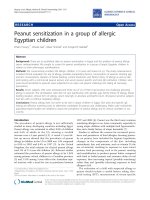
actions as shown in figure 1. The G1 sub-network has a
diameter of 5, and average path length of 2.7, which are
consistent with a small-world network.
Several features in the network G1 and other sub-net-
works of G1 described below, suggest a significant role for
PRKDC(DNA-PKcs). The maximum core (a group of pro-
teins with the most intra-group interactions) of G1 is 6,
and DNA-PKcs is a member of the 5-core; the 5-core is a
highly interacting group of 12 proteins (DNA-PKcs, TOP1,
PCNA, RPA1, DDX9, CDK4, CDKN1A (p21), CDK5,
ADPRT (PARP), XRCC5 (Ku70), XRCC6 (Ku86), NCOA6
(TRBP)), all of which are related to the DNA-repair proc-
ess. Interestingly 6 of these 12 proteins (DNA-PKca,
TOP1, DDX9, ADPRT, XRCC5, XRCC6) were also among
the Tax-binding proteins observed in the mass spectrome-
NFKB2 co-IP LYT-10 Murakami T, Virology 1995 206(2):1066
VAC14 co-IP TAX1BP2; TRX Mireskandari A, BBA 1996 1306(1):9
GPS2 yeast two hybrid; GST pulldown TXBP31 Jin DY, JBC 1997 272(41):25816
CCND3 co-IP Cyclin D3 Haller K, MCB 2002 22(10):3327
PSMB4 yeast two hybrid; co-IP HN3 Haller K, MCB 2002 22(10):3327
PSMA4 yeast two hybrid; co-IP HC9; PSC9 Rousset R, Nature 1996 381(6580):328
CARM1 GST pulldown; co-IP; Colocalization PRMT4 Jeong SJ, J Virol 2006 80(20):10036
GNB2 yeast two hybrid; co-IP; Colocalization transducin beta chain 2 Twizere JC, Blood 2007 109(3):1051
GNB5 co-IP; colocalization GB5 Twizere JC, Blood 2007 109(3):1051
GNB1 co-IP; colocalization transducin beta chain 1 Twizere JC, Blood 2007 109(3):1051
IL16 co-IP, colocalization LCF Wilson KC, Virology 2003 306(1):60
PPP2CA co-IP, GST pulldown PP2A catalytic subunit Fu DX, JBC 2003 278(3):1487
MAP3K14 co-IP NIK Xiao G, EMBO J 2001 20(10):6805
TP53BP1 co-IP, colocalization 53BP1, p202 Haoudi A, JBC 2003 278(39):37736
Table 1: Tax interacting proteins (Continued)
Table 2: Tax binding proteins sorted by number of unique peptides
Protein Unique peptides Protein score Coverage emPAI
DNA-dependent Protein Kinase 25 1391 9% 0.27
Vimentin 11 1387 44% 7.54
Gamma interferon-inducible protein 19 1116 24% 1.7
PARP 15 1414 34% 1.78
H2A.1 7 569 30% 1.25
Retrovirology 2008, 5:92 />Page 5 of 13
(page number not for citation purposes)
The G1 first neighborhood network for Rad51, TOP1, Chk2 and 53BP1Figure 1
The G1 first neighborhood network for Rad51, TOP1, Chk2 and 53BP1. The four initial proteins (yellow) were used
to generate a network via interrogation of the Human Protein Reference Database. Protein-protein interactions are indicated
by lines. Proteins with two or more shared interactions will form a core. PRKDC (DNA-PK) is also highlighted.
Retrovirology 2008, 5:92 />Page 6 of 13
(page number not for citation purposes)
try analysis. We also note that active DNA-PK consists of
the catalytic subunit (DNA-PKcs) and the two regulatory
subunits (Ku70 and Ku86) each of which is a member of
this highly interactive core. Furthermore, DNA-PKcs ranks
eighth in degree (the number of interactions) and in the
top 30% in two centrality measures (betweenness and
closeness).
We next considered the structure of the G1 sub-network
after the removal of the four initial Tax-binding proteins
comprising C1. This would allow for an assessment of the
degree and centrality of neighbors without interference
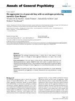
from the original four proteins. The largest connected
component of the resulting network consisted of 29 pro-
teins and 60 interactions as shown in figure 2. This net-
work has a diameter of 6 and a small average path length
of 2.6. In this sub-network, DNA-PKcs is among the top
six proteins in degree and betweenness centrality. Thus,
the critical role of DNA-PKcs as determined through our
clustering process is independent of the presence of the
four initial proteins.
We then created a sub-network of G1 restricted to those
involved in DNA repair response, referred to as G1*. Spe-
cifically, we removed those proteins that lacked the pri-
mary function of DNA repair as listed in the HRPD. This
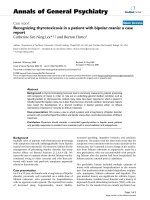
network consisted of 26 proteins and 42 interactions as
shown in figure 3. The G1* network has a diameter of 5
and an average path length of 2.5. In this restricted net-
work, DNA-PKcs ranks fourth in degree and ninth in
betweenness centrality. The maximum core of this net-
work is the 4-core, which consists of six proteins of which
DNA-PKcs is a member (DNA-PKcs, PCNA, PARP, Ku70,
Ku86, and TRBP). Thus, DNA-PKcs demonstrates an
increased rank when consideration is refocused toward
protein interactions involved in DNA damage response.
Definition of the second neighbors of C1 refined to DNA
repair
In our next exercise, we attempt to assign value to the pro-
teins identified in the prior networks by examining their
context in the "larger world" of second neighbors. Our
assumption was that key proteins from the first neighbor
analysis should retain their central role as defined by
The largest interacting network remaining in G1 after removal of Rad51, TOP1, Chk2 and 53BP1Figure 2
The largest interacting network remaining in G1 after removal of Rad51, TOP1, Chk2 and 53BP1. The compo-
nents that populated the first neighborhood network were depleted of rad51, top1, chk2 and 53bp1. The remaining compo-
nents with the highest degree of interaction are shown. DNA-PK (PRKDC) is indicated (yellow).
Retrovirology 2008, 5:92 />Page 7 of 13
(page number not for citation purposes)
interactions in the large second neighbor population. Spe-
cifically, in this exercise we first extend the database of
Tax-interacting proteins outward to include second neigh-
bor proteins (a protein that binds a protein that is known
to bind Tax). We considered the first and second neigh-
borhood of the initial set of proteins in C1, which we refer
to as G2 = 2NN (C1). The G2 network consisted of 667
proteins and 3827 interactions. From the proteins in the
G2 network, we created a smaller network by restricting to
proteins involved in DNA repair, and refer to this sub-net-
work as G2*. There were 114 proteins in G2*. Once this
group is developed we use a clustering analysis in an
attempt to identify the presumed most critical members of
the Tax-interacting world restricted to DNA repair
response proteins. The clustering process ranks compo-
nents of the network based upon the intra-group interac-
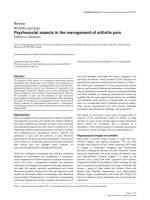
tions. We show the 3-core of the G2* network, which
consists of 54 proteins, in figure 4. All 3-core proteins will
have three or more interactions in order to be included in
the network. By application of our clustering approach,
we expose the structure of this subnetwork. It consists of
five clusters of proteins, with the largest cluster having 22
proteins, and the smallest cluster consisting of 3 proteins.
Adding proteins of lower degree clearly generates a larger
G2* network, but did not change the integrity of the struc-
ture of the network (data not shown). We can also observe
from the clustering that three proteins, DNA-PKcs, PCNA,
and P53 (TP53) link the various clusters to each other. We
call these three proteins "bridges", since they connect the
different clusters together. Hence, DNA-PKcs is a bridge
protein in this second neighborhood network that links
The G1* first neighborhood network restricted to proteins documented to play a role in the DNA-repair responseFigure 3
The G1* first neighborhood network restricted to proteins documented to play a role in the DNA-repair
response. The components of the entire first neighborhood network were filtered to remove those not known to have a role
in the DNA-repair response. The remaining components are displayed to reveal interactions and a central core.
Retrovirology 2008, 5:92 />Page 8 of 13
(page number not for citation purposes)
clusters 1, 4, and 5, and is also linked to the bridge protein
PCNA.
The five clusters depicted in figure 4, anchored to the three
prominent bridge proteins (TP53, PCNA and PRKDC),
include proteins that play key roles in DNA repair, stress-
induced signaling pathways and cell cycle controls. In
general, these proteins are discretely associated with the
clusters. For example, Cluster 1 includes four members of
the Fanconi anemia complementation group (FANCA,
D2, E and G). FANC genes mediate a stress related signal-
ing pathway that allows a normal cell to surmount certain
types of damage induced in DNA, principally interstrand
crosslinks [20]. In contrast, Cluster 2 includes key genes
whose proteins mediate cell cycle arrest in response to
genotoxic and other cellular stresses. Thus, if these protein
The 3-core representation of the G2* second neighborhood network restricted to DNA damage repair responseFigure 4
The 3-core representation of the G2* second neighborhood network restricted to DNA damage repair
response. Shown is the result of clustering the components of the G2* second neighborhood network arising from the origi-
nal four Tax binding proteins known to be involved in the cellular DNA damage response. There are five clusters with three
bridge proteins; DNA-PK is one of the bridge proteins. For clarity in drawing the network, we do not show edges from these
three proteins to the individual proteins in the clusters. The numbers on the edges from these proteins to the clusters count
the number of edges from each protein to proteins in each cluster.
Retrovirology 2008, 5:92 />Page 9 of 13
(page number not for citation purposes)
interactions reflect a true subset of the proteins that are
directly, or indirectly, affected by Tax-1, then this key viral
protein has command over some of the principal cellular
stress response pathways that might otherwise inhibit cell
growth following HTLV1 infection.
Endogenous DNA-PK co-precipitates with affinity isolated
Tax
As a final verification of the binding between Tax and
DNA-PKcs, we performed an affinity pull-down of endog-
enous cellular Tax protein complexes. In this study, we
expressed either S-Tax or S-GFP via transient transfection
of 293T cells and normalized for S-fusion protein
amount. The extracts were then isolated by affinity purifi-
cation of the S peptide and the complexes separated on
SDS-PAGE and subjected to immunoblotting with anti-
DNA-PKcs. Endogenous DNA-PKcs specifically associates
with the Tax containing protein complex and is detected
by staining with anti-DNA-PKcs (Figure 5). These results
confirm the identification of DNA-PKcs as a Tax-binding
protein.
Discussion
The HTLV-1 Tax protein has been defined by the proteins
with which it interacts [21]. Therefore, it stands to reason
that defining the functional properties of this protein will
require an understanding of which cellular proteins it
interacts with. Clearly, uncovering all potential interac-
tions will include those with functional significance.
However, determining which interactions support func-
tion and which interactions are of no consequence is an
obvious and critical question. We have taken the
approach that if we assume that Tax impacts the DNA
damage repair process, as many studies support, then
those interactions that are critical to the DNA damage
repair process will hold greater promise of functional sig-
nificance. Given this hypothesis, we devised a computa-
tional biology approach to help define which physical
interactions warrant further study.
One of the challenges in computational systems biology is
to create a tool to identify functional modules and the
interactions among them from large-scale protein interac-
tion networks. There are three major clustering
approaches that have been employed to identify func-
tional modules in proteomic networks. The first approach
searches for sub-graphs with specified connectivity, called
network motifs, and characterizes these as functional
modules or parts of them. This approach is not scalable
for finding larger clusters in large-scale networks. The sec-
ond approach, an example of which is work by Bader and
Hogue [22], identifies a seed vertex, around which to grow
a cluster. The seed vertex is identified by choosing a vertex
of largest weight, where the weight of a vertex is a measure
of the number of edges that join the neighbors of the ver-
tex, the clustering coefficient. A vertex in the neighbor-
hood of a cluster is added to it as long as its weight is close
(within a threshold) to the weight of the seed vertex. Once
a cluster has been identified, the procedure is repeated
with a vertex of largest weight that currently does not
belong to a cluster as the seed vertex. However, our expe-
rience comparing this approach with the spectral algo-
rithms we employed in this study indicates that this
method is less stable (i.e., the clusters obtained depend
strongly on the seed vertices chosen). We used an
improved clustering method [23] to reveal proteins that
form functional modules, i.e., multiple proteins involved
in the same biological function. This approach was used
to apply an objective measure to the functional signifi-
cance of a protein. Specifically we use this to both cluster
proteins into specific functional domains as well as to
objectively measure each individual protein's value to that
functional domain.
When we compared these results to the Tax-binding pro-
teins generated from our physical mapping efforts, DNA-
PK was in the top five best represented binding proteins
and occupied a top tier ranking via our functional cluster-
ing for DNA damage proteins. Clearly, DNA-PK is a criti-
cal component in cellular processes that mediate response
to damage and thus the fact that our clustering analysis
places high value on this protein is as much a validation
HTLV-1 Tax binds to DNA-PKcsFigure 5
HTLV-1 Tax binds to DNA-PKcs. The fusion proteins S-
Tax and S-GFP were isolated from 293T cells as described
and analyzed for co-precipitation with DNA-PKcs. Shown is
the pre-isolated total cell extract (input) for S-GFP (lane 1)
and S-Tax (lane 3). Also shown is the affinity purified protein
complexes for S-GFP (lane 2) and S-Tax (lane 4). Experimen-
tal normalization was achieved by using equal amounts of
purified protein.
,36EHDGV
7D[.G
'1$3.FV.G
*)3.G
Retrovirology 2008, 5:92 />Page 10 of 13
(page number not for citation purposes)
of the process as it is novel information. However, we
began with a network of known Tax-binding proteins and
their neighbors and second-neighbors, and DNA-PK was
selected, through our functional clustering approach,
whereas other equally critical damage response proteins
were not. For instance, among the PI3K protein family
members ATM and ATR hold positions of prominence in
the DNA damage-response arena equal to DNA-PK [24].
In fact, the three proteins are considered redundant in spe-
cific pathways and are sometimes able to substitute func-
tionally [25-27]. However, neither of the other two
proteins was reflected in the upper tier interactions when
using the Tax-designated protein networks. Furthermore,
ATM and ATR were not found among the list of Tax-bind-
ing proteins identified in the physical isolation of Tax
complexes, again verifying the novelty of the DNA-PK
finding.
This is not the first time that DNA-PK has been targeted as
a cellular protein through which Tax might mediate
genomic instability [28]. It is clear that DNA-PK is known
to mediate functions associated with reported Tax activi-
ties. Specifically, Tax has been shown to cause constitutive
activation of Chk2, a downstream target of DNA-PK [19].
DNA-PK can phosphorylate the tumor suppressor p53 at
S15 and S37 [29] whereas Tax expression results in phos-
phorylation at S15 and S392 [30,31]. In addition, we have
recently shown that Tax interaction with DNA-PK results
in saturation of the damage response (manuscript submit-
ted). Thus, the Tax-DNA-PK interaction satisfies several
previous observations regarding Tax function and pro-
vides a unifying model for all of these activities. Thus,
although Van et al. [32] demonstrated that the Tax-p53
nexus was intact in a DNA-PK knock-out line, it may well
be worth examining this protein as a mediator of other
Tax activities.
Clearly HTLV-1 Tax presents a biological model for an
interesting protein with an overwhelming amount of
associated published literature. A recent review by Boxus
et al highlights this complexity and presents an exhaustive
compilation of all known Tax-interacting proteins [33].
The growth in the Tax knowledge base requires constant
surveillance and verification if this body of work is to be
useful in understanding how Tax functions. Additionally,
as proteomic techniques continue to mature, the data gen-
erated in experimental studies is increasing exponentially.
We have described a parallel process for combining in sil-
ico analysis with experimental proteomic analysis so that
information gained in each process facilitates data mining
of the orthogonal process. Further building of the Tax
interactome should reveal other critical proteins that play
key roles in mediating the biologically significant Tax
functions within the host cell.
Methods
Cell culture and transfection
293T cells were maintained at 37°C in a humidified
atmosphere of 5% CO
2
in air, in Iscove's modified Dul-
becco's medium supplemented with 10% fetal bovine
serum and 1% penicillin-streptomycin. Transient trans-
fections were performed by standard calcium phosphate
precipitation. The plasmid used for expression of S-Tax-
GFP has been described previously [18]. For expression of
S-Tax and S-GFP the tax or EGFP open reading frame was
inserted into the SmaI site of pTriEx4-Neo (Novagen, Mad-
ison, WI). Cells were plated in 150-mm plates at 4 × 10
6
cells per plate. The following day, 20 μg of plasmid DNA
in 2 M CaCl
2
and 2X HBS were added drop wise to cells in
fresh medium. Cells were incubated at 37°C for 5 h and
fresh medium was added. The cells were harvested 48 h
later.
Purification of S-fusion proteins
S-Tax-GFP, S-Tax, or S-GFP protein was isolated following
a single wash with 1X PBS, in 500 μl M-Per mammalian
protein extraction reagent (Pierce, Rockford, IL) supple-
mented with protease inhibitor cocktail (Roche, Palo
Alto, CA) and immediately frozen at -80°C. The cell lysate
(2.5 mL) was incubated with 200 μl bed volume of S-pro-
tein™ agarose (Novagen, Madison, WI) for 30 min at
room temperature as per manufacturer's suggestion. The
bound S-tagged protein was then washed 3 times with 1
mL Bind/Wash Buffer (20 mM Tris-HCl pH 7.5, 150 mM
NaCl, 0.1% TritonX-100).
Isolation of Tax-complexes
Freshly prepared S-Tax-GFP or S-GFP beads were washed
3× in incubation buffer (25 mM HEPES, pH 7.5, 150 mM
NaCl, 1% NP-40, 10 mM MgCl2, 1 mM EDTA, 1% glyc-
erol) and placed on ice. A working stock of Jurkat nuclear
lysate (Active Motif, Carlsbad CA) was prepared by dilut-
ing 25 μg lysate to a total volume of 75 μL in incubation
buffer. The lysate was pre-cleared by adding 30 μL of S-
bead slurry and incubating on ice for 30 minutes with
occasional mixing. The pre-cleared slurry was spun down
at 2000 g for 3 minutes and the lysate (70 μL) transferred
to a fresh 0.5 ml tube containing 10 μL of the S-Tax-GFP
or S-GFP protein bound to beads. This slurry was incu-
bated at 4°C for 60 minutes on a shaker. The beads were
centrifuged at 2000 g for 3 minutes, lysate removed, and
beads washed 1× with 250 μL incubation buffer followed
by 4 washes with 250 μL ice cold PBS.
Isolation of endogenous DNA-PK-Tax protein complex
In some cases, S-Tax or S-GFP expression plasmids were
transfected into 293T and protein complexes isolated as
described above from a single T75 flask. In these experi-
ments no nuclear extracts were added. The protein lysates
were subjected to purification on S-beads, 50 μL of sample
Retrovirology 2008, 5:92 />Page 11 of 13
(page number not for citation purposes)
loading buffer (Bio-Rad, Hercules, CA) with β-mercap-
toethanol was added to the S-bead pellet and boiled for
10 min. The whole protein sample that was bound to the
S-bead was separated by 4–12% SDS-PAGE and analyzed
by Western Blot as described below.
LC-MS/MS of protein complexes
S-Tax-GFP or S-GFP beads were washed 3X with ice cold
50 mM ammonium bicarbonate, pH 8 and subsequently
resuspended in 50 μL of 50 mM ammonium bicarbonate,
10% acetonitrile containing 3.12 ng/μL sequencing grade
modified trypsin (Promega Corp., Madison, WI). The
digest was incubated for 6 hours at 37°C with occasional
mixing, transferred to a 0.2 μm centrifuge tube filter and
spun at 5000 rpm for 3 minutes. The flow through was
recovered and peptides dried in a speed vac. Digests were
resuspended in 20 μl Buffer A (5% Acetonitrile, 0.1% For-
mic Acid, 0.005% heptafluorobutyric acid) and 10 μl were
loaded onto a 12-cm × 0.075 mm fused silica capillary
column packed with 5 μM diameter C-18 beads (The Nest
Group, Southborough, MA) using a N2 pressure vessel at
1100 psi. Peptides were eluted over 300 minutes, by
applying a 0–80% linear gradient of Buffer B (95% Ace-
tonitrile, 0.1% Formic Acid, 0.005% HFBA) at a flow rate
of 150 μl/min with a pre-column flow splitter resulting in
a final flow rate of ~200 nl/min directly into the source. A
LTQ™ Linear Ion Trap (ThermoFinnigan, San Jose, CA)
was run in an automated collection mode with an instru-
ment method composed of a single segment and 5 data-
dependent scan events with a full MS scan followed by 4
MS/MS scans of the highest intensity ions. Normalized
collision energy was set at 28%, activation Q was 0.250
with minimum full scan signal intensity at 1 × 10
5
with no
minimum MS
2
intensity specified. Dynamic exclusion was
turned on utilizing a three minute repeat count of 2 with
the mass width set at 1.0 m/z. Protein searches were per-
formed with MASCOT version 2.2.0 v (Matrix Sciences,
London GB) using the SwissProt version 51.3 database.
Parent ion mass tolerance was set at 1.5 and MS/MS toler-
ance 0.5 Da.
Western analysis
Total protein concentrations were determined by Protein
Assay (Bio-Rad, Hercules, CA). An equal volume of sam-
ple loading buffer (Bio-Rad, Hercules, CA) with β-mercap-
toethanol was added to the lysate and boiled for 5 min.
Samples were normalized to total protein and separated
through a 10% SDS-polyacrylamide gel. The proteins were
transferred onto Immobilon-P (Millipore, Billerica, MA)
membrane using a Trans-blot SD semi-dry transfer cell
(Bio-Rad, Hercules, CA) at 400 mA for 50 min. Following
blocking in 5% non-fat milk in PBS/0.1% Tween-20, blots
were incubated in primary antibody overnight, followed
by 1 h incubation in secondary horseradish-peroxidase
conjugated anti-mouse or anti-rabbit antibody (Bio-Rad,
Hercules, CA). Immunoreactivity was detected via
Immunstar enhanced chemiluminescence protein detec-
tion (Bio-Rad, Hercules, CA). The following primary anti-
bodies were used in the analysis: mouse monoclonal
antibody of DNA-PKcs (Upstate), 1:1000; rabbit polyclo-
nal antibody of Tax, 1:5000; mouse monoclonal antibody
of GFP (Santa Cruz), 1: 2000.
Sources of data for in silico analysis
Interaction data were gathered from three types of infor-
mation sources: manual extraction from Pubmed, labora-
tory derived physical interactions, and protein interaction
databases. In the first database source, the information
was extracted by manually searching the Pubmed litera-
ture to obtain a list of known Tax binding proteins. The
criterion for acceptance in this group was physical verifi-
cation of binding in the referenced publication. For the
second database source, the physical interactions utilized
in this study were all derived from the experimental efforts
described elsewhere in this article. For the final database
source, we queried a human protein interaction database;
The Human Protein Reference Database (HPRD) [34].
The HPRD
contains interactions of
proteins in the human proteome manually extracted from
the literature by expert biologists who read, interpret and
analyze the published data.
Terms and definitions for in silico analysis
For our topological studies of interaction networks, we
utilized a novel overlapping clustering approach [23] that
exposes the modular structure of the network. We define
bridges as proteins that belong to multiple clusters due to
the overlap among them. We also employed centrality
measures of networks known as betweenness and close-
ness. To define these measures, first we need to define
some network concepts. The distance of a protein v from
another protein w is the number of edges in a shortest
path between them. The diameter of a network is the max-
imum distance between any pair of vertices. The average
path length of a network is the average distance over all
pairs of vertices. The closeness centrality measure for a
protein, v, is the reciprocal of the sum of the distances of
v to all other proteins in the network.
The dependence of a protein s on a protein v is the sum
over all proteins t in the network of the ratio of the
number of distinct shortest paths between proteins s and
t that includes v as an intermediate vertex, and the number
of distinct shortest paths between s and t. The between-
ness value of a protein v is the sum of the dependence val-
ues of all proteins s on the protein v. This is equivalent to
the following equation for betweenness.
Retrovirology 2008, 5:92 />Page 12 of 13
(page number not for citation purposes)
Here V is the set of proteins in the network. The numera-
tor in the fraction shows the number of distinct shortest
paths joining s and t on which v is an intermediate vertex;
the denominator is the number of distinct shortest paths
joining s and t. Further details on centrality measures are
available in [35].
As in earlier work [36], we define hubs as all proteins that
are ranked in the top 20% with respect to degree in the
network (the number of interactions a protein is involved
in). Similarly bottlenecks are all the proteins that are
ranked in the top 20% of betweenness values. To calculate
betweenness values for proteins, we used an algorithm
provided by Yu et al. [37].
In the clustering approach to be described next, we use the
concept of a k-core of a graph. The k-core of a graph is
obtained by repeatedly deleting all vertices which are
joined to the vertices remaining in the graph by fewer than
k edges. This procedure begins by deleting all vertices
whose degree is less than k. The deletion of such vertices
could decrease the degrees of the remaining vertices. If
some of these vertices have degrees less than k, they would
be deleted as well. This process is repeated until the sub-
graph that remains has every vertex with degree at least k;
this subgraph is the k-core of the graph. All the deleted
vertices belong to the (k-1)-shell. Computing the k-core of
a graph helps with denoising the interaction network by
removing many false positives, and also reduces the initial
size of the network to be clustered. The deleted vertices
will be added to the clustering obtained in a subsequent
step.
Spectral clustering and modules identification
We now summarize the technique we used for clustering
the protein interaction networks [23]. The protein interac-
tion network is represented by a graph G = (V, E), with the
proteins constituting a set of proteins V, and interactions
constituting the set of edges E. We obtain clusters in the
interaction network by identifying a number of subgraphs
of G that have a relatively large number of edges joining
vertices in each subgraph and fewer edges to vertices out-
side the subgraph. We permit these clusters to overlap
(have some vertices in common), since proteins have
multiple functions and could be involved in more than
one biological process.
The details of the clustering algorithm will be described
elsewhere, but here we provide an overview. Clusters are
obtained by dividing a subgraph at each step into two sub-
graphs based on the ratio of the number of edges that join
vertices in the subgraph to the total number of edges, a
measure called the cohesion of the subgraph. Given the ini-
tial graph G, we recursively split it into subgraphs until the
value of cohesion of a subgraph is above a threshold
value, or the subgraph has number of vertices fewer than
a threshold size. We have used a spectral algorithm that
uses the components of an eigenvector of the Laplacian
matrix of the graph to divide each subgraph into two.
Once the eigenvector is computed (its components corre-
spond to the vertices of the graph), those vertices whose
component values are below some specified value are
included in one subgraph and the others belong to the
second subgraph. The choice of the value where the split
should be made is based on computing the cohesion.
We have found that the overall clustering approach
described above needed to be adapted to protein interac-
tion networks, which are small-world and modified
power-law networks. Initially we decompose the vertices
of the network into three sets; hubs or high degree vertices
(those in the top 20% of the degrees); low-shell vertices
(vertices not in the 3-core of the network); and the resid-
ual sub-network, which forms a 3-core of the network
from which the hubs have been removed. We call the last
subnetwork as the local network. We have found it advan-
tageous to cluster the local and hub sub-networks sepa-
rately using the spectral clustering method described
above. The clusters from both sub-networks are then
merged together if a large number of edges join clusters
from the two networks. We check to see if nodes that
belong to a cluster are significantly connected to other
clusters, and if so, they are included in such clusters as
well. The statistical significance of the connections is com-
puted using a p-value based on the hypergeometric distri-
bution. Finally, the low-shell nodes are added to clusters;
each such node could be added to none, one, or more
than one cluster, based on whether it has a statistically sig-
nificant number of connections to the clusters that have
been found. If a node belongs to three or more clusters,
we call it a bridge node.
Competing interests
The authors declare that they have no competing interests.
Authors' contributions
ER performed the computational experiments on the
interaction networks. MW performed all mass spectrome-
try analysis. XG and SD conducted the Tax-DNA-PKcs
binding experiments. AS contributed to the compilation
of Tax binding proteins. MV was responsible for study
design and interpretation of results. CO was involved in
aspects of study design and manuscript preparation. AP
designed the network algorithms and helped with the
Bv
st
v
st
tv
ts
tv
sv
sv
()
()
,
=
∈
≠
≠
∈
≠
∑∑
σ
σ
Retrovirology 2008, 5:92 />Page 13 of 13
(page number not for citation purposes)
writing. OJ designed the study, interpreted results and
contributed to manuscript preparation.
Acknowledgements
We thank Kurt Maly and Mohammed Zubair of Old Dominion University,
our collaborators on the Human Virus Interactome Resource (HVIR)
project, who designed a digital library for representing protein interactions
involving viral and human proteins. This study was supported, in part, by the
United States Public Service Grant CA076595 from the National Cancer
Institute, National Institutes of Health, awarded to OJS and a multi-discipli-
nary research initiative grant from the Old Dominion University Research
Foundation, awarded to AP, CO, and OJS.
References
1. Gessain A, Barin F, Vernant JC, Gout O, Maurs L, Calender A, de The
G: Antibodies to human T-lymphotropic virus type-I in
patients with tropical spastic paraparesis. Lancet 1985,
2:407-410.
2. Osame M, Usuku K, Izumo S, Ijichi N, Amitani H, Igata A, Matsumoto
M, Tara M: HTLV-I associated myelopathy, a new clinical
entity. Lancet 1986, 1:1031-1032.
3. Poiesz BJ, Ruscetti FW, Gazdar AF, Bunn PA, Minna JD, Gallo RC:
Detection and isolation of type C retrovirus particles from
fresh and cultured lymphocytes of a patient with cutaneous
T-cell lymphoma. Proc Natl Acad Sci USA 1980, 77:7415-7419.
4. Takatsuki K: Discovery of adult T-cell leukemia. Retrovirology
2005, 2:16.
5. Yoshida M, Seiki M, Yamaguchi K, Takatsuki K: Monoclonal inte-
gration of human T-cell leukemia provirus in all primary
tumors of adult T-cell leukemia suggests causative role of
human T-cell leukemia virus in the disease. Proc Natl Acad Sci
USA 1984, 81:2534-2537.
6. Yoshida M, Miyoshi I, Hinuma Y: Isolation and characterization
of retrovirus from cell lines of human adult T-cell leukemia
and its implication in the disease. Proc Natl Acad Sci USA 1982,
79:2031-2035.
7. Giam CZ, Jeang KT: HTLV-1 Tax and adult T-cell leukemia.
Front Biosci 2007, 12:1496-1507.
8. Marriott SJ, Semmes OJ: Impact of HTLV-I Tax on cell cycle
progression and the cellular DNA damage repair response.
Oncogene 2005, 24:5986-5995.
9. Peloponese JM Jr, Kinjo T, Jeang KT: Human T-cell leukemia virus
type 1 Tax and cellular transformation. Int J Hematol 2007,
86:101-106.
10. Jin DY, Spencer F, Jeang KT: Human T cell leukemia virus type 1
oncoprotein Tax targets the human mitotic checkpoint pro-
tein MAD1. Cell 1998, 93:81-91.
11. Harhaj EW, Sun SC: IKKgamma serves as a docking subunit of
the IkappaB kinase (IKK) and mediates interaction of IKK
with the human T-cell leukemia virus Tax protein.
J Biol Chem
1999, 274:22911-22914.
12. Jin DY, Giordano V, Kibler KV, Nakano H, Jeang KT: Role of
adapter function in oncoprotein-mediated activation of NF-
kappaB. Human T-cell leukemia virus type I Tax interacts
directly with IkappaB kinase gamma. J Biol Chem 1999,
274:17402-17405.
13. Gatza ML, Dayaram T, Marriott SJ: Ubiquitination of HTLV-I Tax
in response to DNA damage regulates nuclear complex for-
mation and nuclear export. Retrovirology 2007, 4:95.
14. Ishioka K, Higuchi M, Takahashi M, Yoshida S, Oie M, Tanaka Y, Taka-
hashi S, Xie L, Green PL, Fujii M: Inactivation of tumor suppres-
sor Dlg1 augments transformation of a T-cell line induced by
human T-cell leukemia virus type 1 Tax protein. Retrovirology
2006, 3:71.
15. Liu B, Hong S, Tang Z, Yu H, Giam CZ: HTLV-I Tax directly binds
the Cdc20-associated anaphase-promoting complex and
activates it ahead of schedule. Proc Natl Acad Sci USA 2005,
102:63-68.
16. Merling R, Chen C, Hong S, Zhang L, Liu M, Kuo YL, Giam CZ:
HTLV-1 Tax mutants that do not induce G1 arrest are disa-
bled in activating the anaphase promoting complex. Retrovi-
rology 2007, 4:35.
17. Wu K, Bottazzi ME, de la Fuente C, Deng L, Gitlin SD, Maddukuri A,
Dadgar S, Li H, Vertes A, Pumfery A, Kashanchi F: Protein profile
of tax-associated complexes. J Biol Chem 2004, 279:495-508.
18. Durkin SS, Ward MD, Fryrear KA, Semmes OJ: Site-specific phos-
phorylation differentiates active from inactive forms of the
human T-cell leukemia virus type 1 Tax oncoprotein. J Biol
Chem 2006, 281:31705-31712.
19. Gupta SK, Guo X, Durkin SS, Fryrear KF, Ward MD, Semmes OJ:
Human T-cell leukemia virus type 1 Tax oncoprotein pre-
vents DNA damage-induced chromatin egress of hyperphos-
phorylated Chk2. J Biol Chem 2007, 282:29431-29440.
20. Niedernhofer LJ, Lalai AS, Hoeijmakers JH: Fanconi anemia
(cross)linked to DNA repair. Cell 2005, 123:1191-1198.
21. Wycuff DR, Marriott SJ: The HTLV-I Tax oncoprotein: hyper-
tasking at the molecular level. Front Biosci 2005,
10:620-642.
22. Bader GD, Hogue CW: An automated method for finding
molecular complexes in large protein interaction networks.
BMC Bioinformatics 2003, 4:2.
23. Ramadan E, Osgood C, Pothen A: The architecture of a pro-
teomic network in the yeast. Lecture Notes in Bioinformatics 2005,
3695:265-276.
24. Abraham RT: PI 3-kinase related kinases: 'big' players in stress-
induced signaling pathways. DNA Repair (Amst) 2004, 3:883-887.
25. Marone R, Cmiljanovic V, Giese B, Wymann MP: Targeting phos-
phoinositide 3-kinase: moving towards therapy. Biochim Bio-
phys Acta 2008, 1784:159-185.
26. Pommier Y, Sordet O, Rao VA, Zhang H, Kohn KW: Targeting
chk2 kinase: molecular interaction maps and therapeutic
rationale. Curr Pharm Des 2005, 11:2855-2872.
27. Yang J, Yu Y, Hamrick HE, Duerksen-Hughes PJ: ATM, ATR and
DNA-PK: initiators of the cellular genotoxic stress
responses. Carcinogenesis 2003, 24:1571-1580.
28. Majone F, Luisetto R, Zamboni D, Iwanaga Y, Jeang KT: Ku protein
as a potential human T-cell leukemia virus type 1 (HTLV-1)
Tax target in clastogenic chromosomal instability of mam-
malian cells. Retrovirology 2005, 2:45.
29. Lees-Miller SP, Sakaguchi K, Ullrich SJ, Appella E, Anderson CW:
Human DNA-activated protein kinase phosphorylates ser-
ines 15 and 37 in the amino-terminal transactivation domain
of human p53. Mol Cell Biol 1992, 12:5041-5049.
30. Pise-Masison CA, Mahieux R, Jiang H, Ashcroft M, Radonovich M,
Duvall J, Guillerm C, Brady JN: Inactivation of p53 by human T-
cell lymphotropic virus type 1 Tax requires activation of the
NF-kappaB pathway and is dependent on p53 phosphoryla-
tion. Mol Cell Biol 2000, 20:3377-3386.
31. Pise-Masison CA, Radonovich M, Sakaguchi K, Appella E, Brady JN:
Phosphorylation of p53: a novel pathway for p53 inactivation
in human T-cell lymphotropic virus type 1-transformed cells.
J Virol 1998, 72:6348-6355.
32. Van PL, Yim KW, Jin DY, Dapolito G, Kurimasa A, Jeang KT: Genetic
evidence of a role for ATM in functional interaction between
human T-cell leukemia virus type 1 Tax and p53. J Virol 2001,
75:396-407.
33. Boxus M, Twizere JC, Legros S, Dewulf JF, Kettmann R, Willems L:
The HTLV-1 Tax interactome. Retrovirology 2008, 5:76.
34. Peri S, Navarro JD, Kristiansen TZ, Amanchy R, Surendranath V,
Muthusamy B, Gandhi TK, Chandrika KN, Deshpande N, Suresh S, et
al.: Human protein reference database as a discovery
resource for proteomics. Nucleic Acids Res 2004, 32:D497-501.
35. Brandes U: A faster algorithm for betweeness centrality. Jour-
nal of Methematical Sociology 2001, 25:163-177.
36. Barabasi AL, Oltvai ZN: Network biology: understanding the
cell's functional organization. Nat Rev Genet 2004, 5:101-113.
37. Yu H, Kim PM, Sprecher E, Trifonov V, Gerstein M: The impor-
tance of bottlenecks in protein networks: correlation with
gene essentiality and expression dynamics. PLoS Comput Biol
2007, 3:e59.