Báo cáo y học: "Isolation and characterization of human cells resistant to retrovirus infection" pdf
Bạn đang xem bản rút gọn của tài liệu. Xem và tải ngay bản đầy đủ của tài liệu tại đây (592.15 KB, 11 trang )
BioMed Central
Page 1 of 11
(page number not for citation purposes)
Retrovirology
Open Access
Research
Isolation and characterization of human cells resistant to retrovirus
infection
Patrycja Lech
1
and Nikunj V Somia*
2
Address:
1
Molecular, Cellular, Developmental Biology and Genetics Graduate Program, University of Minnesota, Minneapolis, Minnesota, USA
and
2
Dept. of Genetics, Cell Biology and Development and the Institute of Human Genetics, University of Minnesota, Minneapolis, Minnesota,
USA
Email: Patrycja Lech - ; Nikunj V Somia* -
* Corresponding author
Abstract
Background: Identification of host cell proteins required for HIV-1 infection will add to our
knowledge of the life cycle of HIV-1 and in the development of therapeutics to combat viral
infection. We and other investigators have mutagenized rodent cells and isolated mutant cell lines
resistant to retrovirus infection. Since there are differences in the efficiency of single round
infection with VSVG pseudotyped HIV-1 on cells of different species, we conducted a genetic
screen to isolate human cells resistant to HIV-1 infection. We chemically mutagenized human HeLa
cells and validated our ability to isolate mutants at test diploid loci. We then executed a screen to
isolate HeLa cell mutants resistant to infection by an HIV-1 vector coding for a toxic gene product.
Results: We isolated two mutant cell lines that exhibit up to 10-fold resistance to infection by HIV-
1 vectors. We have verified that the cells are resistant to infection and not defective in gene
expression. We have confirmed that the resistance phenotype is not due to an entry defect. Fusion
experiments between mutant and wild-type cells have established that the mutations conferring
resistance in the two clones are recessive. We have also determined the nature of the block in the
two mutants. One clone exhibits a block at or before reverse transcription of viral RNA and the
second clone has a retarded kinetic of viral DNA synthesis and a block at nuclear import of the
preintegration complex.
Conclusion: Human cell mutants can be isolated that are resistant to infection by HIV-1. The
mutants are genetically recessive and identify two points where host cell factors can be targeted to
block HIV-1 infection.
Background
Intensive studies of the structure and function of HIV-1
encoded genes has led to the development of a number of
small molecule drugs to combat HIV-1. However, the
mutation rate of HIV-1 is high (about one mutation in
every 3 new genomes produced [1]) which leads to the
evolution of viruses that are resistant to the drug blockade.
Indeed some antiviral drugs may accelerate the mutation
rate of HIV-1 [1]. This necessitates the development of
new drugs and strategies to combat HIV-1 infection. In
this regard, a novel approach is to target cellular factors
required by HIV-1 to complete its lifecycle [2]. One
method of identifying cellular factors essential for retrovi-
ral infection is through genetic screening of mutagenized
Published: 3 July 2007
Retrovirology 2007, 4:45 doi:10.1186/1742-4690-4-45
Received: 18 December 2006
Accepted: 3 July 2007
This article is available from: />© 2007 Lech and Somia; licensee BioMed Central Ltd.
This is an Open Access article distributed under the terms of the Creative Commons Attribution License ( />),
which permits unrestricted use, distribution, and reproduction in any medium, provided the original work is properly cited.
Retrovirology 2007, 4:45 />Page 2 of 11
(page number not for citation purposes)
cells and identifying clones resistant to infection. Comple-
mentation cloning could then be used to identify genes
that confer infection susceptibility to the mutant clone.
The development of high titer retroviral vectors (based on
MLV and HIV-1) that recapitulate the early lifecycle of ret-
rovirus infection greatly facilitates such screens [3]. For
example, Gao and Goff (1999) isolated and characterized
two mutagenized rat fibroblasts clones (R3-2 and R4-7)
that are resistant to infection by MLV and HIV-1 viruses
[4]. The resistance phenotype in R3-2 is due to the over
expression of the FEZ1 gene [5]. Consistent with the
reported block in R3-2 (after reverse transcription but
before nuclear entry) FEZ1 over expression presumably
interferes with transport of the reverse transcription com-
plex or pre-integration complex in the cell. Indeed this has
been demonstrated for FEZ1 overexpression and intracel-
lular trafficking of the human polyoma JC virus [6]. The
mutations responsible for the resistance in the R4-7 cell
line have not been identified but can be rescued by two
non-protein coding RNA suppressors: an anti-sense tran-
script of the transcription coactivator CAPER and a central
portion of the VL30 endogenous retrovirus like element
[7]. The mechanisms by which these suppressors act are
not known. In another study Bruce and colleagues (2005)
isolated five clones from mutagenized Chinese hamster
ovary (CHO) cells that are specifically resistant to murine
MLV and are not resistant to HIV-1 based vectors [8]. In
our laboratory we have mutagenized hamster lung fibro-
blast cells (V79-4) and isolated two mutants that are (i)
resistant to MLV and HIV-1 infection (ii) are blocked at
pre and post reverse transcription steps and (iii) are dom-
inant and recessive for the resistance genotype [9,10].
Studies with VSVG pseudotyped retroviral vectors (that
enables infection of a wide variety of cells) have revealed
differences in the efficiency of single round infection in
cells of differing types and species [11,12]. Therefore, to
build upon and extend the rodent cell studies, and to
identify cellular factors in human cells required for the
early phase of infection we have executed a genetic screen
in HeLa cells to isolate mutants resistant to HIV-1 infec-
tion. HeLa cells were subjected to mutagenesis and clones
resistant to infection were isolated by infecting muta-
genized cells with an HIV-1 vector encoding a toxic bar-
nase gene [9]. Successful infection results in cell death
enabling the isolation of rare virus resistant clones. We
isolated two resistant clones designated 30-2 and 42-7.
These clones are genetically recessive for the resistance
phenotype. Infection of clone 30-2 is blocked at or before
virus reverse transcription. Infection in 42-7 is perturbed
during reverse transcription and is impaired for nuclear
import of proviral DNA.
Results
HeLa cell mutagenesis and validation
We exposed HeLa cells to the acridine half-mustard muta-
gen ICR-191 which results in frameshift mutations and
chromosomal re-arrangements [13]. We used a concentra-
tion of ICR-191 that killed 90% of cells and surviving cells
were allowed to recover before being subjected to another
round of mutagenesis. After each round of mutagenesis,
the mutation efficiency was determined at the hypoxan-
thine guanine phosphoribosyl transferase (HPRT) locus
and at the adenine phosphoribosyltransferase locus by
plating in medium containing 6-thioguanine (6-TG) or
diaminopurine (DAP), respectively. These drugs select
against the expression of the HPRT and APRT gene prod-
ucts since expression of these proteins results in the incor-
poration of the toxic purine analogues into DNA. The
genes coding for these enzymes (HPRT X-chromosome
and APRT human chromosome 16) are diploid and pos-
sibly polyploid in HeLa cells [14]. Table 1 shows the
kinetics of the appearance of 6-TG and DAP resistant col-
onies after 7 rounds of mutagenesis. These results demon-
strate that the mutagenesis procedure affected all alleles of
diploid test loci HPRT and APRT in a significant portion
of the cell population (1 in 10
6
) and validated the efficacy
of our mutagenesis protocol.
Isolation of cell clones resistant to infection by HIV-1
The mutagenized round 6 HeLa cells were multiply
infected with a VSVG pseudotyped HIV-1 Barnase vector
[9] to select for mutants that were resistant to infection.
Barnase expression results in apoptotic cell death, there-
fore cells that survive after incubation with virus have sim-
ply escaped infection, are mutant in expression of the
barnase gene or are resistant to infection by the HIV-1 vec-
tor. A total of 10
7
round 6 mutagenized Hela cells were
Table 1: Rounds of mutagenesis to generate mutations at diploid
loci.
6-thioguanine
resistant (HPRT-)
colonies per 10
7
cells
Diaminopurine
resistant (APRT-)
colonies per 10
7
cells
Spontaneous 0 0
Round 1 mutagenesis NA 0
Round 2 mutagenesis NA 0
Round 3 mutagenesis NA 0
Round 4 mutagenesis NA 0
Round 5 mutagenesis NA 1
Round 6 mutagenesis 31 10
Round 7 mutagenesis 26 31
NA = not assayed
Appearance of Diaminopurine and 6-thioguanine resistant colonies
examined at each round of mutagenesis with ICR-191. Mutagenized
HeLa cells were selected in the presence of 6-thioguanine (6-TG) and
Diaminopurine (DAP) to isolate APRT (-) and HPRT (-) colonies
respectively, which serve as indicators of mutagenesis at diploid loci.
Retrovirology 2007, 4:45 />Page 3 of 11
(page number not for citation purposes)
infected with an HIV-1 barnase vector at a moi ≤ 2, eight
times on consecutive days. Cell death became apparent on
day 3 and since we infected with the same volume of virus
on subsequent days the effective moi increased on subse-
quent infections. Cells that survived the selection were
isolated and expanded. We expanded 119 clones and
infected with a VSVG psuedotyped HIV-1 viral vector
transducing EGFP (HIV-1 GFP/VSVG). Infection efficiency
was initially semi-quantified visually by examining cells
under an inverted fluorescence microscope and compar-
ing cell clones to wild-type cells and to each other. Two
clones (30 and 42) were chosen for further analysis on the
basis of their resistance to infection and growth rates sim-
ilar to the mutagenized round 6 HeLa cells (parental pop-
ulation). Each clone was further subcloned to ensure that
the line is truly clonal and stable for the resistance pheno-
type. Subclones that displayed the latter qualities were
designated 30-2 and 42-7. The variation between sub-
clones was 2-fold with respect to infection by HIV GFP.
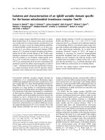
The relative efficiency of infection of the clones is visually
illustrated in Figure 1.
Growth rates of parental and mutant cells and extent of
HIV integration
We tested if the refraction to infection could be explained
by differences in the growth rates between parental and
mutant 30-2 and 42-7 cells. Figure 2A illustrates that the
growth rates are not significantly different between the
parental and mutant cells. To examine if the defect in
infection was in the early stages of the life-cycle we next
examined the extent of integration of HIV-1 DNA after
infection of parental and mutant cells. Figure 2B illus-
trates the results of a qPCR analysis for HIV-1 in genomic
DNA of parental and mutant cells that were infected (at an
moi = 1) and passaged 3 times before DNA extraction.
This analysis reveals over a 10-fold reduction in the
amount of DNA integrated into the genome of mutant
cells.
Clones 30-2 and 42-7 are resistant to MLV and HIV-1
infection
We then quantified the resistance to infection of clones
30-2 and 42-7 by fluorescence cytometry relative to the
parental population. We further determined if the resist-
ance is specific to HIV-1 or common to other evolutionar-
ily distinct retroviruses such as murine leukemia virus
(MLV). The clones and parental round 6 cell lines were
infected with VSVG pseudotyped HIV-1 EGFP or MLV
EGFP vector at an moi of 0.01, 0.1, 1 and 10 (the moi were
Growth rates of parental and mutant cells and extent of HIV integrationFigure 2
Growth rates of parental and mutant cells and extent
of HIV integration. (A) Growth rates. Parental and
mutant 30-2 and 42-7 cells were seeded and growth meas-
ured over time with the MTT assay. (B) HIV-1 integra-
tion. The extent of integrated HIV-1 vector was measured
by infection of cells at moi = 1. The cells were passaged 3
times and the quantity of stable HIV-1 DNA was measured
by quantitative real time PCR.
0
0.05
0.1
0.15
0.2
0.25
0.3
0.35
0.4
0244872
Time (hrs)
Parental
30-2
42-7
Parental 42-7
30-2
0
6.0 x
10
5
5.0 x
10
5
4.0 x
10
5
3.0 x
10
5
2.0 x
10
5
1.0 x
10
5
A)
B)
Infection of parental and mutant HeLa cells (30-2 and 42-7) cells with an HIV-EGFP vector at a moi = 0.5Figure 1
Infection of parental and mutant HeLa cells (30-2 and
42-7) cells with an HIV-EGFP vector at a moi = 0.5.
Transmission light phase microscopy of cells is illustrated in
the top panel and the corresponding field with fluorescence
microscopy is illustrated in the bottom panel.
Parental 30-2 42-7
Retrovirology 2007, 4:45 />Page 4 of 11
(page number not for citation purposes)
determined by infection of non-mutagenized HeLa cells).
This range of moi ensured that the infection was in a lin-
ear range for quantification. Infections were analyzed by
fluorescence cytometry 72 hours later. Typical results from
this analysis are illustrated in Figure 3. The range is con-
sidered linear where increase in moi yields a correspond-
ing increase in the number of cells infected and the
geometric means of fluorescence (a measure of multiple
infections) are also comparable. By this analysis clone 30-
2 is 12 fold less infectable with the HIV-1 vector (Fig 3A,
at an moi = 1) and approximately 10 fold less infectable
with the MLV vector (Fig 3B at an moi = 0.1). Clone 42-7
is 10 fold less infectable by HIV-1 EGFP (Fig 3C at moi =
1) and 5 fold less infectable by MLV EGFP (Fig 3D at an
moi = 0.1). This phenotypic analysis of HIV GFP infection
correlates with the molecular analysis of the extent of inte-
gration with a 10-fold reduction in the mutant cells (Fig.
2B). Strikingly both clones remain resistant to HIV-1 at
high MOI whereas they become almost as sensitive to
MLV as wild type cells. This might indicate a greater
dependence for HIV-1 on, or sensitivity to, the factors that
have been altered by the mutagenesis.
Resistance is independent of the reporter and is not a
defect in gene expression of the reporter
We next examined if the resistance phenotype is due to a
defect of the reporter or due to defects in expression of the
reporter. To verify that the resistance is not due to a defect
of the EGFP reporter used we next infected the parental
cells and the 30-2 and 42-7 mutant clones with an HIV-1
based vector transducing a gene coding for secreted alka-
line phosphatase (SEAP). Infection was quantified by the
amount of SEAP secreted into the media by infected cells
[15]. Subclone 30-2 was 20 fold resistant and 42-7 was 6
fold resistant to HIV-1 viral vector infection using this
assay (Figure 4A). We conclude that the observed resist-
ance to infection is independent of the reporter used. We
next investigated if the observed resistance is due to
defects in expression of the reporter. We transfected wild-
type and mutant cells with the HIV-1 EGFP vector (in
Clones 30-2 and 42-7 are resistant to MLV and HIV-1 infectionFigure 3
Clones 30-2 and 42-7 are resistant to MLV and HIV-1 infection. Parental (diamond point and black line), 30-2 and 42-7
(square point and grey line) cells were infected with HIV GFP/VSVG (2A, C) and MLV GFP/VSVG (2B, D) at an increasing moi
of 0.01, 0.1, and 1. Data is expressed as % of GFP positive cells determined by fluorescence cytometry.
A) 30-2: Infected with HIV-1 CSII EGFP
6.11
45.84 (236)
93.29 (840)
0.28
3.63 (181)
20.22 (197)
0
20
40
60
80
100
0.01 0.1 1 10
7.1
41.67(286)
90.96 (961)
0.31
3.56 (169)
20.41 (200)
0
20
40
60
80
100
0.01 0.1 1 10
C) 42-7: Infected with HIV-1 CSII EGFP
Multiplicity of infection (MOI)
2.96
29.7
92.18
85.1
0.45
3.52
35.97
73.43
0
20
40
60
80
100
0.01 0.1 1 10
B) 30-2: Infected with MLV MFG EGFP
D) 42-7: Infected with MLV MFG EGFP
2.33
20.66
88.41
97.69
0.55
3.78
38.59
94.63
0
20
40
60
80
100
0.01 0.1 1 10
Multiplicity of infection (MOI)
Parental
Mutant
Retrovirology 2007, 4:45 />Page 5 of 11
(page number not for citation purposes)
which the human EF1α promoter dictates EGFP expres-
sion) or the MLV vector (where EGFP expression is
directed by the early human CMV promoter). Fig. 4B illus-
trates the results from this experiment. While the transfec-
tion efficiency can vary between cell types (compare HeLa
cells to 30-2 cells) the overall gene expression (as deter-
mined by mean fluorescence intensity) is similar between
HeLa and mutant cells for both the HIV-1 and MLV EGFP
vectors. Hence the block seen on infection is not due to
alterations in gene expression in the 30-2 and 42-7
mutant cells.
Resistance is independent of receptor use and accessory
factors
Lentivirus encoded accessory factors can mitigate infec-
tion of certain cell types [16,17]. The HIV-1 packaging
construct used in this study, ΔNRF retains Tat, Rev and
Vpu coding [18]. To test the effect of Nef, Vif and Vpr
(accessory proteins that are packaged into virons [19-21])
we generated vectors with packaging plasmids providing
all these accessory proteins. Furthermore to determine if
the mutant clones are deficient for VSVG mediated entry,
we pseudotyped the HIV-1 based vectors with the MLV
amphotropic envelope, 10A1. The envelope protein 10A1
of the amphotropic retrovirus binds to phosphate trans-
porter proteins Pit-1 or Pit-2 [22] and enters using a pH
independent pathway [23], while VSVG is thought to bind
a phospholipid [24] and infects using a pH dependent
pathway [23]. Figure 4C illustrates the analysis from infec-
tion of wild type and mutant cells using 10A1 pseudo-
typed HIV-1 virus produced in the presence of all HIV-1
accessory proteins. Both mutant cell lines retain the resist-
ance to infection. We conclude that (i) HIV-1 accessory
proteins cannot rescue the resistance to infection in the
30-2 and 42-7 mutant cell type and (ii) the resistance is
independent of the receptor used for entry or the route of
entry (pH dependent or independent pathways).
Analysis of proviral DNA synthesis in mutant cells
To further characterize the block to infection we next fol-
lowed the formation of viral DNA products over time in
infected wild-type and mutant clones. Subclone 30-2, 42-
7 and the parental cell line were infected and total DNA
extracted at different times post infection. Viral DNA was
amplified using real-time qPCR and primers were used to
amplify specific reverse transcription intermediates by
hybridizing to particular regions of the viral genome. This
allows discrimination of strong stop and full products of
the reverse transcription process. The number of mole-
cules of reverse transcription product formed was calcu-
lated from the quantity of PCR product by reference to a
standard curve. The results of this analysis are illustrated
in Figure 5 for 30-2 and Figure 6 for 42-7. qPCR analysis
of subclone 30-2 revealed that over a 36 hour period the
strong stop primers amplified 2 to 16 fold less initial
HeLa mutants are resistant to infectionFigure 4
HeLa mutants are resistant to infection. (A) Resist-
ance to infection is independent of the reporter.
Clones 30-2 and 42-7 were infected (moi ~ 0.5) with HIV-1
viral vector transducing the gene for secreated alkaline phos-
phatase (SEAP). The amount of SEAP released by infected
cells was measured 72 hours latter and is expressed as the
V
max
of SEAP activity. (B) Resistance to infection is not
due to a defect in reporter gene expression. HeLa,
Parental (Round 6 mutagenesis), 30-2 and 42-7 cell lines
were transfected with an HIV vector plasmid where EGFP
expression is dictated by the human EF1α promoter and with
the MLV viral vector where EGFP expression is controlled by
the CMV promoter. Although the cells differ in their trans-
fection efficiencies they show comparable levels of GFP
expression. The x-geo mean intensity of EGFP expression is
indicated above each bar. (C) Resistance to infection is
independent of receptor use and HIV-1 accessory
proteins. Parental, 30-2 and 42-7 were infected with a fully
accessorized HIV-1 viral vector psuedotyped with a 10A1
envelope, which uses a pH independent pathway of entry.
The line graph depicts that x-geo mean intensity of each sam-
ple.
0
5
1
0
1
5
2
0
2
5
3
0
3
5
4
0
30-2
42-7
Parental
0
20
40
60
80
100
120
0
20
40
60
80
100
Hela
30-2
42-7
A)
B)
MLV
HIV
0
Hela
Parental
30-2
42-7
373
446
608
534
128
304
213
300
C)
20
25
15
10
5
Retrovirology 2007, 4:45 />Page 6 of 11
(page number not for citation purposes)
minus strand DNA product when compared to the control
cells (Figure 5A). A similar trend was revealed by the full
product primer sets (figure 5B), suggesting that the virus is
blocked before or at the stage of reverse transcription. In
clone 42-7, the formation of viral DNA intermediates is
also initially decreased – on average a 2-fold decrease in
the amount of products formed for the strong stop (Figure
6A). This decrease is also apparent at earlier time points
for the full-product. However the difference is less appar-
ent at the latter (36 hr) timepoint (Figure 6B). We con-
clude from this that the synthesis of proviral DNA is
retarded in 42-7 cells. Notably, even though the molecular
analysis reveals that there is near equivalence of proviral
DNA synthesis this does not correlate to the titer of virus
on 42-7 cells (10 fold less than wild type cells, see Figure
3) or the level of integration (Figure 2B). Indeed the titer
does not increase even if infection (% EGFP infected cells)
is measured at 144 hrs rather than 72 hrs (data not
shown). We conclude from this that one of the blocks to
infection in 42-7 cells is due to a slower completion or
aberrant reverse transcription.
42-7 cells are further impaired for nuclear entry of viral
DNA
The accumulation of 2 LTR circles is a product of intra-
molecular ligation of the linear reverse transcription prod-
uct and can be a surrogate molecular marker for nuclear
entry of viral DNA [25]. Hence we next asked if the
nuclear accumulation of viral DNA was impaired in 42-7
cells. PCR primers were used to probe the extent of 2LTR
circle accumulation. Results of this analysis (Fig 6C)
Synthesis and localization of proviral DNA in wild type and mutant 42-7 cellsFigure 6
Synthesis and localization of proviral DNA in wild
type and mutant 42-7 cells. Parental (black bars) and 42-7
(grey bars) cells were infected with HIV-1 GFP/VSVG at a
moi of 0.5 and viral DNA products quantified at the indicated
times. (A) amplification of strong stop early product; (B) full
product; (C) 2LTR circles 36 hours after infection; (D) bio-
chemical fractionation of cytoplasmic and nuclear extracts
and measurement of the amount of full product 36 hours
after infection in parental (black bars) and 42-7 (grey bars)
cells; (E) Parental and 42-7 cells were infected with HIV/GFP/
VSVG (black bars) or with a viral vector generated with a
mutant integrase (grey bars) to examine expression 72 hrs
latter of the EGFP reporter from circles and other uninte-
grated viral products.
0
2hrs 4hrs 6hrs 12hrs 24hrs 36hrs
length of infection (hours)
0
2hrs 4hrs 6hrs
12hrs 24hrs 36hrs
length of infection (hours)
A) Strong Stop
B) Full Product
8.0 x
10
6
4.0 x
10
6
6.0 x
10
6
2.0 x
10
6
2.5 x
10
6
2.0 x
10
6
1.5 x
10
6
1.0 x
10
6
5.0 x
10
5
0
1x10
4
2x10
4
3x10
4
4x10
4
5x10
4
6x10
4
C)
0
10
20
30
40
50
60
70
80
90
100
Parental 42-7
cell line
D)
E)
NUCLEUS
CYTOPLASM
0
1x10
5
2x10
5
3x10
5
4x10
5
Kinetics of proviral DNA synthesis in wild-type and 30-2 cellsFigure 5
Kinetics of proviral DNA synthesis in wild-type and
30-2 cells. Parental (black bars) and 30-2 (grey bars) cells
were infected with HIV-1 GFP/VSVG at a moi of 0.5 and viral
DNA products quantified at the indicated times. (A) amplifi-
cation of strong stop early product and (B) amplification of
late full product. The absence of contamination was con-
firmed by the failure to amplify viral replication intermediates
from water and a heat inactivated viral vector (data not
shown).
0
2hrs 4hrs 6hrs 12hrs 24hrs 36hrs
length of infection (hours)
0
2hrs 4hrs 6hrs 12hrs 24hrs 36hrs
length of infection (hours)
A) Strong Stop
B) Full Product
8.0 x
10
6
6.0 x
10
6
4.0 x
10
6
2.0 x
10
6
1.6 x
10
6
1.2 x
10
6
4.0 x
10
5
8.0 x
10
5
Retrovirology 2007, 4:45 />Page 7 of 11
(page number not for citation purposes)
reveal that accumulation of 2LTR circles is impaired in 42-
7 cells suggesting a defect in nuclear entry of viral DNA.
However the ratio of linear and 2 LTR circles has been
reported to be altered by certain cell factors (i.e RAD 52
[26]). Hence to exclude this possibility we quantified HIV
DNA 36 hours after infection in cytoplasmic and nuclear
extracts of parental and 42-7 cells. This analysis (Fig 6D)
reveals up to a 4 fold difference in the accumulation of
HIV DNA in nuclear extracts between parental and mutant
cells. This difference correlates with the deficiency in the
accumulation of 2 LTR circles in 42-7 cells (Fig 6C). To
examine expression of the EGFP reporter from these cir-
cles and other unintegrated viral products we infected
cells with a viral vector generated with a mutant integrase
deficient helper plasmid. This analysis (Fig 6D) reveals
EGFP is expressed in only 10% of cells compared to wild-
type parental cells which is comparable to integrase profi-
cient vector. We conclude from this analysis that although
reverse transcription may reach completion (albeit
slower, see Fig 6B) that the product of this reaction is not
comparably detected in the nucleus.
Resistance to infection in 30-2 and 42-7 cells is recessive
We next asked if the mutations conferring resistance to
infection in the mutant cells were dominant or recessive.
To address this question we performed cell fusion experi-
ments between wild type parental and mutant cells. Figure
7 illustrates an example of such an analysis. Parental, 30-
2 or 42-7 cells were labeled with the membrane dyes Ore-
gon Green or Vybrant DID that mark cells green and blue
respectively. These differentially marked cells were then
mixed (either as self-self or as parental and mutant com-
binations) and fused by addition of polyethelene glycol
(PEG). The fused cells were then infected with a dsRED
marked HIV-1 virus and the homo and hetrokaryons ana-
lyzed by fluorescence activated cytometry. While mutant
homokaryons exhibit a resistance to infection compared
to parental homokaryons (3.5% for 42-7 to 42-7 fusions;
3.7% for 30-2 to 30-2; and 20% for parental fusions) this
resistance is rescued in the parental and mutant hetrokary-
ons (16.4% and 24.5%) in the reciprocal staining experi-
ments for 42-7 and parental fusions and 23.8% and
20.5% for the 30-2 and parental fusions). In repeat exper-
iments we routinely observed that in the reciprocal dyeing
experiments the rescue is more pronounced when the
mutant cells are labeled with Vybrant DID. We conclude
from this experiment that the mutations causing the
resistance in 30-2 and 42-7 are recessive.
Discussion
In this study we report on the isolation of two human
clones that are resistant to infection by HIV-1 and MLV
viruses. Somatic cell mutagenesis and complementation
cloning is a powerful approach for the identification of
host cell factors involved in the retroviral lifecycle. An
early application of this approach (by Hillman and col-
leagues (1990) [27]) used EMS mutagenesis in a screen to
derive human T-cell clones with varying CD4 expression
levels. This group also characterized some mutants with
normal CD4 expression but a reduced capacity for HIV-1
replication due to defective in NFκB signaling [28,29].
These mutants were from a screen that initially targeted
CD4 expression. Gao and Goff (1999) [4] isolated mutant
cell clones on the basis of resistance to MLV and observed
that the cells were also resistant to HIV-1 vectors. In con-
trast Bruce et al., (2005) [8] isolated clones from muta-
genized Chinese Hamster Ovary cells that are uniquely
resistant to MLV infection but are infected normally by
HIV-1 and ASLV vectors. We have isolated clones from
mutagenized hamster lung fibroblasts (V79-4) cells that
are refractory to both MLV and HIV-1 vectors ([9,10]).
Taken together these studies imply that evolutionary dis-
tant retroviruses utilize common and distinct host cell fac-
tors. In this study we have extended these observations to
human cells. Here we report two clones that are resistant
to both HIV and MLV vectors although the resistance is
more pronounced for HIV-1 (Figure 3). We note the
42-7 and 30-2 are recessive mutantsFigure 7
42-7 and 30-2 are recessive mutants. Parental, 30-2 or
42-7 cells were labeled with the membrane dyes Oregon
Green or Vybrant DID that mark cells green and blue
respectively. The cells were then mixed (either as self-self or
as parental and mutant combinations) and fused with poly-
ethelene glycol (PEG). The fused cells were infected with
HIV-1 virus transducing DsRed and analyzed by flourecence
cyometry. The bar graph illustrates the percentage of fused
cells expressing DsRed and line graphs the geometic mean
intensity of DsRed expression.
0
5
10
15
20
25
30
% Infected
Geo Mean
0
50
100
150
200
250
300
350
Retrovirology 2007, 4:45 />Page 8 of 11
(page number not for citation purposes)
clones that we and others have isolated are not entirely
resistant to infection but rather refractory to infection.
There are several possible hypotheses that may explain
this: (i) Gene mutations that would make cells totally
resistant are also lethal for cell viability (ii) the screens are
not saturating and totally resistant clones have been
missed (iii) HIV-1 and MLV use redundant, but saturable
pathways for infection, and these clones are mutant in
only one pathway and (iv) the clones are "leaky" and pro-
duce reduced amounts of protein needed for infection.
We demonstrate that the resistance to infection is not at
the level of gene expression by transfection of the vector
DNA into mutant cells (Figure 4B). Notably, isolated
clones vary in transfection efficiency compared to the
parental population and hence interpretation of these
results are in the context of both transfection efficiency
and level of expression (as judged by the geometric mean
fluorescence). All the studies reported thus far have uti-
lized the VSVG envelope to pseudotype MLV and HIV-1
vectors during the selection procedure. To date no clones
have been reported that are due to an entry block to VSVG.
The resistance in the two human mutants reported here is
also independent of the envelope used (Figure 4C). We
further characterized the blocks to infection and identi-
fied a block at or before reverse transcription in the 30-2
clone (Figure 5). We also identified a block in 42-7 cells
with retarded kinetics of reverse transcription with a sub-
sequent block to nuclear import (Figure 6). There is a dis-
parity in the number of molecules in the nucleus
(approximately 1/4 of wild-type in the mutant cells), the
expression and integration analysis (Fig 6D and Fig 2B)
reveals a log difference in infection between wild type and
42-7 cells. While this may be due to differences in the lev-
els of detection between the PCR analysis and fluores-
cence cytometry, the slower synthesis and reduced nuclear
import suggests that the products of the reverse transcrip-
tion reaction may be aberrant. We are currently examining
this hypothesis. Although we and others have identified
blocks pre and post reverse transcription the 42-7 mutant
represents a novel phenotype in the slower kinetic of
reverse transcription. Cell fusion experiments have also
allowed us to conclude that the block to infection is reces-
sive and can be rescued by fusion with wild-type cells (Fig-
ure 7). This analysis does not suggest a mechanism for the
resistance to infection. For example it is possible that a
mutation of a transcriptional repressor may activate the
expression of a restriction factor [5]. However this experi-
ment does suggest that complementation cloning by
transfer of cDNA libraries derived from wild-type cells
[30,7] is a feasible approach and should yield novel host
cell factors involved in the early stages of retrovirus infec-
tion.
Conclusion
Human cell mutants can be isolated that are resistant to
infection by HIV-1 and MLV. The mutants are genetically
recessive and blocked at or before reverse transcription
and in nuclear import.
Methods
Tissue culture
293T cells, HeLa and derived cell lines (30-2 and 42-7)
were maintained in Dulbecco's modified Eagle's medium,
DMEM (Cellgro) supplemented with 10% Fetal Bovine
serum, FBS (Gemini Bioproducts). During heterokaryon
experiments HeLa and derived cells lines were maintained
in DMEM without phenol red supplemented with 20%
FBS.
Virus production
MLV and HIV-1 vectors were generated by transient trans-
fection of multiple plasmids into 293T cells as described
previously [30,31]. Briefly, for MLV based vector 10 μg of
CMVgp, 5 μg of pMDG and 15 μg of vector DNA were
transfected using the method of Chen and Okayama [32].
72 hrs after transfection virus was collected, filtered
through a 0.45 μ membrane and stored at -80°C. HIV-1
based vectors were similarly generated using 10 μg of
ΔNRF (a kind gift from Dr. Tal Kafri, [18]), 5 μg pMDG
[31] or pRK510A1 (N.S unpublished) and 15 μg vector
DNA (CSII EGFP, CSII DsRed, CSII Barnase [9] or CSII
SEAP (N.S. unpublished)). An integrase-defective packag-
ing plasmid ΔR8.2 (INT-) with a point mutation in the
integrase (D64V) was kindly provided by Dr. Tal Kafri.
Viral titers for EGFP transducing vectors were determined
by infecting 10
5
HeLa cells with serial (10 fold) dilutions
of the vector preparation. The medium was changed after
12 hours incubation of the viral vector with the cells, and
the extent of EGFP expression was quantified 72 hours
after infection by flow cytometry on a Becton-Dickinson
FACScalibur. HIV-1 based viral vectors utilized for qPCR
analysis were treated with 25 U/ml DNaseI at room tem-
perature for 1 hour.
Mutagenesis of HeLa cells
10
8
HeLa cells were mutagenized for 10 hours with 10 μg/
ml ICR-191 (Sigma), followed by a media change and a
recovery period. Mutagenesis was repeated for 7 rounds.
After each round an aliquot (10
7
cells) was incubated with
6-thioguanine (10 mg/ml) or 2-aminopurine (50 mg/ml)
and resistant clones were quantified when visible colonies
appeared. Aliquots of cells were frozen at -80°C after each
round of mutagenesis.
Screening of HIV-1 resistant clones
HeLa cells that were mutagenized for 6 rounds were
infected 8 times with a VSVG pseudotyped HIV-1 vector
encoding Barnase [9] at an initial moi = 2, on 8 consecu-
Retrovirology 2007, 4:45 />Page 9 of 11
(page number not for citation purposes)
tive days. The 119 colonies that survived the selection
were isolated and resistance to infection was assessed by
infecting with VSVG pseudotyped HIV-1 and MLV viral
vectors transducing EGFP. The efficiency of infection was
assessed visually using an inverted fluorescence micro-
scope, and the most resistant clones (as compared to the
wild-type parental cells and to each other) were selected
for further study. The clones were further sub-cloned by
limiting dilution to ensure that the clones were homoge-
neous, and that the resistant phenotype was stable.
Growth analysis
1 × 10
4
cells were seeded in 24 well plates and at given
time points viable cells were measured using the MTT
assay [33]. Briefly, at given time points media was
replaced with 500 μl 1X MTT solution and cells were incu-
bated for 1 hr at 37°C and the MTT solution was removed.
Cells were lysed in acetic isopropanol (400 ul Isopropanol
+ 40 mM HCl) and the absorbance measured at 540 nm.
Flow Cytometry analysis
Infected or transfected cells expressing EGFP or Ds Red
proteins were quantified by Fluorescence cytometry on a
Becton-Dickinson FACScan and analyzed using Becton-
Dickinson CellQuest 3.1 software at the Flow Cytometry
Core Facility of the University of Minnesota Cancer
Center.
Secreted alkaline phosphatase (SEAP) assay of viral
infection
10
5
cells were seeded in triplicate in 6 well plates and
infected with a VSVG psuedotyped HIV-1 vector transduc-
ing SEAP (CSII-EF-SEAP, N.S, unpublished). 12 hrs post
infection the media was changed to remove the viral
supernatant. 72 hrs post infection SEAP activity within the
media of infected and uninfected cells was assayed as pre-
viously described [34]. Briefly, media was collected from
each well and heated at 65°C to inactivate endogenous
phosphatases. Serial dilutions of the heat inactivated sam-
ples were made in DMEM. Samples were mixed at a 1:1
ratio with 2 × SEAP buffer (2 M Diethanolamine; 1 mM
MgCl2; 20 mM L-homoarginine). The substrate (120 mM
p-nitrophenol phosphate) was dissolved in 1 × SEAP
buffer and 1/10 sample volume was added to each sam-
ple. The kinetics of the reaction was measured as absorb-
ance at 450 nm every 5 min for 30 min at 37°C using a
plate reader (Bio-Tek Synergy HT).
Cell fusion assay
Cells were stained with Oregon Green (Invitrogen probes
Cat # O34550) or with Vybrant DID (Invitrogen probes
Cat # V22887) for 15 min according to the manufactures
protocol. The stained cells were gently washed 3 times
with PBS buffer and between each wash the cells were
incubated for 10 min at 37°C. The stained cells were left
to recover for 4 hrs in DMEM (without phenol red) sup-
plemented with 10% FBS. Cell fusions were performed by
removing the cells from the plate with a non-trypsin dis-
sociation media and self-self or parental and mutant cells
were mixed in 15 ml conical tubes (Falcon) and pelleted
by centrifugation for 5 min at 500 g. The pelleted cells
were incubated in 1 ml of a sterile PBS solution contain-
ing 50% Polyethelene glycol (PEG 3000–3700 Da)
(Sigma) and 2% Glucose for 45 seconds. The cell suspen-
sion was then diluted with 1 ml of PBS and incubated for
another 45 seconds. The PEG solution was further diluted
with 3 mls of wash buffer (PBS + 2% FBS) before being
centrifuged at 500 g for 5 min. Gentle resuspension of
cells in wash buffer and pelleting of cells by centrifugation
was repeated 3 times before resuspending the cells in
DMEM media without phenol red supplemented with
20% FBS. Cells were allowed to recover and settle for 6–8
hours in 10 cm tissue culture plates before being infected
with HIV vector transducing DsRed. 48 hours post infec-
tion the cells were analyzed by fluorescence cytometry
using 4 color differentiation on a Becton-Dickinson FAC-
SCalibur. Background leakage through the channels was
compensated by subtraction of the background value
from all samples.
Reverse transcription product qPCR assay
3.5 × 10
5
cells were plated into 6 well dishes and infected
at a moi= 0.5 with DNaseI treated viral supernatant. To
control for DNA contamination, DNaseI treated virus was
placed in a boiling water bath for 30 minutes to serve as a
heat inactivated sample control. Cells were incubated
with virus for 6, 12, 24, or 36 hours. Controls consisted of
uninfected cells or cells infected with heat inactivated
virus for 36 hours. Infection was stopped by harvesting
the cells and washing them with PBS buffer. Total cell
lysate was prepared by resuspending the cell pellet in lysis
buffer (Tris pH 8.0, 25 mM EDTA pH 8.0, 100 mM NaCl,
1% Triton X-100, and 2 mg/ml proteinase K) and incubat-
ing at 55°C overnight. The next day, the proteinase K was
heat inactivated at 95°C for 15 minutes. Lysates were used
directly for PCR analysis. The following primers were used
for qPCR [35] : 5' β-actin-ATC ATG TTT GAG ACC TTC AA,
3' β-actin-AGA TGG GCA CAG TGT GGG T, LTR9 – GCC
TCA ATA AAG CTT GCC TTG, 5NC2 – CCG AGT CCT
GCG TCG AGA GAG C, AA55 -CTG CTA GAG ATT TTC
CAC ACT GAC, LTR8 TCC CAG GCT CAG ATC TGG TCT
AAC. LTR9 and AA55 were used to amplify the strong stop
product, LTR9 and 5NC2 amplified the full product and
LTR8 and LTR9 amplified the 2 LTR circle products. Quan-
titative PCR reactions using SYBR green were performed
using a Biorad iCycler equipped with an optical module
and BioRad SuperMix (without ROX) following the man-
ufacturer's protocol. Cycling conditions used were 95°C
for 3 min, followed by 35 cycles of 95°C 30s, 58°C 30s,
and 72°C 30s, and a final extension (5 minute 72°C) to
Retrovirology 2007, 4:45 />Page 10 of 11
(page number not for citation purposes)
complete all the PCR products. Quantification of the
amount of DNA was calculated from the cycle threshold
(C
T
) determined using the Bio-Rad software. The melt
curve as well as analysis of the PCR products by agarose
gel electrophoresis confirmed the presence of one product
at the expected size (data not shown). DNA input was
controlled by qPCR amplification of a fragment of the β-
actin gene. The number of molecules amplified in test
samples was extrapolated from a standard curve generated
from the viral vector DNA of known concentration for
strong stop and full product primer sets. Standard curves
for the 2LTR product was generated using PCR generated
2LTR product that was purified to homogeneity and quan-
tified by spectrometry.
Analysis of integrated HIV DNA
Parentals, 30-2 and 42-7 cells were infected with HIV GFP
at moi = 1. Virus was removed from the cells 24 hrs latter
and fresh media added. After three passages (with each
passage constituting a 1/10 split) that dilute out all non
integrated viral DNA, cells lysates were prepared as out-
lined above. 500 ng of DNA was used as a template to
amplify full product by real-time quantitative PCR as out-
lined above.
Nuclear and Cytoplasmic separation
Nuclear and cytoplasmic fractions were isolated as previ-
ously described [36]. Briefly, 3 × 10
5
parental and 42-7
cells were infected with HIV GFP vector at an MOI = 0.5
and 36 hrs latter washed with PBS and lysed on ice with
100 μl hypotonic buffer (10 mM Tris, pH 7.5; 10 mM
NaCl; 1 mM EDTA; 100 g of digitonin per ml) for 5 min-
utes. The lysates were centrifuged for 5 min at 1,500 rpm
(Eppendorf microfuge) and the pelleted nuclear fraction
was resuspended in 100 μl hypotonic buffer. The superna-
tant was further centrifuged for 5 min at 13,000 rpm and
the supernatant constituted the cytoplasmic fraction.
Real-time quantitative PCR was used to detect full product
in the nuclear and cytoplasmic fractions as outlined
above.
Abbreviations
HIV-1, human immunodeficiency virus type 1; MLV,
murine leukemia virus; VSVG, vesicular stomatitis virus G
protein; HPRT, hypoxanthine guanine phosphoribosyl
transferase; APRT, adenine phosphoribosyltransferase; 6-
TG, 6 thioguanine; DAP, diamino purine; EGFP,
enhanced green fluorescent protein; SEAP, secreted alka-
line phosphatase; EF1α, elongation factor 1 alpha; CMV,
cytomegalovirus; LTR, Long terminal repeat; PEG, poly-
ethylene glycol; EMS, ethylmethanesulfonate; ASLV,
Avian sarcoma and leukosis virus.
Competing interests
The author(s) declare that they have no competing inter-
ests.
Authors' contributions
PL and NS conceived of and executed the experiments. PL
and NS wrote the manuscript. All authors read and
approved the final manuscript.
Acknowledgements
We would like to acknowledge the assistance of the Flow Cytometry Core
Facility of the University of Minnesota Cancer Center, a comprehensive
cancer center designated by the National Cancer Institute, supported in
part by P30 CA77598. We thank Scott McIvor and Kathleen Conklin for
helpful suggestions. We thank Lou Mansky and Perry Hackett for use of
equipment for the qPCR analysis. This work was supported by startup funds
from the Institute of Human Genetics, University of Minnesota, the Univer-
sity of Minnesota Medical School and a NIH grant (R21 AI60470) to NS.
References
1. Chen R, Quinones-Mateu ME, Mansky LM: Drug resistance, virus
fitness and HIV-1 mutagenesis. Curr Pharm Des 2004,
10(32):4065-4070.
2. Kwong AD, Rao BG, Jeang KT: Viral and cellular RNA helicases
as antiviral targets. Nat Rev Drug Discov 2005, 4(10):845-853.
3. Somia N: Gene transfer by retroviral vectors: an overview.
Methods Mol Biol 2004, 246:463-490.
4. Gao G, Goff SP: Somatic cell mutants resistant to retrovirus
replication: intracellular blocks during the early stages of
infection. Mol Biol Cell 1999, 10(6):1705-1717.
5. Naghavi MH, Hatziioannou T, Gao G, Goff SP: Overexpression of
fasciculation and elongation protein zeta-1 (FEZ1) induces a
post-entry block to retroviruses in cultured cells. Genes Dev
2005, 19(9):1105-1115.
6. Suzuki T, Okada Y, Semba S, Orba Y, Yamanouchi S, Endo S, Tanaka S,
Fujita T, Kuroda S, Nagashima K, Sawa H: Identification of FEZ1 as
a protein that interacts with JC virus agnoprotein and micro-
tubules: role of agnoprotein-induced dissociation of FEZ1
from microtubules in viral propagation. J Biol Chem 2005,
280(26):24948-56. Epub 2005 Apr 20
7. Gao G, Goff SP: Isolation of suppressor genes that restore ret-
rovirus susceptibility to a virus-resistant cell line. Retrovirology
2004, 1(1):30.
8. Bruce JW, Bradley KA, Ahlquist P, Young JA: Isolation of cell lines
that show novel, murine leukemia virus-specific blocks to
early steps of retroviral replication. J Virol 2005,
79(20):12969-12978.
9. Agarwal S, Nikolai B, Yamaguchi T, Lech P, Somia NV: Construction
and use of retroviral vectors encoding the toxic gene barnase.
Mol Ther 2006, 14(4):555-63. Epub 2006 Jun 30
10. Agarwal S, Harada J, Schreifels J, Lech P, Nikolai B, Yamaguchi T,
Chanda SK, Somia NV: Isolation, characterization, and genetic
complementation of a cellular mutant resistant to retroviral
infection. Proc Natl Acad Sci U S A 2006, 103(43):15933-8. Epub 2006
Oct 16
11. Hofmann W, Schubert D, LaBonte J, Munson L, Gibson S, Scammell J,
Ferrigno P, Sodroski J: Species-specific, postentry barriers to pri-
mate immunodeficiency virus infection. J Virol 1999,
73(12):10020-10028.
12. Saenz DT, Teo W, Olsen JC, Poeschla EM: Restriction of feline
immunodeficiency virus by Ref1, Lv1, and primate
TRIM5alpha proteins. J Virol 2005, 79(24):15175-15188.
13. Taft SA, Liber HL, Skopek TR: Mutational spectrum of ICR-191 at
the hprt locus in human lymphoblastoid cells. Environ Mol Muta-
gen 1994, 23(2):96-100.
14. Isfort RJ, Cody DB, Lovell GJ, Gioeli D, Weissman BE, Doersen CJ:
Analysis of oncogene, tumor suppressor gene, and chromo-
somal alterations in HeLa x osteosarcoma somatic cell
hybrids. Mol Carcinog 1999, 25(1):30-41.
15. Berger J, Hauber J, Hauber R, Geiger R, Cullen BR: Secreted placen-
tal alkaline phosphatase: a powerful new quantitative indica-
tor of gene expression in eukaryotic cells. Gene 1988,
66(1):1-10.
Publish with BioMed Central and every
scientist can read your work free of charge
"BioMed Central will be the most significant development for
disseminating the results of biomedical research in our lifetime."
Sir Paul Nurse, Cancer Research UK
Your research papers will be:
available free of charge to the entire biomedical community
peer reviewed and published immediately upon acceptance
cited in PubMed and archived on PubMed Central
yours — you keep the copyright
Submit your manuscript here:
/>BioMedcentral
Retrovirology 2007, 4:45 />Page 11 of 11
(page number not for citation purposes)
16. Aiken C: Pseudotyping human immunodeficiency virus type 1
(HIV-1) by the glycoprotein of vesicular stomatitis virus tar-
gets HIV-1 entry to an endocytic pathway and suppresses
both the requirement for Nef and the sensitivity to
cyclosporin A. J Virol 1997, 71(8):5871-5877.
17. Mangeot PE, Duperrier K, Negre D, Boson B, Rigal D, Cosset FL, Dar-
lix JL: High levels of transduction of human dendritic cells with
optimized SIV vectors. Mol Ther 2002, 5(3):283-290.
18. Xu K, Ma H, McCown TJ, Verma IM, Kafri T: Generation of a stable
cell line producing high-titer self-inactivating lentiviral vec-
tors. Mol Ther 2001, 3(1):97-104.
19. Welker R, Kottler H, Kalbitzer HR, Krausslich HG: Human immun-
odeficiency virus type 1 Nef protein is incorporated into virus
particles and specifically cleaved by the viral proteinase. Virol-
ogy 1996, 219(1):228-236.
20. Camaur D, Trono D: Characterization of human immunodefi-
ciency virus type 1 Vif particle incorporation. J Virol 1996,
70(9):6106-6111.
21. Cohen EA, Dehni G, Sodroski JG, Haseltine WA: Human immuno-
deficiency virus vpr product is a virion-associated regulatory
protein. J Virol 1990, 64(6):3097-3099.
22. Miller AD, Chen F: Retrovirus packaging cells based on 10A1
murine leukemia virus for production of vectors that use mul-
tiple receptors for cell entry. J Virol 1996, 70(8):5564-5571.
23. McClure MO, Sommerfelt MA, Marsh M, Weiss RA: The pH inde-
pendence of mammalian retrovirus infection. J Gen Virol 1990,
71(Pt 4):767-773.
24. Schlegel R, Tralka TS, Willingham MC, Pastan I: Inhibition of VSV
binding and infectivity by phosphatidylserine: is phosphatidyl-
serine a VSV-binding site? Cell 1983, 32(2):639-646.
25. Shank PR, Varmus HE: Virus-specific DNA in the cytoplasm of
avian sarcoma virus-infected cells is a precursor to covalently
closed circular viral DNA in the nucleus. J Virol 1978,
25(1):104-104.
26. Lau A, Kanaar R, Jackson SP, O'Connor MJ: Suppression of retrovi-
ral infection by the RAD52 DNA repair protein. Embo J 2004,
23(16):3421-9. Epub 2004 Aug 5
27. Hillman K, Shapira-Nahor O, Gruber MF, Hooley J, Manischewitz J,
Seeman R, Vujcic L, Geyer SJ, Golding H: Chemically induced CD4
mutants of a human T cell line. Evidence for dissociation
between binding of HIV I envelope and susceptibility to HIV I
infection and syncytia formation. J Immunol 1990,
144(6):2131-2139.
28. Hillman K, Qian J, Siegel JN, Roderiquez G, Blackburn R, Manischewitz
J, Norcross M, Golding H: Reduced susceptibility to HIV-1 infec-
tion of ethyl-methanesulfonate-treated CEM subclones cor-
relates with a blockade in their protein kinase C signaling
pathway. J Immunol 1992, 148(12):3991-3998.
29. Qian J, Bours V, Manischewitz J, Blackburn R, Siebenlist U, Golding H:
Chemically selected subclones of the CEM cell line demon-
strate resistance to HIV-1 infection resulting from a selective
loss of NF-kappa B DNA binding proteins. J Immunol 1994,
152(8):4183-4191.
30. Somia NV, Schmitt MJ, Vetter DE, Van Antwerp D, Heinemann SF,
Verma IM: LFG: an anti-apoptotic gene that provides protec-
tion from Fas-mediated cell death. Proc Natl Acad Sci U S A 1999,
96(22):12667-12672.
31. Naldini L, Blomer U, Gallay P, Ory D, Mulligan R, Gage FH, Verma IM,
Trono D: In vivo gene delivery and stable transduction of non-
dividing cells by a lentiviral vector. Science 1996,
272(5259):263-267.
32. Chen C, Okayama H: High-efficiency transformation of mam-
malian cells by plasmid DNA. Mol Cell Biol 1987, 7(8):2745-2752.
33. Mosmann T: Rapid colorimetric assay for cellular growth and
survival: application to proliferation and cytotoxicity assays. J
Immunol Methods 1983, 65(1-2):55-63.
34. Cullen BR, Malim MH: Secreted placental alkaline phosphatase
as a eukaryotic reporter gene. Methods Enzymol 1992,
216:362-368.
35. Zack JA, Arrigo SJ, Weitsman SR, Go AS, Haislip A, Chen IS: HIV-1
entry into quiescent primary lymphocytes: molecular analysis
reveals a labile, latent viral structure. Cell 1990, 61(2):213-222.
36. Delelis O, Saib A, Sonigo P: Biphasic DNA synthesis in spumavi-
ruses. J Virol 2003, 77(14):8141-8146.