Báo cáo y học: "Analysis of the contribution of cellular and viral RNA to the packaging of APOBEC3G into HIV-1 virions" docx
Bạn đang xem bản rút gọn của tài liệu. Xem và tải ngay bản đầy đủ của tài liệu tại đây (738.89 KB, 11 trang )
BioMed Central
Page 1 of 11
(page number not for citation purposes)
Retrovirology
Open Access
Research
Analysis of the contribution of cellular and viral RNA to the
packaging of APOBEC3G into HIV-1 virions
Mohammad A Khan, Ritu Goila-Gaur, Sandrine Opi, Eri Miyagi,
Hiroaki Takeuchi, Sandra Kao and Klaus Strebel*
Address: Laboratory of Molecular Microbiology, Viral Biochemistry Section, National Institute of Allergy and Infectious Diseases, National
Institutes of Health, Building 4, Room 310, 4 Center Drive, MSC 0460, Bethesda, MD 20892-0460, USA
Email: Mohammad A Khan - ; Ritu Goila-Gaur - ; Sandrine Opi - ;
Eri Miyagi - ; Hiroaki Takeuchi - ; Sandra Kao - ;
Klaus Strebel* -
* Corresponding author
Abstract
Background: Efficient incorporation of the cellular cytidine deaminase APOBEC3G (APO3G) into
HIV-1 virions is necessary for its antiviral activity. Even though cellular RNAs are known to be non-
specifically incorporated into virus particles, we have previously found that encapsidation of
APO3G into HIV-1 virions is specifically enhanced by viral genomic RNA. Intracellularly, APO3G
was found to form large RNA-protein complexes involving a variety of cellular RNAs. The goal of
this study was to investigate the possible contribution of host RNAs recently identified in
intracellular APO3G ribonucleoprotein complexes to APO3G's encapsidation into HIV-1 virions.
Results: Our results show that 7SL RNA, a component of signal recognition particles, and hY1,
hY3, hY4, hY5 RNAs were present in intracellular APO3G complexes and were packaged into HIV-
1 particles lacking viral genomic RNA unlike APO3G, which was not packaged in significant amounts
into genomic RNA-deficient particles. These results indicate that packaging of 7SL or hY RNAs is
not sufficient for the packaging of APO3G into HIV-1 virions. We also tested the encapsidation of
several other cellular RNAs including β-actin, GAPDH, α-tubulin, and small nuclear RNAs and
determined their effect on the packaging of APO3G into nascent virions. Again, we were unable to
observe any correlation between APO3G encapsidation and the packaging of any of these cellular
RNAs.
Conclusion: The results from this study support our previous conclusion that viral genomic RNA
is a critical determinant for APO3G incorporation into HIV-1 virions. While most cellular RNAs
tested in this study were packaged into viruses or virus-like particles we failed to identify a
correlation between APO3G encapsidation and the packaging of these cellular RNAs.
Background
APOBEC3G (APO3G) is a member of the family of cyti-
dine deaminases that in humans include APOBEC1,
APOBEC2, seven APOBEC3 variants designated
APOBEC3A through 3H, as well as activation-induced
deaminase (AID) [1-4]. The protein has potent antiretro-
viral properties and is expressed in all major target cells
susceptible to HIV-1. A crucial prerequisite for antiretrovi-
Published: 16 July 2007
Retrovirology 2007, 4:48 doi:10.1186/1742-4690-4-48
Received: 4 May 2007
Accepted: 16 July 2007
This article is available from: />© 2007 Khan et al; licensee BioMed Central Ltd.
This is an Open Access article distributed under the terms of the Creative Commons Attribution License ( />),
which permits unrestricted use, distribution, and reproduction in any medium, provided the original work is properly cited.
Retrovirology 2007, 4:48 />Page 2 of 11
(page number not for citation purposes)
ral activity is the packaging of APO3G into assembling vir-
ions. APO3G is efficiently packaged into vif-deficient HIV-
1 particles but is largely absent from wild type virions [5-
11]. A number of studies have shown that packaging of
APO3G into virus-like particles (VLP) is mediated
through an interaction with the viral Gag precursor [9,11-
17]. In vitro studies demonstrated that the APO3G-Gag
interaction is sensitive to RNase-treatment suggesting a
possible role of RNA in APO3G encapsidation
[9,11,14,17]. Consistent with these studies, we previously
observed that efficient packaging of APO3G into vif-defi-
cient HIV-1 particles required the presence of viral
genomic RNA [18]. Furthermore, even though small
amounts of APO3G were packaged into particles in the
absence of viral genomic RNA, such APO3G was sensitive
to detergent treatment of the virus and therefore not sta-
bly associated with the viral nucleoprotein complex [18].
HIV-1 virions containing genomic RNA packaged approx-
imately 3 times more APO3G and the APO3G found in
such virions was largely detergent resistant, indicative of
stable association with the viral nucleoprotein complex
[18]. Other studies support the significance of viral
genomic RNA for the encapsidation of APO3G into HIV-
1 particles [16,19,20].
APO3G is an RNA binding protein [21] and recent studies
demonstrated that intracellular APO3G can assemble into
high molecular mass (HMM) RNA-protein complexes
[19,22,23]. Intracellular HMM complexes of APO3G are
thought to lack cytidine-deaminase activity and are una-
ble to restrict retrovirus replication [20,22]. Recent analy-
sis of APO3G complexes identified a variety of cellular
RNAs including Alu and hY retroelements as well as
mRNAs encoding APO3G, ubiquitin, and protein phos-
phatase 2A [19,23]. On the other hand, messenger RNA
encoding α-tubulin was not identified in APO3G com-
plexes [23]. Similarly, β-actin mRNA was found to be
absent from [23] or underrepresented in APO3G com-
plexes [19].
Retroviruses including HIV-1 package small cellular RNAs
in addition to two copies of viral genomic RNA [24-32]. It
is not clear how cellular RNAs are packaged into virions;
however, most cellular RNAs appear to be packaged ran-
domly and independent of genomic RNA [28,32]. Fur-
thermore, the efficiency of encapsidation of most of the
cellular RNAs seems to reflect their cellular abundance
[28,32,33]. One of the first cellular RNAs identified in
murine and avian retroviruses is 7SL RNA [34-39]. 7SL
RNA is a critical component of the signal recognition par-
ticle and is involved in the recognition of the signal pep-
tide during protein translocation across the endoplasmic
reticulum [40]. More recently, 7SL RNA was also identi-
fied in HIV-1 virions [28,32]; however, so far no func-
tional significance has been associated with the presence
of 7SL RNA in retroviral particles.
The current study aimed at the investigation of the possi-
ble involvement of cellular RNAs in the encapsidation of
APO3G into HIV-1 virions. We focused on RNAs previ-
ously identified in intracellular APO3G complexes (e.g.
human Y RNAs [23] or HIV-1 RNA [19]) or previously
found in retroviral particles (7SL [27,28,32]; snRNAs (U1-
U6) [41]). We also analyzed mRNAs previously reported
to be excluded from intracellular APO3G complexes (α-
tubulin and β-actin [19,23]) and we randomly chose glyc-
eraldehyde-3-phosphate dehydrogenase (GAPDH) to
study APO3G binding and virus encapsidation of its
mRNA. Our results confirmed the presence of hY1 and
hY3 RNAs in intracellular APO3G complexes. In addition,
we identified 7SL RNA, U6 snRNA, and GAPDH mRNA as
novel components of intracellular APO3G complexes.
Only small amounts of α-tubulin mRNA were recovered
from APO3G immune complexes as reported before [23];
On the other hand, β-actin mRNA was clearly associated
with APO3G complexes in our analysis thus contrasting
earlier reports. Most of these RNAs were also packaged
into HIV-1 virions. Interestingly, packaging of hY RNAs
appeared to be inhibited by the presence of genomic RNA
while packaging of other cellular RNAs including 7SL
RNA was largely independent of viral genomic RNA.
Taken together, our data strongly support a role of viral
genomic RNA in the specific encapsidation of APO3G.
Our results also demonstrate that cellular RNAs are not
sufficient for the encapsidation of APO3G into HIV-1 par-
ticles and for the functional association with viral nucleo-
protein complexes.
Results
Association of APO3G with cellular RNAs
Cellular APO3G is present in HMM ribonucleoprotein
complexes. Analysis of the RNAs in these complexes
revealed the presence of Alu RNAs and small Y RNAs, two
of the most prominent non-autonomous mobile genetic
elements in human cells [23,42]. We wanted to confirm
and extend these observations by further investigating the
association of APO3G with other small cellular RNAs such
as 7SL RNA, Y RNAs, and U RNAs. Messenger RNAs
encoding β-actin, GAPDH, or α-tubulin were included as
additional controls for the specificity of APO3G-RNA
interactions. HeLa cells were transfected with pcDNA-
Apo3G-MycHis DNA. Cells were harvested 24 h after
transfection, washed with PBS and divided into two frac-
tions: 30% of the transfected cells were used to isolate
total cellular RNA as described in Methods; the remaining
70% of the cells were lysed in Triton X-100 lysis buffer. A
sample of the lysate (10%) was used as total protein con-
trol for the subsequent immunoblot analysis (Fig. 1A,
Total). Equal fractions of the remaining lysate (45% of
Retrovirology 2007, 4:48 />Page 3 of 11
(page number not for citation purposes)
total lysate each) were either immunoprecipitated with a
myc-specific polyclonal antibody (Fig. 1A/B, α-myc) or
were exposed to Protein-A beads without antibody (Fig.
1A/B, mock). Half of the immunoprecipitated samples
were used for immunoblotting to identify APO3G protein
(Fig. 1A). Immunoblot analysis revealed the presence of
APO3G in the cell extract (Fig. 1A, Total) and the APO3G
immune complex (Fig. 1A, α-myc). As expected, APO3G
was absent in the mock immunoprecipitated sample (Fig.
1A, mock). The second half of the precipitated samples
was used for RNA extraction. RT-PCR was performed on
total RNA and RNA from the immune complexes as
described in Methods using a series of primer sets as listed
in table 1. All RT-PCR reactions were done simultane-
ously. RT-PCR of total RNA identified all RNAs in the total
cellular extract (Fig. 1B, Total). None of the RNAs was
amplified from the mock precipitated sample demonstrat-
ing the lack of non-specific binding of these RNAs to Pro-
tein A beads (Fig. 1B, mock). In contrast, several of the
RNAs, including 7SL, β-actin, and GAPDH, as well as hY3
and U6 RNA were recovered from APO3G immune com-
plexes (Fig. 1B, α-myc). Alpha-tubulin mRNA, as well as
hY1, hY4, and U4 RNAs were amplified only inefficiently
from the APO3G immune complexes suggesting weak
interaction of these RNAs with APO3G (Fig. 1B, α-myc).
In contrast, hY5 cytoplasmic RNAs and U1 and U2 small
nuclear RNAs did not appear to associate with APO3G
immune complexes (Fig. 1B, α-myc).
To rule out non-specific binding of RNAs to the myc-spe-
cific antibody in figure 1B, plasmids encoding epitope
tagged or untagged APO3G were separately transfected
into HeLa cells. Cell extracts were subjected to immunob-
lot analysis and RT-PCR as described for figure 1B. Myc-
tagged and untagged APO3G were efficiently expressed in
the transfected cells (Fig. 1C, top panel, lanes 1–2). As
expected, untagged APO3G was not immunoprecipitated
by the myc-specific antibody (Fig. 1C, top panel, lane 4)
while epitope-tagged APO3G-MycHis was identified in
the immune complexes (Fig. 1C, top panel, lane 3). A
shorter form of APO3G-MycHis co-migrating with the
untagged form of APO3G in figure 1C presumably repre-
sents C-terminally truncated protein missing part or all of
the epitope tag as it was not recognized by epitope-tag-
specific antibodies (data not shown). To test non-specific
binding of RNA to the myc-specific antibody, we per-
formed RT-PCR as described for figure 1B using 7SL-spe-
cific primers. As expected, 7SL RNA was identified in
immune complexes of myc-tagged APO3G (Fig. 1C, lower
panel, lane 3). However, 7SL RNA was not amplified by
RT-PCR from samples containing untagged APO3G (Fig.
1C, lower panel, lane 4). These results demonstrate that
the presence of 7SL RNA in immune complexes of myc-
tagged APO3G was due to the presence of APO3G and not
caused by non-specific binding of the RNA to the myc
antibody. Finally, the RT-PCR reaction was sensitive to
treatment with RNase A as exemplified by the lack of 7SL
RNA amplification in RNase-treated samples (Fig. 1D).
Cellular RNAs are not sufficient to target APO3G into HIV-
1 virions
Previous studies on murine and avian retroviruses found
that these viruses encapsidate a variety of host RNAs
[24,25,28-31,33,43]. More recent studies have similarly
identified cellular RNAs in HIV-1 particles [28,32]. The
experiments described above are both consistent with our
Table 1: Primer sets for RT PCR
Target Primer Sets
forward reverse
α-tubulin
1)
cacccgtcttcagggcttcttggttt catttcaccatctggttggctggctc
GAPDH
2)
gaaggtgaaggtcggagtc gaagatggtgatgggatttc
β-actin
3)
atggatgatgatatcgccgcg ctagaagcatttgcggtggacg
7SL
3)
gggctgtagtgcgctatgc cccgggaggtcaccatatt
Vif gatggcaggtgatgattgtgtgg ctgtccattcattgtatggc
hY1
4)
ggctggtccgaaggtagtga aaagactagtcaagtgcagtagtgag
hY3
4)
ggctggtccgagtgcagtg aaaggctagtcaagtgaagcagtgg
hY4
4)
ggctggtccgagtgcagtg aaagccagtcaaatttagcagtggg
hY5
4)
agttggtccgagtgttgtggg aaaacatgcaagctagtcaagcgcg
U1
5)
cctggcaggggagataccatgatcacg ggggaaagcgcgaacgcagtccccc
U2
5)
cttcttggccttttagctaagatc ggtgcactgttcctggaggtactgc
U4
5)
gctttgcgcagtggcagtatcg cagtctccgtagagactgtcaaaaattg
U6
5)
gtgctcgcttcggcagcacatatac ggaacgcttcacgaatttgcg
1) Eppendorf cMaster RTplusPCR system (Eppendorf Inc. Westbury, NY)
2) Funaki et al. 2003 [54]
3) Onafuwa-Nuga et al. 2006 [28]
4) Chiu et al 2006 [22]
5) Giles et al. 2004 [41]
Retrovirology 2007, 4:48 />Page 4 of 11
(page number not for citation purposes)
previous finding that APO3G has RNA binding properties
in vitro [21] and other studies demonstrating association
of APO3G with cellular RNAs as well as HIV-1 RNA
[19,23]. Furthermore, we and others previously reported
that viral genomic RNA enhances the encapsidation of
APO3G into HIV-1 virions [16,18]. Contrasting these
findings, other reports concluded that Gag is sufficient for
the encapsidation of APO3G into VLP [9,11-14,16]. Inter-
estingly, the APO3G-Gag interaction was found to be
either RNA independent [13] or to be sensitive to RNase-
treatment [14] and several studies concluded that nonspe-
cific RNA was critical for APO3G packaging [9,11]. Thus,
the parameters determining APO3G packaging into HIV-
1 virions remained unclear and warranted further investi-
gation.
In our next experiment, we compared the packaging of
APO3G and cellular RNAs into HIV-1 virions or VLP in an
attempt to identify a possible correlation between APO3G
packaging and encapsidation of cellular RNAs. Four types
of particles were analyzed as shown in Fig. 2. All particles
lacked a functional vif gene to prevent degradation of
APO3G, which would make interpretation of our results
more difficult. NL4-3∆Vif served as a positive control; C-
Help∆Vif is a helper virus construct lacking both LTRs and
carrying deletions in env and in the 5' untranslated region
[44]. C-Help∆Vif particles do not package detectable
quantities of genomic RNA and we previously found that
packaging of APO3G into C-Help∆Vif virions was
impaired [18]. The mS.1∆Vif construct carries mutations
in stem-loop 1 of the 5' untranslated region of the viral
RNA [18] mS.1∆Vif particles contain viral genomic RNA
but are impaired in APO3G packaging due to the muta-
tions in the stem loop 1 motif [18]. Finally, DB653∆Vif
was included to control for the requirement of NC in RNA
and APO3G packaging. DB653∆Vif was derived from
DB653 [18,45] and carries SSHS/SSHS mutations in the
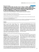
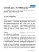
Intracellular association of APO3G and host RNAsFigure 1
Intracellular association of APO3G and host RNAs. (A)
Expression and immunoprecipitation of APO3G. HeLa cells
(5 × 10
6
) were transfected with 5 µg of pcDNA-Apo3G-
MycHis plasmid DNA. Cells were harvested 24 h post trans-
fection. An aliquot of the transfected cells was used for the
analysis of APO3G expression as follows: Cell lysates were
immunoprecipitated with a polyclonal antibody to the myc
epitope tag (α-myc) or were mock immunoprecipitated
(mock). Immunoprecipitated samples and total cell lysate
(Total) were analyzed for the presence of APO3G by immu-
noblotting using an APO3G-specific polyclonal peptide anti-
body. (B) The remaining cells from above were used for RT-
PCR analysis as follows: Total cellular RNA (Total) or RNA
present in the immune complexes (α-myc and mock, respec-
tively) was extracted and used for RT-PCR analysis as
described in Methods. Primer pairs were selected for the
specific amplification of the RNAs as indicated on the left.
Primer sequences are listed in table 1. All RT-PCR reactions
were performed simultaneously to minimize experimental
error. RT-PCR products were analyzed on 1% agarose gels
and visualized by staining with ethidium bromide. (C) HeLa
cells (5 × 10
6
) were transfected with 5 µg of pcDNA-Apo3G-
MycHis plasmid DNA (lanes 1 & 3) or 5 µg of pcDNA-
Apo3G (lanes 2 & 4). Cells were harvested 24 h post trans-
fection and analyzed as in panels A and B. (D) The specificity
of the RT-PCR reaction was validated using 7SL RNA as a
substrate. Total cellular RNA from panel B was either left
untreated (-) or treated with RNase A (50 µg/ml) for 60 min
at 37°C (+) prior to RT-PCR.
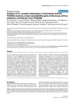
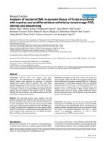
Schematic representation of constructs used in the studyFigure 2
Schematic representation of constructs used in the study.
Constructs are discussed in the text. All constructs carry an
out-of-frame deletion in the vif gene as described previously
[50]. The nucleotide changes in the stem portion of stem-
loop 1 region in mS.1∆Vif and the alignment of wild type and
DB653 zinc finger residues are shown.
Retrovirology 2007, 4:48 />Page 5 of 11
(page number not for citation purposes)
NC zinc finger motifs. The genomic RNA content of
DB653 particles was reported to be less than 10% of wild
type virus [45].
Particles were produced by transfecting HeLa cells with
appropriate plasmid DNAs in the presence of APO3G.
Viruses were purified and concentrated as described in
Methods. Aliquots were used for immunoblot analysis to
determine viral protein content and to verify APO3G
packaging (Fig. 3A). Other aliquots of the concentrated
viruses were used to extract particle-associated RNA,
which was then used for RT-PCR analysis (Fig. 3C). Con-
sistent with our previous report, immunoblot analysis
showed that NL4-3∆Vif packaged significantly higher
amounts of APO3G than C-Help∆Vif and mS.1∆Vif parti-
cles (Fig. 3A). Packaging of APO3G was quantified by den-
sitometric scanning of the APO3G bands. Results were
corrected for fluctuations in capsid (CA) levels and are
presented as percentage of APO3G packaged into NL4-
3∆Vif particles, which was defined as 100% (Fig. 3B).
Consistent with our previous data [18], packaging of
APO3G into C-Help∆Vif and mS.1∆Vif particles was
reduced to about 25–30% of wild type levels.
Equal numbers of particles, as judged by reverse tran-
scriptase activity, were used for extraction of RNA, which
was then used for RT-PCR using a series of primers as
shown in figure 3C and detailed in table 1. All RT-PCR
reactions shown in figure 3C were done simultaneously.
Amplification by an HIV-1 specific primer confirmed the
presence of genomic RNA in NL4-3∆Vif and mS.1∆Vif
particles and verified the lack of detectable amounts of
genomic RNA in C-Help∆Vif preparations (Fig. 3C, HIV-
1). In contrast, amplification of 7SL RNA as well as β-
actin, GAPDH, and α-tubulin mRNAs yielded comparable
amounts of PCR products indicative of the presence of
similar levels of these cellular RNAs in all three particle
preparations. These results suggest that packaging of these
RNAs was independent of the presence or absence of viral
genomic RNA (Fig. 3C). Similarly, U1, U2, U4, and U6
small nuclear RNAs were amplified with similar efficiency
from all three particle preparations. while human Y5 RNA
was virtually absent from the particles. On the other hand,
hY1, hY3, and hY4 RNAs appeared to be packaged more
efficiently into C-Help∆Vif particles than into NL4-3∆Vif
and mS.1∆Vif virions. The less efficient packaging of hY1,
hY3, and hY4 RNAs into NL4-3∆Vif and mS.1∆Vif parti-
cles is unrelated to APO3G encapsidation as APO3G lev-
els in mS.1∆Vif particles were as low as in C-Help∆Vif
(Fig. 3A &3B). Importantly, there was no obvious correla-
tion between APO3G packaging and encapsidation of any
of the tested cellular RNAs.
Packaging of hY RNAs requires the NC zinc finger domains
The increased packaging of hY RNAs into particles lacking
genomic RNA could indicate a competitive mechanism in
which viral genomic RNA competes for a common pack-
aging domain. Since viral genomic RNA is packaged
through an interaction with the NC zinc finger domain,
we investigated the impact of zinc finger mutations on the
packaging of hY RNAs. In addition, we assessed the
impact of zinc finger mutations on the packaging of
genomic RNA and 7SL RNA as well as APO3G (Fig. 4).
NL4-3∆Vif and DB653∆Vif particles were produced from
transfected HeLa cells as described for figure 3. Cell lysates
and concentrated cell-free virions were subjected to
immunoblot analysis to verify comparable amounts of
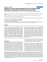
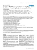
Correlation between cellular and viral RNA encapsidation and APO3G packagingFigure 3
Correlation between cellular and viral RNA encapsidation
and APO3G packaging. HeLa cells were co-transfected with
pcDNA-APO3G-MycHis together with vif-defective variants
of either pNL4-3 (43∆Vif), pC-Help (C-Help∆Vif), or mS.1
(mS.1∆Vif). Viruses were harvested 24 h after transfection
and purified as described in Methods. (A) Virus production
and packaging of APO3G was monitored by immunoblot
analysis using an aliquot of the purified, concentrated virus
preparations. APO3G encapsidation was identified using a
polyclonal APO3G-specific peptide antibody. Viral capsid
proteins (CA) were identified using an HIV-positive human
patient serum (APS). (B) APO3G-specific bands in panel A
were quantified by densitometric scanning and corrected for
fluctuations in capsid levels. Results were calculated relative
to APO3G associated with NL4-3∆Vif particles, which was
defined as 100%. (C) RNAs were extracted from purified,
concentrated viruses and amplified by RT-PCR using primer
pairs specific for HIV-1 RNA or host RNAs as indicated on
the left and detailed in table 1. RT-PCR products were sepa-
rated on 1% agarose gels and visualized by staining with
ethidium bromide.
Retrovirology 2007, 4:48 />Page 6 of 11
(page number not for citation purposes)
viral Gag proteins and to assess the encapsidation of
APO3G into NC zinc finger mutant particles. Consistent
with previous reports [9,11-14,16] we found that muta-
tion of the NC zinc finger domain abolished packaging of
APO3G into virus-like particles (Fig. 4A, DB653∆Vif).
For RT-PCR analysis, C-Help∆Vif RNA from figure 3C was
included for comparison. As before particles were normal-
ized for equal reverse transcriptase activity. RT-PCR analy-
sis using HIV-1-specific primers confirmed the absence of
viral genomic RNA in C-Help∆Vif and the DB653∆Vif zinc
finger mutant (Fig. 4B). As before, hY5 RNA was virtually
absent from all particle preparations including the zinc
finger mutant. Interestingly, packaging of 7SL RNA was
not affected by mutation of the NC zinc fingers suggesting
that 7SL RNA is packaged in an NC-independent manner.
In contrast, packaging of hY1, hY3, and hY4 RNAs was
critically dependent on intact NC zinc finger domains
(Fig. 4B). Thus, packaging of hY RNAs is indeed NC-
dependent and the absence of hY RNAs from NL4-3∆Vif
particles is best explained by competitive binding of viral
genomic RNA and hY RNA to NC.
7SL RNA does not promote SRP54 encapsidation
7SL RNA (also referred to as SRP RNA) is a component of
the signal recognition particle (SRP), which is critical for
the targeting of nascent secretory and membrane proteins
to the endoplasmic reticulum membrane (for review see
[46]). SRP54 is one of six protein subunits that constitute
mammalian SRPs and is responsible for high affinity
assembly of 7SL RNA into the SRP complex (reviewed in
[47]). Given the fact that 7SL RNA was efficiently pack-
aged into HIV-1 virions, we wanted to test whether intra-
cellular high affinity 7SL RNA-SRP54 interactions would
result in the recruitment of SRP54 rather than APO3G
into HIV-1 virions.
First, we verified the association of 7SL RNA with SRP54
in normal HeLa cells. For that purpose, HeLa cell lysates
were adsorbed to SRP54 reactive autoantibodies and
immunoprecipitation of SRP54 was confirmed by immu-
noblotting using an SRP54-specific antibody (Fig. 5A, top
panel, SRP). The specificity of the reaction was verified by
the absence of SRP54 protein in mock-immunoprecipi-
tated samples (Fig. 5A, mock) and by the absence of α-
tubulin in SRP54-specific and mock precipitates (Fig. 5A,
middle panel). Total RNA extracted from the immunopre-
cipitates revealed the presence of 7SL RNA in SRP54-spe-
cific but not in mock immunoprecipitated samples (Fig.
5A, lower panel).
Next, the packaging of SRP54 protein into HIV-1 virions
was tested. Virus particles were produced as described for
figure 3 except that APO3G was omitted in these samples.
Cell lysates and concentrated virus preparations were used
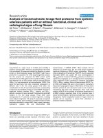
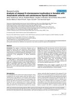
Packaging of hY RNAs requires the NC zinc finger domainsFigure 4
Packaging of hY RNAs requires the NC zinc finger domains.
HeLa cells were co-transfected with pcDNA-APO3G-
MycHis together with pNL4-3∆Vif (43∆Vif) or pDB653∆Vif.
Viruses were harvested 24 h after transfection and purified
as described in Methods. (A) Virus production and packaging
of APO3G was monitored by immunoblot analysis using an
aliquot of the purified, concentrated virus preparations.
APO3G encapsidation was identified using a polyclonal
APO3G-specific peptide antibody. Viral capsid proteins (CA)
were identified using an HIV-positive human patient serum
(APS). (B) RNAs were extracted from purified, concen-
trated viruses and amplified by RT-PCR using primer pairs
specific for HIV-1 RNA or host RNAs as indicated on the left
and detailed in table 1. RNA extracted from C-Help∆Vif
preparations in figure 3 was included as control. RT-PCR
products were separated on 1% agarose gels and visualized
by staining with ethidium bromide.
Retrovirology 2007, 4:48 />Page 7 of 11
(page number not for citation purposes)
for immunoblotting and for RT-PCR analysis as described
for figure 3. The results are shown in figure 5B. All cell
lysates contained equal amounts of SRP54 and viral cap-
sid proteins as well as 7SL RNA (Fig. 5B, cell). Further-
more, all samples produced comparable amounts of cell-
free virions as judged from the immunoblot (Fig. 5B, CA)
and packaged comparable amounts of 7SL RNA (Fig. 5B,
7SL). Of note, SRP54 was virtually absent from the virus
preparations (Fig. 5B, SRP54), thus confirming and
extending a recent study that also did not find SRP54 pro-
tein in HIV-1 virions [28]. These results demonstrate that
intracellular RNA-protein interactions are not a predictor
for subsequent targeting of the proteins into viral parti-
cles.
Discussion
There is general agreement in the literature that APO3G
can severely impair replication of HIV-1 and other pri-
mate lentiviruses lacking functional Vif proteins. It is also
uncontested that the antiviral activity of APO3G – with
the notable exception of resting CD4+ T cells [22] –
requires the encapsidation of APO3G into nascent virions
(for review see [48,49]). However, the mechanism of
APO3G encapsidation is not fully understood. In vitro
studies demonstrated the ability of APO3G to interact
with viral Gag protein and the nucleocapsid region of the
viral Gag precursor was identified as the likely APO3G
binding site [9,12-14,16]. Consistent with this model,
studies on virus-like particles demonstrated efficient pack-
aging of APO3G in the absence of viral genomic RNA
[9,11-14,16] although some of these studies proposed
that non-specific cellular RNA may contribute to APO3G
encapsidation [9,11,14,16]. Our own data confirm the
importance of NC for encapsidation as APO3G was not
encapsidated into a zinc finger mutant (Fig. 4). The
absence of APO3G from DB653∆Vif particles combined
with the presence of low levels of APO3G in C-Help∆Vif
virions (Fig. 3) suggests that APO3G/NC interactions –
either with or without support from NC-dependent cellu-
lar RNAs – are sufficient for low level packaging of APO3G
into virus-like particles. However, the presence of
genomic RNA invariably increased the efficiency of
APO3G packaging (Figs. 3 &4). Importantly, our previous
analysis of helper virus-associated APO3G demonstrated
that APO3G packaged through genomic RNA-independ-
ent mechanism(s) is sensitive to detergent treatment and
thus most likely not associated with the viral nucleopro-
tein complex [18].
The current study was stimulated by recent reports on the
presence of cellular 7SL RNA and snRNAs in HIV-1 virions
or retroviral particles [28,32,41] as well as the characteri-
zation of cellular RNAs associated with intracellular
APO3G [19,23]. Our goal was to test the possible contri-
bution of these or other host RNAs towards the packaging
7SL RNA interaction is insufficient for incorporation of SRP54 protein into HIV-1 particlesFigure 5
7SL RNA interaction is insufficient for incorporation of
SRP54 protein into HIV-1 particles. (A) Cell lysates of
untransfected HeLa cells were immunoprecipitated with an
SRP54-specific antibody (IP) or were mock-precipitated
(Ctrl). Aliquots of total cell lysate (Total) and immunoprecip-
itates were subjected to immunoblot analysis using antibod-
ies to SRP54 (α-SRP54), α-tubulin (α-tubulin). RNA was
extracted from remaining cell lysate and immunoprecipitates
and used for RT-PCR amplification of 7SL RNA. (B) HeLa
cells were transfected vif-defective variants of either pNL4-3
(43∆Vif), pC-Help (C-Help∆Vif, or mS.1 (mS.1∆Vif). Trans-
fected cells and virus-containing supernatants were harvested
24 h after transfection. Virus-containing supernatants were
purified and concentrated as described in Methods. Cell and
viral lysates were analyzed by immunoblotting for virus pro-
duction using an HIV-positive patient serum (APS). Expres-
sion and packaging of SRP54 was analyzed using an SRP54-
specific antibody oα-SRP54). Total cellular RNA and RNA
extracted from concentrated viruses was used for RT-PCR
amplification of 7SL RNA.
Retrovirology 2007, 4:48 />Page 8 of 11
(page number not for citation purposes)
of APO3G into HIV-1 particles. Of the four hY RNAs pre-
viously identified in APO3G complexes [23], hY3 was
clearly identified in APO3G complexes while hY1 and
hY4 only weakly interacted with APO3G in our analysis
(Fig. 1B). Among the snRNAs tested, only U6 clearly co-
purified with APO3G complexes and U4 showed weak
interaction. This finding is interesting since U6 snRNA
localizes primarily to the nucleus and does not have a
known cytoplasmic function. Surprisingly β-actin mRNA,
which was previously reported to be absent from APO3G
complexes [19,23] as well as GADPH mRNA clearly co-
purified with APO3G in our study. In contrast, we con-
firmed that α-tubulin mRNA only poorly associated with
APO3G. The reasons for these discrepancies are not clear
and could be due to differences in experimental condi-
tions. Importantly, however, most RNAs tested in our
study were packaged into NL4-3∆Vif virions as well as
helper virus and mS.1∆Vif particles (Fig. 3C). Interest-
ingly, comparative RT-PCR analysis demonstrated that
hY1, hY3, and hY4 RNAs were more efficiently packaged
into C-Help∆Vif particles lacking viral genomic RNA than
into particles containing viral genomic RNA (Fig. 3C).
Subsequent analysis of an NC mutant revealed that these
hY RNAs are packaged through an NC-dependent mecha-
nism. Thus, their inefficient packaging into NL4-3∆Vif
and mS.1∆Vif particles may be explained by competitive
binding of viral genomic RNA to NC.
U6 snRNA was previously identified in RSV particles [41].
Interestingly, however, U1, U2, and U4 snRNA, all of
which were identified in our HIV preparations, were either
absent from RSV particles or only present in trace
amounts [41]. While it is possible that RSV and HIV differ
in the packaging of cellular RNAs, it is also possible that
the greater sensitivity of the RT-PCR approach used in our
study versus the northern blot analysis employed in the
RSV analysis contributed to the different findings. Of
note, 7SL RNA despite being packaged in molar excess rel-
ative to viral genomic RNA [28] did not promote the pack-
aging of SRP54 protein (Fig. 3B) consistent with a recent
report [28]. Thus, despite the high affinity interaction of
7SL RNA with SRP54, such intracellular interaction was
insufficient to promote packaging of SRP54 into cell-free
virions. Similarly, packaging of RNAs previously found in
association with intracellular APO3G complexes was
insufficient to support APO3G encapsidation. Thus, we
did not observe a correlation between the packaging of
cellular RNAs into HIV-1 particles and encapsidation of
APO3G. The exclusion of APO3G from C-Help∆Vif parti-
cles lacking genomic RNA but containing high levels of
cellular RNAs and the absence of APO3G from mS.1∆Vif
particles containing genomic RNA with mutations in the
stem-loop 1 region of the 5' untranslated region point to
a role of viral genomic RNA in the packaging of APO3G.
We cannot formally rule out that other, thus far unidenti-
fied cellular RNA species contribute to the packaging of
APO3G into virus particles; however, this seems unlikely
since we would have to posit that such RNAs are specifi-
cally excluded from C-Help∆Vif and mS.1∆Vif particles.
Conclusion
We have demonstrated that vif-defective HIV-1 particles
package a variety of cellular RNAs. Most of the cellular
RNAs tested, except hY RNAs, were packaged independent
of viral genomic RNA. Packaging of hY RNAs was NC-
dependent and inhibited by viral genomic RNA. In all
experiments, APO3G packaging correlated well with the
presence of viral genomic RNA but not with the presence
of any of the cellular RNAs tested. Thus, our data do not
support a model in which APO3G is packaged through
non-specific or specific interaction with cellular RNAs. In
particular, we can rule out that packaging of 7SL RNA is
sufficient for the encapsidation of APO3G. Instead, we
propose that packaging of APO3G into virus particles is
mediated through interaction with viral genomic RNA.
Methods
Plasmids
The vif-defective molecular clone pNL4-3∆Vif [50] was
used for the production of vif-defective HIV-1 virus stocks.
Plasmid pC-Help∆Vif was used for the production of vif-
defective Ψ
-
virus-like particles (VLP). These particles con-
tain undetectable levels of viral genomic RNA [18]. Plas-
mid pNL4-3mS.1∆Vif carries mutations in stem-loop 1 of
the 5'-untranslated region [51] and was constructed by
subcloning the mutated stem-loop 1 region into the vif-
defective pNL4-3 vector [18]. NL4-3mS.1∆Vif particles are
Ψ
+
but do not support the encapsidation of APO3G [18].
A vif-defective variant of DB653 [45] was constructed by
transferring the Gag region of DB653 into pNL4-3Vif(-)
using standard cloning techniques. The structures of these
constructs are schematically shown in figure 2. Construc-
tion of pcDNA-Apo3G-MycHis for the expression of C-ter-
minally epitope-tagged wild type human APO3G proteins
was described previously [7] and untagged version,
pcDNA-Apo3G, was construction by introducing a stop
coding at the end of the APO3G gene [52].
Tissue culture and transfection
HeLa cells, which do not express endogenous APO3G,
were propagated in Dulbecco's modified Eagles medium
(DMEM) containing 10% fetal bovine serum (FBS). For
transfection, HeLa cells were grown in 25 cm
2
flasks to
about 80% confluency. Cells were transfected using Lipo-
fectAMINE PLUS™ (Invitrogen Corp, Carlsbad CA) fol-
lowing the manufacturer's recommendations. A total of 5
µg of plasmid DNA per 25 cm
2
flask (5 × 10
6
cells) was
generally used. Cells were harvested 24 h post transfec-
tion.
Retrovirology 2007, 4:48 />Page 9 of 11
(page number not for citation purposes)
Preparation of virus stocks
Virus stocks were prepared by transfecting HeLa cells with
pNL4-3∆Vif, pC-Help∆Vif, or pNL4-3mS.1∆Vif DNAs in
the presence or absence of APO3G expression vector as
indicated in the text. Virus-containing supernatants were
harvested 24 h after transfection. Cellular debris was
removed by centrifugation (5 min, 1500 rpm) and clari-
fied supernatants were filtered (0.45 µm) to remove resid-
ual cellular contaminants. For immunoblot analysis of
viral proteins and RNA extraction, virus-containing super-
natants (7 ml) were concentrated by ultracentrifugation
through 2 ml of 20% sucrose in PBS as described previ-
ously [7].
Antisera
APO3G was identified using a polyclonal rabbit serum
against a synthetic peptide comprising the 17 C-terminal
residues of APO3G. Serum from an HIV-positive patient
(APS) was used to detect HIV-1-specific capsid (CA) pro-
teins. Tubulin was identified using an α-tubulin-specific
monoclonal antibody (Sigma-Aldrich, Inc., St. Louis
MO). SRP54 protein was detected with a SRP54-specific
monoclonal antibody (BD Biosciences, San Jose, CA).
Immunoprecipitation of APO3G was done using a poly-
clonal antibody raised against the myc tag (Sigma-Aldrich,
Inc., St. Louis, MO). A human SRP54-reactive autoim-
mune serum was used for immunoprecipitation of SRP54
protein (kind gift of Frederick W. Miller, NIEHS, NIH,
Bethesda, MD, USA).
Immunoblotting
HeLa cells transfected with APO3G were used to detect
cellular APO3G expression and untransfected HeLa cells
were used for the detection of endogenous SRP54 protein
by immunoblotting with appropriate antibodies. For
immunoblot analysis of cellular proteins, whole cell
lysates were prepared as follows. Cells were washed once
with PBS, suspended in 450 µl/10
7
cells with X-100 lysis
buffer (50 mM Tris-HCL pH7.5, 150 mM NaCl, 0.5% Tri-
ton X-100). For Western blot analysis 50 µl aliquot was
taken and mixed with equal volume of sample buffer (4%
sodium dodecyl sulfate [SDS], 25 mM Tris-HCL, pH 6.8,
10% 2-mercaptoethanol, 10% glycerol, and 0.002%
bromphenol blue). Proteins were solubilized by boiling
for 5 min at 95°C with occasional vortexing of the sam-
ples to shear chromosomal DNA. Residual insoluble
material was removed by centrifugation (2 min, 15,000
rpm in an Eppendorf Minifuge). For immunoblot analysis
of virus-associated proteins, concentrated viral pellets
were suspended in a 1:1 mixture of PBS and sample buffer
and boiled. Cell lysates and viral extracts were subjected to
SDS-polyacrylamide gel electrophoresis; proteins were
transferred to polyvinylidene difluoride membranes and
reacted with appropriate antibodies as described in the
text. Membranes were then incubated with horseradish
peroxidase-conjugated secondary antibodies (Amersham
Bioscience, Piscataway, NJ) and visualized by enhanced
chemiluminescence (Amersham Bioscience).
Immunoprecipitation analysis
HeLa cells were transfected with pcDNA-APO3G-MycHis.
Cells were harvested at 24 h post transfection cell lysates
were prepared as follows: Cells were divided into two une-
qual fractions (30% and 70%). The larger fraction was
used for immunoprecipitation studies and the smaller
fraction was used for RNA extraction (see below). For
immunoprecipitation, cells were washed once with PBS
and lysed in 450 µl of lysis buffer (50 mM Tris, pH7.5, 150
mM, NaCl 0.5% and Triton X-100). The cell extracts were
clarified by centrifugation (13,000 × g, 3 min) and the
supernatant was incubated on a rotating wheel for 1 h at
4°C with protein A-Sepharose beads (Sigma-Aldrich, Inc.,
St. Louis MO) coupled with (IP) or without (Ctrl) anti-
myc rabbit polyclonal antibody (Sigma-Aldrich, Inc., St.
Louis MO). Immunocomplexes were washed three times
with wash buffer (50 mM Tris, 300 mM NaCl, and 0.1%
Triton X-100 (pH 7.4). Bound proteins were eluted form
beads by heating in sample buffer for 5 min at 95°C and
analyzed by immunoblotting using antibodies as indi-
cated in the text. For immunoprecipitation of APO3G-
RNA complexes, cell extracts were subjected to immuno-
precipitation by antibody covered beads or control beads
as described above and washed three times with RNA-pro-
tein binding buffer (20 mM HEPES, 25 mM KCl, 7 mM 2-
Mercaptoethanol, 5% Glycerol and 0.1% NP-40). Bound
RNA was then extracted as described below.
RNA extraction
Total cellular RNA was extracted from untransfected and
transfected HeLa cells using the RNeasy RNA extraction kit
(QIAGEN, Valencia, CA) following the manufacturer's
instructions. To isolate RNA from immunocomplexes,
beads were washed three times with RNA-protein binding
buffer (20 mM HEPES, 25 mM KCl, 7 mM 2-Mercaptoeth-
anol, 5% Glycerol and 0.1% NP-40). RNA was then
extracted using RNeasy RNA extraction kit. For isolation
of SRP54-associated RNA, SRP54 was precipitated with
SRP54-reactive human autoantibodies derived from a
patient with polymyositis ([53]; gift of Frederick W Miller,
NIEHS, NIH, Bethesda, MD, USA). RNA was then
extracted from the immunocomplexes as before
RT-PCR
RNA extracted from cells, viruses, or immunocomplexes
was treated with RNase-free DNase I (10 units, 30 min,
37°C) prior to the RT-PCR reaction. RNA concentrations
were determined by spectrophotometry. RT-PCR was per-
formed using equal amounts of RNA and the one-step RT-
PCR kit (QIAGEN, Valencia, CA) according to the manu-
facturer's instruction. Primers for the amplification of spe-
Retrovirology 2007, 4:48 />Page 10 of 11
(page number not for citation purposes)
cific RNAs are listed in table 1. RNA was first reverse
transcribed at 50°C for 30 minutes followed by 30 PCR
cycles (denaturation at 94°C; 15 sec; annealing at 55°C,
30 sec; and extension at 72°C, 1 min) and one 10-minute
extension cycle at 72°C. RT-PCR products were mixed
with DNA loading buffer (EDTA 20 mM, TAE 5×, Glycerol
50% and 0.002% Bromphenol Blue dye), electrophoresed
in 1% agarose gels, and visualized by staining with ethid-
ium bromide. A DNA size marker was run in parallel.
Competing interests
The author(s) declare that they have no competing inter-
ests.
Authors' contributions
M.K. conceived the study, was leading the execution of the
experiments, and participated in the writing of the manu-
script. K.S. coordinated and supervised the study and was
involved in the writing of the manuscript. R.G., S.O., E.M.,
H.T., and S.K. participated in virus production and sample
preparation and provided critical comments on the man-
uscript. All authors read and approved the final manu-
script.
Acknowledgements
We are grateful to Frederick Miller (NIEHS, NIH) for providing SRP54-
reactive human autoimmune serum. We thank Jared Clever and Tristram
Parslow for the mS.1 mutant. Plasmid DB653 was a generous gift of Robert
Gorelick (AIDS Vaccine Research Program, NCI). Part of this work was
supported by a grant from the NIH Intramural AIDS Targeted Antiviral
Program to K.S. and by the Intramural Research Program of the NIH,
NIAID to K.S.
Table Refs [23,28,41,54]
References
1. Conticello SG, Thomas CJ, Petersen-Mahrt SK, Neuberger MS: Evo-
lution of the AID/APOBEC family of polynucleotide
(deoxy)cytidine deaminases. Mol Biol Evol 2005, 22:367-377.
2. Jarmuz A, Chester A, Bayliss J, Gisbourne J, Dunham I, Scott J, Navar-
atnam N: An anthropoid-specific locus of orphan C to U RNA-
editing enzymes on chromosome 22. Genomics 2002,
79:285-296.
3. Rogozin IB, Basu MK, Jordan IK, Pavlov YI, Koonin EV: APOBEC4,
a New Member of the AID/APOBEC Family of Polynucle-
otide (Deoxy)cytidine Deaminases Predicted by Computa-
tional Analysis. Cell Cycle 2005, 4:1281-1285.
4. Wedekind JE, Dance GS, Sowden MP, Smith HC: Messenger RNA
editing in mammals: new members of the APOBEC family
seeking roles in the family business. Trends Genet 2003,
19:207-216.
5. Mariani R, Chen D, Schrofelbauer B, Navarro F, Konig R, Bollman B,
Munk C, Nymark-McMahon H, Landau NR: Species-specific exclu-
sion of APOBEC3G from HIV-1 v irions by Vif. Cell 2003,
114:21-31.
6. Marin M, Rose KM, Kozak SL, Kabat D: HIV-1 Vif protein binds
the editing enzyme APOBEC3G and induces its degradation.
Nat Med 2003, 9:1398-1403.
7. Kao S, Khan MA, Miyagi E, Plishka R, Buckler-White A, Strebel K: The
human immunodeficiency virus type 1 Vif p rotein reduces
intracellular expression and inhibits packaging of
APOBEC3G (CEM15), a cellular inhibitor of virus infectivity.
J Virol 2003, 77:11398-11407.
8. Yu X, Yu Y, Liu B, Luo K, Kong W, Mao P, Yu XF: Induction of
APOBEC3G ubiquitination and degradation by an HIV-1 Vif-
Cul5-SCF complex. Science 2003, 302:1056-1060.
9. Zennou V, Perez-Caballero D, Gottlinger H, Bieniasz PD:
APOBEC3G incorporation into human immunodeficiency
virus type 1 particles. J Virol 2004, 78:12058-12061.
10. Mangeat B, Turelli P, Caron G, Friedli M, Perrin L, Trono D: Broad
antiretroviral defence by human APOBEC3G through lethal
editing of nascent reverse transcripts. Nature 2003,
424:99-103.
11. Svarovskaia ES, Xu H, Mbisa JL, Barr R, Gorelick RJ, Ono A, Freed EO,
Hu WS, Pathak VK: Human apolipoprotein B mRNA-editing
enzyme-catalytic polypeptide-like 3G (APOBEC3G) is incor-
porated into HIV-1 virions through interactions with viral
and nonviral RNAs. J Biol Chem 2004, 279:35822-35828.
12. Alce TM, Popik W: APOBEC3G is incorporated into virus-like
particles by a direct interaction with HIV-1 Gag nucleocapsid
protein. J Biol Chem 2004, 279:34083-34086.
13. Cen S, Guo F, Niu M, Saadatmand J, Deflassieux J, Kleiman L: The
interaction between HIV-1 Gag and APOBEC3G. J Biol Chem
2004, 279:33177-33184.
14. Schafer A, Bogerd HP, Cullen BR: Specific packaging of
APOBEC3G into HIV-1 virions is mediated by the nucleo-
capsid domain of the gag polyprotein precursor. Virology 2004,
328:163-168.
15. Douaisi M, Dussart S, Courcoul M, Bessou G, Vigne R, Decroly E:
HIV-1 and MLV Gag proteins are sufficient to recruit
APOBEC3G into virus-like particles. Biochem Biophys Res Com-
mun 2004, 321:566-573.
16. Luo K, Liu B, Xiao Z, Yu Y, Yu X, Gorelick R, Yu XF: Amino-termi-
nal region of the human immunodeficiency virus type 1
nucleocapsid is required for human APOBEC3G packaging.
J Virol 2004, 78:11841-11852.
17. Burnett A, Spearman P: APOBEC3G Multimers Are Recruited
to the Plasma Membrane for Packaging into Human Immu-
nodeficiency Virus Type 1 Virus-Like Particles in an RNA-
Dependent Process Requiring the NC Basic Linker. J Virol
2007, 81:5000-5013.
18. Khan MA, Kao S, Miyagi E, Takeuchi H, Goila-Gaur R, Opi S, Gipson
CL, Parslow TG, Ly H, Strebel K: Viral RNA is required for the
association of APOBEC3G with human immunodeficiency
virus type 1 nucleoprotein complexes. J Virol 2005,
79:5870-5874.
19. Kozak SL, Marin M, Rose KM, Bystrom C, Kabat D: The anti-HIV-
1 editing enzyme APOBEC3G binds HIV-1 RNA and mes-
senger RNAs that shuttle between polysomes and stress
granules. J Biol Chem 2006, 281:29105-29119.
20. Soros VB, Yonemoto W, Greene WC: Newly synthesized
APOBEC3G is incorporated into HIV virions, inhibited by
HIV RNA, and subsequently activated by RNase H. PLoS
Pathog 2007, 3:e15.
21. Iwatani Y, Takeuchi H, Strebel K, Levin JG: Biochemical Activities
of Highly Purified, Catalytically Active Human APOBEC3G:
Correlation with Antiviral Effect. J Virol 2006, 80:5992-6002.
22. Chiu YL, Soros VB, Kreisberg JF, Stopak K, Yonemoto W, Greene
WC: Cellular APOBEC3G restricts HIV-1 infection in resting
CD4+ T cells. Nature 2005, 435:108-114.
23. Chiu YL, Witkowska HE, Hall SC, Santiago M, Soros VB, Esnault C,
Heidmann T, Greene WC: High-molecular-mass APOBEC3G
complexes restrict Alu retrotransposition. Proc Natl Acad Sci
USA 2006, 103:15588-15593.
24. Adkins B, Hunter T: Identification of a packaged cellular mRNA
in virions of rous sarcoma virus. J Virol 1981, 39:471-480.
25. Aronoff R, Linial M: Specificity of retroviral RNA packaging. J
Virol 1991, 65:71-80.
26. Berkowitz R, Fisher J, Goff SP: RNA packaging. Curr Top Microbiol
Immunol 1996, 214:177-218.
27. Onafuwa-Nuga AA, King SR, Telesnitsky A: Nonrandom packag-
ing of host RNAs in moloney murine leukemia virus. J Virol
2005, 79:13528-13537.
28. Onafuwa-Nuga AA, Telesnitsky A, King SR: 7SL RNA, but not the
54-kd signal recognition particle protein, is an abundant
component of both infectious HIV-1 and minimal virus-like
particles. RNA 2006, 12:542-546.
Publish with BioMed Central and every
scientist can read your work free of charge
"BioMed Central will be the most significant development for
disseminating the results of biomedical research in our lifetime."
Sir Paul Nurse, Cancer Research UK
Your research papers will be:
available free of charge to the entire biomedical community
peer reviewed and published immediately upon acceptance
cited in PubMed and archived on PubMed Central
yours — you keep the copyright
Submit your manuscript here:
/>BioMedcentral
Retrovirology 2007, 4:48 />Page 11 of 11
(page number not for citation purposes)
29. Muriaux D, Mirro J, Harvin D, Rein A: RNA is a structural ele-
ment in retrovirus particles. Proc Natl Acad Sci USA 2001,
98:5246-5251.
30. Jiang M, Mak J, Ladha A, Cohen E, Klein M, Rovinski B, Kleiman L:
Identification of tRNAs incorporated into wild-type and
mutant human immunodeficiency virus type 1. J Virol 1993,
67:3246-53.
31. Linial M, Medeiros E, Hayward WS: An avian oncovirus mutant
(SE 21Q1b) deficient in genomic RNA: biological and bio-
chemical characterization. Cell 1978, 15:1371-1381.
32. Rulli SJ Jr, Hibbert CS, Mirro J, Pederson T, Biswal S, Rein A: Selec-
tive and Nonselective Packaging of Cellular RNAs in Retro-
virus Particles. J Virol 2007, 81:6623-6631.
33. Gallis B, Linial M, Eisenman R: An avian oncovirus mutant defi-
cient in genomic RNA: characterization of the packaged
RNA as cellular messenger RNA. Virology 1979, 94:146-161.
34. Erikson RL: Studies on the RNA from avian myeloblastosis
virus. Virology 1969, 37:124-131.
35. Bishop JM, Levinson WE, Sullivan D, Fanshier L, Quintrell N, Jackson
J: The low molecular weight RNAs of Rous sarcoma virus. II.
The 7 S RNA. Virology 1970, 42:927-937.
36. Erikson E, Erikson RL, Henry B, Pace NR: Comparison of oligonu-
cleotides produced by RNase T1 digestion of 7 S RNA from
avian and murine oncornaviruses and from uninfected cells.
Virology 1973, 53:40-46.
37. Faras AJ, Garapin AC, Levinson WE, Bishop JM, Goodman HM: Char-
acterization of the low-molecular-weight RNAs associated
with the 70S RNA of Rous sarcoma virus. J Virol 1973,
12:334-342.
38. Sawyer RC, Dahlberg JE: Small RNAs of Rous sarcoma virus:
characterization by two-dimensional polyacrylamide gel
electrophoresis and fingerprint analysis. J Virol 1973,
12:1226-1237.
39. Walker TA, Pace NR, Erikson RL, Erikson E, Behr F: The 7S RNA
common to oncornaviruses and normal cells is associated
with polyribosomes. Proc Natl Acad Sci USA 1974, 71:3390-3394.
40. Walter P, Blobel G: Signal recognition particle contains a 7S
RNA essential for protein translocation across the endoplas-
mic reticulum. Nature 1982, 299:691-698.
41. Giles KE, Caputi M, Beemon KL: Packaging and reverse tran-
scription of snRNAs by retroviruses may generate pseudo-
genes. RNA 2004, 10:299-307.
42. Perreault J, Noel JF, Briere F, Cousineau B, Lucier JF, Perreault JP,
Boire G: Retropseudogenes derived from the human Ro/SS-A
autoantigen-associated hY RNAs. Nucleic Acids Res 2005,
33:2032-2041.
43. Ikawa Y, Ross J, Leder P: An association between globin messen-
ger RNA and 60S RNA derived from Friend leukemia virus.
Proc Natl Acad Sci USA 1974, 71:1154-1158.
44. Mochizuki H, Schwartz JP, Tanaka K, Brady RO, Reiser J: High-titer
human immunodeficiency virus type 1-based vector systems
for gene delivery into nondividing cells. J Virol 1998,
72:8873-8883.
45. Guo J, Wu T, Anderson J, Kane BF, Johnson DG, Gorelick RJ, Hend-
erson LE, Levin JG: Zinc finger structures in the human immu-
nodeficiency virus type 1 nucleocapsid protein facilitate
efficient minus- and plus-strand transfer. J Virol 2000,
74:8980-8988.
46. Keenan RJ, Freymann DM, Stroud RM, Walter P: The signal recog-
nition particle. Annu Rev Biochem 2001, 70:755-775.
47. Doudna JA, Batey RT: Structural insights into the signal recog-
nition particle. Annu Rev Biochem 2004, 73:539-557.
48. Hache G, Mansky LM, Harris RS: Human APOBEC3 proteins,
retrovirus restriction, and HIV drug resistance. AIDS Rev
2006, 8:148-157.
49. Chiu YL, Greene WC: APOBEC3 Cytidine Deaminases: Dis-
tinct Antiviral Actions along the Retroviral Life Cycle. J Biol
Chem 2006, 281:8309-8312.
50. Karczewski MK, Strebel K: Cytoskeleton association and virion
incorporation of the human immunodeficiency virus type 1
Vif protein. J Virol 1996, 70:494-507.
51. Clever JL, Parslow TG: Mutant human immunodeficiency virus
type 1 genomes with defects in RNA dimerization or encap-
sidation. J Virol 1997, 71:3407-3414.
52. Opi S, Takeuchi H, Kao S, Khan MA, Miyagi E, Goila-Gaur R, Iwatani
Y, Levin JG, Strebel K: Monomeric APOBEC3G Is Catalytically
Active and Has Antiviral Activity. J Virol 2006, 80:4673-4682.
53. Romisch K, Miller FW, Dobberstein B, High S: Human autoanti-
bodies against the 54 kDa protein of the signal recognition
particle block function at multiple stages. Arthritis Res Ther
2006, 8:R39.
54. Funaki H, Nishimura G, Harada S, Ninomiya I, Terada I, Fushida S,
Tani T, Fujimura T, Kayahara M, Shimizu K, Ohta T, Miwa K: Expres-
sion of vascular endothelial growth factor D is associated
with lymph node metastasis in human colorectal carcinoma.
Oncology 2003, 64:416-422.