Báo cáo y học: " Human Immunodeficiency Virus Type 1 Nef protein modulates the lipid composition of virions and host cell membrane microdomains" pot
Bạn đang xem bản rút gọn của tài liệu. Xem và tải ngay bản đầy đủ của tài liệu tại đây (481.06 KB, 12 trang )
BioMed Central
Page 1 of 12
(page number not for citation purposes)
Retrovirology
Open Access
Research
Human Immunodeficiency Virus Type 1 Nef protein modulates the
lipid composition of virions and host cell membrane microdomains
Britta Brügger
1
, Ellen Krautkrämer
2
, Nadine Tibroni
2
, Claudia E Munte
3
,
Susanne Rauch
2
, Iris Leibrecht
1
, Bärbel Glass
2
, Sebastian Breuer
4
,
Matthias Geyer
4
, Hans-Georg Kräusslich
2
, Hans Robert Kalbitzer
3
,
Felix T Wieland
1
and Oliver T Fackler*
2
Address:
1
Heidelberg University Biochemistry Center (BZH), Heidelberg, Germany,
2
Abteilung Virologie, Universität Heidelberg, Heidelberg,
Germany,
3
Institut für Biophysik und Physikalische Biochemie, Universität Regensburg, Regensburg, Germany and
4
Max-Planck-Institut für
molekulare Physiologie, Abteilung Physikalische Biochemie, Dortmund, Germany
Email: Britta Brügger - ; Ellen Krautkrämer - ;
Nadine Tibroni - ; Claudia E Munte - ;
Susanne Rauch - ; Iris Leibrecht - ;
Bärbel Glass - ; Sebastian Breuer - ;
Matthias Geyer - ; Hans-Georg Kräusslich - ;
Hans Robert Kalbitzer - ; Felix T Wieland - ;
Oliver T Fackler* -
* Corresponding author
Abstract
Background: The Nef protein of Human Immunodeficiency Viruses optimizes viral spread in the infected host
by manipulating cellular transport and signal transduction machineries. Nef also boosts the infectivity of HIV
particles by an unknown mechanism. Recent studies suggested a correlation between the association of Nef with
lipid raft microdomains and its positive effects on virion infectivity. Furthermore, the lipidome analysis of HIV-1
particles revealed a marked enrichment of classical raft lipids and thus identified HIV-1 virions as an example for
naturally occurring membrane microdomains. Since Nef modulates the protein composition and function of
membrane microdomains we tested here if Nef also has the propensity to alter microdomain lipid composition.
Results: Quantitative mass spectrometric lipidome analysis of highly purified HIV-1 particles revealed that the
presence of Nef during virus production from T lymphocytes enforced their raft character via a significant
reduction of polyunsaturated phosphatidylcholine species and a specific enrichment of sphingomyelin. In contrast,
Nef did not significantly affect virion levels of phosphoglycerolipids or cholesterol. The observed alterations in
virion lipid composition were insufficient to mediate Nef's effect on particle infectivity and Nef augmented virion
infectivity independently of whether virus entry was targeted to or excluded from membrane microdomains.
However, altered lipid compositions similar to those observed in virions were also detected in detergent-
resistant membrane preparations of virus producing cells.
Conclusion: Nef alters not only the proteome but also the lipid composition of host cell microdomains. This
novel activity represents a previously unrecognized mechanism by which Nef could manipulate HIV-1 target cells
to facilitate virus propagation in vivo.
Published: 1 October 2007
Retrovirology 2007, 4:70 doi:10.1186/1742-4690-4-70
Received: 17 July 2007
Accepted: 1 October 2007
This article is available from: />© 2007 Brügger et al; licensee BioMed Central Ltd.
This is an Open Access article distributed under the terms of the Creative Commons Attribution License ( />),
which permits unrestricted use, distribution, and reproduction in any medium, provided the original work is properly cited.
Retrovirology 2007, 4:70 />Page 2 of 12
(page number not for citation purposes)
Background
The Nef protein of Human Immunodeficiency Viruses is a
multifunctional protein critical for high virus titers in
vivo. Consequently, disease progression in individuals
infected with nef deficient viruses is at least significantly
delayed [1-3]. These effects are thought to mirror inde-
pendent activities of Nef that prevent immune recognition
of virally infected cells and directly boost the replicative
potential of HIV [4,5]. To achieve such optimized spread
in the infected host, Nef manipulates a variety of transport
and signal transduction processes in cells infected by HIV-
1. Modulation of cellular transport paths by Nef affects
the surface presentation of an increasing number of cell
receptors like e.g. CD4, MHC class I and II molecules and
chemokine receptors [6-9]. Equally wide spread are Nef
effects on host cell signalling, including various altera-
tions of the TCR cascade in T lymphocytes. According to
an emerging view Nef can act as an intracellular inducer of
TCR distal events in the absence of exogenous stimulation
while signalling by exogenous TCR stimulation is tuned
down in the presence of the viral protein [10-14]. Finally,
during production of progeny virus, Nef augments the
intrinsic infectivity of cell-free HIV particles by a factor 5–
10 via a poorly characterized mechanism [15-17].
Associated with cellular membranes by virtue of its N-ter-
minal myristoylation and additional membrane targeting
motifs, a subpopulation of Nef resides in detergent resist-
ant membrane microdomains (DRMs) or lipid rafts [18-
23]. Lipid rafts are defined as highly dynamic microdo-
mains in cellular membranes that are enriched in sphin-
golipids, cholesterol and raft-targeted proteins. This
particular lipid and protein composition is thought to
facilitate protein-protein interactions to create microdo-
mains with distinct biological properties. Lipid rafts have
been implicated as platforms for central cellular processes
such as signal transduction and protein trafficking but are
also utilized as preferred sites for entry and egress of a
number of viruses, including HIV-1 [24-27]. Originally
defined as resistant to extraction with cold detergent, the
existence of these membrane microdomains in living cells
has been subject to intense debates [28-32]. This contro-
versy stemmed primarily from the lack of both, appropri-
ate live cell imaging techniques to visualize such
assemblies and detergent-free biochemical purification
protocols. Over the past years, the application of new dyes
such as Laurdan and the real-time visualization of protein
dynamics during signalling processes have largely corrob-
orated the membrane microdomain concept [33-37].
Moreover, our previous lipidome analysis of highly puri-
fied HIV-1 particles provided an example of a biological
membrane generated in the absence of detergent that dis-
plays a lipid composition with striking similarity to DRMs
[38].
Recent studies suggested that DRM incorporation of Nef
spatially separates its individual activities in infected cells.
The use of mutated Nef proteins that are enriched in
DRMs due to an additional palmitoylation signal or that
lack a di-lysine motif that facilitates DRM incorporation
of the viral protein, respectively, revealed that Nef activi-
ties in receptor transport are largely independent of its raft
association [19,21]. In contrast, signal transduction prop-
erties of Nef such as the association with the activated
form of the cellular Pak2 kinase strictly occurs within
membrane microdomains, thus providing spatial com-
partmentalization of individual Nef activities [19,20,39].
This concept is in line with a recent proteomic analysis
that revealed significant alterations in the DRM recruit-
ment of TCR machinery by Nef [40]. In contrast, conflict-
ing results exist as to what extent DRM association of Nef
determines its ability to enhance virion infectivity. An
early report demonstrated that this Nef activity depends
on raft integrity of the producer cell and suggested this to
reflect the Nef-mediated recruitment of HIV budding
structures into DRMs [23]. In line with a role for lipid rafts
in Nef-mediated infectivity enhancement, disruption of
DRM association of Nef correlates with a loss of infectivity
enhancement [19]. However, DRM recruitment of HIV
structural proteins was not observed in another study and
DRM enrichment of Nef failed to further boost particle
infectivity [21].
The biological properties of Nef were thus far largely
explained by its interactions with host cell proteins. A few
recent reports however suggest that Nef might also affect
biosynthesis and transport of select host cell lipid species.
These reports primarily focus on cellular cholesterol and
suggest a direct interaction of Nef with this sterol as well
as the induction of cholesterol biosynthesis genes by Nef
[41,42]. Via a mechanism specific to macrophages, Nef
was also reported to alter cholesterol efflux by interacting
with the ABCA1 transporter [43]. These results raised the
possibility that Nef might influence not only the protein
but also the overall lipid composition of host cell mem-
branes to optimize virus replication. To test this hypothe-
sis, we compared in this study the impact of Nef on the
lipidome of HIV-1 virions and T lymphocytes DRMs.
Results
Association of Nef with membrane microdomains is not
limiting for its effects on virion infectivity and viral
replication
We set out to analyze the lipidome of HIV-1 particles pro-
duced from MT-4 T lymphocytes in the presence (HIV-1
wt, wt) or absence (HIV-1∆Nef, ∆Nef) of Nef. As addi-
tional control, we generated a corresponding proviral
HIV-1 clone that encodes for a palmitoylated and thus
DRM enriched Nef variant [20,21] (PalmNef). Since the
effects of Nef on particle infectivity can depend on the
Retrovirology 2007, 4:70 />Page 3 of 12
(page number not for citation purposes)
nature of the producer cell [44,45], we first analyzed viri-
ons produced from infected MT-4 T lymphocytes for their
infectivity in a single round of replication on CD4-posi-
tive HeLa cells (Fig. 1A). As expected, infection with wt
resulted in approx. 7-fold more productively infected cells
per ng p24CA virus input than the ∆Nef virus. PalmNef
was only slightly more efficient (approx. 1.5-fold) than wt
Nef in this assay. Similar results were obtained when HIV-
1 replication was monitored over several rounds on
human primary T lymphocytes: while wt and PalmNef
expressing HIV-1 variants replicated with indistinguisha-
ble kinetics, spread of HIV-1∆Nef was delayed early post
infection (Fig. 1B). Thus, Nef robustly enhances the infec-
tivity of virions produced in MT-4 cells and DRM associa-
tion of the viral protein is not limiting for this activity.
Nef does not recruit HIV Gag into detergent resistant
microdomains in infected MT-4 T lymphocytes
To address potential reasons for the Nef-mediated
increase in virion infectivity in our experimental system,
we performed raft flotation experiments with lysates of
MT4 T lymphocytes infected with cell free virus stocks
(Fig. 1C). Cells were lysed in the presence of cold Tx-100
and, following a standard raft flotation procedure [20],
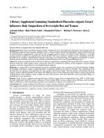
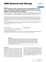
Nef boosts HIV-1 infectivity and replication without increasing microdomain association of Gag in producer cellsFigure 1
Nef boosts HIV-1 infectivity and replication without increasing microdomain association of Gag in producer
cells. (A) Single round of replication analysis on TZM cells. TZM cells were infected with 0.5 ng CA of the indicated virus
stocks. 36 hours post infection, the cells were fixed, stained for β-galactosidase activity and the number of blue cells was
counted. Data represent average values from three independent experiments with triplicate measurements each with the indi-
cated standard error of the mean. Depicted is the relative virion infectivity (number of blue cells per ng CA) with values for
HIV-1
NL4-3
NefSF2 (wt) arbitrarily set to 100%. (B) HIV-1 replication in PBL. HIV replication was measured in 96 well plates on
1 × 10
5
PBL per well and 1 ng CA virus input. Freshly isolated, non-activated cells were infected (day -6) for three days and
subsequently activated by PHA/IL-2 for three days. Starting from day 0, cells were kept in the presence of IL-2 and cell culture
supernatants were collected each day to monitor CA production. CA values represent the average from quadruplicate infec-
tions performed in parallel. (C-D) Lipid raft flotation analysis from infected MT-4 (C) or transfected Jurkat T lymphocytes (D).
Cell lysates (1% Triton X-100) were separated by Optiprep gradient ultracentrifugation, and eight fractions were collected
from the top (fraction 1) to the bottom (fraction 8) of the gradient. The detergent resistant membrane fraction (DRM, fraction
2) and the pooled nonraft (soluble) fractions (S, fractions 7 and 8) were analyzed together with the unfractionated cell lysate
(L) by Western Blotting for the distribution of Gag (top), Nef (middle) and TfR (bottom).
0
20
40
60
80
100
120
140
160
wt
'Nef
PalmNef
wt
'Nef
PalmNef
p24 [ng/ml]
20
40
60
80
12347
days p.i.
Relative virion infectivity [%]
AB
CD
0
wt
'Nef PalmNef
83 -
62 -
47 -
32 -
p55
p48
p41
p24
32 -
Nef/PalmNef
Infection (MT4)
DRM S L
DRM S L DRM S L
TfR
DRM S L
DRM S L DRM S L
83 -
62 -
47 -
32
-
32 -
wt
Transfection (Jurkat)
'Nef PalmNef
83 -
83 -
Retrovirology 2007, 4:70 />Page 4 of 12
(page number not for citation purposes)
equal volumes DRM (DRM) and soluble (S) fractions as
well as total cell lysate (L) were analyzed by Western Blot-
ting. The DRM excluded transferrin receptor (TfR) was
used as loding control (bottom panel). Only small
amounts of Nef were detected in the DRM fraction and the
microdomain association of PalmNef was significantly
more pronounced. Pr55
Gag
precursor, processing interme-
diates as well as fully processed p24CA were detected with
the anti-p24CA antibody (upper panel). The low levels of
p24CA in DRM fractions do not reflect a processing defect
but rather the lack of membrane targeting after physical
separation of CA from MA by protease cleavage. No Nef-
mediated enrichment of Pr55
Gag
in DRMs was detected in
infected MT-4 T lymphocytes (ratio of DRM-associated
relative to total Gag: wt: 39.4%; ∆Nef: 51.4%; PalmNef:
36.1%). These results are in agreement with a study by
Sol-Foulon et al. [21] that used HIV-1 infected Jurkat T
lymphocytes, however are in conflict with a report on Nef-
mediated DRM recruitment of Gag in 293T cells that were
transfected with proviral DNA [23]. To assess if this dis-
crepancy stems from the different ways of provirus deliv-
ery, we performed the same analysis in Jurkat T
lymphocytes transfected with HIV proviral plasmids (Fig.
1D), that expressed higher levels of Gag and Nef than
infected MT-4 cells. The presence of Nef resulted in a
slightly more pronounced accumulation of Pr55
Gag
in the
DRM fraction under these conditions (ratio of DRM-asso-
ciated relative to total Gag: wt: 29.8%; ∆Nef: 13.4%; Palm-
Nef: 32.3%), suggesting that in Jurkat cells, Nef-mediated
DRM recruitment by Nef is only observed upon transfec-
tion of proviral DNA. This most likely reflects the unphys-
iologically high levels of HIV-1 gene products per cell
following provirus transfection. Thus, Nef-mediated
recruitment of Gag into DRMs does not occur in the con-
text of T lymphocyte infection and is dispensable for Nef's
effects on virion infectivity.
Purification and characterization of HIV-1 virions
The above results together with previous reports on the
ability of Nef to interfere with cellular cholesterol biosyn-
thesis, homeostasis and transport [41-43] suggested that
Nef might increase virion infectivity by altering the com-
position of the lipid envelope of the particles. Using a pre-
viously validated purification scheme that yields particle
preparations that are essentially free of vesicle contamina-
tion [46], we recently established quantitative lipid mass
spectrometry of highly purified HIV particles from
infected MT-4 T lymphocytes to determine the lipid com-
position of HIV virions [38]. We employed this experi-
mental setup to analyze potential differences imprinted
by Nef and first assessed the relative incorporation of viral
proteins into purified wt, ∆Nef and PalmNef particles by
Western Blotting (Fig. 2A). No significant difference was
detected in the amounts of isolated Gag proteins (MA,
CA), viral glycoprotein (Env), viral enzymes (RT) and the
virion associated factor Vpr. Comparable amounts of both
wt and palmitoylated Nef were also detected, indicating
that DRM enrichment does not cause the accumulation of
virion-associated Nef under these experimental condi-
tions. Silver staining revealed comparable purity of all
preparations analyzed and no significant differences in
the virion incorporation of cellular proteins were
detected. Notably, the differences in relative infectivity
between wt, ∆Nef and PalmNef particles seen in cell cul-
ture supernatants were preserved following velocity gradi-
ent purifications of the particles (Fig. 2B). Furthermore,
based on the recovery of viral antigen relative to input
amounts prior to the purification, the lack of Nef did not
significantly alter the stability of HIV-1 particles (Fig. 2C).
HIV-1 Nef increases the raft character of virus particles
We next determined the full lipid composition of the var-
ious HIV-1 particle preparations (Fig. 3A). As we reported
recently [38], HIV-1 particles display a raft-like lipid com-
position. In line with the results on DRM incorporation of
HIV-1 Gag, the presence or absence of Nef had no global
effect on the lipid composition of HIV-1 particles. How-
ever, the analysis of individual lipid classes relative to
phosphatidylcholine (PC) (that was found to be constant
in all particle preparations) revealed a slight but signifi-
cant enrichment of sphingomyelin (SM) in virions pro-
duced in the presence of Nef or PalmNef relative to the
∆Nef controls (Fig. 3A) (average SM to PC ratio ∆Nef: 1.6,
wt: 2.1; p = 0.003 by student's t-test). Alterations in cellu-
lar SM levels in a similar range have recently been implied
to play important roles in Alzheimer's disease [47]. These
alterations were specific as Nef had no effect on the virion
incorporation of phosphatidylethanolamine (PE), plasm-
alogen-PE (pl-PE), or phosphatidylserine (PS). Further
Nef-specific differences were revealed by the quantitative
analysis of PC molecular species (Fig. 3B, C): The presence
of Nef and PalmNef during virus production increased the
virion amounts of mono- (wt: 39.8% vs. ∆Nef: 33.1%, p =
0.01) and di-unsaturated PC species (wt: 11.7% vs. ∆Nef:
9.1%, p = 0.0015) while both Nef proteins reduced the
virion incorporation of polyunsaturated PC by more than
two-fold (wt: 11.8% vs. ∆Nef: 23.7%, p = 0.0002).
Together these results demonstrate that Nef is not a key
determinant for the overall lipid composition of HIV-1
particles but enhances the incorporation of SM and trig-
gers the exclusion of polyunsaturated PC from HIV-1 par-
ticles, thereby enhancing their raft microdomain
character.
HIV-1 Nef has no effect on the incorporation of cholesterol
into HIV-1 particles
Based on the reported direct virion recruitment of choles-
terol by Nef, the upregulation of cholesterol biosynthesis
in Nef expressing cells, and the importance of virion cho-
lesterol for particle infectivity [41,42,48,49], we specifi-
Retrovirology 2007, 4:70 />Page 5 of 12
(page number not for citation purposes)
cally addressed the cholesterol content of the isolated
HIV-1 particles. As depicted in Fig. 4A, the presence of Nef
had no effect on the amounts of cholesterol present in our
HIV-1 virion preparations. This prompted us to analyze
the proposed direct interaction between Nef and choles-
terol in vitro using NMR spectroscopy. This method
detects ligand binding by chemical shift perturbation with
high sensitivity. The interaction of Nef with cholesterol
has been reported to occur via a cholesterol recognition
motif Leu
198
X
1–5
Tyr
202
X
1–5
Lys
204
[42] at the c-terminus
of the well folded core domain of Nef. Thus, cholesterol
dissolved in ethanol or cholesterol complexed with
methyl-β-cyclodextrine was added in increasing concen-
trations up to a final ratio of 1:2 to a solution containing
the
13
C/
15
N-labeled core domain structure (residues 44–
210) of Nef. Even at the highest concentrations choles-
terol specific shifts were neither observed in the
1
H spec-
trum (Fig. 4B) nor in the
1
H/
15
N HSQC spectrum showing
the main chain amide signals (Fig. 4C). Thus, no physical
interaction between Nef and cholesterol was detected
with this highly sensitive in vitro approach.
Besides SM, HIV-1 virions are also highly enriched in the
unusual sphingolipid dihydrosphingomyelin (DHSM)
and inhibition of sphingolipid synthesis resulted in a 5-
fold reduction in virion infectivity [38]. The magnitude of
this effect is remarkable close to that Nef exerts on HIV-1
infectivity. However, when we determined the DHSM lev-
els in HIV-1 virions produced in the absence of Nef, no
significant change in DHSM virion incorporation was
detected (data not shown). Together we conclude that the
presence of Nef alters the PC species distribution and SM
content of virus particles, while cholesterol and DHSM
levels are unaffected.
Increase of virion SM levels by Nef is insufficient for
elevating virion infectivity
We next sought to test whether the Nef-induced altera-
tions of viral envelope lipid composition are instrumental
for the elevated relative infectivity of virions produced in
the presence of Nef. To this end, we analyzed the effect of
Nef variants that were previously shown to lack infectivity
enhancement potential [50] on the viral lipidome. As
shown in Fig. 5A, the V78A, R81A and ED178/179AA var-
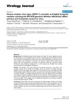
Characterization of purified HIV-1 particlesFigure 2
Characterization of purified HIV-1 particles. HIV-1 virions were purified from cell culture supernatants (see Materials
and Methods for details). (A), Western Blot and silver stain analysis of the indicated virion preparations for major viral particle
constituents. (B) Single round of replication analysis on TZM cells with the particle preparations analyzed in A. The assay was
performed analogous to that described in Fig. 1A. (C) Relative amounts of total cell culture supernatant p24 recovered after
the optiprep procedure. Depicted are average p24 amounts recovered in the virion preparation procedure relative to the total
input from four independent purifications with the indicated standard error of the mean.
RT
0
50
100
150
200
wt
'Nef
PalmNef
Relative virion infectivity [%]
0
5
10
15
20
wt
'Nef
PalmNef
Yield p24 [% of input]
Env
175 -
83 -
CA
25 -
MA
16 -
62 -
47 -
Vpr
16 -
Nef
25 -
wt
'Nef
PalmNef
p66
p51
AB
C
25 -
32 -
47 -
62 -
83 -
Silver
stain
Western
Blot
Retrovirology 2007, 4:70 />Page 6 of 12
(page number not for citation purposes)
iants of Nef failed to augment particle infectivity when
compared to ∆Nef. A Nef deletion mutant ∆12-39 dis-
played intermediate activity in this assay. When we ana-
lyzed the lipid composition of particles purified from cell
culture supernatants used for infection in A, all virion
preparations contained SM to PC ratios that were signifi-
cantly higher than ∆Nef particles (Fig. 5B). We therefore
conclude that the ability to augment relative SM levels in
virus particles is not sufficient to mediate Nef's effects on
virion infectivity.
Enhancement of virion infectivity by Nef is independent of
DRM association of the viral entry receptor CD4
Entry of HIV-1 into target cells occurs at the plasma mem-
brane and several but not all studies suggested that mem-
brane microdomains represent a preferential local
environment for this process [51-56]. While modulation
of the virion lipid composition was insufficient to explain
the positive effects of the viral protein on particle infectiv-
ity, this effect of Nef occurs via a microdomain-dependent
mechanism [23]. We therefore reasoned that the increased
microdomain character of virions produced in the pres-
ence of Nef might specifically increase infection events
that occur via lipid raft domains, e.g. by augmenting bind-
ing affinity or fusion efficiency of particles at target cell
microdomains. To test this hypothesis, we made use of
mutations in the primary entry receptor for HIV-1, CD4
that specifically target the receptor to or exclude it from
membrane microdomains and tested whether Nef's effect
on virion infectivity depends on the DRM localization of
CD4. While wt CD4 is distributed approximately equally
between DRM and detergent-soluble fractions, mutation
of the palmitoylation acceptor in the 2Cm CD4 variant
significantly reduced its raft association (approx. 25% in
DRM) and additional mutation of the Lck binding motif
in the cytoplasmic tail in the 4C CD4 variant almost com-
pletely disrupted its DRM association (approx. 5% in
DRM) [57]. Wt, 2Cm and 4C CD4 variants were tran-
siently expressed in HeLa cells which were subsequently
infected with wt or ∆Nef HIV-1. Control experiments con-
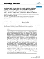
Lipid analysis of virus particlesFigure 3
Lipid analysis of virus particles. Quantitative lipid analysis was performed as described in Methods. Data are displayed as
molar ratio of individual lipid classes to PC. Values present the average from at least three independent experiments with error
bars indicating the standard deviation of the mean. (A) PC molecular species distribution given in % of total. (B) Number of
species in % of total either containing none, one, or two or more than two double bonds in both fatty acids. (C) Analysis of all
PC species. X:Y values on the x-axis denote the total number of C- atoms of both fatty acids (X) and the total number of dou-
ble bonds (Y), respectively.
0
5
10
15
20
25
30
3
0
:
0
3
2
:
2
3
2
:
1
3
2
:
0
3
4
:
3
3
4
:
2
3
4
:
1
3
4
:
0
3
6
:
5
3
6
:
4
3
6
:
3
3
6
:
2
3
6
:
1
3
6
:
0
3
8
:
7
3
8
:
6
3
8
:
5
3
8
:
4
3
8
:
3
3
8
:
2
3
8
:
1
3
8
:
0
4
0
:
7
4
0
:
6
4
0
:
5
40
:
4
wt
'Nef
[%] of total PC species
C
PalmNef
0
1
2
3
4
SM/PC PE/PC PS/PC pl-PE/PC
lipid species/PC
[mol/mol]
saturated monoun-
saturated
diun-
saturated
polyun-
saturated
[%] of total PC species
B
A
wt
PalmNef
'Nef
wt
PalmNef
'Nef
0
10
20
30
40
Retrovirology 2007, 4:70 />Page 7 of 12
(page number not for citation purposes)
firmed the expected segregation of these CD4 variants
between DRM and detergent-soluble fractions as well as
comparable cell surface levels for all three proteins (data
not shown). The percentage of productively infected cells
was determined by flow cytometry of intracellular p24CA
at 36 h post infection. Using this experimental set-up, Nef
positive virions were approx. 3-fold more infectious than
their ∆Nef counterparts when wt CD4 was used as entry
receptor (Fig. 6, CD4 wt). Of note, comparable differences
between the relative infectivity of both viruses were
detected when virus entry occurred via raft-excluded or
raft enriched CD4 variants (Fig. 6, CD42cm, CD4 4c) and
the relative infectivity of wt was comparable irrespective
of which CD4 variant was used. Thus, HIV-1 entry does
not strictly depend on the raft localization of its primary
entry receptor and enhancement of virion infectivity by
Nef is independent of whether or not virus entry occurs
via raft microdomains.
Nef modulates the lipid composition of DRMs in infected
T lymphocytes
We finally sought to address whether Nef selectively
changes the incorporation of individual lipid classes into
lipid microdomains of the host cell plasma membrane. To
this end, we performed a lipid analysis of different frac-
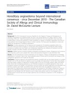
Nef does not affect the cholesterol content of HIV-1 particlesFigure 4
Nef does not affect the cholesterol content of HIV-1 particles. (A) Quantitative lipid analysis was performed as
described in Methods. Data are displayed as molar ratio of cholesterol to PC. Error bars represent standard deviation of the
mean. (B) Selected high-field region of the
1
H spectra of 0.4 mM Nef-SF2 (∆
1–43
, C210A) in the absence (black) and presence of
0.4 mM water soluble cholesterol (red), in comparison with water soluble cholesterol dissolved in the same buffer as the pro-
tein (green).(C)
1
H,
15
N-TROSY-HSQC spectra of 0.4 mM Nef-SF2 (∆
1–43
, C210A) highlighting an overlay of the C-terminal
residues including the putative cholesterol binding site in the absence (black) and presence of 0.4 mM water soluble cholesterol
(red) or 0.4 mM methyl-β-cyclodextrine (green).
0
1
2
3
4
5
6
7
8
Chol/PC [mol/mol]
wt
'Nef
A
C
B
Elevation of SM/PC ratios is insufficient for Nef-mediated enhancement of virion infectivityFigure 5
Elevation of SM/PC ratios is insufficient for Nef-medi-
ated enhancement of virion infectivity. (A) Single round
of infection analysis on TZM cells with cell culture superna-
tants from MT-4 T lymphocytes infected with the indicated
viruses. The assay was performed analogous to that
described in Fig. 1A. (B) Quantitative lipid analysis of virion
SM/PC ratios. HIV particles purified from the cell culture
supernatants analyzed in A were subjected to lipidome analy-
sis. Depicted are the SM to PC ratios.
0
50
100
150
200
250
Relative virion infectivity [%]
A
wt
'Nef
V78A
R81A
EDAA
'12-
39
0
0.5
1.0
1.5
2.0
2.5
SM/PC [mol/mol]
B
wt
'Nef
V78A R81A EDAA
'12-
39
Retrovirology 2007, 4:70 />Page 8 of 12
(page number not for citation purposes)
tions of HIV-1 infected MT4 T lymphocytes harvested in
parallel to the cell culture supernatants that served as
source for infectious virions in the previous analyses (i.e.
at a time point where over 90% of all cells are productively
infected). Total cell lysates (L) as well as soluble (S) and
detergent-resistant (DRM) fractions of flotation gradients
were compared regrading their SM to PC ratio (Fig. 7). No
significant differences in the SM to PC ratio of the L or S
fractions were observed in the absence or presence of Nef.
In contrast, DRMs were significantly enriched in SM rela-
tive to PC in cells infected with Nef or PalmNef expressing
HIV-1 when compared to cells infected with the ∆Nef
virus. This scenario was remarkably similar to the results
obtained with highly purified HIV-1 virions. We conclude
that Nef not only emphasizes the microdomain like lipid
composition of HIV-1 virions but also alters that of mem-
brane microdomains of virus producing T lymphocytes.
Discussion
Building on our previous lipidome analysis of highly puri-
fied HIV-1 virions, this study tested the hypothesis that
Nef affects the lipid composition of HIV-1 particles.
Quantitative mass spectrometry indeed revealed that Nef
underscores the microdomain character of the viral lipid
envelope by increasing virion SM levels and reducing the
amounts of polyunsaturated PC in HIV-1 particles. As
these effects were also detected in DRM preparations from
virus producing cells, the reported modulation of lipid
composition of complex membrane bilayers adds a novel
mechanism by which Nef can manipulate HIV-1 host
cells.
Based on previous reports on the interaction with Nef and
the resulting virion incorporation [42] we expected cho-
lesterol to serve as a positive control for Nef effects in our
lipidome analysis. Surprisingly however, we failed to
detect any appreciable impact of Nef on virion cholesterol
levels. These opposing results may reflect different experi-
mental settings in both studies: Zheng et al. measured the
uptake of radiolabeled cholesterol while in the present
study overall cholesterol levels were quantified by highly
sensitive nano-mass spectrometry. In line with our find-
ings, we also failed to detect a physical interaction
between Nef and cholesterol in solution by NMR spectros-
copy. Such an interaction had been concluded from com-
petition experiments of [17α-methyl-
3
H]-promegestone
with water-soluble cholesterol [42]. Using even higher
concentrations of up to 800 µM water soluble cholesterol
or cholesterol dissolved in ethanol we could not detect a
specific interaction of cholesterol with Nef, however. In
particular, no chemical shifts were observed for the puta-
tive C-terminal cholesterol binding motif L
198
HPEYYK in
the presence of cholesterol (Fig. 4C). Irrespective of the
basis for these differences, our results clearly demonstrate
that Nef augments virion infectivity in the absence of ele-
vating cholesterol levels of particles produced from
infected T lymphocytes. Our study however does not
exclude that Nef affects cholesterol homeostasis e.g. by
upregulation of cholesterol biosynthesis genes [41,42],
which may not be reflected in bulk virion levels. Addition-
ally, effects of Nef on cholesterol transport were recently
Nef enhances virion infectivity independently of whether HIV-1 entry occurs via membrane microdomainsFigure 6
Nef enhances virion infectivity independently of
whether HIV-1 entry occurs via membrane microdo-
mains. Single round infection analysis on HeLa cells tran-
siently expressing the indicated CD4 variants. Productively
infected, p24CA positive cells were quantified 36 hours post
infection by flow cytometry. Data represent average values
from three independent experiments with the indicated
standard error of the mean. Depicted is the relative virion
infectivity with values for HIV-1
NL4-3
NefSF2 (wt) arbitrarily
set to 100%.
0
50
100
Relative Infectivity [%]
wt
'Nef
CD4 wt CD4 2cm CD4 4c
Nef alters the lipid composition of DRMs in HIV-1 infected T lymphocytesFigure 7
Nef alters the lipid composition of DRMs in HIV-1
infected T lymphocytes. DRM flotation analysis was per-
formed from MT4 cells infected with the indicated viruses as
described for Fig. 1D and quantitative lipid analysis was per-
formed from L, S and DRM fractions. Depicted are the SM to
PC ratios.
0
1
2
3
SM/PC ratio
wt
PalmNef
'Nef
L S DRM
Retrovirology 2007, 4:70 />Page 9 of 12
(page number not for citation purposes)
reported in macrophages, where the interaction of Nef
with the ABCA1 transporter and the resulting reduction of
cholesterol efflux were suggested to be instrumental for
Nef-mediated enhancement of virion infectivity [43].
While this particular mechanism is not in place in T lym-
phocytes due to the lack of ABCA1 expression in this cell
type, these results emphasize that Nef can affect lipid
transport in HIV target cells.
Conflicting previous results caused a controversy as to
whether the association of Nef with membrane microdo-
mains contributes to its enhancement of virion infectivity
[18,21,23]. We confirm here that in the context of T lym-
phocyte infection, Nef does not facilitate recruitment of
HIV-1 Gag into DRMs and that an experimental increase
in Nef's DRM association does not augment its effect on
particle infectivity [21]. The results presented also indicate
that analysis of DRM incorporation of viral gene products
following transfection of proviral DNA, which results in
much higher copy numbers of these proteins per cell than
following virus infection, should be interpreted with care.
Even in the absence of elevated DRM recruitment of Gag,
microdomain integrity is a prerequisite for Nef-mediated
infectivity enhancement and specific disruption of Nef's
microdomain association interferes with this activity
[19,23]. Together these results suggest that while micro-
domain association of Nef is critical for the positive effect
of Nef on particle infectivity, this effect is already saturated
at the physiological, moderate levels of Nef microdomain
incorporation. Consistent with the well accepted model
that Nef exerts its effects on virion infectivity during virus
production [58], the viral protein augmented particle
infectivity independently of whether virus entry occurred
via microdomain targeted or excluded entry receptors. We
cannot exclude that the microdomain association of the
CD4 variants used changed upon engagement by the
virus. Nevertheless our results are consistent with the con-
cept that Nef modifies HIV-1 particles during production
in a microdomain dependent manner to facilitate early,
microdomain independent, post entry steps in the new
target cells. Based on the low amounts of Nef present in
microdomains, this scenario would be best compatible
with a catalytic post entry event that is affected by Nef. In
this context Qi and Aiken recently presented an intriguing
model that predicts Nef to counteract proteasomal degra-
dation of HIV-1 particles following entry into new target
cells [59]. Our analysis of Nef mutants revealed that the
observed alterations in particle lipid composition were
not sufficient to mediate Nef's effect on virion infectivity.
However, as the ratio of e.g. cholesterol and SM critically
determines the infectivity of HIV-1 particles
[41,42,48,49], these results do not exclude a contribution
of the virion lipid composition to this Nef function. Such
changes in lipid composition, possibly in conjunction
with altered incorporation of host cell proteins, could
thus affect post translational modifications and/or con-
formation of virion determinants that are recognized by
the degradation machinery early post entry into a new tar-
get cell. This might also involve changes in the membrane
microdomain organization of the virions, a parameter
that is also critical for optimal particle infectivity
[41,42,48,49]. It will be of interest to understand in the
future how microdomain association of Nef affects the
early post entry fate of HIV virions in the recipient cell.
An important finding of this study is that Nef altered not
only the lipid composition of HIV particles but also that
of host cell membrane microdomains. Thus, in addition
to the well established changes in microdomain protein
composition [20,40,60], Nef also affects their lipid micro-
environment. The overlap in changes induced by Nef on
the lipid composition of bulk host cell microdomains and
purified virions implies that the altered particle composi-
tion is a direct consequence of the modifications observed
in the virus producing cell. The mechanism of this lipid
modulatory activity of Nef as well as the molecular deter-
minants for this activity remain unclear. In light of the
effects of Nef on lipid transport discussed above, the lack
of differences in the lipid composition in total cell lysates
suggest effects of Nef on lipid sorting and/or microdo-
main incorporation as a major determinant of this func-
tion. Additionally, Nef may affect microdomain lipid
composition by local changes in the turnover of select
lipid classes e.g. via modulation of host cell signal trans-
duction pathways and its direct association with lipid
kinases such as PI3-K [61]. As lipids potently regulate
microdomain activitiy in membrane traffic and signal
transduction [62], this mechanism could potentially con-
tribute to a number of Nef's biological properties. The reg-
ulatory role of microdomain lipids is particularly
prominent during T cell receptor signaling [32,63-65], a
process that is altered by Nef in a membrane microdo-
main dependent manner [19,20,39,40]. Importantly,
with Pak2 and PI3 kinases as well as the actin regulator
Wasp, these effects of Nef involve its physical or func-
tional association with cellular components whose activ-
ity is either subject to regulation by the local lipid
microenvironment or causes alterations in the local lipid
composition [66-68]. Finally, the altered lipid composi-
tion of microdomains in the presence of Nef might have
direct consequences of protein incorporation into these
domains [69]. Future studies will focus on the functional
consequences this novel lipid modulatory activity of Nef
exhibits on host cell microdomain function.
Conclusion
This study describes alterations of the lipidome as a new
activity of the HIV pathogenicity factor Nef that might
contribute to its multifaceted strategies to manipulate HIV
Retrovirology 2007, 4:70 />Page 10 of 12
(page number not for citation purposes)
target cells for the optimization of virus spread in the
infected host.
Methods
Reagents and plasmids
Isogenic proviral constructs expressing various nef genes
based on the HIV-1 SF2 nef sequence in the backbone of
the HIV-1 NL4-3 proviral clone were described earlier
[50]. The provirus encoding for a Nef protein with palmi-
toylation site (G3C mutation) [20] was constructed anal-
ogously. The complete nucleotide sequences of all nef
inserts of these proviral clones were verified by sequenc-
ing of both DNA strands. The expression plasmids for wt
CD4 and the 2Cm and 4C CD4 mutants were kindly pro-
vided by Dr. Jin and were described elsewhere [57]. 2Cm
lacks the palmitoylation acceptor in the cytoplasmic tail
of CD4 while in 4C, the Lck interaction motif is addition-
ally removed. Gag and Nef were detected using the poly-
clonal rabbit serum CA1 against p24CA [70] and the
polyclonal anti-Nef sheep serum Arp444 [71], respec-
tively. Antibodies against Env, RT, Vpr are described else-
where [50].
Mass spectrometric lipid analysis of purified HIV particles
HIV-1 virions were purified from cell culture supernatants
of infected MT-4 lymphocyte cultures and subjected to
quantitative lipid analysis by nano-electrospray ioniza-
tion tandem mass spectrometry, similarly as described
[38].
Western blotting
For Western blot analysis, samples were boiled in SDS
sample buffer, separated by 10% SDS-PAGE and trans-
ferred to a nitrocellulose membrane. Protein detection
was performed following incubation with appropriate
first and secondary antibodies using the super signal pico
detection kit (Pierce, Bonn, Germany) according to the
manufacturer's instructions.
HIV replication, single round infectivity, p24 ELISA
The relative infectivity of HIV-1 particles was determined
by CA ELISA and a standardized 96 well TZM blue cell
assay as described [72]. Briefly, infections were carried out
in triplicates with 0.5 ng CA input virus. 36 hours post
infection, cells were fixed, stained for β-galactosidase
activity and the number of blue cells was determined by
microscopy. To analyze the effects of Nef on HIV replica-
tion in primary human T lymphocytes [50], peripheral
blood mononuclear cells were isolated from healthy
donors by Ficoll gradients using Ficoll-Paque Plus (Amer-
sham Biosciences, Uppsala, Sweden). For infection, cells
were thawed and kept in bulk cultures in RPMI, 10% FCS
at 1 × 10
6
cells/ml over night. 1 × 10
5
cells/well were then
seeded in V-bottom 96 well plates and infected with 1 ng
CA virus input per well the following day. 3 days later,
cells were washed and stimulated with 2 µg/ml PHA
(Sigma) and 20 nM IL-2 (Chiron, Emeryville, CA) for 3
days. After stimulation, the PBL were washed and resus-
pended in 200 µl of RPMI containing FCS and IL-2. Each
day, 100 µl of cell culture supernatant was replaced with
fresh medium and amounts of CA in the cell culture
supernatant were quantified to monitor virus replication
using an in-house p24 ELISA [72].
NMR spectroscopy
15
N,
13
C-labelled HIV-1 Nef-SF2 (∆
1–43
, C210A) of HIV-1
was expressed and purified as described previously [73].
The main-chain amide resonance assignments were taken
from the HIV-1 Nef BH10 (∆
2–39, 159–173
, C206A), whose
structure was previously solved by high-resolution NMR
spectroscopy [74]. For binding studies 0.4 mM
15
N,
13
C-
labeled Nef (∆
1–43
, C210A) was used in 5 mM Tris/HCl,
pH 8.0, 5 mM DTE, 8% D
2
O, 92% H
2
O, and 0.05 mM
DSS (4,4-dimethyl-4-silapentane-sulphonic acid). Cho-
lesterol, water soluble cholesterol (cholesterol in methyl-
β-cyclodextrin) and methyl-β-cyclodextrine were pur-
chased from Sigma (C3045, C4951 and C4555, respec-
tively), and used to prepare stock solutions of 40 mM
cholesterol in ethanol, 7.5 mM water soluble cholesterol
in 5 mM Tris/HCl buffer pH 8.0, and 7.5 mM methyl-β-
cyclodextrine in the same buffer.
NMR measurements were performed at 308 K on Bruker
Avance 800 MHz spectrometer operating at 800 MHz pro-
ton resonance frequency.
1
H,
15
N-TROSY-HSQC were
recorded as described by Pervushin et al. [75]. The spectra
were recorded with 4 K data points and a spectral width of
14 ppm in the
1
H dimension, and with 128 data points
and a spectral width of 40 ppm in the
15
N dimension. Pro-
ton chemical shifts were referenced to DSS used as inter-
nal reference. The
15
N chemical shifts were indirectly
referenced to DSS using the frequency ratio given by
Wishart et al. [76]. Spectra were acquired and processed
with Topspin 1.3 (Bruker Biospin, Karlsruhe), and ana-
lysed with the program AUREMOL [77].
Four identical 0.5 ml samples of 0.4 mM Nef solution
were titrated with ethanol, cholesterol in ethanol, cyclo-
dextrine and water soluble cholesterol. The total amount
of protein was held constant. ethanol/cholesterol/cyclo-
dextrine was added in increasing amounts up to a molar
ratio of protein to ligand of 1 to 2.
Competing interests
The author(s) declare that they have no competing inter-
ests.
Authors' contributions
BB, EK, NT, CEM, SR, IL, BG and SB performed the exper-
imental work. BB, MG, HGK, HRK, FTW and OTF con-
Retrovirology 2007, 4:70 />Page 11 of 12
(page number not for citation purposes)
ceived the experimental strategies and OTF, HRK and BB
designed individual experiments. BB and OTF analyzed
the data and OTF wrote the manuscript. All authors read
and approved the final manuscript.
Acknowledgements
We thank Dr. Yong-Jiu Jin for CD4 expression plasmids. This work was
supported by grants from the Deutsche Forschungsgemeinschaft within
Transregio 13 to O.T.F., F.T.W., B.B. and H.R.K., within SFB638 to H.G.K.,
as well as a group leader fellowship from the C.H.S. Stiftung to O.T.F.
References
1. Deacon NJ, Tsykin A, Solomon A, Smith K, Ludford-Menting M,
Hooker DJ, McPhee DA, Greenway AL, Ellett A, Chatfield C, et al.:
Genomic structure of an attenuated quasi species of HIV-1
from a blood transfusion donor and recipients. Science 1995,
270(5238):988-991.
2. Kestler HW 3rd, Ringler DJ, Mori K, Panicali DL, Sehgal PK, Daniel
MD, Desrosiers RC: Importance of the nef gene for mainte-
nance of high virus loads and for development of AIDS. Cell
1991, 65(4):651-662.
3. Kirchhoff F, Greenough TC, Brettler DB, Sullivan JL, Desrosiers RC:
Brief report: absence of intact nef sequences in a long-term
survivor with nonprogressive HIV-1 infection. N Engl J Med
1995, 332(4):228-232.
4. Geyer M, Fackler OT, Peterlin BM: Structure function relation-
ships in HIV-1 Nef. EMBO Rep 2001, 2(7):580-585.
5. Tolstrup M, Ostergaard L, Laursen AL, Pedersen SF, Duch M: HIV/
SIV escape from immune surveillance: focus on Nef. Curr HIV
Res 2004, 2(2):141-151.
6. Garcia JV, Miller AD: Serine phosphorylation-independent
downregulation of cell-surface CD4 by nef. Nature 1991,
350(6318):508-511.
7. Michel N, Allespach I, Venzke S, Fackler OT, Keppler OT: The Nef
protein of human immunodeficiency virus establishes super-
infection immunity by a dual strategy to downregulate cell-
surface CCR5 and CD4. Curr Biol 2005, 15(8):714-723.
8. Roeth JF, Collins KL: Human immunodeficiency virus type 1
Nef: adapting to intracellular trafficking pathways. Microbiol
Mol Biol Rev 2006, 70(2):548-563.
9. Schwartz O, Marechal V, Le Gall S, Lemonnier F, Heard JM: Endocy-
tosis of major histocompatibility complex class I molecules
is induced by the HIV-1 Nef protein. Nat Med 1996,
2(3):338-342.
10. Fackler OT, Alcover A, Schwartz O: Modulation of the immuno-
logical synapse: a key to HIV-1 pathogenesis? Nat Rev Immunol
2007, 7(4):310-317.
11. Haller C, Rauch S, Michel N, Hannemann S, Lehmann MJ, Keppler OT,
Fackler OT: The HIV-1 pathogenicity factor Nef interferes
with maturation of stimulatory T-lymphocyte contacts by
modulation of N-Wasp activity. J Biol Chem 2006,
281(28):19618-19630.
12. Schindler M, Munch J, Kutsch O, Li H, Santiago ML, Bibollet-Ruche F,
Muller-Trutwin MC, Novembre FJ, Peeters M, Courgnaud V, Bailes E,
Roques P, Sodora DL, Silvestri G, Sharp PM, Hahn BH, Kirchhoff F:
Nef-mediated suppression of T cell activation was lost in a
lentiviral lineage that gave rise to HIV-1. Cell 2006,
125(6):1055-1067.
13. Schrager JA, Marsh JW: HIV-1 Nef increases T cell activation in
a stimulus-dependent manner. Proc Natl Acad Sci U S A 1999,
96(14):8167-8172.
14. Thoulouze MI, Sol-Foulon N, Blanchet F, Dautry-Varsat A, Schwartz
O, Alcover A: Human immunodeficiency virus type-1 infection
impairs the formation of the immunological synapse. Immu-
nity 2006, 24(5):547-561.
15. Aiken C, Trono D: Nef stimulates human immunodeficiency
virus type 1 proviral DNA synthesis. J Virol 1995,
69(8):5048-5056.
16. Chowers MY, Spina CA, Kwoh TJ, Fitch NJ, Richman DD, Guatelli JC:
Optimal infectivity in vitro of human immunodeficiency
virus type 1 requires an intact nef gene. J Virol 1994,
68(5):2906-2914.
17. Schwartz O, Marechal V, Danos O, Heard JM: Human immunode-
ficiency virus type 1 Nef increases the efficiency of reverse
transcription in the infected cell. J Virol 1995, 69(7):4053-4059.
18. Alexander M, Bor YC, Ravichandran KS, Hammarskjold ML, Rekosh
D: Human immunodeficiency virus type 1 Nef associates
with lipid rafts to downmodulate cell surface CD4 and class I
major histocompatibility complex expression and to
increase viral infectivity. J Virol 2004, 78(4):1685-1696.
19. Giese SI, Woerz I, Homann S, Tibroni N, Geyer M, Fackler OT: Spe-
cific and distinct determinants mediate membrane binding
and lipid raft incorporation of HIV-1(SF2) Nef. Virology 2006,
355(2):175-191.
20. Krautkramer E, Giese SI, Gasteier JE, Muranyi W, Fackler OT:
Human immunodeficiency virus type 1 Nef activates p21-
activated kinase via recruitment into lipid rafts. J Virol 2004,
78(8):4085-4097.
21. Sol-Foulon N, Esnault C, Percherancier Y, Porrot F, Metais-Cunha P,
Bachelerie F, Schwartz O: The effects of HIV-1 Nef on CD4 sur-
face expression and viral infectivity in lymphoid cells are
independent of rafts. J Biol Chem 2004, 279(30):31398-31408.
22. Wang JK, Kiyokawa E, Verdin E, Trono D: The Nef protein of HIV-
1 associates with rafts and primes T cells for activation. Proc
Natl Acad Sci U S A 2000, 97(1):394-399.
23. Zheng YH, Plemenitas A, Linnemann T, Fackler OT, Peterlin BM: Nef
increases infectivity of HIV via lipid rafts. Curr Biol 2001,
11(11):875-879.
24. Rauch S, Fackler O: Viruses, lipid rafts and signal transduction.
Signal Transduction 2007, 7:53-63.
25. Chazal N, Gerlier D: Virus entry, assembly, budding, and mem-
brane rafts. Microbiol Mol Biol Rev 2003, 67(2):226-37, table of con-
tents.
26. Rawat SS, Viard M, Gallo SA, Rein A, Blumenthal R, Puri A: Modula-
tion of entry of enveloped viruses by cholesterol and sphin-
golipids (Review). Mol Membr Biol 2003, 20(3):243-254.
27. Suomalainen M: Lipid rafts and assembly of enveloped viruses.
Traffic 2002, 3(10):705-709.
28. Mayor S, Rao M: Rafts: scale-dependent, active lipid organiza-
tion at the cell surface. Traffic 2004, 5(4):231-240.
29. Munro S: Lipid rafts: elusive or illusive? Cell 2003,
115(4):377-388.
30. Nichols B: Cell biology: without a raft. Nature 2005,
436(7051):638-639.
31. Simons K, Ehehalt R: Cholesterol, lipid rafts, and disease. J Clin
Invest 2002, 110(5):597-603.
32. Simons K, Toomre D: Lipid rafts and signal transduction. Nat
Rev Mol Cell Biol 2000, 1(1):31-39.
33. Gaus K, Chklovskaia E, Fazekas de St Groth B, Jessup W, Harder T:
Condensation of the plasma membrane at the site of T lym-
phocyte activation. J Cell Biol 2005, 171(1):121-131.
34. Gaus K, Zech T, Harder T: Visualizing membrane microdo-
mains by Laurdan 2-photon microscopy. Mol Membr Biol 2006,
23(1):41-48.
35. Plowman SJ, Muncke C, Parton RG, Hancock JF: H-ras, K-ras, and
inner plasma membrane raft proteins operate in nanoclus-
ters with differential dependence on the actin cytoskeleton.
Proc Natl Acad Sci U S A 2005, 102(43):15500-15505.
36. Sharma P, Varma R, Sarasij RC, Ira, Gousset K, Krishnamoorthy G,
Rao M, Mayor S: Nanoscale organization of multiple GPI-
anchored proteins in living cell membranes. Cell 2004,
116(4):577-589.
37. Douglass AD, Vale RD: Single-molecule microscopy reveals
plasma membrane microdomains created by protein-pro-
tein networks that exclude or trap signaling molecules in T
cells. Cell 2005, 121(6):937-950.
38. Brugger B, Glass B, Haberkant P, Leibrecht I, Wieland FT, Krausslich
HG: The HIV lipidome: a raft with an unusual composition.
Proc Natl Acad Sci U S A 2006, 103(8):2641-2646.
39. Pulkkinen K, Renkema GH, Kirchhoff F, Saksela K: Nef associates
with p21-activated kinase 2 in a p21-GTPase-dependent
dynamic activation complex within lipid rafts. J Virol 2004,
78(23):12773-12780.
40. Simmons A, Gangadharan B, Hodges A, Sharrocks K, Prabhakar S,
Garcia A, Dwek R, Zitzmann N, McMichael A: Nef-mediated lipid
raft exclusion of UbcH7 inhibits Cbl activity in T cells to pos-
itively regulate signaling. Immunity 2005, 23(6):621-634.
Retrovirology 2007, 4:70 />Page 12 of 12
(page number not for citation purposes)
41. van 't Wout AB, Swain JV, Schindler M, Rao U, Pathmajeyan MS, Mul-
lins JI, Kirchhoff F: Nef induces multiple genes involved in cho-
lesterol synthesis and uptake in human immunodeficiency
virus type 1-infected T cells. J Virol 2005, 79(15):10053-10058.
42. Zheng YH, Plemenitas A, Fielding CJ, Peterlin BM: Nef increases the
synthesis of and transports cholesterol to lipid rafts and HIV-
1 progeny virions. Proc Natl Acad Sci U S A 2003,
100(14):8460-8465.
43. Mujawar Z, Rose H, Morrow MP, Pushkarsky T, Dubrovsky L,
Mukhamedova N, Fu Y, Dart A, Orenstein JM, Bobryshev YV, Bukrin-
sky M, Sviridov D: Human immunodeficiency virus impairs
reverse cholesterol transport from macrophages. PLoS Biol
2006, 4(11):e365.
44. Tokunaga K, Kojima A, Kurata T, Ikuta K, Akari H, Koyama AH,
Kawamura M, Inubushi R, Shimano R, Adachi A: Enhancement of
human immunodeficiency virus type 1 infectivity by Nef is
producer cell-dependent. J Gen Virol 1998, 79 ( Pt
10):2447-2453.
45. Tokunaga K, Kojima A, Kurata T, Ikuta K, Inubushi R, Shimano R,
Kawamura M, Akari H, Koyama AH, Adachi A: Producer cell-
dependent requirement of the Nef protein for efficient entry
of HIV-1 into cells. Biochem Biophys Res Commun 1998,
250(3):565-568.
46. Welker R, Hohenberg H, Tessmer U, Huckhagel C, Krausslich HG:
Biochemical and structural analysis of isolated mature cores
of human immunodeficiency virus type 1. J Virol 2000,
74(3):1168-1177.
47. Grimm MO, Grimm HS, Patzold AJ, Zinser EG, Halonen R, Duering
M, Tschape JA, De Strooper B, Muller U, Shen J, Hartmann T: Regu-
lation of cholesterol and sphingomyelin metabolism by amy-
loid-beta and presenilin. Nat Cell Biol 2005, 7(11):1118-1123.
48. Campbell S, Gaus K, Bittman R, Jessup W, Crowe S, Mak J: The raft-
promoting property of virion-associated cholesterol, but not
the presence of virion-associated Brij 98 rafts, is a determi-
nant of human immunodeficiency virus type 1 infectivity. J
Virol 2004, 78(19):10556-10565.
49. Campbell SM, Crowe SM, Mak J: Virion-associated cholesterol is
critical for the maintenance of HIV-1 structure and infectiv-
ity. Aids 2002, 16(17):2253-2261.
50. Fackler OT, Moris A, Tibroni N, Giese SI, Glass B, Schwartz O, Kraus-
slich HG: Functional characterization of HIV-1 Nef mutants in
the context of viral infection. Virology 2006, 351(2):322-339.
51. Del Real G, Jimenez-Baranda S, Lacalle RA, Mira E, Lucas P, Gomez-
Mouton C, Carrera AC, Martinez AC, Manes S: Blocking of HIV-1
infection by targeting CD4 to nonraft membrane domains. J
Exp Med 2002, 196(3):293-301.
52. Liu NQ, Lossinsky AS, Popik W, Li X, Gujuluva C, Kriederman B,
Roberts J, Pushkarsky T, Bukrinsky M, Witte M, Weinand M, Fiala M:
Human immunodeficiency virus type 1 enters brain microv-
ascular endothelia by macropinocytosis dependent on lipid
rafts and the mitogen-activated protein kinase signaling
pathway. J Virol 2002, 76(13):6689-6700.
53. Nguyen DH, Giri B, Collins G, Taub DD: Dynamic reorganization
of chemokine receptors, cholesterol, lipid rafts, and adhe-
sion molecules to sites of CD4 engagement. Exp Cell Res 2005,
304(2):559-569.
54. Percherancier Y, Lagane B, Planchenault T, Staropoli I, Altmeyer R,
Virelizier JL, Arenzana-Seisdedos F, Hoessli DC, Bachelerie F: HIV-1
entry into T-cells is not dependent on CD4 and CCR5 locali-
zation to sphingolipid-enriched, detergent-resistant, raft
membrane domains. J Biol Chem 2003, 278(5):3153-3161.
55. Popik W, Alce TM: CD4 receptor localized to non-raft mem-
brane microdomains supports HIV-1 entry. Identification of
a novel raft localization marker in CD4. J Biol Chem 2004,
279(1):704-712.
56. Popik W, Alce TM, Au WC: Human immunodeficiency virus
type 1 uses lipid raft-colocalized CD4 and chemokine recep-
tors for productive entry into CD4(+) T cells. J Virol 2002,
76(10):4709-4722.
57. Fragoso R, Ren D, Zhang X, Su MW, Burakoff SJ, Jin YJ: Lipid raft
distribution of CD4 depends on its palmitoylation and asso-
ciation with Lck, and evidence for CD4-induced lipid raft
aggregation as an additional mechanism to enhance CD3 sig-
naling. J Immunol 2003, 170(2):913-921.
58. Pandori MW, Fitch NJ, Craig HM, Richman DD, Spina CA, Guatelli JC:
Producer-cell modification of human immunodeficiency
virus type 1: Nef is a virion protein. J Virol 1996,
70(7):4283-4290.
59. Qi M, Aiken C: Selective restriction of Nef-defective human
immunodeficiency virus type 1 by a proteasome-dependent
mechanism. J Virol 2007, 81(3):1534-1536.
60. Djordjevic JT, Schibeci SD, Stewart GJ, Williamson P: HIV type 1
Nef increases the association of T cell receptor (TCR)-sign-
aling molecules with T cell rafts and promotes activation-
induced raft fusion. AIDS Res Hum Retroviruses 2004,
20(5):547-555.
61. Blagoveshchenskaya AD, Thomas L, Feliciangeli SF, Hung CH, Thomas
G: HIV-1 Nef downregulates MHC-I by a PACS-1- and PI3K-
regulated ARF6 endocytic pathway. Cell 2002, 111(6):853-866.
62. Krauss M, Haucke V: Phosphoinositide-metabolizing enzymes
at the interface between membrane traffic and cell signal-
ling. EMBO Rep 2007, 8(3):241-246.
63. Kabouridis PS: Lipid rafts in T cell receptor signalling. Mol
Membr Biol 2006, 23(1):49-57.
64. Geyeregger R, Zeyda M, Zlabinger GJ, Waldhausl W, Stulnig TM: Pol-
yunsaturated fatty acids interfere with formation of the
immunological synapse. J Leukoc Biol 2005, 77(5):680-688.
65. Zeyda M, Szekeres AB, Saemann MD, Geyeregger R, Stockinger H,
Zlabinger GJ, Waldhausl W, Stulnig TM: Suppression of T cell sig-
naling by polyunsaturated fatty acids: selectivity in inhibition
of mitogen-activated protein kinase and nuclear factor acti-
vation. J Immunol 2003, 170(12):6033-6039.
66. Bokoch GM, Reilly AM, Daniels RH, King CC, Olivera A, Spiegel S,
Knaus UG: A GTPase-independent mechanism of p21-acti-
vated kinase activation. Regulation by sphingosine and other
biologically active lipids. J Biol Chem 1998, 273(14):8137-8144.
67. Papayannopoulos V, Co C, Prehoda KE, Snapper S, Taunton J, Lim
WA: A polybasic motif allows N-WASP to act as a sensor of
PIP(2) density. Mol Cell 2005, 17(2):181-191.
68. Hawkins PT, Anderson KE, Davidson K, Stephens LR: Signalling
through Class I PI3Ks in mammalian cells. Biochem Soc Trans
2006, 34(Pt 5):647-662.
69. Zeyda M, Staffler G, Horejsi V, Waldhausl W, Stulnig TM: LAT dis-
placement from lipid rafts as a molecular mechanism for the
inhibition of T cell signaling by polyunsaturated fatty acids. J
Biol Chem 2002, 277(32):28418-28423.
70. Muller B, Daecke J, Fackler OT, Dittmar MT, Zentgraf H, Krausslich
HG: Construction and characterization of a fluorescently
labeled infectious human immunodeficiency virus type 1
derivative. J Virol 2004, 78(19):10803-10813.
71. Coates K, Cooke SJ, Mann DA, Harris MP: Protein kinase C-medi-
ated phosphorylation of HIV-I nef in human cell lines. J Biol
Chem 1997, 272(19):12289-12294.
72. Keppler OT, Allespach I, Schuller L, Fenard D, Greene WC, Fackler
OT: Rodent cells support key functions of the human immu-
nodeficiency virus type 1 pathogenicity factor Nef. J Virol
2005, 79(3):1655-1665.
73. Breuer S, Gerlach H, Kolaric B, Urbanke C, Opitz N, Geyer M: Bio-
chemical Indication for Myristoylation-Dependent Confor-
mational Changes in HIV-1 Nef. Biochemistry 2006,
45(7):2339-2349.
74. Grzesiek S, Bax A, Hu JS, Kaufman J, Palmer I, Stahl SJ, Tjandra N,
Wingfield PT: Refined solution structure and backbone
dynamics of HIV-1 Nef. Protein Sci 1997, 6(6):1248-1263.
75. Pervushin K, Riek R, Wider G, Wuthrich K: Attenuated T2 relax-
ation by mutual cancellation of dipole-dipole coupling and
chemical shift anisotropy indicates an avenue to NMR struc-
tures of very large biological macromolecules in solution.
Proc Natl Acad Sci U S A 1997, 94(23):12366-12371.
76. Wishart DS, Bigam CG, Yao J, Abildgaard F, Dyson HJ, Oldfield E,
Markley JL, Sykes BD: 1H, 13C and 15N chemical shift referenc-
ing in biomolecular NMR. J Biomol NMR 1995, 6(2):135-140.
77. Gronwald W, Kalbitzer HR: Automated Structure Determina-
tion of Proteins by NMR Spectroscopy. Progr NMR Spectr 2004,
44:33-96.