Báo cáo y học: "Localization of HIV-1 Vpr to the nuclear envelope: Impact on Vpr functions and virus replication in macrophages" potx
Bạn đang xem bản rút gọn của tài liệu. Xem và tải ngay bản đầy đủ của tài liệu tại đây (4.2 MB, 15 trang )
BioMed Central
Page 1 of 15
(page number not for citation purposes)
Retrovirology
Open Access
Research
Localization of HIV-1 Vpr to the nuclear envelope: Impact on Vpr
functions and virus replication in macrophages
Guillaume Jacquot
†1,2
, Erwann Le Rouzic
†1,2
, Annie David
3
,
Julie Mazzolini
1,2
, Jérôme Bouchet
1,2
, Serge Bouaziz
4
, Florence Niedergang
1,2
,
Gianfranco Pancino
3
and Serge Benichou*
1,2
Address:
1
Institut Cochin, Université Paris Descartes, CNRS (UMR 8104), Paris, France,
2
Inserm U567, Paris, France,
3
Unité de Régulation des
Infections Rétrovirales, Institut Pasteur, Paris, France and
4
Inserm U640, CNRS UMR 8151, Paris, France
Email: Guillaume Jacquot - ; Erwann Le Rouzic - ; Annie David - ;
Julie Mazzolini - ; Jérôme Bouchet - ; Serge Bouaziz - ;
Florence Niedergang - ; Gianfranco Pancino - ; Serge Benichou* -
* Corresponding author †Equal contributors
Abstract
Background: HIV-1 Vpr is a dynamic protein that primarily localizes in the nucleus, but a
significant fraction is concentrated at the nuclear envelope (NE), supporting an interaction between
Vpr and components of the nuclear pore complex, including the nucleoporin hCG1. In the present
study, we have explored the contribution of Vpr accumulation at the NE to the Vpr functions,
including G2-arrest and pro-apoptotic activities, and virus replication in primary macrophages.
Results: In order to define the functional role of Vpr localization at the NE, we have characterized
a set of single-point Vpr mutants, and selected two new mutants with substitutions within the first
α-helix of the protein, Vpr-L23F and Vpr-K27M, that failed to associate with hCG1, but were still
able to interact with other known relevant host partners of Vpr. In mammalian cells, these mutants
failed to localize at the NE resulting in a diffuse nucleocytoplasmic distribution both in HeLa cells
and in primary human monocyte-derived macrophages. Other mutants with substitutions in the
first α-helix (Vpr-A30L and Vpr-F34I) were similarly distributed between the nucleus and
cytoplasm, demonstrating that this helix contains the determinants required for localization of Vpr
at the NE. All these mutations also impaired the Vpr-mediated G2-arrest of the cell cycle and the
subsequent cell death induction, indicating a functional link between these activities and the Vpr
accumulation at the NE. However, this localization is not sufficient, since mutations within the C-
terminal basic region of Vpr (Vpr-R80A and Vpr-R90K), disrupted the G2-arrest and apoptotic
activities without altering NE localization. Finally, the replication of the Vpr-L23F and Vpr-K27M
hCG1-binding deficient mutant viruses was also affected in primary macrophages from some but
not all donors.
Conclusion: These results indicate that the targeting of Vpr to the nuclear pore complex may
constitute an early step toward Vpr-induced G2-arrest and subsequent apoptosis; they also suggest
that Vpr targeting to the nuclear pore complex is not absolutely required, but can improve HIV-1
replication in macrophages.
Published: 26 November 2007
Retrovirology 2007, 4:84 doi:10.1186/1742-4690-4-84
Received: 13 August 2007
Accepted: 26 November 2007
This article is available from: />© 2007 Jacquot et al; licensee BioMed Central Ltd.
This is an Open Access article distributed under the terms of the Creative Commons Attribution License ( />),
which permits unrestricted use, distribution, and reproduction in any medium, provided the original work is properly cited.
Retrovirology 2007, 4:84 />Page 2 of 15
(page number not for citation purposes)
Background
In contrast to oncoretroviruses that replicate only in divid-
ing cells and require nuclear envelope (NE) disassembly
during mitosis to integrate their genetic material into the
host cell genome, HIV-1 and other lentiviruses have the
ability to productively infect non-dividing cells, such as
terminally-differentiated macrophages [1]. In the case of
HIV-1, these cell populations represent important targets
during the initial steps of infection and largely contribute
to the establishment of viral reservoirs [2]. The ability of
HIV-1 to infect non-dividing cells relies on mechanisms
allowing active transport of the so-called "preintegration
complex" (PIC), the nucleoprotein complex containing
the viral DNA, from the cytoplasm to the nuclear com-
partment through the intact NE. While nuclear import of
the PIC is essential for virus replication in non-dividing
cells, it was also proposed that uncoating of the viral cap-
sid after virus entry might rather be the rate-limiting step
in the ability of HIV-1 to infect such non-dividing cells
[3]. The molecular details underlying this process are still
unknown, but a certain body of evidence suggests that the
PIC may be transported along the microtubule network to
accumulate at the nuclear periphery before anchoring to
the NE (for review, see Ref. [4]).
Although the composition of the HIV-1 PIC changes dur-
ing its travel to the nucleus, three viral proteins, namely
the matrix protein (MA), integrase (IN) and the auxiliary
viral protein R (Vpr), remain tightly associated with the
viral DNA and have thus been proposed as potential
mediators of the nuclear import of the PIC. The central
DNA flap structure generated upon completion of the
reverse transcription process has been involved in this
active process. While the exact contribution of these dis-
tinct viral determinants in the nuclear import of the PIC is
still controversial (for review, see Ref. [4]), HIV-1 Vpr spe-
cifically facilitates virus replication in non-dividing cells
and differentiated macrophages [5-8]. In addition, it was
recently reported that some tRNA species incorporated
into virus particles may also promote nuclear import of
the viral DNA [9].
HIV-1 Vpr is a highly conserved 96-amino acid (a.a.) basic
protein (14 kDa). The analysis of the soluble full length
Vpr polypeptide by nuclear magnetic resonance (NMR)
allowed the three-dimensional (3D) structure determina-
tion of the protein. Vpr consists of an hydrophobic central
core domain, with three α-helices (H1 a.a. 17–33, H2 a.a.
38–50 and H3 a.a. 55–77), that are connected by loops
and surrounded by two flexible N- and C-terminal
domains negatively and positively charged, respectively
[10]. By contrast with other HIV-1 auxiliary proteins, Vpr
is specifically incorporated at a high copy number in virus
particles [11-15], and is consequently present in the cyto-
plasm of newly infected cells, indicating that it certainly
plays specific roles in the early post-entry steps of viral
replication [16]. In addition to its role in the nuclear
import of the viral PIC, Vpr displays several other activi-
ties, including an effect on the fidelity of the reverse-tran-
scription process, an arrest of the cell cycle at the G2/M
transition, an induction of apoptosis and the transactiva-
tion of the HIV-1 LTR as well as host cell genes (for review,
see Ref. [17]. Although the exact contribution of these
activities along the virus life cycle is still debated, Vpr-
induced G2-arrest has been proposed to provide a favora-
ble cellular environment for optimal transcription of HIV-
1 [18], while the modulation of the virus mutation rate
seems required for efficient spreading of HIV-1 in primary
macrophages [19].
When expressed either in dividing or non-dividing cells,
HIV-1 Vpr displays evident karyophilic properties and is
clearly concentrated at the NE at steady state [20-23]. This
latter observation was correlated with its binding to sev-
eral components of the nuclear pore complex (NPC)
which selectively regulates the trafficking of macromole-
cules or complexes between the nucleus and cytoplasm
[24-26]. The NPC is a large supramolecular structure
embedded into the NE and composed of around 30
unique proteins termed nucleoporins (Nups) [27]. About
half of these Nups contain Phe-Gly repeats (FG-repeats)
that contribute directly to the active nucleo-cytoplasmic
transport. While initial studies supported the idea that Vpr
could bind the FG-rich regions of several Nups, including
the human Nup54 and Nup58 [24], the rodent Pom121
[26] and the yeast Nsp1p [25], a more recent study
described a direct interaction between Vpr and the human
CG1 nucleoporin [28]. This interaction does not require
the FG-rich region of hCG1 but rather a region without
consensus motif found in the N-terminal domain of the
protein. Using an in vitro nuclear import assay, it has been
demonstrated that hCG1 contributed in the accumulation
of Vpr to the NE [28].
Only a few reports have tried so far to evaluate the virolog-
ical impact related to the property of HIV-1 Vpr to localize
at the NE [25,29]. In the present study, we have explored
the role of Vpr accumulation at the NE for the Vpr func-
tions, including G2-arrest and pro-apoptotic activities,
and for virus replication in primary macrophages. Single-
point Vpr mutants, including two new independent
mutants that specifically failed to interact with hCG1,
were characterized. Like other mutants with substitutions
within the first α-helix of Vpr, they failed to localize at the
NE and were impaired for G2-arrest and cell death induc-
tion, indicating a functional link between these activities
and the Vpr accumulation at the NE. Finally, the replica-
tion of the hCG1-binding deficient Vpr mutant viruses
was impaired in monocyte-derived macrophages (MDMs)
from some but not all donors, suggesting that Vpr target-
Retrovirology 2007, 4:84 />Page 3 of 15
(page number not for citation purposes)
ing to the nuclear pore complex is not absolutely required,
but can improve HIV-1 replication in macrophages.
Results
Identification of Vpr mutants deficient for hCG1-binding
Previous studies have established that the localization of
HIV-1 Vpr to the NE is related to its ability to interact with
components of the NPC [23,25,26], including the nucle-
oporin hCG1 [28]. In order to identify single-point muta-
tions that altered the Vpr binding to hCG1, we generated
a library of random Vpr mutants and used the yeast two-
hybrid system to screen for hCG1-binding deficient Vpr
mutants. Only mutants which retained the capacity to
interact with UNG2 and HHR23A, two other known rele-
vant host partners of Vpr [30,31] but failed to bind hCG1
were selected. Two Vpr mutants (clones 11 and 35) that
still interacted with UNG2 and HHR23A were isolated
(Fig. 1A and data not shown, respectively), as evidenced
by growth of yeast-transformed cells on medium without
histidine (-His) and β-gal activity. In contrast, these
mutants did not bind to hCG1, since no growth on -His
medium and β-gal activity was observed. Used as controls,
the VprR90K mutant, which is known to abolish Vpr-
induced G2-arrest [31], still bound both to hCG1 and
UNG2, while the W54R mutant, which is deficient for
binding to UNG2 [32], still interacted with hCG1 (Fig. 1A,
lower panel). These results show that this yeast two-
hybrid strategy is a powerful system to isolate specific
hCG1-binding deficient Vpr mutants.
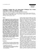
DNA Sequencing of clone 11 revealed 3 substitutions
within the VprLai primary sequence (Leu23Phe,
Leu67Gln and Arg73Gly), while clone 35 contained a sin-
gle substitution (Lys27Met) (Fig. 1B). Each substitution
from clone 11 was introduced in the Vpr sequence and the
3 single-point mutants were analyzed again for binding to
hCG1 and UNG2. As shown in Fig. 1C, the L23F and
K27M substitutions were sufficient to abrogate hCG1
binding without significant alteration of binding to
UNG2. In contrast, the L67Q and R73G Vpr mutants still
interacted with both hCG1 and UNG2. These results
reveal that the L23F and K27M Vpr variants are specifically
altered for the binding to hCG1.
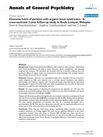
As deduced from the 3D structure organization of Vpr
resolved by NMR (see on Fig. 2A), the Leu23 and Lys27
residues are located in the first N-terminal α-helix H1 (res-
idues 17–33) of Vpr which has amphipathic properties.
Leu23 and Lys27 are separated by 3 residues and are thus
located on the same face of the first α-helix (Fig. 2D). The
connection between these two residues is favored by the
formation of a hydrogen-bonding network through the
O19/NH23, O23/NH27 and O27/NH31 atoms maintain-
ing the structure of the α-helix. Moreover, the Corey, Paul-
ing, and Koltun (CPK) representation, indicates that the
Leu23 and Lys27 residues are located at the bottom of a
pocket that is easily accessible to the solvent (Fig. 2B) and
could constitute a binding site for hCG1. In addition, the
Leu23 residue is hydrophobic and is surrounded by rather
hydrophobic residues (Leu20, Trp54, Gly51 and Tyr47)
that border one edge of the pocket (Fig. 2C), whereas the
Lys27 residue is hydrophilic, positively charged and bor-
dered by hydrophilic residues (Gln44, His40, Asn28 and
Glu24) that constitute the second edge of the pocket. The
potential structural modifications induced by substitution
of Leu23 and Lys27 in Phe and Met, respectively, have
been calculated by homology with the wild type Vpr pro-
tein using the Swiss-Model program [33-35]. The analysis
indicated that the structure of the first α-helix (residues
17–33) is conserved as well as the hydrogen-bonding net-
work allowing the stabilization of the 3 helices of HIV-1
Vpr. This supports the notion that the global 3D structure
of the protein is not modified in these two Vpr mutants,
as suggested from the yeast two-hybrid analysis.
Intracellular distribution of the Vpr mutants
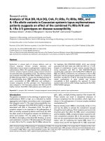
Since HIV-1 Vpr localizes predominantly in the nucleus
but also concentrates at the NE as a nuclear rim staining
(Fig. 3A, middle panel) where it co-localizes with the
nucleoporin hCG1 (left panel) [28], the cellular distribu-
tion of the two hCG1-binding deficient Vpr mutants was
first analyzed. In contrast to the wt Vpr-GFP fusion, both
Vpr-L23F and -K27M equally distributed between the
cytoplasm and the nucleus (Fig. 3B), but they were
excluded from the nucleolus. When expressed as HA-
tagged proteins, these Vpr mutants similarly co-distrib-
uted in the cytoplasm and the nucleus, whereas wt HA-
Vpr was concentrated into the nucleus and at the NE (data
not shown). These data support that mutations of Vpr
which alter its binding to hCG1 also impair its accumula-
tion at the NE.
In order to explore whether substitutions in the first α-
helix had a general impact on the localization of Vpr, the
cellular distribution of two other Vpr mutants (Vpr-A30L
and -F34I) was also analyzed (Fig. 3C). In contrast with
published observations [36], we found that Vpr-A30L was
distributed between the nucleus and the cytoplasm and
failed to concentrated at the NE. As previously reported
[25], Vpr-F34I displayed a nucleocytoplasmic distribu-
tion. In contrast, other Vpr mutants with substitutions in
the third α-helix or in the C-terminal flexible basic region
of the protein, such as Vpr-W54R, -R80A and -R90K, were
concentrated at the NE as efficiently as the wt Vpr-GFP
fusion (Fig. 3C). Altogether, these results indicate that the
first α-helix of Vpr contains the major determinants
required for the nuclear localization of the protein.
Retrovirology 2007, 4:84 />Page 4 of 15
(page number not for citation purposes)
Identification of Vpr mutants deficient for binding to the nucleoporin hCG1Figure 1
Identification of Vpr mutants deficient for binding to the nucleoporin hCG1. A) Screening for Vpr mutants defective
for the interaction with hCG1. The L40 yeast reporter strain expressing the wt or mutated (clones 11 and 35, and Vpr-R90K
and -W54R single-point mutants) HIV-1 Vpr fused either to LexABD (upper panels) or to the Gal4 DNA binding domain
(Gal4BD) (lower panels), in combination with each of the Gal4AD-hybrids indicated on the top was analyzed for histidine aux-
otrophy and β-Gal activity. Double transformants were patched on selective medium with histidine (+His) and then replica-
plated on medium without histidine (-His) and on Whatman filters for β-Gal assay. Growth in the absence of histidine and
expression of β-galactosidase indicated an interaction between hybrid proteins. B) Amino acid substitutions found in the
hCG1-binding deficient Vpr mutants (clones 11 and 35). Mutants were derived by error prone PCR-mediated mutagenesis
from the primary sequence of the VprLai strain that is shown at the top. C) Isolation of single-point Vpr mutants defective for
the interaction with hCG1. Single-point mutants derived from Vpr clones 11 and 35 fused to LexABD were expressed in L40
strain in combination with each of the Gal4AD-hybrids indicated on the top. Double transformants were assessed as described
in A).
Retrovirology 2007, 4:84 />Page 5 of 15
(page number not for citation purposes)
G2-arrest activity and cell death induction of the Vpr
mutants
Since a functional link was reported between the targeting
at the NE and the Vpr-induced cell cycle arrest [36,37], the
G2-arrest activity of the Vpr-L23F and Vpr-K27M mutants
was first assessed in T lymphocytes. HPB-ALL T lymphoid
cells were transfected with wt or mutated HA-tagged Vpr
expression vector together with a GFP expression vector
(see Fig. 4C), and the DNA content was analyzed 48 h
later by flow cytometry on GFP-positive cells after staining
with propidium iodide. The results of four independent
experiments are recapitulated on Fig. 4A. The Vpr-L23F
mutant was affected but retained about 50% of the activity
measured for the wt protein, while the Vpr-K27M mutant
was more severely affected leading to a residual G2-arrest
activity. Consistent with previous observations, the Vpr-
F34I mutant was partially altered for the G2-arrest activity
[25], while the Vpr-A30L mutant was completely defective
[20,36] (Fig. 4A). As controls, the Vpr-R80A and -R90K
variants, which still accumulated at the NE (Fig. 3C), were
Impact of the Vpr-L23F and -K27M substitutions on the three-dimensional structure of VprFigure 2
Impact of the Vpr-L23F and -K27M substitutions on the three-dimensional structure of Vpr. A) 3D structure of
HIV-1 Vpr [10], showing the three α-helices (residues 17–33, 38–50 and 54–77) represented in light blue, yellow and purple,
respectively. The L23, K27, A30 and F34 residues are colored in red. The unstructured N- and C-terminal domains are repre-
sented in dark blue. B) CPK representation of Vpr. Residues are colored according to their hydrophobicity, except for L23 and
K27 which are colored in yellow. The yellow box is enlarged in C), and this region shows a pocket that is organized around the
L23 and K27 residues within the first α-helix and may represent a site for hCG1 binding. D) Helical-wheel diagram of the first
α-helix of Vpr extending from a.a. D17 to F34. Residues L23, K27, A30 and F34 which have been mutated in the present study
are indicated. Hydrophilic residues are in blue, whereas hydrophobic residues are in red.
Retrovirology 2007, 4:84 />Page 6 of 15
(page number not for citation purposes)
unable to induce a G2-arrest (Fig. 4A and Refs. [31,37]).
The pro-apoptotic activity of the wt Vpr protein and the
mutants was also assayed, 72 h after transfection, by flow
cytometry analysis of the cell surface exposure of phos-
phatidylserine (PS) after staining with phycoerythrin-
labeled Annexin V (Fig. 4B). Interestingly, the Vpr-
induced pro-apoptotic activity of all the Vpr mutants,
including Vpr-L23F and -K27M, strictly paralleled the
results obtained in the cell cycle experiments (compare
Fig. 4A and 4B), suggesting that induction of G2-arrest
and apoptosis by HIV-1 Vpr are functionally related. As
evidenced on Fig. 4C, the reduction in G2-arrest and cell
death induction observed with the Vpr mutants could not
be explained by important differences in their expression
levels, since all mutants were correctly expressed in HPB-
ALL T lymphoid cells.
Altogether, these observations indicate that accumulation
of Vpr at the NE is required but is not sufficient for its
action on the cell cycle progression and the subsequent
cell death. They also confirm that these two Vpr functions
are functionally related.
Intracellular localization of Vpr mutants in primary human
monocyte-derived macrophages
In order to confirm that Vpr also accumulated at the
nuclear envelope in target cells relevant for HIV-1 replica-
tion, the distribution of both wt and mutated Vpr proteins
was then analyzed in primary macrophages derived from
monocytes (MDMs) isolated from buffy coats of healthy
donors. As previously shown in HeLa cells (see Fig. 3), the
wt Vpr-GFP fusion localized in the nucleus of MDMs but
also concentrated at the NE as a punctuate staining likely
corresponding to NPC structures (Fig. 5). A similar punc-
tuate staining at the NE was observed in a myeloid cell
line, such as THP-1 cells, expressing the Vpr-GFP fusion
(not shown). Again, both Vpr-L23F and -K27M mutants
failed to concentrate at the NE and predominantly local-
ized in the cytoplasm as a diffuse staining. These data con-
firm that Vpr mutants deficient for hCG1-binding also fail
to accumulate at the NE in primary macrophages.
Replication in primary macrophages of the hCG1-binding
deficient Vpr HIV-1 mutants
Finally, the relationship between the Vpr docking at the
NE and HIV-1 replication in non-dividing cells was
explored by analyzing the impact of the hCG1-binding
deficient Vpr-L23F and -K27M mutations on viral replica-
tion in primary macrophages. The requirement of Vpr for
early stages of the virus life cycle, including nuclear trans-
port of the viral DNA (for review, see Ref. [17]), has been
associated with its packaging into virions and the result-
ant presence in the cytoplasm of newly infected cells.
Using a transient Vpr packaging assay in which HA-tagged
Vpr is expressed in trans in virus producing cells [32], we
therefore analyzed whether the two Vpr mutants were
incorporated into virions. As evidenced in Fig. 6A, both
Vpr-L23F and -K27M were efficiently packaged into puri-
fied virions, but a slight difference in the level of incorpo-
ration was repeatedly observed.
Subcellular distribution of the Vpr mutantsFigure 3
Subcellular distribution of the Vpr mutants. A) Colo-
calization of Vpr and hCG1 at the NE. HeLa cells co-express-
ing Vpr-GFP (middle row) and Myc-hCG1 (left row) fusion
proteins were permeabilized with digitonin, fixed, and subse-
quently stained with an anti-Myc monoclonal antibody. B and
C) Localization of wt and mutated Vpr-GFP fusions. HeLa
cells expressing either GFP, wt Vpr-GFP, or the indicated
Vpr-GFP mutants were fixed and directly examined. Cells
were analyzed by epifluorescence microscopy, and images
were acquired using a CCD camera. Scale bar, 10 µm.
Retrovirology 2007, 4:84 />Page 7 of 15
(page number not for citation purposes)
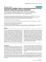
G2-arrest and pro-apoptotic activities of the Vpr mutantsFigure 4
G2-arrest and pro-apoptotic activities of the Vpr mutants. HPB-ALL T cells were transfected with the HA-tagged Vpr
(wt or mutated) expression vectors in combination with the GFP expression vector. A) G2-arrest activity. The DNA content
was analyzed 48 h after transfection by flow cytometry on GFP-positive cells after staining with propidium iodide. Results are
expressed as the percentage of the G
2
M/G1 ratio relative to that of the wt HA-Vpr. Values are the means of four independent
experiments. Error bars represent 1 standard deviation from the mean. B) Pro-apoptotic activity. Cell surface PS exposure was
analyzed 72 h after transfection by flow cytometry on GFP-positive cells after staining with phycoerythrin-labelled Annexin V.
Results are expressed as the percentage of GFP-positive cells displaying surface PS exposure relative to that measured with wt
HA-Vpr. Values are the means of four independent experiments. Error bars represent 1 standard deviation from the mean. C)
Expression of wt and mutated HA-tagged Vpr proteins. Lysates from HPB-ALL transfected cells were analyzed by western-
blotting using anti-GFP (upper panels) and anti-HA antibodies (lower panels).
Retrovirology 2007, 4:84 />Page 8 of 15
(page number not for citation purposes)
Subcellular localization of wild type Vpr and Vpr mutants in human monocyte-derived-macrophagesFigure 5
Subcellular localization of wild type Vpr and Vpr mutants in human monocyte-derived-macrophages. MDMs
expressing either GFP, wt Vpr-GFP, or the indicated Vpr-GFP mutants were fixed and analyzed by wide-field microscopy. Z
stacks of fluorescent images were acquired using a piezo with a 0.2 µm increment and one medial section is shown (left panels).
Phase contrast images of the same cells were acquired to identify the nucleus (right panels). Scale bar, 5 µm.
Retrovirology 2007, 4:84 />Page 9 of 15
(page number not for citation purposes)
Impact of the Vpr mutations on HIV-1 replication in monocyte-derived macrophagesFigure 6
Impact of the Vpr mutations on HIV-1 replication in monocyte-derived macrophages. A) Packaging assay of the wt
and mutated HA-tagged HIV-1 vpr into virus like particles. 293T cells were transfected with an HIV-1-based packaging vector
lacking the vpr gene in combination with vectors for expression of the wt or mutated HA-tagged Vpr protein. 48 h later, pro-
teins from cell and virion lysates were separated by SDS-PAGE and analyzed by Western blotting with anti-HA and anti-CAp24
antibodies. B and C) The L23F or K27M mutations were introduced into the vpr gene of the HIV-1YU-2 molecular clone. In B)
Lysates from transfected 293T cells and virions isolated from cell supernatants were subjected to SDS-PAGE followed by
Western blotting, using a rabbit polyclonal anti-Vpr and a mouse anti-CAp24 (provided from the NIH AIDS Research and Ref-
erence Reagent Program). In C) Replication of wild type and mutated HIV-1 in monocyte-derived macrophages. The wild type
HIV-1YU-2 (WT, open diamonds) and the vpr-defective (∆Vpr, open squares), Vpr-L23F (black circles) and -K27M (black trian-
gles) mutant viruses were produced by transfection of 293T cells with proviral DNAs. Monocyte-derived macrophages from
four healthy donors were infected in triplicates with 0.5 ng of CAp24. Virus production was then monitored by measuring the
p24 antigen by ELISA 10, 14 and 17 days after infection. Results are expressed as the level of p24 in the supernatants of infected
cells. Values are the means of four experiments and error bars represent 1 standard deviation from the mean.
Retrovirology 2007, 4:84 />Page 10 of 15
(page number not for citation purposes)
The L23F and K27M mutations were thus introduced into
the vpr gene of the macrophage-tropic HIV-1YU-2 molec-
ular clone. As a negative control, we used the isogenic vpr-
defective mutant HIV-1YU-2∆Vpr, which contains two
stop codons in frame without altering the vif open reading
frame. As shown in Fig. 6B, both mutated VprYU-2 pro-
teins were efficiently incorporated into purified HIV-1YU-
2 virus particles, even if the slight difference in the level of
incorporation of the two Vpr mutants evidenced in panel
A was still apparent. We first verified that the Vpr mutant
viruses did not show replication defects in cells permissive
for vpr-defective virus replication in vitro. HeLa-CD4 cells
or primary lymphocytes were first infected with equiva-
lent inocula of wt or mutant viruses. Similar replication
kinetics were observed for wt HIV-1YU-2, HIV-1∆Vpr, and
the Vpr-L23F and -K27M mutant viruses (data not
shown). We then infected monocyte-derived macro-
phages (MDMs) from 4 healthy seronegative donors with
the same viral inocula. Consistent with previous reports
[5-8], the vpr-defective virus showed a marked replication
defect in MDMs from all donors (Fig. 5B). The Vpr-L23F
and -K27M mutant viruses exhibited differential replica-
tion abilities according to the donor. Compared to the wt
virus, a significant decrease of replication levels of the
mutant viruses was observed in MDMs from three out of
four donors (Fig. 5B, donors 1, 2 and 4). Conversely, the
levels of replication of the Vpr-L23F and -K27M mutant
viruses were similar to that of the wt virus in MDMs from
another donor (Fig. 5B, donor 3). Although we cannot
exclude that the replication defect observed in donors 1, 2
and 4 may be related to the differential virion incorpora-
tion evidenced in Fig. 5A, one can note that the levels of
incorporation were sufficient, at least in donor 3, for effi-
cient replication of the Vpr-L23F and -K27M mutant
viruses. While these results confirm that the absence of
Vpr expression consistently affects HIV-1 replication in
primary macrophages, the Vpr-L23F and -K27M muta-
tions lead to a replication defect in macrophages from
most of the donors.
Discussion
Although several studies have suggested a specific role for
HIV-1 Vpr in facilitating the nuclear localization of the
viral DNA during infection of non-dividing cells, such as
macrophages, there is still no evident correlation reported
in the literature between its real contribution to this proc-
ess and the known functions of Vpr in vitro, including its
ability to accumulate at the NE. Given that Vpr is dynam-
ically associated with the NE [25,28], this subcellular dis-
tribution may be a pre-requirement for one or more of its
known functions. Based on the findings that Vpr is able to
interact directly with some proteins of the NPC, including
the nucleoporin hCG1 [28], we have now identified two
Vpr mutants, Vpr-L23F and -K27M, that are specifically
deficient for hCG1-binding. Both mutations similarly
abrogate Vpr concentration at the NE both in HeLa cells
and in primary human monocyte-derived macrophages,
supporting the hypothesis that this nucleoporin partici-
pates in the docking of the protein to the NPC. To our
knowledge, it is the first report confirming that HIV-1 Vpr
efficiently accumulates at the NE in primary macrophages.
However, direct evidences regarding the specific role of
hCG1 in the NE concentration of Vpr are still missing,
since we failed so far to significantly deplete the endog-
enous hCG1 protein by using the RNA interference tech-
nology.
While substitutions of the Leu23 residue have been
described previously [37,38], the mutation at position 27
(K27M) was not yet identified and is particularly interest-
ing since the K27 is well-conserved among HIV-1 isolates
and constitutes the only lysine residue along the whole
Vpr sequence. This residue may potentially constitute a
site for post-translation modifications, such as methyla-
tion, acetylation, hydroxylation, sumoylation or ubiquiti-
nation [39]. None of these modifications have been
described previously for Vpr and our western blot analysis
did not reveal any change in the level of expression and/
or stability of the Vpr-K27M mutant compared to the wt
protein. Interestingly, both Leu23 and Lys27 residues are
located in the first N-terminal α-helix H1 (residues 17–
33) of Vpr which has amphipathic properties (see on Fig.
2). The structural analysis confirms that the 3D structure
and the stability of the three α-helices of Vpr are not sig-
nificantly affected by the L23F and K27M substitutions, as
indicated by the overall conservation of the hydrogen-
bonding network of the protein. This analysis also reveals
that the Leu23 and Lys27 residues are located in a close
proximity at the end of the first α-helix, in a pocket that is
easily accessible for protein-protein interaction with cellu-
lar partners, such as nucleoporins. We can notice that the
two other mutations, A30L and F34I, impairing the NE
accumulation of Vpr involve amino acids located on the
same face of the first α-helix of the protein (see on Fig.
2D). However, Ala30 and Phe34 are not accessible and are
rather involved in the stability of the structure by estab-
lishing hydrophobic interactions with residues of the
third α-helix (55–77) of Vpr (see on Fig. 2A). Proximities
between Ala30 and Leu64/Leu68 and between Phe34 and
Leu64/Leu67/Leu68 have been identified from NMR
experiments, indicating that Ala30 and Phe34 are directly
involved in the interaction between the first and the third
α-helix. Any mutation of the residues found at this inter-
face will likely perturb the structure of the Vpr protein.
Our functional analysis raises intriguing questions con-
cerning the functional link between Vpr accumulation at
the NE and the in vitro properties of the protein, namely
G2-arrest and cell death induction. Like other Vpr
mutants that fail to localize at the NE, such as Vpr-A30L
Retrovirology 2007, 4:84 />Page 11 of 15
(page number not for citation purposes)
and -F34I, both L23F and K27M substitutions affect the
Vpr-induced G2-arrest and cell death. These observations
highlight two important points: i) the functional link
between Vpr docking at the NE and its ability to cause a
G2-arrest, and ii) the link between the G2-arrest and the
pro-apoptotic activities of Vpr. First, it is possible that the
accumulation at the NE could constitute a prerequisite for
the Vpr-induced G2-arrest [40]. It was reported that Vpr
provoked herniations and transient ruptures of the NE,
resulting in a mixing of cytoplasmic and nuclear compo-
nents that could contribute to the cell cycle arrest. None-
theless, the molecular mechanism underlying this process
is still unknown, and the local bursting caused by Vpr at
the NE in this report has not been confirmed to date, even
in imaging experiments performed on living cells [28].
Alternatively, the concentration of Vpr at the NE, or in a
close vicinity of the nuclear pore complexes, could be
required for the establishment of local interactions with
some cellular partners involved in the regulation of the
cell cycle. Because no mutant of HIV-1 Vpr has been
described so far as disrupting the NE accumulation but
keeping intact G2-arrest activity, our results confirm these
observations and suggest that Vpr accumulation at the NE
may be required. However, targeting to the NE is not suf-
ficient for the G2-arrest activity, since several Vpr point
mutants with substitutions in the C-terminal basic region
of the protein, such as Vpr-R90K and Vpr-R80A, have been
reported as defective for the G2-arrest activity (present
study and Refs. [31,40,41]), while we show that both can
still accumulate at the NE. Recent studies have shown that
these residues may be involved in the direct recruitment of
an unknown cellular factor that is required for G2-to-M
transition [42-46].
Second, our results show that the pro-apoptotic activity of
Vpr totally parallels the Vpr-induced G2-arrest, confirm-
ing previous reports suggesting that apoptosis is a direct
consequence of the prolonged cell cycle arrest induced in
Vpr-expressing cells [47-51]. Whereas other authors have
suggested that these two Vpr properties were separated
[22,41,52-54], it was recently demonstrated that the Vpr-
induced apoptosis is directly dependent on the cell cycle
arrest [55]. Both Vpr activities are directly related to the
activation of G2 checkpoint ATR-initiated DNA damage-
signaling pathway.
Though it is admitted that Vpr displays evident affinity for
the NE [21-23,25,28], the biological significance of this
property for efficient virus replication in non-dividing
cells has been explored only in a few reports [25,29].
While a previous study showed that virus expressing sin-
gle-point mutants (Vpr-F34I and -H71R), which failed to
concentrate at the NE, displayed decreased infectivity in
macrophages [25], it was recently reported that a Vpr
mutant with multiple substitutions, in which the 4 Leu
residues, including Leu23, found within the first α-helix
of the protein were replaced by Ala, failed to localize in
the nucleus and rendered the virus unable to replicate in
MDMs from two donors [29]. Here, we show that the sub-
stitution of the Leu23 residue is sufficient to impair both
nuclear accumulation of the Vpr protein and virus replica-
tion in primary macrophages. In addition, mutation of
the Lys27 residue found in a close proximity within the
first helix similarly impaired HIV-1 replication in MDMs
from most donors. While the Vpr-L23F and -K27M
mutant viruses displayed a wild-type level of replication
in only one out of four donors, we cannot formally
exclude that the replication defect observed in the three
other donors may be related to the slight difference in the
level of virion incorporation of the two Vpr mutants evi-
denced in Fig. 6A and 6B. As reported previously, the first
α-helix of HIV-1 Vpr contains the main determinants
required for efficient incorporation into virions
[12,20,36,37].
Primary cells and especially MDMs are heterogeneous cell
populations that greatly vary in their susceptibility to HIV
infection [5,56]. Moreover, it is most likely that the con-
tribution of Vpr for virus replication in macrophages relies
on more than one of its activities. In addition to its role in
nuclear import of the viral DNA, Vpr displays several
other activities that could be important to maintain effi-
cient virus replication in macrophages. Since it was
reported that the manipulation of the cell cycle by Vpr
increased viral expression in both dividing and non-divid-
ing cells [18,57], the replication defect observed here with
the mutant viruses could be, at least in part, related to the
impairment of the Vpr-mediated G2-arrest. Additionally,
the Vpr effect on the fidelity of the reverse-transcription
process was also directly correlated with the ability of
HIV-1 to efficiently replicate in primary macrophages
[19]. These findings confirm that the absence of Vpr
expression is deleterious for HIV-1 replication in primary
macrophages, but additional analyses are required to re-
evaluate the relationship between the multiple functions
of Vpr characterized in vitro and its requirement in vivo for
efficient replication in the non-dividing target cells of
HIV-1.
Conclusion
The present study shows that the targeting of Vpr to the
nuclear pore complex may constitute an early step toward
Vpr-induced G2-arrest and subsequent apoptosis. How-
ever, this localization at the NE is not absolutely required,
but could improve HIV-1 replication in macrophages.
Methods
Expression plasmids and proviral DNAs
Most of the yeast and mammalian expression plasmids
used in this study have been described previously
Retrovirology 2007, 4:84 />Page 12 of 15
(page number not for citation purposes)
[12,19,28,31], except plasmids for expression of the Vpr
mutants with L23F and K27M substitutions; the Vpr
mutant with the R80A substitution was kindly provided
by E. Cohen (Universite de Montreal, Montreal, Canada).
The L23F and K27M mutants were constructed by PCR-
mediated site-directed mutagenesis using specific primers
containing the desired mutations. The PCR product was
then cloned between the BamHI-SalI restriction sites of
the pLex10 plasmid [12,31] and BamHI-XhoI of the pAS1b
plasmid [28] to obtain vectors for expression of Vpr fused
either to the LexA DNA binding domain (LexABD) or to
the HA-tag, in yeast and in mammalian cells, respectively.
For expression of Vpr mutants as N-terminal fusions with
the green fluorescent protein (GFP), vpr mutated
sequences were amplified by PCR with specific primers
and cloned into pEGFP-N1 (Clontech).
Mutations of the vpr gene from the HIV-1 YU-2 molecular
clone (obtained from the NIH AIDS Research & Reference
Reagent Program) have been done using a shuttle vector
containing the NdeI fragment (from nucleotide 5121 to
6401 according to HIVYU2X accession number) cloned
into the pUC19 plasmid. Mutations (L23F, K27M) were
thus performed using pUC19-VprYU-2 as a matrix by site-
directed mutagenesis with specific primers: L23F-F
ACACTAGAG CTTTTT
GAGGAGCTTAAG, L23F-R CTTAA
GCTCCTCAAAAAGCTCTAGTGT, K27M-F CTTTTAGAGG
AGCTTATG
AGAGAAGCTGTTAG, K27M-R CTAACAGCT-
TCTCTCATAA GCTCCTCTAAAAG. HIV-1YU-2∆vpr has
been constructed using the same procedure by insertion of
two stop codons within the vpr gene without altering the
vif gene using the following set of primers: Forward
GATAGATGGAATAA
GCCCCAGAAGACTAAG-
GGCCACAGAG
G; Reverse CCTCTGTGGCCCTTAGTCTTCTGGGGCTTAT-
TCCATCTATC. All constructs were verified by nucleotide
sequencing with a DYEnamic ET Terminator kit (Amer-
sham) and a Genetic Analyzer (ABI3100 Applied Biosys-
tems).
Yeast two-hybrid assay
The library of HIV-1 Vpr mutants fused to LexABD and the
two-hybrid screening procedure of the library have been
described [12,31]. Briefly, the vpr gene from the HIV-1 Lai
isolate was amplified with error-prone Taq DNA polymer-
ase (Promega Inc.) using previously described conditions
[31,58], and the fragments were then inserted between
BamHI and SalI sites of pLex10. Library plasmid from
about 10
5
independent E. coli clones, representing the
complexity of the vpr mutant library, was prepared and
used to transform the L40-MATα yeast strain. About 10
5
yeast clones were then screened to select Vpr mutants
defective for binding to hCG1, by mating with the
AMR70-MATα yeast strain previously transformed with
the Gal4AD-hCG1 expression vector [28]. The yeast
clones were also mated with the AMR70 strain previously
transformed with the Gal4AD-UNG2 expression vector
[19]. Plasmids from two mutants unable to interact with
hCG1, but still binding to UNG2, were rescued and their
insert was completely sequenced. Since one variant con-
tained several point mutations (L23F, L67Q, and R73G;
see on Fig. 1B), the single-point variants were constructed
by site-directed mutagenesis. The L40 yeast reporter strain
containing the two LexA-inducible genes, HIS3 and LacZ,
was cotransformed with the indicated LexABD and
Gal4AD hybrid expression vectors and plated on selective
medium lacking tryptophan and leucine as previously
reported [12]. Double transformants were patched on the
same medium and replica-plated on selective medium
lacking tryptophan, leucine, and histidine for auxotrophy
analysis, and on Whatman 40 filters for β-galactosidase
(β-gal) activity assay [12]. This latter assay was monitored
by incubation for 1 h to 4 h at 30°C, and the reaction was
then stopped with 1 M Na2CO3.
Cell culture and transfections
HeLa cells were maintained in Dulbecco's modified
Eagle's medium (DMEM, Invitrogen) supplemented with
10% fetal bovine serum (Invitrogen), 50 U/mL penicillin/
streptomycin and 125 ng/mL amphotericin B (Gibco-
BRL), at 37°C under 5% CO2. The cells were grown onto
coverslips in 6-well plates to 30–50% confluence on the
day of transfection. Myc-hCG1 and Vpr-GFP (wild-type or
mutated) expression vectors were co-transfected using the
calcium phosphate method. Briefly, 4 µg of both plasmids
were diluted in 60 µL of a 0.24 M CaCl2 solution. The
CaCl2-DNA mix was slowly added dropwise to 60 µL of 2
× HBS buffer (50 mM HEPES pH 7.1, 1.5 mM Na2HPO4,
0.28 M NaCl). After 20 min at room temperature, DNA-
containing precipitates were slowly dropped onto the sur-
face of the cell culture medium. Cells were incubated at
37°C with 5% CO2 for 6 h, rinsed once with PBS and
fresh medium was added before returning to the incuba-
tor until analysis. CD4-positive human T cells (HPB-ALL
cell line) were kindly provided by G. Bismuth (Institut
Cochin, Paris, France), and were maintained in RPMI
1640 medium with Glutamax-1 (Invitrogen) supple-
mented with 10% fetal bovine serum, 10 mM HEPES
buffer, 50 U/mL penicillin/streptomycin and 125 ng/mL
amphotericin B (GibcoBRL) at 37°C under 5% CO2. For
transient transfections, 1 × 10
7
HPB-ALL cells were electro-
porated as described [59] with 4 µg of a GFP expression
vector as a transfection marker, and 16 µg of HA-tagged
Vpr (wild-type or mutated) expression vectors. For locali-
zation analysis in MDMs, PBMCs were isolated from buffy
coats of healthy donors (Etablissement Français du Sang
Ile-de-France, Site Saint Vincent-de-Paul) and derived into
macrophages for 5 days in complete culture medium
[MEM with non essential amino acids (Invitrogen/Gibco)
supplemented with 10% FCS, 100 µg/ml streptomycin/
Retrovirology 2007, 4:84 />Page 13 of 15
(page number not for citation purposes)
penicillin, 1 mM sodium pyruvate, 2 mM L-glutamin and
10 ng/ml rhM-CSF (R&D systems)]. Cells were transfected
with the Nucleofector II device (Amaxa GmbH Europe/
World), and nucleofection was performed with the
Human Macrophage Nucleofector Kit according to the
manufacturer's recommendations. Briefly, 6 × 10
5
macro-
phages were nucleofected with 4 µg of plasmid and then
cultured in Macrophage-SFM medium (Invitrogen/Gibco)
supplemented with 10% FCS and 2 mM L-glutamine for 6
hours. The HEK-293T cells used in the virion packaging
assay were maintained in DMEM supplemented with 10%
fetal bovine serum, 50 U/mL penicillin/streptomycin and
125 ng/mL amphotericin B (GibcoBRL), at 37°C under
5% CO2. Cells were then co-transfected as described [12],
using the calcium phosphate method with 10 µg of the
pCMV∆8.3 proviral vector [60] and 5 µg of wild-type or
mutated HA-Vpr expression vector.
Immunofluorescence staining
18 h after transfection, HeLa cells grown onto coverslips
were fixed with 4% paraformaldehyde (PFA) for 20 min
and permeabilized with 0.1% Triton X-100 for 10 min.
Alternatively, cells were permeabilized for 5 min at 4°C
with 55 µg/ml digitonin (Sigma) in transport buffer [61]
and then fixed with 4% PFA. Monoclonal antibody to the
Myc tag (9E10, Roche) was applied for 30 min followed
by a 30-min incubation with Texas Red-conjugated don-
key anti-mouse IgG (Jackson). Cells were mounted in PBS
containing 50% glycerol. Images were acquired with a
Leica DMRB epifluorescence microscope equipped with a
CCD camera (Princeton) controlled by Metamorph
V5.0r6 software. Optical sections were done using Adobe
Photoshop software.
Cell cycle and apoptosis analysis
Two days after transfection, half of HPB-ALL T cells were
collected, rinsed once with PBS and fixed in 1% PFA for 20
min. After two washes in PBS, cells were permeabilized in
cold 70% ethanol for 1 h at 4°C. Finally, cells were
washed once with PBS, resuspended in PBS containing
200 µg/ml RNase A and 50 µg/ml propidium iodide, and
incubated for 15 min at room temperature prior to analy-
sis of DNA content by flow cytometry as described [62].
Three days after transfection, the remaining HPB-ALL cells
were rinsed in PBS and analyzed by flow cytometry for
exposure of phosphatidylserines (PS) at the cell surface, as
an early marker of apoptosis, using phycoerythrin-conju-
gated annexin V (Annexin V-PE, Bender MedSystems) as
described [59]. Cell cycle and PS-exposure profiles were
analyzed on a minimum of 5,000 GFP-positive cells using
a Cytomics FC 500 instrument (Beckman Coulter).
Assay for incorporation of Vpr into HIV-1 particles
Incorporation of the Vpr variants was first analyzed using
a packaging assay in which HA-tagged Vpr was expressed
in trans and incorporated into virions [32]. 293T cells
were co-transfected with 10 µg of the HIV-1-based packag-
ing vectors pCMV∆R8.3 lacking the vpr auxiliary gene, 5
µg of pAS1B-Vpr (wt or mutated) using the calcium phos-
phate procedure. Cell culture supernatants were harvested
48 h after transfection and filtered through 0.45-µm-pore-
size filters. Virions were collected by ultracentrifugation
for 1.5 h at 120,000 × g at 4°C and suspended in ice-cold
lysis buffer containing 1% NP40, 0.5% sodium deoxycho-
late, 0.05% SDS in PBS and an antiprotease mixture
(Roche). For preparation of cell lysates, cells were
trypsinized, collected by centrifugation and suspended in
ice-cold lysis buffer and clarified by centrifugation. Pro-
tein samples from cell and virion lysates were separated by
SDS-PAGE and analyzed by Western blotting as previ-
ously described [32], using a rat monoclonal anti-HA
(3F10, Roche), a mouse monoclonal anti-tubulin (DM1A,
Sigma) and a mouse anti-CAp24 (provided from the NIH
AIDS Research and Reference Reagent Program).
Preparation of monocyte-derived macrophages (MDMs)
Peripheral blood mononuclear cells (PBMCs) were iso-
lated from buffy coats of healthy seronegative donors
(Centre de Transfusion Sanguine Ile-de-France, Rungis
and Hôpital de la Pitié-Salpêtrière, Paris, France), using
lymphocyte separation medium (PAA laboratories
GmbH, Haidmannweg) density gradient centrifugation.
Monocytes were isolated from PBMC by plastic adherence
as previously described [63], and non adherent cells
(PBLs) were collected, frozen in heat-inactivated fetal calf
serum 10% DMSO and stored at -80°C. Monocytes were
differentiated in macrophages by culturing for 7–11 days
in MDM medium (RPMI 1640 medium supplemented
with 200 mM L-glutamine, 100 U penicillin, 100 µg strep-
tomycin, 10 mM HEPES, 10 mM sodium pyruvate, 50 µM
β-mercaptoethanol, 1% minimum essential medium vita-
mins, and 1% nonessential amino acids) supplemented
with 15% of human AB serum in hydrophobic Teflon
dishes (Lumox™ D Dutcher, Brumath, France). MDMs
were then harvested, washed and resuspended in MDM
medium containing 10% fetal calf serum. The purity of
CD14
+
macrophages was usually more than 95% as
assessed by immunofluorescence staining and flow
cytometry analysis (not shown). Three days before infec-
tion, PBLs were thawed and cultured in PBL medium
(RPMI 1640 medium supplemented with 200 mM L-
glutamine, 100 U penicillin, 100 µg streptomycin, 10%
FCS, 100 U/ml of interleukin 2 (Proleukin, Chiron,
France) and activated 3 days in presence of 1 µg/ml of
PHA (Sigma).
Virus production and infection
Viruses were produced by transfection of 293T cells with
the wild type or mutated YU-2 HIV-1 molecular clones
[19] using SuperFect (Qiagen GmbH, Hilden, Germany).
Retrovirology 2007, 4:84 />Page 14 of 15
(page number not for citation purposes)
Viral supernatants were harvested 72 h after transfection
and stored at -80°C. Viral supernatants were centrifuged
in Vivaspin centrifugal concentrators (MW cut off 100 000
kDa) against medium to eliminate free p24Gag and
cytokines, and then normalized for viral-bound p24
before analysis of Vpr incorporation into virus particles by
SDS-PAGE and Western blotting, using a rabbit polyclo-
nal anti-Vpr (provided from the NIH AIDS Research and
Reference Reagent Program) and the mouse anti-CAp24.
MDMs (0.8 × 10
5
cells/well in 96 well plates) and PHA-
activated PBLs (10
5
cells/well in 96 well plates) were then
infected in triplicate with 0.5 ng of p24 using a spinocula-
tion protocol (1 h centrifugation at room temperature at
1,200 × g followed by 1 h incubation at 37°C). Cells were
then washed with PBS and cultured in MDM or PBL
medium. The supernatant of each well was harvested
every 3 or 4 days and fresh medium was added. p24 levels
in viral stocks and in infected culture supernatants were
measured using a commercial ELISA kit (Beckman Coul-
ter, Paris, France).
Competing interests
The author(s) declare that they have no competing inter-
ests.
Authors' contributions
GJ, ELR, AD, JM and SBO performed the experimental
work. GJ, ELR, AD, FN, GP and SB conceived the experi-
mental strategies and designed individual experiments.
SBO, GF and SB analyzed the data and GJ and SB wrote
the manuscript. All authors read and approved the final
manuscript.
Acknowledgements
This work was supported in part by INSERM, CNRS, Université Paris-Des-
cartes and the French National Agency for AIDS Research (ANRS), Sidac-
tion and Fondation de France (SB, GP and FN). GJ is a recipient fellowship
of the French Ministery of Research.
We thank C. Dargemont and Alexandre Benmerah for active supports, dis-
cussion, and critical reading of the manuscript.
References
1. Yamashita M, Emerman M: Retroviral infection of non-dividing
cells: old and new perspectives. Virology 2006, 344:88-93.
2. Crowe S, Zhu T, Muller WA: The contribution of monocyte
infection and trafficking to viral persistence, and mainte-
nance of the viral reservoir in HIV infection. J Leukoc Biol 2003,
74:635-41.
3. Yamashita M, Emerman M: The cell cycle independence of HIV
infections is not determined by known karyophilic viral ele-
ments. PLoS Pathog 2005, 1:e18.
4. Fassati A: HIV infection of non-dividing cells: a divisive prob-
lem. Retrovirology 2006, 3:74.
5. Balliet JW, Kolson DL, Eiger G, Kim FM, McGann KA, Srinivasan A,
Collman R: Distinct effects in primary macrophages and lym-
phocytes of the human immunodeficiency virus type 1 acces-
sory genes vpr, vpu, and nef: mutational analysis of a primary
HIV-1 isolate. Virology 1994, 200:623-31.
6. Connor RI, Chen BK, Choe S, Landau NR: Vpr is required for effi-
cient replication of human immunodeficiency virus type-1 in
mononuclear phagocytes. Virology 1995, 206:935-44.
7. Eckstein DA, Sherman MP, Penn ML, Chin PS, De Noronha CM,
Greene WC, Goldsmith MA: HIV-1 Vpr enhances viral burden
by facilitating infection of tissue macrophages but not nondi-
viding CD4+ T cells. J Exp Med 2001, 194:1407-19.
8. Heinzinger NK, Bukinsky MI, Haggerty SA, Ragland AM, Kewalramani
V, Lee MA, Gendelman HE, Ratner L, Stevenson M, Emerman M: The
Vpr protein of human immunodeficiency virus type 1 influ-
ences nuclear localization of viral nucleic acids in nondividing
host cells. Proc Natl Acad Sci USA 1994, 91:7311-5.
9. Zaitseva L, Myers R, Fassati A: tRNAs promote nuclear import
of HIV-1 intracellular reverse transcription complexes. PLoS
Biol 2006, 4:e332.
10. Morellet N, Bouaziz S, Petitjean P, Roques BP: NMR structure of
the HIV-1 regulatory protein VPR. J Mol Biol 2003, 327:215-27.
11. Muller B, Tessmer U, Schubert U, Krausslich HG: Human immun-
odeficiency virus type 1 Vpr protein is incorporated into the
virion in significantly smaller amounts than gag and is phos-
phorylated in infected cells. J Virol 2000, 74:9727-31.
12. Selig L, Pages JC, Tanchou V, Preveral S, Berlioz-Torrent C, Liu LX,
Erdtmann L, Darlix J, Benarous R, Benichou S: Interaction with the
p6 domain of the gag precursor mediates incorporation into
virions of Vpr and Vpx proteins from primate lentiviruses. J
Virol 1999, 73:592-600.
13. Cohen EA, Dehni G, Sodroski JG, Haseltine WA: Human immun-
odeficiency virus vpr product is a virion-associated regula-
tory protein. J Virol 1990, 64:3097-9.
14. Paxton W, Connor RI, Landau NR: Incorporation of Vpr into
human immunodeficiency virus type 1 virions: requirement
for the p6 region of gag and mutational analysis. J Virol 1993,
67:7229-37.
15. Yu XF, Matsuda M, Essex M, Lee TH: Open reading frame vpr of
simian immunodeficiency virus encodes a virion-associated
protein. J Virol 1990, 64:5688-93.
16. Yuan X, Matsuda Z, Matsuda M, Essex M, Lee TH: Human immun-
odeficiency virus vpr gene encodes a virion-associated pro-
tein. AIDS Res Hum Retroviruses 1990, 6:1265-71.
17. Le Rouzic E, Benichou S: The Vpr protein from HIV-1: distinct
roles along the viral life cycle. Retrovirology 2005, 2:11.
18. Goh WC, Rogel ME, Kinsey CM, Michael SF, Fultz PN, Nowak MA,
Hahn BH, Emerman M: HIV-1 Vpr increases viral expression by
manipulation of the cell cycle: a mechanism for selection of
Vpr in vivo. Nat Med 1998, 4:65-71.
19. Chen R, Le Rouzic E, Kearney JA, Mansky LM, Benichou S: Vpr-
mediated incorporation of UNG2 into HIV-1 particles is
required to modulate the virus mutation rate and for repli-
cation in macrophages. J Biol Chem 2004, 279(27):28419-28425.
20. Di Marzio P, Choe S, Ebright M, Knoblauch R, Landau NR: Muta-
tional analysis of cell cycle arrest, nuclear localization and
virion packaging of human immunodeficiency virus type 1
Vpr. J Virol 1995, 69:7909-16.
21. Kamata M, Aida Y: Two putative alpha-helical domains of
human immunodeficiency virus type 1 Vpr mediate nuclear
localization by at least two mechanisms. J Virol 2000,
74:7179-86.
22. Waldhuber MG, Bateson M, Tan J, Greenway AL, McPhee DA: Stud-
ies with GFP-Vpr fusion proteins: induction of apoptosis but
ablation of cell-cycle arrest despite nuclear membrane or
nuclear localization. Virology 2003, 313:91-104.
23. Depienne C, Roques P, Creminon C, Fritsch L, Casseron R, Dormont
D, Dargemont C, Benichou S: Cellular distribution and kary-
ophilic properties of matrix, integrase, and Vpr proteins
from the human and simian immunodeficiency viruses. Exp
Cell Res 2000, 260:387-95.
24. Popov S, Rexach M, Ratner L, Blobel G, Bukrinsky M: Viral protein
R regulates docking of the HIV-1 preintegration complex to
the nuclear pore complex. J Biol Chem 1998, 273:13347-52.
25. Vodicka MA, Koepp DM, Silver PA, Emerman M: HIV-1 Vpr inter-
acts with the nuclear transport pathway to promote macro-
phage infection. Genes Dev 1998, 12:175-85.
26. Fouchier RA, Meyer BE, Simon JH, Fischer U, Albright AV, Gonzalez-
Scarano F, Malim MH: Interaction of the human immunodefi-
ciency virus type 1 Vpr protein with the nuclear pore com-
plex. J Virol 1998, 72:6004-13.
Retrovirology 2007, 4:84 />Page 15 of 15
(page number not for citation purposes)
27. Tran EJ, Wente SR: Dynamic nuclear pore complexes: life on
the edge. Cell 2006, 125:1041-53.
28. Le Rouzic E, Mousnier A, Rustum C, Stutz F, Hallberg E, Dargemont
C, Benichou S: Docking of HIV-1 Vpr to the nuclear envelope
is mediated by the interaction with the nucleoporin hCG1. J
Biol Chem 2002, 277:45091-8.
29. Nitahara-Kasahara Y, Kamata M, Yamamoto T, Zhang X, Miyamoto Y,
Muneta K, Iijima S, Yoneda Y, Tsunetsugu-Yokota Y, Aida Y: A novel
nuclear import of Vpr promoted by importin {alpha} is cru-
cial for HIV-1 replication in macrophages. J Virol 2007.
JVI.01928-06
30. Withers-Ward ES, Jowett JB, Stewart SA, Xie YM, Garfinkel A,
Shibagaki Y, Chow SA, Shah N, Hanaoka F, Sawitz DG, Armstrong
RW, Souza LM, Chen IS: Human immunodeficiency virus type 1
Vpr interacts with HHR23A, a cellular protein implicated in
nucleotide excision DNA repair. J Virol 1997, 71:9732-42.
31. Selig L, Benichou S, Rogel ME, Wu LI, Vodicka MA, Sire J, Benarous R,
Emerman M: Uracil DNA glycosylase specifically interacts with
Vpr of both human immunodeficiency virus type 1 and sim-
ian immunodeficiency virus of sooty mangabeys, but binding
does not correlate with cell cycle arrest. J Virol 1997,
71:4842-6.
32. Mansky LM, Preveral S, Selig L, Benarous R, Benichou S: The inter-
action of vpr with uracil DNA glycosylase modulates the
human immunodeficiency virus type 1 In vivo mutation rate.
J Virol 2000, 74:7039-47.
33. Schwede T, Kopp J, Guex N, Peitsch MC: SWISS-MODEL: An
automated protein homology-modeling server. Nucleic Acids
Res 2003, 31:3381-5.
34. Guex N, Peitsch MC: SWISS-MODEL and the Swiss-Pdb-
Viewer: an environment for comparative protein modeling.
Electrophoresis 1997, 18:2714-23.
35. Peitsch MC, Wells TN, Stampf DR, Sussman JL: The Swiss-
3DImage collection and PDB-Browser on the World-Wide
Web. Trends Biochem Sci 1995, 20:82-4.
36. Mahalingam S, Ayyavoo V, Patel M, Kieber-Emmons T, Weiner DB:
Nuclear import, virion incorporation, and cell cycle arrest/
differentiation are mediated by distinct functional domains
of human immunodeficiency virus type 1 Vpr. J Virol 1997,
71:6339-47.
37. Yao XJ, Subbramanian RA, Rougeau N, Boisvert F, Bergeron D,
Cohen EA: Mutagenic analysis of human immunodeficiency
virus type 1 Vpr: role of a predicted N-terminal alpha-helical
structure in Vpr nuclear localization and virion incorpora-
tion. J Virol 1995, 69:7032-44.
38. Thotala D, Schafer EA, Tungaturthi PK, Majumder B, Janket ML, Wag-
ner M, Srinivasan A, Watkins S, Ayyavoo V: Structure-functional
analysis of human immunodeficiency virus type 1 (HIV-1)
Vpr: role of leucine residues on Vpr-mediated transactiva-
tion and virus replication. Virology 2004, 328:89-100.
39. Yang XJ: Multisite protein modification and intramolecular
signaling. Oncogene 2005, 24:1653-62.
40. de Noronha CM, Sherman MP, Lin HW, Cavrois MV, Moir RD, Gold-
man RD, Greene WC: Dynamic disruptions in nuclear envelope
architecture and integrity induced by HIV-1 Vpr. Science 2001,
294:1105-8.
41. Somasundaran M, Sharkey M, Brichacek B, Luzuriaga K, Emerman M,
Sullivan JL, Stevenson M: Evidence for a cytopathogenicity
determinant in HIV-1 Vpr. Proc Natl Acad Sci USA 2002,
99:9503-8.
42. Le Rouzic E, Belaidouni N, Estrabaud E, Morel M, Rain JC, Transy C,
Margottin-Goguet F: HIV1 Vpr arrests the cell cycle by recruit-
ing DCAF1/VprBP, a receptor of the Cul4-DDB1 ubiquitin
ligase. Cell Cycle 2007, 6:182-8.
43. Hrecka K, Gierszewska M, Srivastava S, Kozaczkiewicz L, Swanson
SK, Florens L, Washburn MP, Skowronski J: Lentiviral Vpr usurps
Cul4-DDB1[VprBP] E3 ubiquitin ligase to modulate cell
cycle. Proc Natl Acad Sci USA 2007, 104:11778-83.
44. Belzile JP, Duisit G, Rougeau N, Mercier J, Finzi A, Cohen EA: HIV-1
Vpr-Mediated G2 Arrest Involves the DDB1-
CUL4A(VPRBP) E3 Ubiquitin Ligase. PLoS Pathog 2007, 3:e85.
45. Tan L, Ehrlich E, Yu XF: DDB1 and Cul4A are required for
human immunodeficiency virus type 1 Vpr-induced G2
arrest. J Virol 2007, 81(19):10822-10830.
46. DeHart JL, Zimmerman ES, Ardon O, Monteiro-Filho CM, Arganaraz
ER, Planelles V: HIV-1 Vpr activates the G2 checkpoint through
manipulation of the ubiquitin proteasome system. Virol J
2007, 4:57.
47. Andersen JL, Zimmerman ES, DeHart JL, Murala S, Ardon O, Blackett
J, Chen J, Planelles V: ATR and GADD45alpha mediate HIV-1
Vpr-induced apoptosis. Cell Death Differ 2005, 12:326-34.
48. Muthumani K, Hwang DS, Desai BM, Zhang D, Dayes N, Green DR,
Weiner DB: HIV-1 Vpr induces apoptosis through caspase 9 in
T cells and peripheral blood mononuclear cells. J Biol Chem
2002, 277:37820-31.
49. Stewart SA, Poon B, Song JY, Chen IS: Human immunodeficiency
virus type 1 vpr induces apoptosis through caspase activa-
tion. J Virol 2000, 74:3105-11.
50. Yuan H, Xie YM, Chen IS: Depletion of Wee-1 kinase is neces-
sary for both human immunodeficiency virus type 1 Vpr- and
gamma irradiation-induced apoptosis. J Virol 2003, 77:2063-70.
51. Yuan H, Kamata M, Xie YM, Chen IS: Increased levels of Wee-1
kinase in G(2) are necessary for Vpr- and gamma irradiation-
induced G(2) arrest. J Virol 2004, 78:8183-90.
52. Chen M, Elder RT, Yu M, O'Gorman MG, Selig L, Benarous R,
Yamamoto A, Zhao Y: Mutational analysis of Vpr-induced G2
arrest, nuclear localization, and cell death in fission yeast. J
Virol 1999, 73:3236-45.
53. Nishizawa M, Kamata M, Mojin T, Nakai Y, Aida Y: Induction of
apoptosis by the Vpr protein of human immunodeficiency
virus type 1 occurs independently of G(2) arrest of the cell
cycle. Virology 2000, 276:16-26.
54. Lum JJ, Cohen OJ, Nie Z, Weaver JG, Gomez TS, Yao XJ, Lynch D,
Pilon AA, Hawley N, Kim JE, Chen Z, Montpetit M, Sanchez-Dardon
J, Cohen EA, Badley AD: Vpr R77Q is associated with long-term
nonprogressive HIV infection and impaired induction of
apoptosis. J Clin Invest 2003, 111:1547-54.
55. Andersen JL, Dehart JL, Zimmerman ES, Ardon O, Kim B, Jacquot G,
Benichou S, Planelles V: HIV-1 Vpr-Induced Apoptosis Is Cell
Cycle Dependent and Requires Bax but Not ANT. PLoS Pathog
2006, 2:e127.
56. Freed EO, Englund G, Martin MA: Role of the basic domain of
human immunodeficiency virus type 1 matrix in macro-
phage infection. J Virol 1995, 69:3949-54.
57. Subbramanian RA, Yao XJ, Dilhuydy H, Rougeau N, Bergeron D,
Robitaille Y, Cohen EA: Human immunodeficiency virus type 1
Vpr localization: nuclear transport of a viral protein modu-
lated by a putative amphipathic helical structure and its rel-
evance to biological activity. J Mol Biol 1998, 278:13-30.
58. Cadwell RC, Joyce GF: Randomization of genes by PCR muta-
genesis. PCR Methods Appl 1992, 2:28-33.
59. Py B, Slomianny C, Auberger P, Petit PX, Benichou S: Siva-1 and an
alternative splice form lacking the death domain, Siva-2,
similarly induce apoptosis in T lymphocytes via a caspase-
dependent mitochondrial pathway. J Immunol 2004,
172:4008-17.
60. Zufferey R, Nagy D, Mandel RJ, Naldini L, Trono D: Multiply atten-
uated lentiviral vector achieves efficient gene delivery in
vivo. Nat Biotechnol 1997, 15:871-5.
61. Adam SA, Marr RS, Gerace L: Nuclear protein import in perme-
abilized mammalian cells requires soluble cytoplasmic fac-
tors. J Cell Biol 1990, 111:807-16.
62. Mansky LM, Preveral S, Le Rouzic E, Bernard LC, Selig L, Depienne C,
Benarous R, Benichou S: Interaction of human immunodefi-
ciency virus type 1 Vpr with the HHR23A DNA repair pro-
tein does not correlate with multiple biological functions of
Vpr. Virology 2001, 282:176-85.
63. Perez-Bercoff D, David A, Sudry H, Barre-Sinoussi F, Pancino G:
Fcgamma receptor-mediated suppression of human immun-
odeficiency virus type 1 replication in primary human mac-
rophages. J Virol 2003, 77:4081-94.