Báo cáo y học: "Early and transient reverse transcription during primary deltaretroviral infection of sheep" ppt
Bạn đang xem bản rút gọn của tài liệu. Xem và tải ngay bản đầy đủ của tài liệu tại đây (557.18 KB, 12 trang )
BioMed Central
Page 1 of 12
(page number not for citation purposes)
Retrovirology
Open Access
Research
Early and transient reverse transcription during primary
deltaretroviral infection of sheep
Carole Pomier
1
, Maria T Sanchez Alcaraz
2
, Christophe Debacq
2
,
Agnes Lançon
1
, Pierre Kerkhofs
4
, Lucas Willems
2
, Eric Wattel
†1,3
and
Franck Mortreux*
†1
Address:
1
CNRS FRE3011-Université Claude Bernard, Oncovirologie et Biothérapies, Centre Léon Bérard, Lyon, France,
2
FUSAGx, Molecular and
cellular biology, Gembloux, Belgium,
3
Hôpital Edouard Herriot, Service d'Hématologie, Pavillon E, Lyon, France and
4
Veterinary and
Agrochemical Research Centre, Department of Virology, Uccle, Belgium
Email: Carole Pomier - ; Maria T Sanchez Alcaraz - ;
Christophe Debacq - ; Agnes Lançon - ; Pierre Kerkhofs - ;
Lucas Willems - ; Eric Wattel - ; Franck Mortreux* -
* Corresponding author †Equal contributors
Abstract
Background: Intraindividual genetic variability plays a central role in deltaretrovirus replication
and associated leukemogenesis in animals as in humans. To date, the replication of these viruses
has only been investigated during the chronic phase of the infection when they mainly spread
through the clonal expansion of their host cells, vary through a somatic mutation process without
evidence for reverse transcriptase (RT)-associated substitution. Primary infection of a new
organism necessary involves allogenic cell infection and thus reverse transcription.
Results: Here we demonstrate that the primary experimental bovine leukemia virus (BLV)
infection of sheep displays an early and intense burst of horizontal replicative dissemination of the
virus generating frequent RT-associated substitutions that account for 69% of the in vivo BLV
genetic variability during the first 8 months of the infection. During this period, evidence has been
found of a cell-to-cell passage of a mutated sequence and of a sequence having undergone both RT-
associated and somatic mutations. The detection of RT-dependent proviral substitution was
restricted to a narrow window encompassing the first 250 days following seroconversion.
Conclusion: In contrast to lentiviruses, deltaretroviruses display two time-dependent
mechanisms of genetic variation that parallel their two-step nature of replication in vivo. We
propose that the early and transient RT-based horizontal replication helps the virus escape the first
wave of host immune response whereas somatic-dependent genetic variability during persistent
clonal expansion helps infected clones escape the persistent and intense immune pressure that
characterizes the chronic phase of deltaretrovirus infection.
Published: 1 February 2008
Retrovirology 2008, 5:16 doi:10.1186/1742-4690-5-16
Received: 11 July 2007
Accepted: 1 February 2008
This article is available from: />© 2008 Pomier et al; licensee BioMed Central Ltd.
This is an Open Access article distributed under the terms of the Creative Commons Attribution License ( />),
which permits unrestricted use, distribution, and reproduction in any medium, provided the original work is properly cited.
Retrovirology 2008, 5:16 />Page 2 of 12
(page number not for citation purposes)
Background
Retroviruses are unique in that they exist as DNA and/or
RNA species. Their polymerases are reverse transcriptases
devoid of 3' exonucleolytic activity, and genetic variability
is thereby a part of their way of life [1]. Among retrovi-
ruses, deltaretroviruses possess an additional mechanism
of replication that accompanies an original way of genetic
variability. In addition to reverse transcriptase, that gener-
ate an error rate in the same range as those of other retro-
viruses; these lymphotropic viruses encode regulatory
proteins that interfere with many host cell pathways
including cell cycle, apoptosis and DNA repair [2,3]. This
results in the persistent clonal expansion of infected cells
and generates a significant level of genetic variability
resulting from somatic mutations of the proviral sequence
[4-6].
Deltaretroviruses include human T-cell leukemia viruses
type -1 [7] and -2 (HTLV-1 and 2) [8], the recently discov-
ered HTLV-3 [9] and -4 [10], simian T-cell leukemia
viruses (STLV) [11], and the bovine leukemia virus (BLV)
[12]. They infect vertebrates and cause leukemia and lym-
phoma. Two steps characterize the course of deltaretrovi-
ruses infection in vivo, including a brief period of primary
infection followed by chronic and persistent infection
[4,6,13,14]. After experimental infection, primary infec-
tion starts with viral contamination and, at least for HTLV-
1 in squirrel monkey (Saïmiri sciureus) and BLV in sheep,
finishes 1–6 months later, as soon as both humoral and
cellular antiviral host immune responses have been
mounted [6,15]. The second phase of the infection
encompasses the remaining lifespan of infected organ-
isms. It can be clinically latent or associated with the
development of inflammatory or malignant diseases. The
somatic mutation process that governs deltaretroviruses
genetic variability in vivo characterizes the chronic phase
of the infection, including asymptomatic and disease
states. During this period, RT-associated substitutions
have never been detected in transformed or untrans-
formed clones [4,5,14,16]. However, the mechanisms
underlying deltaretroviruses genetic variability, i.e.
somatic versus RT-associated mutations, have not been
investigated in vivo during the primary infection. Here we
investigated for the first time the genetic variability proc-
ess of a deltaretrovirus in vivo during primary infection.
By monitoring BLV replication during early experimental
sheep infection we detected a transient burst of RT-gener-
ated mutations.
Results
Experimental strategy
Four sheep were experimentally infected with BLV infec-
tious molecular clones pBLV344 or pBLVIG4. These
viruses are known to induce persistent infection in this
experimental host. As previously described and shown in
Figure 1A, experimental primary BLV infection in sheep
resulted in transient hyperleukocytosis whereas no signif-
icant fluctuation of circulating leukocyte counts character-
ized control animals [17,18]. Animals #4535, 4536, 4537,
and 4538 seroconverted 79, 28, 31, and 21 days after
experimental infection, respectively. For these 4 experi-
mentally infected sheep, B lymphocytosis, circulating pro-
viral loads, and clonality were investigated at different
times including the date of seroconversion, 3 days before,
and 3 and 50 days after seroconversion, and 240 days after
experimental infection (Figure 1B).
Early BLV replication in experimentally infected sheep
Figure 1B shows that, for each animal, circulating BLV
proviral loads paralleled B cell counts; these two variables
were significantly correlated when data from the 4 experi-
mentally infected sheep were pooled for statistical analy-
sis (p < 0.002 and Spearman's rho = 0.39). The
quadruplicate inverse PCR amplification of 3' BLV inte-
gration sites permitted to estimate both the number of cir-
culating integrated BLV proviruses and their degree of
expansion through the clonal expansion of their host
cells. For each animal, the most abundant clones, i.e.
those detected more than 2 times after quadruplicate
IPCR and corresponding to a clonal frequency of >1/
1200, were distinguished from those harboring a lower
detection frequency (Figure 1B).
Figure 2 represents the temporal fluctuations of the BLV
integration pattern for the 4 experimentally infected
sheep. The animals displayed roughly parallel clonality
patterns (Figure 1B) with an early and transient increase of
the number of clones which subsequently decreased to
reach a relatively stable level ~50 days after seroconver-
sion. Figure 1B shows that the number of polyclonally
expanded clones increased earlier than that of abundant
clones, with, for each animal, a 3-day interval between the
first 2 peaks. Figures 1B and 2 show that during primary
infection a burst of clonal expansion characterized the
period of seroconversion. With the exception of animal
4537 for which the zenith of proviral load coincided with
that of the overall number of clones, figures 1B and 2
show that the number of circulating BLV proviral copies
better correlated with the degree of clonal expansion, i.e.
with the number of abundant clones. This correlation was
statistically significant when these 2 data (circulating BLV
proviral copies and number of abundant clones) were
pooled for the 4 animals (p < 10
-4
and Spearman's rho =
0.76). In animal 4535, the number of abundant clones
increased during the course of the infection and the exten-
sive proliferation of a subset of these clones accounted for
a significant increase of the circulating proviral load over
time (Figure 1B and 2). At distance from the seroconver-
sion date, the clonality pattern of the remaining 3 animals
remained stable over time during the period of the study.
Retrovirology 2008, 5:16 />Page 3 of 12
(page number not for citation purposes)
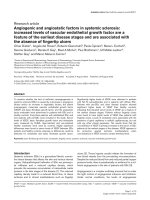
Early bovine leukemia virus replication in experimentally infected sheepFigure 1
Early bovine leukemia virus replication in experimentally infected sheep. Vertical arrows represent the times at which blood samples were collected. A
fluctuation of circulating leukocyte counts over time. mean leukocyte counts of the 4 experimentally BLV-infected sheep aligned relative to the date of
seroconversion -x-x-x-x- leukocyte counts of the two uninfected sheep, aligned relative to the date of injection of the non-infectious solution. B BLV early
replication in experimentally infected sheep. All curves are aligned relative to the date of seroconversion (S). Time (t) is expressed in days. For each animal
the first 2 curves represent the temporal fluctuation of the B cell count (black squares) and proviral loads (open circles); the second 2 curves represent the
clonality of BLV positive circulating cells (black rhombuses, clones = 1200 copies in 1 mcg of circulating DNA; white rhombuses, clones >1200 copies in 1
mcg of circulating DNA); bottom curves represent the frequency of RT-associated substitutions (black circles) and of somatic mutations (open circles); the
blue bars represent, at each time, the number of sequenced BLV integration sites.
Retrovirology 2008, 5:16 />Page 4 of 12
(page number not for citation purposes)
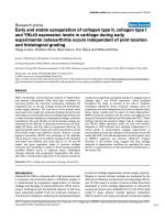
Clonality of BLV-infected cells over time in animals 4535 (A), 4536 (B), 4537 (C), and 4538 (D)Figure 2
Clonality of BLV-infected cells over time in animals 4535 (A), 4536 (B), 4537 (C), and 4538 (D). Each sample was analyzed in quadruplicate by IPCR as
detailed in the Material and Methods section. Each signal on the gel represents a cluster of BLV integration sites having the same length and therefore
belonging to the same cellular clone. The absolute detection threshold of the technique was ~21 copies/150,000 PBMCs while samples harboring 1, 2, 3,
and 4 signals after quadruplicate experiments corresponded to a BLV clonal frequency of 25 to 62.5, 62.5 to 1200, 1200 to 2400, and > 2400 infected cells
per 150,000 PBMCs [4].
-3
MMM M
S +3 +50 +240
A
-3 S +3 +50 +240
M4535 M4536
154
201
220
298
344
396
506
134
B
-3
MMM M
S +3 +50 +240 -3 S +3 +50 +240
M4537 M4538
154
201
220
298
344
396
506
134
154
201
220
298
344
396
506
134
C
D
Retrovirology 2008, 5:16 />Page 5 of 12
(page number not for citation purposes)
These results indicate that BLV primoinfection, i.e. the
first months consecutive to the infection of sheep,
includes a first burst of both polyclonal distribution and
extensive clonal expansion of infected cells, which results
in a transient peak of circulating proviral load.
RT-versus somatically-generated BLV sequence mutations during
early infection in vivo
We searched for RT-versus somatically-associated substi-
tutions of the BLV provirus by comparing the nucleotide
composition of 3'BLV RU5 sequences flanked by distinct
versus identical integration sites, as previously described
for BLV or HTLV-1 [4,6]. For each experimentally infected
animal, IPCR products obtained 3 days before, 50 days
after the date of seroconversion, and 240 days after exper-
imental infection were cloned without size selection. A
total of 842 molecular clones were sequenced (370 kbp of
proviral sequence with 64 kbp of integration site) and
could be arranged into 65 distinct cellular clones based on
cellular flanking sequences. The number of cellular clones
analyzed for the 4 sheep is represented in Figure 1B; at
each time and for each animal, it was correlated with the
overall number of detected clones (Figure 1B). BLV
sequences were aligned with respect to infectious proviral
clone BLV-p344, which was taken as a reference (Figure
3). Fifteen of 65 (23%) cellular clones harbored mutated
3' LTR sequences (16 substitutions), the number of muta-
tions per sequence ranging from 0 in 660 sequences, 1 in
181 sequences and 2 in one sequence. The 16 substitu-
tions were distributed as 14 transitions and 2 transver-
sions (Figure 3). For 10 cellular clones (M35m3-1,
M35p50-2, M50p50-3, M36m3-1, M36m3-3, M36p8-2,
M36p50-1, M36p8-4, M37m3-5, M38-m3-5, Figure 1B,
Figure 3 (shaded in light gray), and Figure 4), all the
3'RU5 sequences defining the clones shared a common
and clone-specific substitution whereas 4 additional cel-
lular clones included only a subset (1/20 to 9/12) of
mutated 3'LTR sequence (dark gray shading, Figure 3).
The distribution of the former corresponded to that of RT-
associated mutations whereas that of the latter possessed
the hallmark of somatically generated mutations [16,19],
which are only harbored by a subset of sequences belong-
ing to a given clone. An additional clone isolated from
sheep #4536 three days before seroconversion (M36m3-
2, Figure 1B and Figure 3) harbored eight 3'RU5
sequences with the same C8203T transition; one of these
sequences had an additional G8351A transition. This
additional clone therefore harbored a RT-mutated 3'RU5
sequence having subsequently undergone a G8351A
somatic substitution. All detected mutations were clearly
beyond the level of PCR errors or artifacts, which were
estimated for this region to be <1 per 30 kb sequenced
[4,16]. For the first time for a deltaretrovirus, these results
provide evidence that early BLV replication is RT-depend-
ent, and generates a mutation load accounting for 69% of
the provirus genetic variability.
RT-associated mutations frequently occur during BLV minus strand
synthesis
As shown in Figure 4A, present RT-associated substitu-
tions possessed the hallmarks of minus-strand synthesis-
associated mutations [16,19]. Those are typically present
on both 3' and 5' LTRs [16,19]. Among these, the G8696A
substitution harbored by clone M36m3-1 (sequence
36m3C1S1, Figure 3) and present 3 days before serocon-
version in sheep 4536 abolished a restriction site for the
Eae I enzyme (YGGCCR->YGACCR where R is a purine
and Y a pyrimidine, Figure 4B). We next searched for this
G->A substitution along the 5' LTR, i.e. at position 511.
Oligonucleotides BLV-s1 and BLV-gag encompassing the
Eae I restriction site at position 511 within the 5' RU5
sequence were used for PCR amplification (see experi-
mental procedures). To specifically amplify the 5' LTR
rather than its 3' counterpart, we chose a 3' primer, BLV-
gag, complementary to the gag gene of the BLV proviral
sequence (Figure 4B). In the absence of Eae I digestion,
PCR amplification of the BLV provirus with BLV-s1 and
BLV-gag primers generated a PCR product of 632 bp (Fig-
ure 4B). In the absence of substitution within the Eae I
restriction site, PCR amplification of Eae I digested DNA
gave no signal whereas, after incubation with Eae I and
PCR amplification, G511A mutated sequences could not
be digested and thereby generated the 632 bp PCR prod-
uct (Figure 4B). As shown in Figure 4B, this signal was
generated after PCR amplification of the DNA of periph-
eral blood cells deriving from sheep 4536 on day 3 before
seroconversion but not on samples deriving from other
infected sheep or from uninfected control. To rule-out the
presence of a PCR inhibitor in samples with negative
results, a control PCR was performed using a primer set
specific for the GAPDH gene, and a specific signal was
obtained with all digested DNA samples (not shown).
Therefore this control experiment confirmed that the
G8696A RT-associated substitutions revealed by cloning
3' IPCR products had occurred during the synthesis of the
BLV provirus minus strand in vivo. The T8617C and
T8651C substitutions revealed in animal 4536 212 days
after seroconversion were harbored by all the sequences
belonging to clones M36p8-2 and M36p8-4 respectively
(Figures 1B and 3), and were thus assumed to have been
generated during RT. Two pairs of primers encompassing
the corresponding positions of these substitutions along
the 5' (BLV-s1 and BLV-gag) and the 3' LTR (BLV-tax and
BLV-U5as) were synthesized (see experimental proce-
dures). After amplification, PCR products corresponding
to the sample collected 212 days after seroconversion
were directly sequenced and both substitutions were iden-
tified along the 3' and 5' LTR. Electropherograms show
that 3' and 5' substitutions were harbored by a similar
Retrovirology 2008, 5:16 />Page 6 of 12
(page number not for citation purposes)
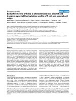
Somatic (miniscule letters) versus reverse-transcriptase (capital letters) associated substitutions of the BLV 3' RU5 sequence during early experimental sheep infectionFigure 3
Somatic (miniscule letters) versus reverse-transcriptase (capital letters) associated substitutions of the BLV 3' RU5 sequence during early experimental
sheep infection. Overall, 65 distinct BLV 3' integration sites were isolated; the first 8 bases of the corresponding flanking cellular sequences are given on
the right. RU5 sequences were aligned according to the sequence of the wild type BLV sequence 344 used for experimental infection of animals 4535 and
4536. Sheep are identified by their unique animal number (UAN). Each cluster of RU5 sequences sharing a common integration site, and therefore belong-
ing to a unique clone of expanded B cells, is identified by its cellular clone number. For each cellular clone the number of non-unique 3' U3RU5 consensus
sequences is indicated between brackets in the third column. Cellular clones harboring a mutated 3'U3RU5 sequence are overlined in grey. A horizontal
double bar separates the clusters of sequences derived from each of the 4 sheep DNA samples. For each animal, cellular clones are sorted according to
their date of isolation, i.e. 3 days before, 50 after seroconversion and 240 days after experimental infection. The two horizontal arrows represent the two
times at which the same C8202T substitution was observed in 2 distinct sequences deriving from animal #4536.
UAN
Cellular
clones
3' U3RU5 sequences
(
n
)
t
8202
8203
8339
8351
8391
8436
8455
8482
8550
8594
8617
8618
8626
8651
8696
pBLV344.12 -
>
3' flanking
sequence
4535 M35m3-1 35m3C1S1 (9) -3
M35m3-2 35m3C2S1 (6) -3
M35m3-3 35m3C3S1 (3) -3
M35m3-4 35m3C4S1 (24) -3
M35m3-5 35m3C5S1 (9) -3
M35m3-6 35m3C6S1 (3) -3
M35m3-7 35m3C7S1 (11) -3
M35m3-8 35m3C8S1 (3) -3
M35p50-1 35p50C1S1 (19) 50
M35p50-1 35p50C1S2 (1) 50
M35p50-2 35p50C2S1 (13) 50
M35p50-3 35p50C3S1 (27) 50
M35p50-4 35p50C4S1 (9) 50
M35p50-5 35p50C5S1 (13) 50
M35p50-6 35p50C6S1 (7) 50
M35p50-7 35p50C7S1 (10) 50
M35p8-1 35p8C1S1 (4) 240
M35p8-2 35p8C2S1 (22) 240
M35p8-3 35p8C3S1 (5) 240
M35p8-4 35p8C4S1 (14) 240
M35p8-5 35p8C5S1 (4) 240
M35p8-6 35p8C6S1 (4) 240
M35p8-7 35p8C7S1 (3) 240
M35p8-7 35p8C7S2 (9) 240
M35p8-8 35p8C8S1 (7) 240
M35p8-9 35p8C9S1 (10) 240
M35p8-10 35p8C10S1 (8) 240
M35p8-10 35p8C10S2 (1) 240
4536 M36m3-1 36m3C1S1 (5) -3
M36m3-2 36m3C2S1 (7) -3
M36m3-2 36m3C2S2 (1) -3
M36m3-3 36m3C3S1 (34) -3
M36m3-4 36m3C4S1 (27) -3
M36m3-5 36m3C5S1 (8) -3
M36m3-6 36m3C6S1 (13) -3
M36m3-7 36m3C7S1 (10) -3
M36m3-8 36m3C8S1 (19) -3
M36p50-1 36p50C1S1 (9) 50
M36p50-2 36p50C2S1 (13) 50
M36p50-3 36p50C3S1 (5) 50
M36p50-4 36p50C4S1 (8) 50
M36p50-5 36p50C5S1 (6) 50
M36p8-1 36p8C1S1 (18) 240
M36p8-2 36p8C2S1 (14) 240
M36p8-3 36p8C3S1 (9) 240
M36p8-4 36p8C4S1 (6) 240
M36p8-5 36p8C5S1 (7) 240
M36p8-6 36p8C6S1 (15) 240
4537 M37m3-1 37m3C1S1 (6) -3
M37m3-2 37m3C2S1 (18) -3
M37m3-3 37m3C3S1 (8) -3
M37m3-3 37m3C3S2 (4) -3
M37m3-4 37m3C4S1 (10) -3
M37m3-5 37m3C5S1 (29) -3
M37p50-1 37p50C1S1 (36) 50
M37p50-2 37p50C2S1 (3) 50
M37p8-1 37p8C1S1 (16) 240
M37p8-2 37p8C2S1 (3) 240
4538 M38m3-1 38m3C1S1 (21) -3
M38m3-2 38m3C2S1 (5) -3
M38m3-3 38m3C3S1 (22) -3
M38m3-4 38m3C4S1 (13) -3
M38m3-5 38m3C5S1 (13) -3
M38p50-1 38p50C1S1 (24) 50
M38p50-2 38p50C2S1 (2) 50
M38p50-3 38p50C3S1 (17) 50
M38p8-1 38p8C1S1 (13) 240
M38p8-2 38p8C2S1 (12) 240
M38p8-3 38p8C3S1 (25) 240
M38p8-4 38p8C4S1 (50) 240
Retrovirology 2008, 5:16 />Page 7 of 12
(page number not for citation purposes)
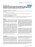
RT-associated mutations frequently occur during BLV minus strand synthesisFigure 4
RT-associated mutations frequently occur during BLV minus strand synthesis. A Generation of U5 substitution during minus strand synthesis. RNA is rep-
resented as a thin line whereas DNA is represented as thick lines. The first 6 horizontal lines represent the synthesis of the provirus. Line 7 represents the
integrated provirus flanked with its two integration sites represented as black boxes. NlaIII restriction sites are represented on both the provirus and the
3' cellular flanking sequence. Lines 8 and 9 represent the first two mitoses of the infected cells harboring the integrated provirus. For each cell, the RU5
sequence and the 3' flanking sequence encompassed by the 2 NlaIII restriction sites, i.e., the sequences obtained after inverse PCR, are represented. Line
10 represents the sequences obtained after inverse PCR, cloning and sequencing. The open circle represents a RT-associated mutation that has occurred
during the synthesis of the 5' RU5 minus strand. As shown in lines 1 to 6, this substitution appears to be harbored by both strands of the two LTRs of the
integrated provirus. Accordingly, all infected cells from the corresponding clone (identified by their common integration site) harbor a provirus with the
same substitution (see lines 8 and 9). As a consequence, all sequences from this clone obtained after inverse PCR harbor the same mutation at the same
position. B PCR detection of the G511A U5 substitution along the 5' LTR. Top: 5' BLV LTR from the wild-type (left) and from clone M36m3-1 (right) car-
rying the putative G511A substitution having occurred during minus strand synthesis, thereby generating the G8696A substitution identified by sequencing
IPCR product derived from the sample harvested in animal 4536 three days before seroconversion. In the absence of digestion, specific 5' LTR PCR ampli-
fies a fragment of 632 bp. EaeI digestion of the wild-type sequence abolishes this signal while incubation with EaeI has no effect on the sequence carrying
the G511A mutation, leading to the detection of the 632 bp fragment. Samples studied in the presence (+) or in the absence (-) of EaeI digestion derived
from BLV infected animals 4536 and 4538, both harvested 3 days before seroconversion, and from the uninfected control animal 4533. C Detection of the
T8617C and T8651C substitutions in their corresponding positions along the 5' LTR. Each LTR was specifically amplified by PCR and substitutions were
detected by direct sequencing of PCR product, as detailed in the experimental procedures.
A
R U5 PBS RU3PPT
RU5
PBS
PBS
PBS
NlaIII
NlaIII
RU5
RU3PPT
RU5
RU3PPT
PBS
RU5
PPT
PBS
PPT RU5
RU5
PBS
PBS
RU5
RU5PBS
U3
U3
U3
RU5PBSU3
U3
RU5U3
RU5U3
R U5 PBSU3
5 ’ 3 ’
+
1
2
3
4
5
6
7
8
9
10
U3 R U5 gag
cggcca
gccggt
Eae I
68 88 679 699
5’ LTR
U3 R U5 gag
cgAcca
gcTggt
Eae I
68 88 679 699509
Eae I digestion
B
PCR
BLV gag
BLV s1
BLV s1 BLV gag
632 pb
632 pb
Wild-type
Clone M36m3-1
Eae I digestion
PCR
PCR PCR
632 pb
MM-+-+-
4536 4538 4533
T466C
T
C
AG
CC
T
C
G
C
TTTT TG TTT
T/C
TT
T8651C
T C AG CCT C GC TTTT TG TTT
T/C
TTT
T8617C
T C AA GC GGC G T C
T/C
GG
C
TT G
T432C
TC AA GC GGC G T C
T/C
GGC TT G
5’ LTR 3’ LTR
BLV provirus
C
Retrovirology 2008, 5:16 />Page 8 of 12
(page number not for citation purposes)
proportion of sequences (Figure 4C). No such signal
could be observed when DNA deriving from sheep #4535
or 4538 was assayed. Therefore, as for the G8696A transi-
tion, these results suggest that T8617C and T8651C tran-
sitions have been RT-generated during minus strand
synthesis. Investigating for the first time the period at
which substitutions occur during BLV reverse transcrip-
tion in vivo, these results suggest that the synthesis of the
minus provirus strand is more error prone than that of the
plus strand.
In vivo cell-to-cell passage of a BLV proviral sequence harboring a RT-
dependent mutation
The C8202T RT-associated substitution harbored by clone
M36m3-3 and isolated from sheep #4536 three days
before seroconversion, i.e. 25 days after experimental
infection (Figure 1B and 3) was subsequently identified in
clone M36p50-1, characterized by a distinct flanking
sequence and isolated 53 days later from the same animal.
This suggests that clonally expanded cells from clone
M36p50-1 shared a RU5 sequence having necessary
undergone at least two rounds of horizontal replication.
Alternatively but less probably, the two C8202T substitu-
tions might have occurred during two distinct RT cycles.
RT-associated substitutions are restricted to early experimental BLV
infection
We next investigated the temporal distribution of somati-
cally-versus RT-generated BLV proviral substitutions. The
proportion of circulating cellular clones harboring somat-
ically mutated BLV proviral sequences at 3 days before
seroconversion, 50 days after seroconversion and 240
days after infection were 7.6%, 5.8%, and 9.5%, respec-
tively. As previously observed with HTLV-1 [16] or BLV
[4], this distribution was time-independent. In contrast,
during primary BLV infection, the proportion of clones
harboring RT-associated mutations was inversely and lin-
early correlated with time (r
2
= 0.99) (Figures 1 and 5). As
a consequence, it could be extrapolated from Figure 5 that
clones with more than 21 infected cells (the lower limit of
IPCR detection) harboring RT-generated mutations could
not been detected in the blood flow of experimentally
infected animals after the 250th day following serocon-
version. These results highlight the ephemeral nature of
RT-generated BLV substitutions in vivo.
Discussion
Deltaretroviruses possess two modes of replication that
include the classical horizontal retrovirus-like spread and
the cell-associated clonal expansion of proviral sequences
[14,20]. The former generates RT-associated substitutions
of the provirus whereas the latter is associated with
somatic mutations of both the provirus and the host cell
sequence [4,16].
Our work was performed in the sheep experimental
model for BLV infection, which is a practical way to study
early infection in vivo. Events occurring in the present
model after injection of proviral DNA may not reflect
what occurs after natural transmission in cows, namely
cell-associated infection followed by horizontal and verti-
cal virus spreads. However, we have demonstrated ([4]
and present results) that the inoculation of infectious
molecular BLV clones in sheep triggered a temporal pat-
tern of infection similar to that observed after experimen-
tal cell-associated infection [6], i.e. the generation of
newly infected host-cells followed by their persistent
clonal expansion. Together these data contribute to vali-
date the present experimental model at the replicative
level.
BLV infection of sheep regularly triggers the formation of
tumors that which occur faster than in the small percent-
age of infected cattle that develop tumors [21-25]. It is
possible that events occurring during early infection in the
sheep model may set the stage for rapid tumor develop-
ment. Alternatively, one can propose that the better adap-
tation of the virus to its natural host might contribute to
the significantly lower incidence of BLV-related malignan-
cies in cattle.
Time-dependent decrease of RT-generated BLV proviral sub-stitutions during early experimental infectionFigure 5
Time-dependent decrease of RT-generated BLV proviral sub-
stitutions during early experimental infection. Data from Fig-
ure 1 served to plot the frequency of clones harboring RT-
dependent substitutions against time in the 4 experimentally
infected sheep.
Retrovirology 2008, 5:16 />Page 9 of 12
(page number not for citation purposes)
The experimental strategy used in our study permitted to
monitor in vivo these two routes of BLV replication and
genetic variability over time. This allowed to show that
experimental primary BLV infection of sheep includes an
early and intense burst of both horizontal and vertical
viral disseminations, generating frequent RT-associated
proviral substitutions that account for 69% of the in vivo
BLV genetic variability during the first months of the
infection (Figure 1, 2, 3). However, all 4 experimentally
infected sheep displayed a rapid shift towards a predomi-
nant vertical route of replication as demonstrated by the
fact that no RT-dependent substitution could be detected
from the 250th day after seroconversion.
Present results about BLV genetic variability are the first
evidence of a RT-dependent mutation process for a del-
taretrovirus in vivo. Typically, both RNA-dependent and
DNA-dependent DNA syntheses by RT contribute to the
genetic variability of retroviruses. In addition, RNA tran-
scription by cellular RNA polymerase II could also partic-
ipate to the mutation process. However the error rate of
RNA polymerase II and its contribution to retroviral
mutation rates remain unknown. In vitro studies of muta-
tion rates during RNA- and DNA-dependent HIV DNA
synthesis have produced conflicting results. They suggest
a higher mutation rate during RNA-dependent DNA syn-
thesis [26], a higher mutation rate during DNA-depend-
ent DNA synthesis [27], or equal mutation rates during
RNA- and DNA-dependent DNA syntheses [28]. In addi-
tion, it appears from in vivo studies that some elements
affecting fidelity in vivo are absent in in vitro assays [29-
33]. In vivo mutation rates have been measured for the
bovine leukemia virus [34] however the contributions of
the various nucleic acid polymerization steps in retroviral
replication to the in vivo retroviral mutation rates have
not been evaluated. From the present experimental study,
in vivo RT-dependent mutations appeared to mainly
occur during BLV minus strand synthesis.
All 4 experimentally infected sheep displayed an early
burst of horizontal BLV replication that generated a burst
of RT-dependent proviral substitutions (Figure 1). In con-
trast, the transient peak of clonal expansion that accompa-
nied the intense horizontal spread was not found to
increase the detection frequency of somatic mutations
(Figures 1 and 2). Previous studies have clearly linked the
degree of clonal expansion with the somatic mutation fre-
quency [4,14,16]. During natural HTLV-1 infection, heav-
ily expanded clones regularly display the highest somatic
mutation frequencies, which culminate at the malignant
stage [6]. Similarly, in experimentally infected sheep, the
premalignant and malignant BLV positive clones harbor
the highest degree of clonal expansion together with the
highest somatic mutation loads, when compared with
other clones of infected cells [4]. Therefore the present
loss of correlation between clonal expansion and somatic
mutations seems to be at odds with these previous results.
However, those were obtained by investigating the
chronic phase of the infection in organisms having
mounted the specific and robust adaptive antiviral
immune response characteristic of deltaretrovirus infec-
tion [4,16]. In the present study, the absence of such a
strong immune response that characterizes early infection
might account for the absence of detected somatic muta-
tions at this stage of the infection. In other words, together
with previous findings, present results are consistent with
the idea that the host immune response might be involved
in the selection of somatic mutations, thus explaining
why the correlation of their frequency with the degree of
clonal expansion is restricted to the chronic phase of the
infection.
The detection of RT-associated proviral substitutions was
confined to a narrow window encompassing the first 250
days following seroconversion. This is in agreement with
our previous works on HTLV-1 [16] and BLV [4], which
regularly failed to identify RT-associated proviral muta-
tions in circulating, infected clones in vivo during the late
phase of the infection [4,14,16]. The question remains of
how these RT-acquired BLV substitutions disappear over
time. The time-dependent decrease in their detection fre-
quency (Figure 5) parallels the time-dependent develop-
ment of the robust and subsequently persistent anti-BLV
immune response [35,36]. Together with present results,
this suggests that RT-generated substitutions and viral
expression could be synonymous. Accordingly, after inte-
gration, RT-dependent mutated proviral sequences
undergo a negative immunological control for clonal
expansion. This hypothesis also helps explain why newly
generated RT-dependent substitutions have never been
detected during the chronic phase of deltaretroviruses
infection in sheep or in humans [4,16]. Alternatively, RT-
dependent substitutions might represent proviruses hav-
ing undergone modification of key genes involved in the
control of host cell multiplication. Finally, given a BLV
IPCR detection threshold of 21 copies per microgram of
DNA [4], present results do not preclude that weakly
expanded sequences harboring RT-dependent substitu-
tions could be generated and/or maintained over time
after experimental BLV infection.
In conclusion, our study suggests that, in contrast to other
retroviruses, deltaretroviruses possess two time-depend-
ent pathways of genetic variation that parallel their two-
step nature of replication over time [6] and correspond to
RT-associated rearrangements and somatic mutations.
The former appears restricted to the first months of the
infection while the latter dominates the prolonged steady-
state step of the infection, with the suggestion that this
Retrovirology 2008, 5:16 />Page 10 of 12
(page number not for citation purposes)
time-dependent pattern of replication depends on the
host immune pressure.
Methods
Experimental BLV infection of sheep
Six one-year-old sheep were kept under controlled condi-
tions at the Veterinary and Agrochemical Research Centre
(Machelen, Belgium). Handling of animals and experi-
mental procedures were approved by the ethics commit-
tee and were conducted in accordance with institutional
and national guidelines for animal care and use. Four
sheep were experimentally infected with BLV infectious
molecular clones as previously described [17]. Briefly, 100
μg of circular plasmid DNA was mixed with 200 μg of
Dotap (Roche Diagnostics) and injected intradermally
into the back of the sheep. Two animals, # 4535 and 4536,
were experimentally infected with a BLV wild-type cloned
provirus (pBLV344) [37]. A plasmid containing the
mutant provirus pBLVIG4, which harbors a stop codon in
the G4 open reading frame [17], was injected in sheep #
4537 and 4538. Two additional animals, # 4533 and
4534, received a non-infectious Dotap solution and
served as uninfected controls. Twice a week, the total leu-
kocyte counts were determined by using a Coulter counter
ZN, and the number of lymphocytes was estimated after
examination under the microscope after staining with
May-Grunwald Giemsa. In parallel, the sera from each
sheep were analyzed for BLV seropositivity using immun-
odiffusion and enzyme-linked immunosorbent assay
(ELISA) techniques [38].
Immunophenotyping of circulating cells
Peripheral blood mononuclear cells (PBMCs) were iso-
lated by Percoll gradient centrifugation and their viability
was estimated by trypan blue dye exclusion [39]. PBMCs
were labeled with monoclonal antibodies (Mabs) directed
against surface immunoglobulin M (anti-sIgMs, clone
1H4, mouse IgG1; Pig45A2, mouse IgG2b), CD4 (ST4,
mouse IgG1), CD8 (CC58, mouse IgG1) provided by C.
Howard (Institute for Animal Health, Compton, United
Kingdom) and by I. Schwartz-Cornil (INRA, Jouy-en-
Josas, France). Cells were then labeled with a rat anti-
mouse IgG1 phycoerythrin (PE)-antibody (Becton Dick-
inson Immunocytometry Systems) or with a goat anti-
mouse IgG2b fluorescein isothiocyanate (FITC)-conjugate
(Caltag Laboratories). Finally, PBMCs were analyzed by
flow cytometry on a Becton Dickinson FACScan flow
cytometer. Ten thousand events were collected for each
sample and data were analyzed with the Cellquest soft-
ware (Becton Dickinson Immunocytometry Systems).
Measurement of circulating BLV proviral Load
The circulating amounts BLV proviral sequences were
measured by LightCycler quantitative PCR as described
[4]. Briefly, the reaction mixture included polymerase
(LightCycler Kit Fast Start DNA Master Hybridization
Probes; Roche), 2 mM MgCl 2, 500 nM primer BLVQF,
500 nM primer BLVQR both targeting Px region and 100
nM donor probe 3' end labeled with fluorescein and 200
nM acceptor probe 5' end labeled with LC Red640. Stand-
ardization of the amount of DNA subjected to quantifica-
tion was performed with quantitation of the sheep beta-
globin gene as an internal standard [40]. The standard
curve for beta-globin was generated using DNA extracted
from BLV negative sheep blood cells.
Detection and quantification of the clonal distribution of
circulating BLV positive cells in vivo
BLV integration was analyzed by Inverse Polymerase
Chain Reaction (IPCR) as described [4]. Briefly, two
micrograms of DNA were digested by 20 U NlaIII and 20
units of MfeI in 1X NlaIII-MfeI buffer for 3 h at 37°C. MfeI
digestion was performed in order to avoid the amplifica-
tion of a 536 bp segment of the 5' LTR complementary to
the set of 3' IPCR primers. Digestion was controlled by 1%
agarose gel electrophoresis and DNA was extracted with
phenol/chloroform (1:1) and precipitated with 100% eth-
anol. One microgram of digested DNA was circularized
for 16 h at 16°C with 20 U of T4 DNA ligase. As there is a
stochastic component to the detection of retrovirus inte-
gration sites using inverse PCR [41], samples were ana-
lyzed in quadruplicate, as previously described for BLV
and HTLV-1 [4,41]: 4 × 500 ng of circularized DNA were
amplified for 39 cycles using 200 μM of the primer pair
BLV3'S and BLV3'AS. Amplifications were performed
using 3.5 units of the Pfu DNA polymerase with thermal
cycling parameters as follows: 95°C 10 min, 35 × (95°C 1
min, 60°C 1 min, 72°C 3 min), and a final elongation
step of 10 min at 72°C. The length polymorphism analy-
sis of 3' BLV flanking sequences was performed by making
a run-off. This method consists in the linear PCR amplifi-
cation of the provirus 3' extremities together with their
flanking sequences. Two microliters of amplified IPCR
products were submitted to 10 cycles of linear PCR with 2
μM of 5'-32P-radiolabeled primer BLV3'RO. Run-off
products were analyzed on 6% sequencing gel. As previ-
ously described [4], the stochastic nature of BLV IPCR was
found to appear at BLV integration site frequencies rang-
ing between 25 and 2400 copies of the BLV provirus per
mcg of blood DNA. At copy numbers ranging from 1200
to 2400, 62.5 to 1200, and 25 to 62.5, detection was 3/4,
2/4, and 1/4, respectively. Accordingly, DNA samples
from BLV infected animals were analyzed in quadrupli-
cate (4 × 0.5 mcg).
Assessment of BLV genetic variability in vivo
The cloning and sequencing of 3'LTR-integration site PCR
fragments were performed as previously described [4].
Briefly, purified IPCR products were ligated with SmaI-
digested and M13mp18 replicative form DNA. After trans-
Retrovirology 2008, 5:16 />Page 11 of 12
(page number not for citation purposes)
formation of Escherichia coli XL1, recombinant M13
plaques were screened by hybridization with the BLV3'RO
or the BLV5'RO LTR-specific 32P-labelled oligonucle-
otides. Single-stranded templates were sequenced using
fluorescent dideoxynucleotides. The sequenced products
were resolved on an Applied Biosystems 377A DNA
sequencer with 377A software. Sequence alignments were
performed with Sequence Navigator Software.
Detection of proviral mutations by direct sequencing of
PCR products
Specific 3' versus 5' LTR oligonucleotides were used for
PCR, and PCR products were directly sequenced. PCR
amplifications of 5'- and 3'-LTRs were performed with oli-
gonucleotides encompassing the 5'-RU5 sequence (BLV-
s1 5'-AGAAAAGCTGGTGACGGCAG-3' and BLV-gag 5'-
GCTTTGCAGAAGGTTGAGCC-3') and the 3' counterpart
(BLV-tax 5'-ACCTGGTCCGAATTGGTTGC-3' and BLV-
U5as 5'-GTTTGCCGGTCTCTCCTG-3') respectively. For
the amplification of the GAPDH gene, the primers
G3PDHS 5'-GACCCCTTCATTGACCTCAACTACA-3' and
G3PDHAS 5'-CTAAGCAGTTGGTGGTGCAG-3' permitted
to rule-out the presence of PCR inhibitor in DNA sample.
Overall, DNA was amplified using 3.5 units of the Pfu
DNA polymerase with thermal cycling parameters as fol-
lows: 95°C 10 min, 35 × (95°C 1 min, 58°C 1 min, 72°C
3 min), and a final elongation step of 10 min at 72°C.
PCR amplified fragments were separated on a 1% agarose
gel and visualized by ethidium bromide staining. PCR
products were purified using a MinElute PCR Purification
kit (QIAGEN, Valencia, CA), and directly sequenced with
BigDyeTM Terminator Cycle Sequencing v2.0 Ready Reac-
tion Kit (Applied Biosystems, Foster City, CA) according
to the manufacturer's instructions. All PCR products were
sequenced directly in both directions with an internal oli-
gonucleotide BLV-2S 5'-CTTCCCCTTTCCCGAAAAAT-3'
and the BLV-gag and BLV-U5as for LTR5' and LTR3'
respectively. To rule out incorporation errors by Taq
polymerase, direct sequencing was repeated from a new
amplification reaction. Sequenced products were resolved
on an Applied Biosystems 377A DNA sequencer as
described above.
Analysis of the 5' LTR restriction fragment length
polymorphism by PCR amplification of digested DNA
The presence of a G511A transition was checked along the
5'LTR. As this G511A substitution abolishes an Eae-I
restriction site, DNA (500 ng) was digested by 1 U of Eae-
I enzyme in 1X Eae-I buffer for 2 h at 37°C. Digested DNA
was subsequently PCR amplified with the BLV-s1 and
BLV-gag 5' LTR specific primers. The presence of the sub-
stitution was evidenced by gel electrophoresis.
Control PCR
To check the accuracy of the IPCR and the absence of PCR-
associated recombination, 3 cloned 3' BLV U3RU5
sequences flanked by there integration sites and harboring
distinct mutations were used as controls. Two hundred
and fifty copies of each of these 3 cloned sequences were
mixed in 1 μg of uninfected DNA. Five hundred nano-
grams of mixed DNA were amplified for 35 cycles using
200 μM of BLV3'S and BLV3'AS primer pair under the
same conditions as used in the analysis of DNA samples
from sheep. Purified PCR products were cloned and
sequenced as described above. Fifty-two sequences were
obtained and analyzed by CLUSTAL alignment with
Sequence Navigator Software.
Statistical analysis
SPSS statistical software version 11 and CA-Cricket Graph
III were used for analyses. The correlation of data was
assessed by Spearman's Rho nonparametric method. P <
0.05 was considered significant in all analyses.
Competing interests
The author(s) declare that they have no competing inter-
ests.
Authors' contributions
CP carried out the most experimental work. MTSA, CD
and FM performed the sample collections. AL, CP and FM
performed the sequencing of IPCR products and the deter-
mination of the proviral loads. PK and LW were responsi-
ble for the sheep studies and participated to interpretation
of data. FM and EW were responsible for the design of the
study and its coordination. CP, EW, and FM wrote the
manuscript. All authors read and approved the final man-
uscript.
Acknowledgements
This work was supported by the Ligue Nationale Contre le Cancer (équipe
labellisée, 2003), the Centre Léon Bérard, the Centre National pour la
Recherche Scientifique (CNRS), the Institut National de la Santé et de la
Recherche Médicale (Inserm) and the Sixth Research Framework Pro-
gramme of the European Union (project INCA LSHC-CT-2005-018704).
CP was supported by a bursary from the Ligue Nationale Contre le Cancer
(Comité de l'Ain). FM is supported by Inserm. LW is supported by the Fond
National pour la Recherche Scientifique (Belgium). The authors thank
Marie-Dominique Reynaud for the preparation of the manuscript.
References
1. Drake JW: Rates of spontaneous mutation among RNA
viruses. Proc Natl Acad Sci U S A 1993, 90(9):4171-4175.
2. Philpott SM, Buehring GC: Defective DNA repair in cells with
human T-cell leukemia/bovine leukemia viruses: role of tax
gene. J Natl Cancer Inst 1999, 91(11):933-942.
3. Johnson JM, Harrod R, Franchini G: Molecular biology and patho-
genesis of the human T-cell leukaemia/lymphotropic virus
Type-1 (HTLV-1). Int J Exp Pathol 2001, 82(3):135-147.
4. Moules V, Pomier C, Sibon D, Gabet AS, Reichert M, Kerkhofs P, Wil-
lems L, Mortreux F, Wattel E: Fate of premalignant clones dur-
ing the asymptomatic phase preceding lymphoid
malignancy. Cancer Res 2005, 65(4):1234-1243.
Retrovirology 2008, 5:16 />Page 12 of 12
(page number not for citation purposes)
5. Leclercq I, Mortreux F, Rabaaoui S, Jonsson CB, Wattel E: Naturally
occurring substitutions of the human T-cell leukemia virus
type 1 3' LTR influence strand-transfer reaction. J Virol Meth-
ods 2003, 109(2):105-117.
6. Mortreux F, Kazanji M, Gabet AS, de Thoisy B, Wattel E: Two-step
nature of human T-cell leukemia virus type 1 replication in
experimentally infected squirrel monkeys (Saimiri sciureus).
J Virol 2001, 75(2):1083-1089.
7. Poiesz BJ, Ruscetti FW, Gazdar AF, Bunn PA, Minna JD, Gallo RC:
Detection and isolation of type C retrovirus particles from
fresh and cultured lymphocytes of a patient with cutaneous
T-cell lymphoma. Proc Natl Acad Sci U S A 1980,
77(12):7415-7419.
8. Kalyanaraman VS, Sarngadharan MG, Robert-Guroff M, Miyoshi I,
Golde D, Gallo RC: A new subtype of human T-cell leukemia
virus (HTLV-II) associated with a T-cell variant of hairy cell
leukemia. Science 1982, 218(4572):571-573.
9. Calattini S, Chevalier SA, Duprez R, Bassot S, Froment A, Mahieux R,
Gessain A: Discovery of a new human T-cell lymphotropic
virus (HTLV-3) in Central Africa. Retrovirology 2005, 2(1):30.
10. Wolfe ND, Heneine W, Carr JK, Garcia AD, Shanmugam V, Tamoufe
U, Torimiro JN, Prosser AT, Lebreton M, Mpoudi-Ngole E,
McCutchan FE, Birx DL, Folks TM, Burke DS, Switzer WM: Emer-
gence of unique primate T-lymphotropic viruses among cen-
tral African bushmeat hunters. Proc Natl Acad Sci U S A 2005,
102(22):7994-7999.
11. Meertens L, Mahieux R, Mauclere P, Lewis J, Gessain A: Complete
sequence of a novel highly divergent simian T-cell lympho-
tropic virus from wild-caught red-capped mangabeys (Cer-
cocebus torquatus) from Cameroon: a new primate T-
lymphotropic virus type 3 subtype. J Virol 2002, 76(1):259-268.
12. Ferrer JF, Abt DA, Bhatt DM, Marshak RR: Studies on the relation-
ship between infection with bovine C-type virus, leukemia,
and persistent lymphocytosis in cattle. Cancer Res 1974,
34(4):893-900.
13. Kazanji M, Ureta-Vidal A, Ozden S, Tangy F, de Thoisy B, Fiette L,
Talarmin A, Gessain A, de The G: Lymphoid organs as a major
reservoir for human T-cell leukemia virus type 1 in experi-
mentally infected squirrel monkeys (Saimiri sciureus): provi-
rus expression, persistence, and humoral and cellular
immune responses. J Virol 2000, 74(10):
4860-4867.
14. Mortreux F, Gabet AS, Wattel E: Molecular and cellular aspects
of HTLV-1 associated leukemogenesis in vivo. Leukemia 2003,
17(1):26-38.
15. Ward WH, Dimmock CK, Eaves FW: T lymphocyte responses of
sheep to bovine leukaemia virus infection. Immunol Cell Biol
1992, 70 ( Pt 5):329-336.
16. Mortreux F, Leclercq I, Gabet AS, Leroy A, Westhof E, Gessain A,
Wain-Hobson S, Wattel E: Somatic mutation in human T-cell
leukemia virus type 1 provirus and flanking cellular
sequences during clonal expansion in vivo. J Natl Cancer Inst
2001, 93(5):367-377.
17. Debacq C, Sanchez Alcaraz MT, Mortreux F, Kerkhofs P, Kettmann
R, Willems L: Reduced proviral loads during primo-infection of
sheep by Bovine Leukemia virus attenuated mutants. Retro-
virology 2004, 1(1):31.
18. Fulton BE Jr., Portella M, Radke K: Dissemination of bovine leuke-
mia virus-infected cells from a newly infected sheep lymph
node. J Virol 2006, 80(16):7873-7884.
19. Mansky LM: Sorting out mutations in human T-cell leukemia
virus type 1 proviruses during in vivo clonal expansion. J Natl
Cancer Inst 2001, 93(5):336-337.
20. Wattel E, Cavrois M, Gessain A, Wain-Hobson S: Clonal expansion
of infected cells: a way of life for HTLV-I. J Acquir Immune Defic
Syndr Hum Retrovirol 1996, 13 Suppl 1:S92-9.
21. Djilali S, Parodi AL, Levy D, Cockerell GL: Development of leuke-
mia and lymphosarcoma induced by bovine leukemia virus in
sheep: a hematopathological study. Leukemia 1987,
1(11):777-781.
22. Gatei MH, Brandon R, Naif HM, Lavin MF, Daniel RC: Lymphosar-
coma development in sheep experimentally infected with
bovine leukaemia virus. Zentralbl Veterinarmed B 1989,
36(6):424-432.
23. Kenyon SJ, Ferrer JF, McFeely RA, Graves DC: Induction of lym-
phosarcoma in sheep by bovine leukemia virus. J Natl Cancer
Inst 1981, 67(5):1157-1163.
24. Mammerickx M, Palm R, Portetelle D, Burny A: Experimental
transmission of enzootic bovine leukosis to sheep: latency
period of the tumoral disease.
Leukemia 1988, 2(2):103-107.
25. Suneya M, Onuma M, Yamamoto S, Hamada K, Watarai S, Mikami T,
Izawa H: Induction of lymphosarcoma in sheep inoculated
with bovine leukaemia virus. J Comp Pathol 1984, 94(2):301-309.
26. Hubner A, Kruhoffer M, Grosse F, Krauss G: Fidelity of human
immunodeficiency virus type I reverse transcriptase in copy-
ing natural RNA. J Mol Biol 1992, 223(3):595-600.
27. Boyer JC, Bebenek K, Kunkel TA: Unequal human immunodefi-
ciency virus type 1 reverse transcriptase error rates with
RNA and DNA templates. Proc Natl Acad Sci U S A 1992,
89(15):6919-6923.
28. Ji JP, Loeb LA: Fidelity of HIV-1 reverse transcriptase copying
RNA in vitro. Biochemistry 1992, 31(4):954-958.
29. Mansky LM, Temin HM: Lower in vivo mutation rate of human
immunodeficiency virus type 1 than that predicted from the
fidelity of purified reverse transcriptase. J Virol 1995,
69(8):5087-5094.
30. Varela-Echavarria A, Garvey N, Preston BD, Dougherty JP: Compar-
ison of Moloney murine leukemia virus mutation rate with
the fidelity of its reverse transcriptase in vitro. J Biol Chem
1992, 267(34):24681-24688.
31. Preston BD, Poiesz BJ, Loeb LA: Fidelity of HIV-1 reverse tran-
scriptase. Science 1988, 242(4882):1168-1171.
32. Laakso MM, Sutton RE: Replicative fidelity of lentiviral vectors
produced by transient transfection. Virology 2006,
348(2):406-417.
33. Bakhanashvili M: p53 enhances the fidelity of DNA synthesis by
human immunodeficiency virus type 1 reverse transcriptase.
Oncogene 2001, 20(52):7635-7644.
34. Mansky LM, Temin HM: Lower mutation rate of bovine leuke-
mia virus relative to that of spleen necrosis virus. J Virol 1994,
68(1):494-499.
35. Mateo L, Gardner J, Suhrbier A: Delayed emergence of bovine
leukemia virus after vaccination with a protective cytotoxic
T cell-based vaccine. AIDS Res Hum Retroviruses 2001,
17(15):1447-1453.
36. Florins A, Gillet N, Asquith B, Boxus M, Burteau C, Twizere JC,
Urbain P, Vandermeers F, Debacq C, Sanchez-Alcaraz MT, Schwartz-
Cornil I, Kerkhofs P, Jean G, Thewis A, Hay J, Mortreux F, Wattel E,
Reichert M, Burny A, Kettmann R, Bangham C, Willems L: Cell
dynamics and immune response to BLV infection: a unifying
model. Front Biosci 2007, 12:1520-1531.
37. Willems L, Thienpont E, Kerkhofs P, Burny A, Mammerickx M, Kett-
mann R: Bovine leukemia virus, an animal model for the study
of intrastrain variability. J Virol 1993, 67(2):1086-1089.
38. Portetelle D, Mammerickx M, Burny A: Use of two monoclonal
antibodies in an ELISA test for the detection of antibodies to
bovine leukaemia virus envelope protein gp51. J Virol Methods
1989, 23(2):211-222.
39. Dequiedt F, Hanon E, Kerkhofs P, Pastoret PP, Portetelle D, Burny A,
Kettmann R, Willems L: Both wild-type and strongly attenuated
bovine leukemia viruses protect peripheral blood mononu-
clear cells from apoptosis. J Virol 1997, 71(1):630-639.
40. Tajima S, Tsukamoto M, Aida Y: Latency of viral expression in
vivo is not related to CpG methylation in the U3 region and
part of the R region of the long terminal repeat of bovine
leukemia virus. J Virol 2003, 77(7):4423-4430.
41. Cavrois M, Wain-Hobson S, Wattel E: Stochastic events in the
amplification of HTLV-I integration sites by linker-mediated
PCR. Res Virol 1995, 146(3):179-184.