A Practical Introduction to Structure, Mechanism, and Data Analysis - Part 10 doc
Bạn đang xem bản rút gọn của tài liệu. Xem và tải ngay bản đầy đủ của tài liệu tại đây (534.16 KB, 41 trang )
calmodulin and subsequent photolysis led to a covalent peptide—calmodulin
complex that could be separated from free calmodulin by SDS-PAGE or
reversed phase HPLC. The same peptide was also synthesized with a H-
containing acetyl cap on the N-terminal lysine to impart a radiolabel to the
peptide and photolysis product. Cleavage of the photoproduct with cyanogen
bromide or S. aureus V8 proteinase led to selective cleavage of amide bonds
within the calmodulin polypeptide without any cleavage of the peptide ligand.
The tritium-containing cleavage product was separated by reversed phase
HPLC and subjected to N-terminal amino acid sequence analysis. From these
studies DeGrado and coworkers were able to identify Met 144 and Met 71 as
the primary sites of photolabeling. These results allowed the researchers to
build a model of the three-dimensional structure of the peptide binding pocket
in calmodulin.
Affinity labeling of enzymes is a common and powerful tool for studying
enzyme structure and mechanism. We have barely scratched the surface in our
brief description of these methods. Fortunately there are several excellent
in-depth reviews of these methods in the literature. General affinity labeling is
covered in a dedicated volume of Methods in Enzymology (Jakoby and
Wilchek, 1977). General chemical modification of proteins is covered well in
the texts by Lundblad (1991) and Glazer et al. (1975). Photoaffinity labeling is
covered in the Methods in Enzymology volume edited by Jakoby and Wilchek
(1977) and also in review articles by Dorman and Prestwich (1994) and by
Chowdhry (1979). These references should serve as good starting points for the
reader who wishes to explore these tools in greater detail.
10.6 SUMMARY
In this chapter we have described the behavior of enzyme inhibitors that elicit
their inhibitory effects slowly on the time scale of enzyme turnover. These slow
binding, or time-dependent, inhibitors can operate by any of several distinct
mechanisms of interaction with the enzyme. Some of these inhibitors bind
reversibly to the enzyme, while others irreversibly inactivate the enzyme
molecule. Irreversible enzyme inactivators that function as affinity labels or
mechanism-based inactivators can provide useful structural and mechanistic
information concerning the types of amino acid residue that are critical for
ligand binding and catalysis.
We discussed kinetic methods for properly evaluating slow binding enzyme
inhibitors, and data analysis methods for determining the relevant rate con-
stants and dissociation constants for these inhibition processes. Finally, we
presented examples of slow binding inhibitors and irreversible inactivators to
illustrate the importance of this class of inhibitors in enzymology.
348 TIME-DEPENDENT INHIBITION
REFERENCES AND FURTHER READING
Anderton, B. H., and Rabin, B. R. (1970) Eur. J. Biochem. 15, 568.
Chowdhry, V. (1979) Annu. Rev. Biochem. 48, 293.
Copeland, R. A. (1994) Methods for Protein Analysis: A Practical Guide to L aboratory Protocols,
Chapman & Hall, New York, pp. 151—160.
Copeland, R. A., Williams, J. M., Giannaras, J., Nurnberg, S., Covington, M., Pinto, D., Pick, S.,
and Trzaskos, J. M. (1994) Proc. Natl. Acad. Sci. USA, 91, 11202.
Copeland, R. A., Williams, J. M., Rider, N. L., Van Dyk, D. E., Giannaras, J., Nurnberg, S.,
Covington, M., Pinto, D., Magolda, R. L., and Trzaskos, J. M. (1995) Med. Chem. Res. 5, 384.
Dorman, G., and Prestwich, G. D. (1994) Biochemistry, 33, 5661.
Glazer, A. N., Delange, R. J., and Sigman, D. S. (1975) Chemical Modification of Proteins, Elsevier,
New York.
Jakoby, W. B., and Wilchek, M., Eds. (1977) Methods in Enzymology, Vol. 46, Academic Press,
New York.
Kauer, J. C., Erickson-Viitanen, S., Wolfe, H. R., Jr., and DeGrado, W. F. (1986) J. Biol. Chem.
261, 10695.
Kettner, C., and Shervi, A. (1984) J. Biol. Chem. 259, 15106.
Kitz, R., Wilson, I. B. (1962) J. Biol. Chem. 237, 3245.
Lundblad, R. (1991) Chemical Reagents for Protein Modification, CRC Press, Boca Raton, FL.
Malcolm, A. D. B., and Radda, G. K. (1970) Eur. J. Biochem. 15, 555.
Morrison, J. F. (1982) Trends Biochem. Sci. 7, 102.
Morrison, J. F., and Walsh, C. T. (1988) Adv. Enzymol. 61, 201.
Norris, R., and Brocklehurst, K. (1976) Biochem. J. 159, 245.
O’Neil, K. T., Erickson-Viitanen, S., and DeGrado, W. F. (1989) J. Biol. Chem. 264, 14571.
Paterson, A. K., and Knowles, J. R. (1972) Eur. J. Biochem. 31, 510.
Picot, D., Loll, P. J., and Garavito, M. R. (1994) Nature, 367, 243.
Rome, L. H., and Lands, W. E. M. (1975) Proc. Natl. Acad. Sci. USA, 72, 4863.
Silverman, R. B. (1988a) Mechanism-Based Enzyme Inactivation: Chemistry and Enzymology, Vols.
I and II, CRC Press, Boca Raton, FL.
Silverman, R. B. (1988b) J. Enzyme Inhib. 2, 73.
Tang, M. S., Askonas, L. J., and Penning, T. M. (1995) Biochemistry, 34, 808.
Tian, W X., and Tsou, C L. (1982) Biochemistry, 21, 1028.
Tipton, K. F. (1973) Biochem. Pharmacol. 22, 2933.
Trzaskos, J. M., Fischer, R. T., Ko, S. S., Magolda, R. L., Stam, S., Johnson, P., and Gaylor, J. L.
(1995) Biochemistry, 34, 9677.
Tsou, C L. (1962) Sci. Sin. Ser. B (English ed.) 11, 1536.
Vane, J. R. (1971) Nature New Biol. 231, 232.
Weissman, G. (1991) Sci. Am. January, p. 84.
REFERENCES AND FURTHER READING 349
11
ENZYME REACTIONS WITH
MULTIPLE SUBSTRATES
Until now we have considered only the simplest of enzymatic reactions, those
involving a single substrate being transformed into a single product. However,
the vast majority of enzymatic reactions one is likely to encounter involve at
least two substrates and result in the formation of more than one product. Let
us look back at some of the enzymatic reactions we have used as examples.
Many of them are multisubstrate and/or multiproduct reactions. For example,
the serine proteases selected to illustrate different concepts in earlier chapters
use two substrates to form two products. The first, and most obvious, substrate
is the peptide that is hydrolyzed to form the two peptide fragment products.
The second, less obvious, substrate is a water molecule that indirectly supplies
the proton and hydroxyl groups required to complete the hydrolysis. Likewise,
when we discussed the phosphorylation of proteins by kinases, we needed a
source of phosphate for the reaction, and this phosphate source itself is a
substrate of the enzyme. An ATP-dependent kinase, for example, requires the
protein and ATP as its two substrates, and it yields the phosphoprotein and
ADP as the two products. A bit of reflection will show that many of the
enzymatic reactions in biochemistry proceed with the use of multiple substrates
and/or produce multiple products. In this chapter we explicitly deal with the
steady state kinetic approach to studying enzyme reactions of this type.
11.1 REACTION NOMENCLATURE
A general nomenclature has been devised to describe the number of substrates
and products involved in an enzymatic reaction, using the Latin prefixes uni,
350
Enzymes: A Practical Introduction to Structure, Mechanism, and Data Analysis.
Robert A. Copeland
Copyright
2000 by Wiley-VCH, Inc.
ISBNs: 0-471-35929-7 (Hardback); 0-471-22063-9 (Electronic)
Table 11.1 General nomenclature for enzymatic
reactions
Reaction Name
A
;
P Uni uni
A ; B
;
P Bi uni
A ; B
;
P
; P
Bi bi
A ; B ; C
;
P
; P
Ter bi
$$
bi, ter, and so on to refer to one, two, three, and more chemical entities. For
example, a reaction that utilizes two substrates to produce two products is
referred to as a bi bi reaction, a reaction using three substrates to form two
products is as a ter bi reaction, and so on (Table 11.1).
Let us consider in some detail a group transfer reaction that proceeds as a
bi bi reaction:
E ; AX ; B
&
E ; A ; BX
The reaction scheme as written leaves several important questions unanswered.
Does one substrate bind and leave before the second substrate can bind? Is the
order in which the substrates bind random, or must binding occur in a specific
sequence? Does group X transfer directly from A to B when both are bound
at the active site of the enzyme, or does the reaction proceed by transfer of the
group from the donor molecule, A, to a site on the enzyme, whereupon there
is a second transfer of the group from the enzyme site to the acceptor molecule
B (i.e., a reaction that proceeds through formation of an E—X intermediate)?
These questions raise the potential for at least three distinct mechanisms for
the generalized scheme; these are referred to as random ordered, compulsory
ordered, and double-displacement or ‘‘Ping-Pong’’ bi bi mechanisms. Often a
major goal of steady state kinetic measurements is to differentiate between
these varied mechanisms. We shall therefore present a description of each and
describe graphical methods for distinguishing among them.
In the treatments that follow we shall use the general steady state rate
equations of Alberty (1953), which cast multisubstrate reactions in terms of the
equilibrium constants that are familiar from our discussions of the Henri—
Michaelis—Menten equation. This approach works well for enzymes that
utilize one or two substrates and produce one or two products. For more
complex reaction schemes, it is often more informative to view the enzymatic
reactions instead in terms of the rate constants for individual steps (Dalziel,
1975). At the end of this chapter we shall briefly introduce the method of King
and Altman (1956) by which relevant rate constants for complex reaction
schemes can be determined diagrammatically.
REACTION NOMENCLATURE 351
11.2 Bi Bi REACTION MECHANISMS
11.2.1 Random Ordered Bi Bi Reactions
In the random ordered bi bi mechanism, either substrate can bind first to the
enzyme, and either product can leave first. Regardless of which substrate binds
first, the reaction goes through an intermediate ternary complex (E·AX·B),as
illustrated:
Here the binding of AX to the free enzyme (E) is described by the dissociation
constant K6, and the binding of B to E is likewise described by K . Note that
the binding of one substrate may very well affect the affinity of the enzyme for
the second substrate. Hence, we may find that the binding of AX to the
preformed E · B complex is described by the constant K6. Likewise, since the
overall equilibrium between E · AX · B and E must be path independent, the
binding of B to the preformed E · AX complex is described by K . When B is
saturating, the value of K6 is equal to the Michaelis constant for AX (i.e.,
K
6
). Likewise, when AX is saturating, K :K
. The velocity of such an
enzymatic reaction is given by Equation 11.1:
v : k
[E · AX · B] :
k
[E
][E · AX · B]
[E] ; [E · AX] ; [E · B] ; [E · AX · B]
(11.1)
If we express the concentrations of the various species in terms of the free
enzyme concentration [E], we obtain:
v :
V
[AX][B]
K6K ;K [AX] ; K6[B] ; [AX][B]
(11.2)
If we fix the concentration of one of the substrates, we can rearrange and
simplify Equation 11.2 significantly. For example, when [B] is fixed and [AX]
varies, we obtain:
v :
V
[AX]
K6
1 ;
K
[B]
; [AX]
1 ;
K
[B]
(11.3)
352 ENZYME REACTIONS WITH MULTIPLE SUBSTRATES
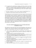
Figure 11.1 Double-reciprocal plot for a random ordered bi bi enzymatic reaction.
At high, fixed concentrations of B, the terms K /[B] and K /[B] go to zero.
Thus, at saturating concentrations of B we find:
v :
V
[AX]
K
6
; [AX]
(11.4)
and likewise, at fixed, saturating [AX]:
v :
V
[B]
K
; [B]
(11.5)
If we measure the reaction velocity over a range of AX concentrations at
several, fixed concentrations of B, the reciprocal plots will display a nest of lines
that converge to the left of the y axis, as illustrated in Figure 11.1. The data
from Figure 11.1 can be replotted as the slopes of the lines as a function of
1/[B], and the y intercepts (i.e., 1/V
) as a function of 1/[B] (Figure 11.2).
The y intercept of the plot of slope versus 1/[B] yields an estimate of
K6/V
, and the x intercept of this plot yields an estimate of 91/K . The
y and x intercepts of the plot of 1/V
versus 1/[B] yield estimates of 1/V
and 91/K , respectively. Thus from the data contained in the two replots,
one can calculate the values of K6, K , and V
simultaneously.
Bi Bi REACTION MECHANISMS 353
Figure 11.2 (A) Slope and (B) y-intercept replots of the data from Figure 11.1, illustrating the
graphical determination of K 6, K , and V
for a random ordered bi bi enzymatic reaction.
11.2.2 Compulsory Ordered Bi Bi Reactions
In compulsory ordered bi bi reactions, one substrate, say AX, must bind to the
enzyme before the other substrate (B) can bind. As with random ordered
reactions, the mechanism proceeds through formation of a ternary intermedi-
354 ENZYME REACTIONS WITH MULTIPLE SUBSTRATES
ate. In this case the reaction scheme is as follows:
E ; AX
&
E·AX
B
&
E·AX·B
&
E·A·BX E·A
&
E ; A
If conversion of the E · AX · B complex to E · A · BX is the rate-limiting step in
catalysis, then E, AX, B, and E · AX · B are all in equilibrium, and the velocity
of the reaction will be given by:
v :
V
[AX][B]
K6K ;K [AX] ; [AX][B]
(11.6)
If, however, the conversion of E · AX · B to E · A · BX is as rapid as the other
steps in catalysis, steady state assumptions must be used in the derivation of
the velocity equation. For a compulsory ordered bi bi reaction, the steady state
treatment yields Equation 11.7:
v :
V
[AX][B]
K6K
; K
[AX] ; K
6
[B] ; [AX][B]
(11.7)
As we have described before, the term K6 in Equation 11.7 is the dissocation
constant for the E · AX complex, and K
6
is the concentration of AX that yields
a velocity of half V
at fixed, saturating [B].
The pattern of reciprocal plots observed for varied [AX] at different fixed
values of [B] is identical to that seen in Figure 11.1 for a random ordered bi
bi reaction (note the similarity between Equations 11.2 and 11.7). Hence, one
cannot distinguish between random and compulsory ordered bi bi mechanisms on
the basis of reciprocal plots alone. It is necessary to resort to the use of isotope
incorporation studies, or studies using product-based inhibitors.
11.2.3 Double Displacement or Ping-Pong Bi Bi Reactions
The double displacement, or Ping-Pong, reaction mechanism involves binding
of AX to the enzyme and transfer of the group, X, to some site on the enzyme.
The product, A, can then leave and the second substrate, B, binds to the E—X
form of the enzyme (in this mechanism, B cannot bind to the free enzyme form).
The group, X, is then transferred (i.e., the second displacement reaction) to the
bound substrate, B, prior to the release from the enzyme of the final product,
BX. This mechanism is diagrammed as follows:
E ; AX
&
E·AX
&
EX · A EX
B
&
EX · B
&
E·BX
&
E ; BX
Bi Bi REACTION MECHANISMS 355
Figure 11.3 Double-reciprocal plot for a double-displacement (Ping-Pong) bi bi enzymatic
reaction.
Using steady state assumptions, the velocity equation for a double-displace-
ment reaction can be obtained:
v :
V
[AX][B]
K
[AX] ; K
6
[B] ; [AX][B]
(11.8)
If we fix the value of [B], then Equation 11.8 for variable [AX] becomes:
v :
V
[AX]
K
6
; [AX]
1 ;
K
[B]
(11.9)
Reciprocal plots of a reaction that conforms to the double-displacement
mechanism for varying concentrations of AX at several fixed concentrations of
B will yield a nest of parallel lines, as seen in Figure 11.3. For each
concentration of substrate B, the values of 1/V
and 91/K
6
can be
determined from the y and x intercepts, respectively, of the double-reciprocal
plot. The data contained in Figure 11.3 can be replotted in terms of 1/V
as
a function of 1/[B], and 1/K
6
as illustrated in Figure 11.4. The value of
91/K
can be determined from the x intercepts of either replot in Figure 11.4.
The y intercepts of the two replots yield estimates of 1/V
(for the 1/V
versus 1/[B] replot) and 1/K
6
(for the 1/K
versus 1/[B] replot) for the
reaction, as seen in Figure 11.4.
356 ENZYME REACTIONS WITH MULTIPLE SUBSTRATES
Figure 11.4 Replots of the data from Figure 11.3 as (A) 1/V
app
max
versus 1/[B] and (B) 1/K
AX,app
m
versus 1/[B], illustrating the graphical determination of K
AX
m
, K
B
m
, and V
max
for a double-
displacement (Ping-Pong) bi bi enzymatic reaction.
11.3 DISTINGUISHING BETWEEN RANDOM AND COMPULSORY
ORDERED MECHANISMS BY INHIBITION PATTERN
It should be clear from Figures 11.1 and 11.3, and the foregoing discussion,
that the qualitative form of the double-reciprocal plots makes it easy to
distinguish between a double-displacement mechanism and a mechanism
DISTINGUISHING BETWEEN RANDOM AND COMPULSORY ORDERED MECHANISMS 357
Table 11.2 Patterns of dead-end inhibition observed for the Bi Bi reaction
E ; AX ; B ; E ; A ; BX for differing reaction mechanisms
Competitive Inhibitor Pattern Observed?
Inhibitor for
Mechanism Substrate For Varied [AX] For Varied [B]
Compulsory ordered with AX Competitive Noncompetitive
[AX] binding first
Compulsory ordered with B Uncompetitive Competitive
[AX] binding first
Compulsory ordered with AX Competitive Uncompetitive
[B] binding first
Compulsory ordered with B Noncompetitive Competitive
[B] binding first
Random ordered AX Competitive Noncompetitive
Random ordered B Noncompetitive Competitive
Double displacement AX Competitive Uncompetitive
Double displacement B Uncompetitive Competitive
?At nonsaturating ([S] : K
) concentration of the fixed substrate.
involving ternary complex formation. But again, it is not possible to further
distinguish between random and compulsory ordered mechanisms on the basis
of reciprocal plots alone. If, however, there is available an inhibitor that binds
to the same site on the enzyme as one of the substrates (i.e., is a competitive
inhibitor with respect to one of the substrates), addition of this compound will
slow the overall forward rate of the enzymatic reaction and can allow one to
kinetically distinguish between random and compulsory ordered reaction
mechanisms. Because of their structural relationship to the substrate, the
product molecules of enzymatic reactions themselves are often competitive
inhibitors of the substrate binding site; this situation is referred to as product
inhibition.
Recall from Chaepter 8 that competitive inhibition is observed when the
inhibitor binds to the same enzyme form as the substrate that is being varied
in the experiment, or alternatively, binds to an enzyme form that is connected
by reversible steps to the form that binds the varied substrate. The pattern of
reciprocal lines observed with different inhibitor concentrations is a nest of
lines that converge at the y intercept (see Chapter 8). For an enzyme that
requires two substrates, a competitive inhibitor of one of the substrate binding
sites will display the behavior of a competitive, noncompetitive, or even
uncompetitive inhibitor, depending on which substrate is varied, whether the
inhibitor is a reversible dead-end (i.e., an inhibitor that does not permit
product formation to occur when it is bound to the enzyme, corresponding to
358 ENZYME REACTIONS WITH MULTIPLE SUBSTRATES
Table 11.3 Pattern of product inhibition observed for the Bi Bi reaction
E ; AX ; B ; E ; A ; BX for differing reaction mechanisms
Inhibitor Pattern Observed?
For Varied [AX] For Varied [B]
Product At At At At
Used As Unsaturated Saturated Unsaturated Saturated
Mechanism Inhibitor [B] [B] [AX] [AX]
Compulsory ordered BX N U N N
with [AX] binding
first
Compulsory ordered A C C N —
with [AX] binding
first
Compulsory ordered BX N — C C
with [B] binding
first
Compulsory ordered A N N N U
with [B] binding
first
Random ordered A C — C —
Random ordered BX C — C —
Double displacement A N — C C
Double displacement BX C C N —
?C, competitive; N, noncompetitive; U, uncompetitive; —, no inhibition.
: 0 for the scheme in Figure 8.1) or product inhibitor, and the mechanism
of substrate interaction with the enzyme. For a bi bi reaction, one observes
specific inhibitor patterns for the different mechanisms we have discussed when
a competitive dead-end inhibitor or a product of the reaction is used as the
inhibitor. The patterns for both dead-end and product inhibition addition-
ally depend on whether the fixed substrate is at a saturating or non-
saturating (typically at [S] : K
) concentration with respect to its apparent K
.
The relationships leading to these differing patterns of dead-end and product
inhibition for bi bi reactions have been derived elsewhere (see, e.g., Segel, 1975).
Rather than rederiving these relationships, we present them as diagnostic tools
for determining the mechanism of reaction. The patterns are summarized in
Tables 11.2 and 11.3 for dead-end and product inhibition, respectively. By
measuring the initial velocity of the reaction in the presence of several
concentrations of inhibitor, and varying separately the concentrations of AX
and B, one can identify the reaction mechanism from the pattern of double-
reciprocal plots and reference to these tables.
DISTINGUISHING BETWEEN RANDOM AND COMPULSORY ORDERED MECHANISMS 359
11.4 ISOTOPE EXCHANGE STUDIES FOR DISTINGUISHING
REACTION MECHANISMS
An alternative means of distinguishing among reaction mechanisms is to look
at the rate of exchange between a radiolabeled substrate and a product
molecule under equilibrium conditions (Boyer, 1959; Segel, 1975).
The first, and simplest mechanistic test using isotope exchange is to ask
whether exchange of label can occur between a substrate and product in the
presence of enzyme, but in the absence of the second substrate. Looking over
the various reaction schemes presented in this chapter, it became obvious that
such an exchange could take place only for a double-displacement reaction:
E;A*X
&
E·A*X
&
EX · A*
—A*
EX
B
&
EX ·B
&
E·BX
&
E;BX
For random or compulsory ordered reactions, the need to proceed through the
ternary complex before initial product release would prevent the incorporation
of radiolabel into one product in the absence of the second substrate.
Next, let us consider what happens when the rate of isotope exchange is
measured under equilibrium conditions for a general group transfer reaction:
AX ; B
&
A ; BX
Under these conditions the forward and reverse reaction rates are equivalent,
and the equilibrium constant is given by:
K
:
[BX][A]
[AX][B]
(11.10)
If under these conditions radiolabeled substrate B is introduced in an amount
so small that it is insufficient to significantly perturb the equilibrium, the rate
of formation of labeled BX can be measured. The measurement is repeated at
increasing concentrations of A and AX, to keep the ratio [A]/[AX] constant
(i.e., to avoid a shift in the position of the equilibrium). As the amounts of A
and AX are changed, the rate of radiolabel incorporation into BX will be
affected.
Suppose that the reaction proceeds through a compulsory ordered mechan-
ism in which B is the first substrate to bind to the enzyme and BX is the last
product to be released. If this is the case, the rate of radiolabel incorporation
into BX will initially increase as the concentrations of A and AX are increased.
As the concentrations of A and AX increase further, however, the formation of
the ternary complexes E · AX · B and E · A · BX will be favored, while dissocia-
tion of the EB and EBX complexes will be disfavored. This will have the effect
360 ENZYME REACTIONS WITH MULTIPLE SUBSTRATES
Figure 11.5 Plots of the equilibrium rate of radioisotope exchange between B and BX as a
function of [AX] for (A) a compulsory ordered bi bi reaction in which B is the first substrate to
bind to the enzyme and BX is the last product to be released, and (B) either a compulsory
ordered bi bi reaction in which AX binds first or a random ordered bi bi reaction.
of lowering the rate of isotope exchange between B and BX. Hence, a plot of
the rate of isotope exchange as a function of [AX] will display substrate
inhibition at high [AX], as illustrated in Figure 11.5A.
The effect of increasing [AX] and [A] on the rate of radiolabel exchange
between B and BX will be quite different, however, in a compulsory ordered
reaction that requires initial binding of AX to the enzyme. In this case,
increasing concentrations of AX and A will disfavor the free enzyme in favor
ISOTOPE EXCHANGE STUDIES FOR DISTINGUISHING REACTION MECHANISMS 361
of the EAX and EA forms. The EAX form will react with B, leading to
formation of BX, while the EA form will not. Hence, the rate of radiolabel
incorporation into BX will increase with increasing [AX] as a hyperbolic
function (Figure 11.5B). The same hyperbolic relationship would also be
observed for a reaction that proceeded through a random ordered mechanism.
In this latter case, however, the hyperbolic relationship also would be seen for
experiments performed with labeled AX and varying [B].
Thus isotope exchange in the absence of the second substrate is diagnostic
of a double-displacement reaction, while compulsory ordered and random
ordered reactions can be distinguished on the basis of the relation of the rate
of radiolabel exchange between one substrate and product of the reaction to
the concentration of the other substrate and product under equilibrium
conditions. (See Segel, 1975, for a more comprehensive treatment of isotope
exchange studies for multisubstrate enzymes.)
11.5 USING THE KING ALTMAN METHOD TO DETERMINE
VELOCITY EQUATIONS
The velocity equations for bi bi reactions can be easily related to the
Henri—Michaelis—Menten equation described in Chapter 5. However, for more
complex reaction schemes, such as those involving multiple intermediate
species, it is often difficult to derive the velocity equation in simple terms. An
alternative method, devised by King and Altman (1956), allows the derivation
of a velocity equation for essentially any enzyme mechanism in terms of the
individual rate constants of the various steps in catalysis. On the basis of the
methods of matrix algebra, King and Altman derived empirical rules for
writing down the functional forms of these rate constant relationships. We
provide a couple of illustrative examples of their use and encourage interested
readers to explore this method further.
To begin with, we shall consider a simple uni uni reaction as first encoun-
tered in Chapter 5:
E ; S
&
ES
-
E ; P
In the King and Altman approach we consider the reaction to be a cyclic
process and illustrate it in a way that displays all the interconversions among
the various enzyme forms involved:
362 ENZYME REACTIONS WITH MULTIPLE SUBSTRATES
For each step in the reaction we can define a term (kappa) which is the
product of the rate constant for that step and the concentration of free
substrate involved in the step. Next, we determine every pathway by which a
particular enzyme species might be formed in the reaction scheme. For the
simple uni uni reaction under consideration we have:
Enzyme Form Pathways to That Form of Kappa Products
EE
k
\
<
k
\
; k
E
k
<
ES
k
[S]
;
ES k
[S]
For any particular enzyme species, the following relationship holds:
[form]
[E]
:
(11.11)
where [form] is the concentration of the particular enzyme form under
consideration,
is the sum of the kappa products for that enzyme form,
and is the sum of the kappa products for all species. Applying this to our
uni uni reaction we obtain:
[E]
[E
]
:
k
\
; k
k
\
; k
; k
[S]
(11.12)
and
[ES]
[E
]
:
k
[S]
k
\
; k
; k
[S]
(11.13)
The overall velocity equation can be written as follows:
v : k
[ES] (11.14)
Substituting the equalities given in Equations 11.12 and 11.13 into Equation
11.14, we obtain:
v :
k
k
[S][E
]
k
\
; k
; k
[S]
:
k
[E
][S]
k
\
; k
k
; [S]
(11.15)
Inspecting Equation 11.15, we immediately see that k
is equivalent to k
, and
(k
\
; k
/k
) is equivalent to the Michaelis constant, K
. If we invoke the
further equality that V
: k
[E], we see that the King—Altman approach
results in the same velocity equation we had derived as Equation 5.24.
Now let us consider the more complex case of a double-displacement bi bi
reaction using the King—Altman approach. Note here that the initial concen-
trations of the two products A and BX are zero, and the release of these
USING THE KING-ALTMAN METHOD TO DETERMINE VELOCITY EQUATIONS 363
products from the enzyme is essentially irreversible. Hence, the cyclic form
of the reaction scheme is:
Consideration of this reaction yields the relationships given in Table 11.4. The
overall rate equation for a double-displacement reaction is:
v : k
[EAX] : k
[EBX] (11.16)
From the preceding relationships, we see that:
[EAX]
[E
]
:
k
k
k
[AX][B]
k
k
[AX][B](k
;k
) ; k
k
[B](k
\
; k
) ; k
k
[AX](k
\
; k
)
(11.7)
Combining Equations 11.16 and 11.17, and performing a few rearrangements
we obtain:
v :
k
k
k
; k
[E
][AX][B]
k
k
k
\
; k
k
; k
[AX] ;
k
k
k
\
; k
k
; k
[B] ; [AX][B]
(11.18)
With the appropriate substitutions, Equation 11.18 can be recast, using the
approach of Alberty, to yield the more familiar form first presented as
Equation 11.8.
With similar considerations, the velocity equations for random ordered and
compulsory ordered bi bi mechanisms can likewise be derived. With some
practice, this seemingly cumbersome approach provides a clear and intuitive
means of deriving the appropriate velocity equation for complex enzymatic
systems. A more thorough treatment of the King—Altman approach can be
found in the text by Segel (1975) as well as in the original contribution by King
and Altman (1956).
11.6 SUMMARY
In this chapter we have briefly introduced the concept of multisubstrate
enzyme reactions and have presented steady state equations to describe the
364 ENZYME REACTIONS WITH MULTIPLE SUBSTRATES
Table 11.4 King Altman relationships for a double displacement Bi Bi reaction
Enzyme Form Pathways to Form of Kappa Products
E k
\
k
k
[B] ; k
k
k
[B] : k
k
[B](k
\
; k
)
E·AX k
k
k
[AX][B]
EX k
k
k
[AX] ; k
k
k
\
[AX] : k
k
[AX](k
\
; k
)
E·BX k
k
k
[AX][B]
365
velocities for these reactions. We have seen that enzyme reactions involving
two substrates and two products can proceed by at least three distinct
mechanisms: random ordered, compulsory ordered, and double-displacement
reactions. Experimental methods were presented to allow the investigator to
distinguish among these mechanisms on the basis of kinetic measurements,
product inhibition studies, and radioisotope exchange studies. We briefly
described the method of King and Altman for deriving the velocity equation
of complex enzymatic reaction, such as those involving multiple substrates.
The importance of multisubstrate enzymatic reactions can hardly be over-
stated. In fact, the vast majority of enzymatic reactions in nature proceed
through the utilization of more than one substrate to yield more than one
product.
REFERENCES AND FURTHER READING
Alberty, R. A. (1953) J. Am. Chem. Soc. 75, 1928.
Boyer, P. D. (1959) Arch. Biochem. Biophys. 82, 387.
Cleland, W. W. (1963) Biochim. Biophys. Acta, 67, 188.
Cornish-Bowden, A., and Wharton, C. W. (1988) Enzyme Kinetics, IRL Press, Oxford, pp. 25—33.
Dalziel, K. (1975) Kinetics and mechanism of nicotinamide-dinucleotide-linked dehydrogenases, in
T he Enzymes, 3rd ed., P. D. Boyer, Ed., Academic Press, San Diego, CA, pp. 1—60.
King, E. L., and Altman, C. (1956) J. Phys. Chem. 60, 1375.
Palmer, T. (1981) Understanding Enzymes, Wiley, New York, pp. 170—189.
Segel, I. H. (1975) Enzyme Kinetics, Wiley, New York, pp. 506—883.
366 ENZYME REACTIONS WITH MULTIPLE SUBSTRATES
12
COOPERATIVITY IN
ENZYME CATALYSIS
As we described in Chapter 3, some enzymes function as oligomeric complexes
of multiple protein subunits, each subunit being composed of copies of the
same or different polypeptide chains. In some oligomeric enzymes, each subunit
contains an active site center for ligand binding and catalysis. In the simplest
case, the active sites on these different subunits act independently, as if each
represented a separate catalytic unit. In other cases, however, the binding of
ligands at one active site of the enzyme can increase or decrease the affinity of
the active sites on other subunits for ligand binding. When the ligand binding
affinity of one active site is affected by ligand occupancy at another active site,
the active sites are said to be acting cooperatively. In positive cooperativity
ligand binding at one site increases the affinity of the other sites, and in negative
cooperativity the affinity of other sites is decreased by ligand binding to the first
site.
For cooperative interaction to occur between two active sites some distance
apart (e.g., on separate subunits of the enzyme complex), ligand binding at one
site must induce a structural change in the surrounding protein that is
transmitted, via the polypeptide chain, to the distal active site(s). This concept
of transmitted structural changes in the protein, resulting in long-distance
communication between sites, has been termed ‘‘allostery,’’ and enzymes that
display these effects are known as allosteric enzymes. (The word ‘‘allosteric,’’
which derives from two Greek words — allos meaning different, and stereos,
meaning structure or solid — was coined to emphasize that the structural
change within the protein mediates the cooperative interactions among differ-
ent sites.)
Allosteric effects can occur between separate binding sites for the same
ligand within a given enzyme, as just discussed, in homotropic cooperativity.
367
Enzymes: A Practical Introduction to Structure, Mechanism, and Data Analysis.
Robert A. Copeland
Copyright
2000 by Wiley-VCH, Inc.
ISBNs: 0-471-35929-7 (Hardback); 0-471-22063-9 (Electronic)
Also, ligand binding at the active site of the enzyme can be affected by binding
of a structurally unrelated ligand at a distant separate site; this effect is known
as heterotropic cooperativity. Thus small molecules can bind to sites other than
the enzyme active site and, as a result of their binding, induce a conformational
change in the enzyme that regulates the affinity of the active site for its
substrate (or other ligands). Such molecules are referred to as allosteric
effectors, and they can operate to enhance active site substrate affinity (i.e.,
serving as allosteric activators) or to diminish affinity (i.e., serving as allosteric
repressors). Both types of allosteric effector are seen in biology, and they form
the basis of metabolic control mechanisms, such as feedback loops.
In this chapter we shall describe some examples of cooperative and allosteric
proteins that not only illustrate these concepts but also have historic signifi-
cance in the development of the theoretical basis for understanding these
effects. We shall then briefly describe two theoretical frameworks for describing
the two effects. Finally, we shall discuss the experimental consequences of
cooperativity and allostery, and appropriate methods for analyzing the kinetics
of such enzymes.
The treatment to follow discusses the effects of cooperativity in terms of
substrate binding to the enzyme. The reader should note, however, that ligands
other than substrate also can display cooperativity in their binding. In fact, in
some cases enzymes display cooperative inhibitor binding, but no cooperativity
is observed for substrate binding to these enzymes. Such special cases are
beyond the scope of the present text, but the reader should be aware of their
existence. A relatively comprehensive treatment of such cases can be found in
the text by Segel (1975).
12.1 HISTORIC EXAMPLES OF COOPERATIVITY AND ALLOSTERY IN
PROTEINS
The proteins hemoglobin and the Trp repressor provide good examples of the
concepts of ligand cooperativity and allosteric regulation, respectively. Hemo-
globin is often considered to be the paradigm for cooperative proteins. This
primacy is in part due to the wealth of information on the structural
determinants of cooperativity in this protein that is available as a result of
detailed crystallographic studies on the ligand-replete and ligand-free states of
hemoglobin. Likewise, much of our knowledge of the regulation of Trp
repressor activity comes from detailed crystallographic studies.
Hemoglobin, as described in Chapter 3, is a heterotetramer composed of
two copies of the subunit and two copies of the subunit. These subunits
fold independently into similar tertiary structures that provide a binding site
for a heme cofactor (i.e., an iron-containing porphyrin cofactor: see Figure
3.19). The heme in each subunit is associated with the protein by a coordinate
bond between the nitrogen of a histidine residue and the central iron atom of
the heme. Iron typically takes up an octahedral coordination geometry
368 COOPERATIVITY IN ENZYME CATALYSIS
Figure 12.1 Plot of bound molecular oxygen as a function of oxygen concentration for the
proteins hemoglobin (Hb) and myoglobin (Mb), illustrating the cooperativity of oxygen binding
for hemoglobin.
composed of six ligand coordination sites. In the heme groups of hemoglobin,
four of these coordination sites are occupied by nitrogens of the porphyrin ring
system and a fifth is occupied by the coordinating histidine, leaving the sixth
coordination site open for ligand binding. This last coordination site forms the
O
binding center for each subunit of hemoglobin.
A very similar pattern of tertiary structure and heme binding motif is
observed in the structurally related monomeric protein myoglobin, which also
binds and releases molecular oxygen at its heme iron center. Based on the
similarities in structure, one would expect each of the four hemes in the
hemoglobin tetramer to bind oxygen independently, and with an affinity
similar to that of myoglobin. In fact, however, when O
binding curves for
these two proteins are measured, the results are dramatically different, as
illustrated in Figure 12.1. Myoglobin displays the type of hyperbolic saturation
curve one would expect for a simple protein—ligand interaction. Hemoglobin,
on the other hand, shows not a simple hyperbolic saturation curve but, instead,
a sigmoidal dependence of O
binding to the protein as a function of O
concentration. This is the classic signature for cooperatively interacting binding
sites. That is, the four heme groups in hemoglobin are not acting as indepen-
dent oxygen binding sites, but instead display positive cooperativity in their
binding affinities. The degree of cooperativity among these distant sites is such
that the data for oxygen binding to hemoglobin are best described by a
two-state model in which all the molecules of hemoglobin contain either 4 or
HISTORIC EXAMPLES OF COOPERATIVITY AND ALLOSTERY IN PROTEINS 369
0 moles of bound O
; under equilibrium conditions, no significant population
of hemoglobin molecules exist with intermediate (i.e., 2 or 3) stoichiometries of
O
binding.
The crystal structures of oxy- (with four O
molecules bound) and deoxy-
(with no O
bound) hemoglobin provide a clear structural basis for this
cooperativity. We know from Chapter 3 that hemoglobin can adopt two
distinct quaternary structures; these are referred to as the R (for relaxed) and
T (for tense) states (see Section 12.2). The differences between the R and T
quaternary structures are relative rotations of two of the subunits, as described
in Figure 3.18. These changes in quaternary structure are mediated by changes
in intersubunit hydrogen bonding at the subunit interfaces. The crystal
structures of oxy- and deoxyhemoglobin reveal that loss of oxygen at the heme
of one subunit induces a change in the strength of the iron—histidine bond that
occupies the fifth coordination site on the heme iron. This change in bond
strength results in a puckering of the porphyrin macrocycle and a displacement
of position for the coordinated histidine (Figure 12.2). The coordinated
histidine is located in a segment of -helical secondary structure in the
hemoglobin subunit, and the motion of the histidine in response to O
binding
or release results in a propagated motion of the entire helix. Ultimately, this
propagated motion produces alterations of the intersubunit hydrogen-bonding
pattern at the
/
subunit interface that acts as a quaternary structure
‘‘switch.’’ The accompanying movements of the other subunits leads to alter-
ations of the oxygen affinities for their associated heme cofactors.
The availability of detailed structural information for both the oxy and
deoxy structures of hemoglobin has made this molecule the classic model of
cooperativity in proteins, illustrating how distant binding sites can interact to
control the overall affinity for a single ligand. Likewise, the structural informa-
tion available for the Trp repressor protein has made this molecule an excellent
example of allosteric regulation in biology. As its name implies, the Trp
repressor protein acts to inhibit the function of the Trp operon, a segment of
DNA that is ultimately responsible for the synthesis of the amino acid
tryptophan. The protein accomplishes this task by binding within the major
groove of the DNA in its tryptophan-bound form and, when not bound by
tryptophan, releasing the DNA. The activity of the Trp repressor is an example
of a negative feedback loop, in which the synthesis of an essential molecule of
the cell is controlled by the concentration of that molecule itself. At low
tryptophan concentrations, the synthesis of tryptophan is required by the cell.
Under these conditions the Trp operon must be functional, and thus the Trp
repressor must not bind to the DNA.
The crystal structures of the tryptophan-depleted protein shows that the
-helical segments of the protein are arranged in a way that precludes effective
DNA binding (Figure 12.3A). Thus, when the tryptophan concentration is low,
the protein is found in a conformation that does not allow for DNA binding,
and the operon is functional, leading to tryptophan synthesis. When the
tryptophan concentration in the cell exceeds some critical concentration,
370 COOPERATIVITY IN ENZYME CATALYSIS
Figure 12.2 Changes in structure of the active site heme that accompany O
2
binding to
hemoglobin, and the associated changes in protein structure at the
1
/
2
subunit interface.
HISTORIC EXAMPLES OF COOPERATIVITY AND ALLOSTERY IN PROTEINS 371
(A) (B)
(B)
Figure 12.3 Cartoons of the interactions of the Trp repressor protein with Trp operon DNA in
the absence (A) and presence (B) of bound tryptophan. This tryptophan-binding-induced
conformational transition is the basis for the negative feedback regulation of tryptophan
synthesis.
however, the Trp repressor binds tryptophan and, as a result, changes its
conformation. The tryptophan-replete form of the protein now has an -helical
arrangement in which two helices are positioned for effective binding to the Trp
operon, via interactions between the helices and the double-stranded DNA
helical structure (Figure 12.3B). When the Trp repressor binds to the operon, it
effectively shuts down the action of this DNA, thus leading to inhibition of further
tryptophan synthesis. This simple method of conformationally controlling the
activity of the Trp repressor, by binding of tryptophan, provides an elegant
mechanism for the metabolic control of the production of an essential amino acid.
Again, we have used hemoglobin and the Trp repressor to illustrate the
concepts of cooperativity and allosteric control in structural terms because of
the wealth of structural information available for these two proteins. The
reader should be aware, however, that the same mechanisms are common in
enzymatic systems as well. Numerous examples of cooperativity and allosteric
control of enzymatic activity can be found in biology, and these control
mechanisms serve vital metabolic roles. For example, many enzymes involved
in de novo biosynthetic cascades display the phenomenon of feedback inhibi-
tion. Here a metabolite that is the ultimate or penultimate product of the
cascade will act as a heterotropic inhibitor of one of the enzymes that occurs
early in the biosynthetic cascade, much as tryptophan controls its own rate of
synthesis by binding to the Trp repressor.
One of the first examples of this phenomenon came from studies of
threonine deaminase from the bacterium E. coli. Abelson (1954) observed that
addition of isoleucine to cultures of the bacterium inhibited the further
biosynthesis of isoleucine. Later workers showed that this effect is due to
specific inhibition by isoleucine of threonine deaminase, the first enzyme in the
biosynthetic route to isoleucine. Further studies with purified threonine
372 COOPERATIVITY IN ENZYME CATALYSIS