DNA Methylation: Basic Mechanisms - Part 3 pptx
Bạn đang xem bản rút gọn của tài liệu. Xem và tải ngay bản đầy đủ của tài liệu tại đây (556.86 KB, 33 trang )
DNA Methyltransferases: Facts, Clues, Mysteries
61
suggest that dsRNA expression, while inducing post-transcriptional silencing
by RNAi, does not induce sequence-specific methylation of the cognate DNA
sequence (Svoboda et al. 2004). Limitations to this study were that the system
used was confined to a specific cell type and that RdDM targeting was analyzed
in a single intronless endogenous gene. Two other reports suggest, on the
contrary, that RNA-mediated DNA methylation can occur in mammals. In
one study on human kidney cells, siRNA targeted to a promoter by means
of lentiviral transduction was found to silence the endogenous EF1A gene,
silencing being associated with DNA methylation (Morris et al. 2004). In
another work, synthetic siRNAs targeted to the E-cadherin gene in human
breast epithelial cells caused its transcriptional repression (Kawasaki and
Taira 2004). Studies in which expression of DNMT genes was suppressed
by means of siRNAs targeting the corresponding messenger (m)RNAs have
shown that DNMT1 and DNMT3B, but not DNMT2, are likely necessary for
siRNA-mediated transcriptional silencing of expression from the E-cadherin
promoter. Bisulfite sequencing revealed a correlation between E-cadherin
silencing correlates and sequence-specific CpG methylation (Kawasaki and
Taira 2004). Thus, RdDM appears also to occur in mammals. Yet from the
few reports available to date, it would already seem that induction of DNA
methylation by siRNA in mammalian cells is not a general phenomenon. If
it turns out to occur in mammals in a limited range of situations, it will be
important to determine which situations, and to explain why only some cells
or some genes are susceptible to RdDM. It will also be essential to unravel the
underlying mechanisms. Key questions will be: How are siRNAs guided to
genomic DNA? How do they gain access to it? Also worthy of special attention,
given the mechanism of RdDM in plants, will be the role played by chromatinmodifying and -remodeling enzymes and the sequence of events leading to
siRNA-directed DNA methylation.
Regarding DNMTs, it will be important to determine how they are mechanistically connected to the RNAi machinery. While these are still early days,
one might imagine, for instance, that RNA molecules serve as cofactors for
DNMTs, thereby guiding CpG methylation to precise sequences (Fig. 3b).
The recent observation that DNMT3A and DNMT3B can interact, at least in
vitro, with RNA molecules is intriguing (Jeffery and Nakielny 2004). Hence,
although highly speculative, the possibility that DNMTs might be targeted directly by an RNA component to establish specific DNA methylation patterns
may deserve future study.
62
C. Brenner · F. Fuks
5
Conclusions
Since the isolation and characterization of the DNMTs in the 1990s, abundant
evidence has established their role as key regulators of DNA methylation. What
is changing is our idea of how DNMTs cause transcriptional repression and
our understanding of how chromatin structure is regulated. It seems almost
certain that chromatin modifications and DNMTs are tightly linked in mammals. As discussed here, clues are emerging that DNMTs may act together with
histone deacetylation and H3-K9 methylation to generate a self-reinforcing
cycle that perpetuates and maintains a repressed chromatin state. Despite
rapid growth of knowledge on the intimate link between chromatin and DNMTs, the picture is still blurred. It will be a notable challenge to untangle the
mutual reinforcements of repression and the different states of chromatin- and
DNA-modifying activities required to silence different genomic regions (e.g.,
highly repetitive elements versus single-copy genes). What’s more, the observation that DNMTs may also silence gene expression by recruiting histone
deacetylase and H3-K9 methyltransferase rather than through their ability to
methylate CpG sites had led to the tempting speculation that DNMTs might
be multifaceted proteins with broader roles in transcriptional repression than
first anticipated.
The origin of DNA methylation patterns is a longstanding mystery. Recent
studies are providing clues that may help explain how DNMTs are targeted
to preferred genomic loci. Like chromatin-modifying enzymes (e.g., HDAC),
DNMTs are recruited to promoters by repressors of transcription, this leading to gene silencing. We anticipate a flurry of research aiming to identify
transcription factors capable of targeting DNMTs to specific genes. If this
mechanism of DNMT targeting turns out to be general, a key issue will be to
understand precisely how specificity is achieved with respect to the DNMTrecruiting transcription factor.
Finally, exciting new evidence suggests a connection between RNAimediated pathways and DNA methylation in mammals. Whether DNMTs
“listen” directly to RNA remains an open question. Work shedding light on
this question is eagerly awaited.
Acknowledgements We thank Luciano Di Croce for critical comments on the
manuscript. C.D. was funded by a grant from the Belgian “Télévie-F.N.R.S”. F.F. is
a “Chercheur Qualifié du F.N.R.S” from the Belgian Fonds National de la Recherche
Scientifique.
DNA Methyltransferases: Facts, Clues, Mysteries
63
References
Ayyanathan K, Lechner MS, Bell P, Maul GG, Schultz DC, Yamada Y, Tanaka K, Torigoe K, Rauscher FJ 3rd (2003) Regulated recruitment of HP1 to a euchromatic
gene induces mitotically heritable, epigenetic gene silencing: a mammalian cell
culture model of gene variegation. Genes Dev 17:1855–1869
Becker PB, Horz W (2002) ATP-dependent nucleosome remodeling. Annu Rev
Biochem 71:247–273
Bender J (2004) Chromatin-based silencing mechanisms. Curr Opin Plant Biol 7:521–
526
Bestor T, Laudano A, Mattaliano R, Ingram V (1988) Cloning and sequencing of a cDNA
encoding DNA methyltransferase of mouse cells. The carboxyl-terminal domain
of the mammalian enzymes is related to bacterial restriction methyltransferases.
J Mol Biol 203:971–983
Bird A (2002) DNA methylation patterns and epigenetic memory. Genes Dev 16:6–21
Bird AP, Wolffe AP (1999) Methylation-induced repression—belts, braces, and chromatin. Cell 99:451–454
Bourc’his D, Bestor TH (2004) Meiotic catastrophe and retrotransposon reactivation
in male germ cells lacking Dnmt3L. Nature 431:96–99
Bourc’his D, Xu GL, Lin CS, Bollman B, Bestor TH (2001) Dnmt3L and the establishment
of maternal genomic imprints. Science 294:2536–2539
Brenner C, Deplus R, Didelot C, Loriot A, Vire E, De Smet C, Gutierrez A, Danovi D,
Bernard D, Boon T, Giuseppe Pelicci P, Amati B, Kouzarides T, de Launoit Y, Di
Croce L, Fuks F (2005) Myc represses transcription through recruitment of DNA
methyltransferase corepressor. EMBO J 24:336–346
Burgers WA, Fuks F, Kouzarides T (2002) DNA methyltransferases get connected to
chromatin. Trends Genet 18:275–277
Cervoni N, Szyf M (2001) Demethylase activity is directed by histone acetylation. J Biol
Chem 276:40778–40787
Chedin F, Lieber MR, Hsieh CL (2002) The DNA methyltransferase-like protein
DNMT3L stimulates de novo methylation by Dnmt3a. Proc Natl Acad Sci U S A
99:16916–16921
Chen T, Ueda Y, Xie S, Li E (2002) A novel Dnmt3a isoform produced from an alternative
promoter localizes to euchromatin and its expression correlates with active de
novo methylation. J Biol Chem 277:38746–38754
Chen T, Ueda Y, Dodge JE, Wang Z, Li E (2003) Establishment and maintenance of
genomic methylation patterns in mouse embryonic stem cells by Dnmt3a and
Dnmt3b. Mol Cell Biol 23:5594–5605
Chen T, Tsujimoto N, Li E (2004) The PWWP domain of Dnmt3a and Dnmt3b is
required for directing DNA methylation to the major satellite repeats at pericentric
heterochromatin. Mol Cell Biol 24:9048–9058
Chuang LS, Ian HI, Koh TW, Ng HH, Xu G, Li BF (1997) Human DNA-(cytosine-5)
methyltransferase-PCNA complex as a target for p21WAF1. Science 277:1996–2000
Dennis K, Fan T, Geiman T, Yan Q, Muegge K (2001) Lsh, a member of the SNF2 family,
is required for genome-wide methylation. Genes Dev 15:2940–2944
Deplus R, Brenner C, Burgers WA, Putmans P, Kouzarides T, de Launoit Y, Fuks F
(2002) Dnmt3L is a transcriptional repressor that recruits histone deacetylase.
Nucleic Acids Res 30:3831–3838
64
C. Brenner · F. Fuks
Di Croce L, Raker VA, Corsaro M, Fazi F, Fanelli M, Faretta M, Fuks F, Lo Coco F,
Kouzarides T, Nervi C, Minucci S, Pelicci PG (2002) Methyltransferase recruitment
and DNA hypermethylation of target promoters by an oncogenic transcription
factor. Science 295:1079–1082
Doherty AS, Bartolomei MS, Schultz RM (2002) Regulation of stage-specific nuclear
translocation of Dnmt1o during preimplantation mouse development. Dev Biol
242:255–266
Freitag M, Lee DW, Kothe GO, Pratt RJ, Aramayo R, Selker EU (2004) DNA methylation
is independent of RNA interference in Neurospora. Science 304:1939
Fuks F, Hurd PJ, Deplus R, Kouzarides T (2003a) The DNA methyltransferases associate
with HP1 and the SUV39H1 histone methyltransferase. Nucleic Acids Res 31:2305–
2312
Fuks F, Hurd PJ, Wolf D, Nan X, Bird AP, Kouzarides T (2003b) The methyl-CpGbinding protein MeCP2 links DNA methylation to histone methylation. J Biol
Chem 278:4035–4040
Ge YZ, Pu MT, Gowher H, Wu HP, Ding JP, Jeltsch A, Xu GL (2004) Chromatin targeting
of de novo DNA methyltransferases by the PWWP domain. J Biol Chem 279:25447–
25454
Geiman TM, Sankpal UT, Robertson AK, Zhao Y, Robertson KD (2004) DNMT3B
interacts with hSNF2H chromatin remodeling enzyme, HDACs 1 and 2, and
components of the histone methylation system. Biochem Biophys Res Commun
318:544–555
Gibbons RJ, McDowell TL, Raman S, O’Rourke DM, Garrick D, Ayyub H, Higgs DR
(2000) Mutations in ATRX, encoding a SWI/SNF-like protein, cause diverse
changes in the pattern of DNA methylation. Nat Genet 24:368–371
Hata K, Okano M, Lei H, Li E (2002) Dnmt3L cooperates with the Dnmt3 family of de
novo DNA methyltransferases to establish maternal imprints in mice. Development 129:1983–1993
Hermann A, Schmitt S, Jeltsch A (2003) The human Dnmt2 has residual DNA(cytosine-C5) methyltransferase activity. J Biol Chem 278:31717–31721
Howell CY, Bestor TH, Ding F, Latham KE, Mertineit C, Trasler JM, Chaillet JR (2001)
Genomic imprinting disrupted by a maternal effect mutation in the Dnmt1 gene.
Cell 104:829–838
Hsieh CL (1999) In vivo activity of murine de novo methyltransferases, Dnmt3a and
Dnmt3b. Mol Cell Biol 19:8211–8218
Jackson JP, Lindroth AM, Cao X, Jacobsen SE (2002) Control of CpNpG DNA methylation by the KRYPTONITE histone H3 methyltransferase. Nature 416:556–560
Jaenisch R, Bird A (2003) Epigenetic regulation of gene expression: how the genome
integrates intrinsic and environmental signals. Nat Genet 33 Suppl:245–254
Jeffery L, Nakielny S (2004) Components of the DNA methylation system of chromatin
control are RNA-binding proteins. J Biol Chem 279:49479–49487
Jones PA, Baylin SB (2002) The fundamental role of epigenetic events in cancer. Nat
Rev Genet 3:415–428
Jones PL, Veenstra GJ, Wade PA, Vermaak D, Kass SU, Landsberger N, Strouboulis J,
Wolffe AP (1998) Methylated DNA and MeCP2 recruit histone deacetylase to
repress transcription. Nat Genet 19:187–191
DNA Methyltransferases: Facts, Clues, Mysteries
65
Kaneda M, Okano M, Hata K, Sado T, Tsujimoto N, Li E, Sasaki H (2004) Essential role
for de novo DNA methyltransferase Dnmt3a in paternal and maternal imprinting.
Nature 429:900–903
Kawasaki H, Taira K (2004) Induction of DNA methylation and gene silencing by short
interfering RNAs in human cells. Nature 431:211–217
Kurdistani SK, Grunstein M (2003) Histone acetylation and deacetylation in yeast. Nat
Rev Mol Cell Biol 4:276–284
Lee JT, Lu N (1999) Targeted mutagenesis of Tsix leads to nonrandom X inactivation.
Cell 99:47–57
Lehnertz B, Ueda Y, Derijck AA, Braunschweig U, Perez-Burgos L, Kubicek S, Chen T,
Li E, Jenuwein T, Peters AH (2003) Suv39h-mediated histone H3 lysine 9 methylation directs DNA methylation to major satellite repeats at pericentric heterochromatin. Curr Biol 13:1192–1200
Leonhardt H, Page AW, Weier HU, Bestor TH (1992) A targeting sequence directs
DNA methyltransferase to sites of DNA replication in mammalian nuclei. Cell
71:865–873
Li E (2002) Chromatin modification and epigenetic reprogramming in mammalian
development. Nat Rev Genet 3:662–673
Li E, Bestor TH, Jaenisch R (1992) Targeted mutation of the DNA methyltransferase
gene results in embryonic lethality. Cell 69:915–926
Maison C, Bailly D, Peters AH, Quivy JP, Roche D, Taddei A, Lachner M, Jenuwein T,
Almouzni G (2002) Higher-order structure in pericentric heterochromatin involves a distinct pattern of histone modification and an RNA component. Nat
Genet 30:329–334
Matzke MA, Birchler JA (2005) RNAi-mediated pathways in the nucleus. Nat Rev Genet
6:24–35
Meehan RR, Stancheva I (2001) DNA methylation and control of gene expression in
vertebrate development. Essays Biochem 37:59–70
Mertineit C, Yoder JA, Taketo T, Laird DW, Trasler JM, Bestor TH (1998) Sex-specific
exons control DNA methyltransferase in mammalian germ cells. Development
125:889–897
Morris KV, Chan SW, Jacobsen SE, Looney DJ (2004) Small interfering RNA-induced
transcriptional gene silencing in human cells. Science 305:1289–1292
Nan X, Ng HH, Johnson CA, Laherty CD, Turner BM, Eisenman RN, Bird A (1998)
Transcriptional repression by the methyl-CpG-binding protein MeCP2 involves
a histone deacetylase complex. Nature 393:386–389
Okano M, Xie S, Li E (1998a) Cloning and characterization of a family of novel
mammalian DNA (cytosine-5) methyltransferases. Nat Genet 19:219–220
Okano M, Xie S, Li E (1998b) Dnmt2 is not required for de novo and maintenance
methylation of viral DNA in embryonic stem cells. Nucleic Acids Res 26:2536–2540
Okano M, Bell DW, Haber DA, Li E (1999) DNA methyltransferases Dnmt3a and
Dnmt3b are essential for de novo methylation and mammalian development. Cell
99:247–257
Qiu C, Sawada K, Zhang X, Cheng X (2002) The PWWP domain of mammalian DNA
methyltransferase Dnmt3b defines a new family of DNA-binding folds. Nat Struct
Biol 9:217–224
Razin A, Cedar H (1977) Distribution of 5-methylcytosine in chromatin. Proc Natl
Acad Sci U S A 74:2725–2728
66
C. Brenner · F. Fuks
Rea S, Eisenhaber F, O’Carroll D, Strahl BD, Sun ZW, Schmid M, Opravil S, Mechtler K,
Ponting CP, Allis CD, Jenuwein T (2000) Regulation of chromatin structure by
site-specific histone H3 methyltransferases. Nature 406:593–599
Rhee I, Jair KW, Yen RW, Lengauer C, Herman JG, Kinzler KW, Vogelstein B, Baylin SB,
Schuebel KE (2000) CpG methylation is maintained in human cancer cells lacking
DNMT1. Nature 404:1003–1007
Rhee I, Bachman KE, Park BH, Jair KW, Yen RW, Schuebel KE, Cui H, Feinberg AP,
Lengauer C, Kinzler KW, Baylin SB, Vogelstein B (2002) DNMT1 and DNMT3b
cooperate to silence genes in human cancer cells. Nature 416:552–556
Rountree MR, Bachman KE, Baylin SB (2000) DNMT1 binds HDAC2 and a new corepressor, DMAP1, to form a complex at replication foci. Nat Genet 25:269–277
Sarraf SA, Stancheva I (2004) Methyl-CpG binding protein MBD1 couples histone H3
methylation at lysine 9 by SETDB1 to DNA replication and chromatin assembly.
Mol Cell 15:595–605
Selker EU (1998) Trichostatin A causes selective loss of DNA methylation in Neurospora. Proc Natl Acad Sci U S A 95:9430–9435
Selker EU, Tountas NA, Cross SH, Margolin BS, Murphy JG, Bird AP, Freitag M (2003)
The methylated component of the Neurospora crassa genome. Nature 422:893–897
Soppe WJ, Jasencakova Z, Houben A, Kakutani T, Meister A, Huang MS, Jacobsen SE,
Schubert I, Fransz PF (2002) DNA methylation controls histone H3 lysine 9 methylation and heterochromatin assembly in Arabidopsis. EMBO J 21:6549–6559
Stec I, Nagl SB, van Ommen GJ, den Dunnen JT (2000) The PWWP domain: a potential
protein-protein interaction domain in nuclear proteins influencing differentiation? FEBS Lett 473:1–5
Svoboda P, Stein P, Filipowicz W, Schultz RM (2004) Lack of homologous sequencespecific DNA methylation in response to stable dsRNA expression in mouse
oocytes. Nucleic Acids Res 32:3601–3606
Ting AH, Jair KW, Suzuki H, Yen RW, Baylin SB, Schuebel KE (2004) CpG island
hypermethylation is maintained in human colorectal cancer cells after RNAimediated depletion of DNMT1. Nat Genet 36:582–584
Tufarelli C, Stanley JA, Garrick D, Sharpe JA, Ayyub H, Wood WG, Higgs DR (2003)
Transcription of antisense RNA leading to gene silencing and methylation as
a novel cause of human genetic disease. Nat Genet 34:157–165
Xin Z, Tachibana M, Guggiari M, Heard E, Shinkai Y, Wagstaff J (2003) Role of histone methyltransferase G9a in CpG methylation of the Prader-Willi syndrome
imprinting center. J Biol Chem 278:14996–15000
Xu GL, Bestor TH, Bourc’his D, Hsieh CL, Tommerup N, Bugge M, Hulten M, Qu X,
Russo JJ, Viegas-Pequignot E (1999) Chromosome instability and immunodeficiency syndrome caused by mutations in a DNA methyltransferase gene. Nature
402:187–191
Yen RW, Vertino PM, Nelkin BD, Yu JJ, el-Deiry W, Cumaraswamy A, Lennon GG,
Trask BJ, Celano P, Baylin SB (1992) Isolation and characterization of the cDNA
encoding human DNA methyltransferase. Nucleic Acids Res 20:2287–2291
Yoder JA, Bestor TH (1998) A candidate mammalian DNA methyltransferase related
to pmt1p of fission yeast. Hum Mol Genet 7:279–284
CTMI (2006) 301:67–122
c Springer-Verlag Berlin Heidelberg 2006
DNA Methylation in Plants
B. F. Vanyushin (u)
Belozersky Institute of Physical and Chemical Biology, Lomonosov Moscow State
University, 119992 Moscow, Russia
1
Introduction . . . . . . . . . . . . . . . . . . . . . . . . . . . . . . . . . . . . . . . . . . . 68
2
2.1
2.2
2.2.1
2.2.2
2.2.3
2.2.4
2.2.5
2.3
2.4
2.5
2.6
2.7
Cytosine DNA Methylation . . . . . . . . . . . . . . . . . . .
Chemical Specificity . . . . . . . . . . . . . . . . . . . . . . . .
Biological Specificity . . . . . . . . . . . . . . . . . . . . . . . .
Species Specificity . . . . . . . . . . . . . . . . . . . . . . . . .
Age Specificity . . . . . . . . . . . . . . . . . . . . . . . . . . . .
Cellular (Tissue) Specificity . . . . . . . . . . . . . . . . . . .
Subcellular (Organelle) Specificity . . . . . . . . . . . . . .
Intragenome Specificity . . . . . . . . . . . . . . . . . . . . .
Replicative DNA Methylation and Demethylation . . . .
Cytosine DNA Methyltransferases . . . . . . . . . . . . . .
Methyl-DNA-Binding Proteins and Mutual Controls
Between DNA Methylation and Histone Modifications
RNA-Directed DNA Methylation . . . . . . . . . . . . . . .
Biological Role of Cytosine DNA Methylation . . . . . .
. . . . . . . . . . . . . . 83
. . . . . . . . . . . . . . 86
. . . . . . . . . . . . . . 90
3
3.1
3.2
3.3
Adenine DNA Methylation . . . . . . . . . . . . . . . . . .
N 6 -Methyladenine in DNA of Eukaryotes . . . . . . . .
Adenine DNA Methyltransferases . . . . . . . . . . . . . .
Putative Role of Adenine DNA Methylation in Plants
.
.
.
.
4
Conclusions . . . . . . . . . . . . . . . . . . . . . . . . . . . . . . . . . . . . . . . . . . . . 103
.
.
.
.
.
.
.
.
.
.
.
.
.
.
.
.
.
.
.
.
.
.
.
.
.
.
.
.
.
.
.
.
.
.
.
.
.
.
.
.
.
.
.
.
.
.
.
.
.
.
.
.
.
.
.
.
.
.
.
.
.
.
.
.
.
.
.
.
.
.
.
.
.
.
.
.
.
.
.
.
.
.
.
.
.
.
.
.
.
.
.
.
.
.
.
.
.
.
.
.
.
.
.
.
.
.
.
.
.
.
.
.
.
.
.
.
.
.
.
.
.
.
.
.
.
.
.
.
.
.
.
.
.
.
.
.
.
.
.
.
.
.
.
.
.
.
.
.
.
.
.
.
.
.
.
.
.
.
.
.
.
.
.
.
.
.
.
.
.
.
.
.
.
.
.
.
.
.
.
.
.
.
.
.
.
.
.
.
.
.
.
.
.
.
.
.
69
69
70
70
71
71
71
72
74
78
97
97
99
102
References . . . . . . . . . . . . . . . . . . . . . . . . . . . . . . . . . . . . . . . . . . . . . . . . . . 105
Abstract DNA in plants is highly methylated, containing 5-methylcytosine (m5 C)
and N 6 -methyladenine (m6 A); m5 C is located mainly in symmetrical CG and CNG
sequences but it may occur also in other non-symmetrical contexts. m6 A but not m5 C
was found in plant mitochondrial DNA. DNA methylation in plants is species-, tissue-,
organelle- and age-specific. It is controlled by phytohormones and changes on seed
germination, flowering and under the influence of various pathogens (viral, bacterial,
fungal). DNA methylation controls plant growth and development, with particular
involvement in regulation of gene expression and DNA replication. DNA replication
is accompanied by the appearance of under-methylated, newly formed DNA strands
including Okazaki fragments; asymmetry of strand DNA methylation disappears until
68
B. F. Vanyushin
the end of the cell cycle. A model for regulation of DNA replication by methylation
is suggested. Cytosine DNA methylation in plants is more rich and diverse compared
with animals. It is carried out by the families of specific enzymes that belong to at
least three classes of DNA methyltransferases. Open reading frames (ORF) for adenine
DNA methyltransferases are found in plant and animal genomes, and a first eukaryotic
(plant) adenine DNA methyltransferase (wadmtase) is described; the enzyme seems
to be involved in regulation of the mitochondria replication. Like in animals, DNA
methylation in plants is closely associated with histone modifications and it affects
binding of specific proteins to DNA and formation of respective transcription complexes in chromatin. The same gene (DRM2) in Arabidopsis thaliana is methylated
both at cytosine and adenine residues; thus, at least two different, and probably interdependent, systems of DNA modification are present in plants. Plants seem to have
a restriction-modification (R-M) system. RNA-directed DNA methylation has been
observed in plants; it involves de novo methylation of almost all cytosine residues in
a region of siRNA-DNA sequence identity; therefore, it is mainly associated with CNG
and non-symmetrical methylations (rare in animals) in coding and promoter regions
of silenced genes. Cytoplasmic viral RNA can affect methylation of homologous nuclear sequences and it may be one of the feedback mechanisms between the cytoplasm
and the nucleus to control gene expression.
1
Introduction
DNA in plants is highly methylated, containing additional methylated bases
such as 5-methylcytosine (m5 C) and N 6 -methyladenine (m6 A). DNA methylation in plants is species-, tissue-, organelle- and age-specific. Specific changes
in DNA methylation accompany the entire life of a plant, starting from seed
germination up to the death programmed or induced by various agents and
factors of biological or abiotic nature. In fact, the ontogenesis and the life itself
are impossible without DNA methylation, because this genome modification
in plants, like in other eukaryotes, is involved in a control of all genetic functions including transcription, replication, DNA repair, gene transposition and
cell differentiation. DNA methylation controls plant growth and development.
On the other hand, plant growth and development are regulated by specific
phytohormones, and modulation of DNA methylation is one of the modes of
the hormonal action in plant.
Plant DNA methylation has many things in common with it in animals but
it has also specific features and even surprises. Plants have a more complicated
system of genome methylations compared with animals; besides, unlike animals, they have the plastids with their own unique DNA modification system
that may control plastid differentiation and functioning; DNA methylation in
plant mitochondria is performed in a different fashion compared with nuclei.
DNA Methylation in Plants
69
Plants seem to have a restriction-modification (R-M) system. Plants supply us
with unique systems or models of living organisms that help us to understand
and decipher the intimate mechanisms and the functional role of genome
modification and functioning in eukaryotes.
Some features and regularities of DNA methylation in plants are described
in this chapter, which cannot be a comprehensive elucidation of many complicated problems associated with this genome modification in the plant
kingdom. An interested reader may find the intriguing details of plant DNA
methylation and its biological consequences also in available reviews (Fedoroff 1995; Meyer 1995; Richards 1997; Dennis et al. 1998; Finnegan et al. 1998b;
Colot and Rossignol 1999; Kooter et al. 1999; Finnegan et al. 2000; Finnegan
and Kovac 2000; Matzke et al. 2000; Sheldon et al. 2000; Wassenegger 2000;
Bender 2001; Chaudhury et al. 2001; Martienssen and Colot 2001; Paszkowski
and Whitham 2001; Vaucheret and Fagard 2001; Bourc’his and Bestor 2002;
Kakutani 2002; Li et al. 2002; Wassenegger 2002; Liu and Wendel 2003; Stokes
2003; Vinkenoog et al. 2003; Matzke et al. 2004; Montgomery 2004; Scott and
Spielman 2004; Steimer et al. 2004; Tariq and Paszkowski 2004).
2
Cytosine DNA Methylation
2.1
Chemical Specificity
5-Methylcytosine in plant DNA is mainly located in symmetrical CG and
CNG sequences (Gruenbaum et al. 1981; Kirnos et al. 1981; Kovarik et al.
1997), but it is found also in various non-symmetrical sequences (Meyer et al.
1994; Oakeley and Jost 1996; Goubely et al. 1999; Pelissier et al. 1999). Some
plant cytosine DNA methyltransferases may methylate any cytosine residue
in DNA except for in CpG, and the specificity of the enzyme is mainly limited
by the availability of certain cytosines in the chromatin structure that can be
modulated essentially by the enzyme itself or its complexes with other proteins
(Wada et al. 2003). The share of m5 C located in CNG sequences in plant DNA
may correspond to up to about 30% of total m5 C content in the genome
(Kirnos et al. 1981). The finding of m5 C in these sequences in plant DNA
was the first safe and widely accepted evidence of the non-CG methylation
in eukaryotes. For a long period many investigators involved in the DNA
methylation research were very sceptical about the existence of this type of
DNA methylation in animals, despite the respective obvious data that were
already available (Salomon and Kaye 1970; Sneider 1972; Woodcock et al. 1987;
70
B. F. Vanyushin
Toth et al. 1990; Clark et al. 1995). The non-CG methylation is carried out by
the Dnmt3a/Dnmt3b enzyme(s) in mammalian cells (Ramsahoye et al. 2000)
and dDnmt2 in Drosophila cells (Lyko 2001) and seems to be guided by RNA
(Matzke et al. 2004). It should be mentioned that attention to the significance
of this particular DNA methylation type for proper genome functioning in
animal cells is still underpaid, and in some modern epigenomic projects
even neglected. But this particular genome modification in animals seems
to have a physiological sense. For example, the histone deacetylase inhibitor
valproate increased 5-lipoxygenase the messenger (m)RNA level and reduced
CNG methylation of the 5-lipoxygenase core promoter in human neuronlike NT2-N but not in NT2 cells (Zhang et al. 2004). The situation with
CNG (non-CG methylation) in plants is better because this modification is
definitely involved in the epigenetic gene silencing including small interfering
(si)RNA-directed silencing (Bartee et al. 2001; Bender 2001; Lindroth et al.
2001).
2.2
Biological Specificity
2.2.1
Species Specificity
Very high m5 C content (up to about 9 mol%) in total DNA is a specific feature
of plants (Vanyushin and Belozersky 1959); in some cases in plant (Scilla sibirica) satellite DNA, the cytosine moiety is almost completely represented by
m5 C. In earlier days, we even could not rule out the possibility that m5 C might
be incorporated into plant DNA in a ready-made form at the template level
during DNA synthesis; there is an indication that 5-methyl-2 -deoxycytidine
5 -triphosphate may be incorporated into DNA in animal cells (Nyce 1991).
But none of any methyl-labelled m5 C derivatives was found to be incorporated
into DNA in an intact plant, and it was concluded that all m5 C present in plant
DNA is a product of DNA methylation (Sulimova et al. 1978). Thus, DNA
in plants compared with other organisms is the most heavily methylated.
m5 C was found in DNA of all archegoniate (mosses, ferns, gymnosperms
and others) and flowering plants (dicots, monocots) investigated. As a rule,
DNA of gymnosperm plants contain less m5 C than DNA of flowering plants
(Vanyushin and Belozersky 1959; Vanyushin et al. 1971). The species differences of phylogenetic significance in the frequency of methylated CNG
sequences in genomes of plants are clearly pronounced (Kovarik et al. 1997;
Fulnecek et al. 2002).
DNA Methylation in Plants
71
2.2.2
Age Specificity
It was known that aging in animals is accompanied by a global DNA demethylation, with the amount of m5 C in the DNA of all organs essentially decreased
(Vanyushin et al. 1973). A similar situation takes place in plants: The amount
of m5 C decreases and its distribution among pyrimidine isopliths in DNA
is essentially changed on seed germination (Sulimova et al. 1978). Some
DNA sequences unmethylated in seeds become methylated in seedlings. The
age changes in DNA methylation may have a regulatory character and seem
to be associated with a developmental switch-over of the gene functioning
(Sulimova et al. 1978). Age differences in the DNA methylation patterns were
found in various plants (Fraga et al. 2002; Baurens et al. 2004; Xiong et al. 1999).
2.2.3
Cellular (Tissue) Specificity
Similarly to animal DNA (Vanyushin et al. 1970), the m5 C content in plant
DNA is tissue (cellular) specific (Vanyushin et al. 1979). This may reflect an
association of DNA methylation with cellular differentiation in plants. There
are many data available now indicating that methylation patterns of total DNA
and distinct genes in various tissues of the same plant are different (Bianchi
and Viotti 1988; Lo Schiavo et al. 1989; Riggs and Chrispeels 1999; Palmgren et
al. 1991; Kutueva et al. 1996; Rossi et al. 1997; Ashapkin et al. 2002; Chopra et al.
2003). The m5 C content in DNA from different plant tissues is associated with
a flowering gradient: It is higher in generative tissues of pea, tobacco, apple
tree and lily-of-the-valley plants compared with vegetative tissues (Chvojka
et al. 1978). The gene silencing associated with DNA methylation is tissue
specific also; methylation of a glucuronidase reporter gene in the transgenic
rice plant accompanied by loss of expression was initially restricted to the
promoter region and observed in the vascular bundle tissue only, the expression character was similar to that of a promoter with a deleted vascular bundle
expression element (Klöti et al. 2002).
2.2.4
Subcellular (Organelle) Specificity
In plant cells the nuclear, mitochondrial and plastid DNAs are methylated
in a different fashion. Contrary to animals (Vanyushin and Kirnos 1974), in
plants m5 C was not found in mitochondrial (mt)DNA (Aleksandrushkina et
al. 1990). Instead, plant mtDNA does contain m6 A, with about 0.5% adenine in
mtDNA from wheat seedlings being methylated (Vanyushin et al. 1988). DNA
72
B. F. Vanyushin
of plastids (chromoplasts, leucoplasts, amyloplasts) contains various methylated bases including m5 C and m6 A, but the chloroplast DNA practically is not
methylated (Ngernprasirtsiri et al. 1988; Ngernprasirtsiri and Akazawa 1990;
Fojtova et al. 2001). It was assumed that plastid DNA (de)methylation is associated with differentiation of plastids and, in particular, with photosynthetic
gene functioning in chloroplasts (Ngernprasirtsiri and Akazawa 1990).
2.2.5
Intragenome Specificity
Plant nuclear DNA is unevenly methylated, since m5 C is mainly located in
GC-enriched and highly repetitive sequences (Guseinov et al. 1975; Guseinov
and Vanyushin 1975). In particular, in petunia the repetitive DNA sequences
(RPS) have hot spots for de novo DNA methylation; for example, the palindromic, moderately to highly RPS-repetitive element that is not predominantly localized to constitutive heterochromatin is a target for strong de novo
methylation. It seems to be due to an intrinsic signal formed by unique DNA
secondary structure. This palindromic element, integrated into the genome
of Arabidopsis thaliana—a plant lacking homology to the RPS—acts as a de
novo hypermethylation site in the non-repetitive genomic background of Arabidopsis, strongly suggesting a signal function of the palindromic RPS unit
(Muller et al. 2002).
The bulk of the repetitive DNA constitutes transposons and retrotransposons; the repeats are primary targets of methylation both in flowering
(ten Lohuis et al. 1995a, b; Muller et al. 2002) and archegoniate (Marchantia
paleacea) (Fukuda et al. 2004) plants. Although the repetitive elements are
methylated in both plants and animals, most mammalian exons are methylated but plant exons are mainly not; there is even an opinion that targeting
of methylation specifically to transposons is restricted to plants (Rabinowicz
et al. 2003).
Usually retrotransposons are hypermethylated (Fukuda et al. 2004) and
their transcription is activated by demethylation (Komatsu et al. 2003). Silent
retrotransposons can be reactivated by ddm1 mutation (Hirochika et al. 2000).
In accordance with methylation patterns, the maize transposable activator
(Ac) elements were divided into two distinct groups. About 50% of the elements are fully unmethylated at cytosine residues through the 256 nucleotides
at the 5 -end (the promoter region), whereas the other half is partially methylated between Ac residues 27 and 92. In contrast, at the 3 -end, all Ac are heavily
methylated between residues 4372 and 4554; the more internally located Ac sequences and the flanking waxy DNA are unmethylated. Methylated cytosines
in Ac are located in both the symmetrical (CG, CNG) and non-symmetrical
DNA Methylation in Plants
73
sequences (Wang et al. 1996). Complex cereal genomes are largely composed
of small gene-rich regions intermixed with 5- to 200-kb blocks of repetitive
DNA. The repetitive DNA blocks are usually methylated at 5 -CG-3 and 5 CNG-3 cytosines in most or all adult tissues, while the genes are generally
unmethylated at these sites (Yuan et al. 2002). The activity and inactivity of
endogenous retrotransposon Tos17 in calli and regenerated rice plants are
accompanied by hypo- and hyper-CG methylation and hemi and full CNG
methylation, respectively, within the element, whereas immobilization of the
element in the other two plant lines is concomitant with near-constant, full
hypermethylation. Treatment with 5-azacytidine (azaCyt) induced both CG
and CNG partial hypomethylation of Tos17 in two lines (Matsumae and RZ35).
A heritable alteration in cytosine methylation patterns occurred in three of
seven genomic regions flanking Tos17 in calli and regenerated plants of RZ35,
but in none of the five regions flanking dormant Tos17 in the other two lines
(Liu et al. 2004). In Arabidopsis, m5 C appears to be differentially distributed
along the major ribosomal (r)RNA gene repeat. The most striking variation
in cytosine methylation in the long arrays of rRNA genes was found at the tips
of chromosomes 2 and 4 (Raddle and Richards 2002). In Brassica napus, S1Bn
short interspersed element (SINE) retroposons are twofold more methylated
than the average methylation level of the nuclear DNA; S1Bn cytosines in
symmetrical CG and CNG sites are methylated at a level of 87% and 44%,
respectively; 5.3% of S1Bn cytosines in non-symmetrical positions were also
methylated. Of this asymmetrical methylation, 57% occurred at a precise motif [Cp(A/T)pA] that only represented 12% of the asymmetrical sites in S1Bn
sequences, suggesting that it represents a preferred asymmetrical methylation
site. This motif is methylated in S1Bn elements at only half the level observed
for the Cp(A/T)pG sites (Goubely et al. 1999).
The methylation patterns of various plant chromosomes are quite different, with even some regions of chromosomes showing enhanced or reduced
methylation (Castilho et al. 1999); DNA in euchromatin is less methylated
compared with heterochromatin DNA (Buzek et al. 1998; Fransz et al. 2002;
Luchniak et al. 2002; Mathieu et al. 2002a). Heterochromatin in Arabidopsis
determined by transposable elements and related tandem repeats is under the
control of the chromatin remodelling ATPase DDM1 (Lippman et al. 2004).
The most methylated repeated family at CG, CNG and asymmetrical sites
was found in the 5S ribosomal DNA. It was highly methylated (Fulnecek et
al. 1998; Fulnecek et al. 2002) even though it is transcribed (Mathieu et al.
2002b). Thus, 5S rRNA gene expression is not inhibited by DNA methylation in
Arabidopsis (Mathieu et al. 2002b). As a rule, centromere regions and satellite
plant DNA are heavily methylated with strand asymmetries (Luo and Preuss
2003). In Vicia faba metaphase chromosomes, the m5 C residues are present
74
B. F. Vanyushin
in different chromosomal sites and are particularly abundant in telomeric
and/or subtelomeric regions and in certain intercalary bands (Frediani et al.
1996). In the Melandrium album male cells, a more intensive methylation on
the shorter arm of the only X chromosome was observed in comparison with
the longer X arm. A global hypermethylation of the male Y chromosome was
not found. But in female cells, the specific cytosine methylation pattern of the
X chromosome was found on a single X chromosome, whereas the other X
displayed an overall higher level of m5 C (Siroky et al. 1998).
At least two CG sequence classes, different in methylation status, were observed in rice genome: Methylation status at the class 1 CG sites was conserved
among genetically diverse rice cultivars, whereas cultivar-specific differential
methylation was frequently detected among the cultivars at the class 2 CG
sites. Five class 2 CG sites were localized on different chromosomes and were
not clustered together in the genome; the differential methylation was stably inherited in a Mendelian fashion over 6 generations, although alterations
in the methylation status at the class 2 CG sites were observed with a low
frequency (Ashikawa 2001).
Usually the individual plant genes and corresponding promoters are
methylated quite unevenly. In Silene latifolia a male reproductive organspecific gene (MROS1) expressed in the late phases of pollen development is
very intensively methylated at CG sites (99%) in the upstream region, whereas
only a low level of CG methylation (7%) was observed in the transcribed
sequence; the asymmetric sequence methylation (2%) in both regions is
quite similar (Janousek et al. 2002). The methylation patterns of cytosine
residues in the Arabidopsis thaliana gene for domain-rearranged methyltransferase (DRM2) were studied in wild-type and several transgene plant
lines containing antisense fragments of the cytosine DNA methyltransferase
gene METI under the control of copper-inducible promoters (Ashapkin
et al. 2002). It was shown that the promoter region of the DRM2 gene is
mostly unmethylated at the internal cytosine residue in CCGG sites, whereas
the 3 -end proximal part of the gene-coding region is highly methylated.
Cytosine methylation in CCGG sites in the DRM2 gene are variable between
wild-type and different transgenic plants. The induction of antisense METI
constructs with copper ions in transgene plants in most cases leads to further
alterations in the DRM2 gene methylation patterns (Ashapkin et al. 2002).
2.3
Replicative DNA Methylation and Demethylation
DNA synthesis in L cells and tobacco cells at a relatively high cell concentration (2–4×10⁵ cells/ml) in a medium is mainly limited to formation of Okazaki
DNA Methylation in Plants
75
fragments (Vanyushin 1984). Thus, it was a unique opportunity to isolate and
investigate the character and level of methylation of the Okazaki fragments
accumulated. It was shown that these fragments do contain m5 C (Bashkite et
al. 1980; Vanyushin 1984), providing evidence that replicative DNA methylation, which starts even at the very early stages of replication, does exist
in plants and animals. The level of methylation of Okazaki fragments was
about twofold lower compared with that of ligated, newly formed and mature
DNAs. The distribution pattern of m5 C among pyrimidine clusters isolated
from the Okazaki fragments and ligated DNA was different. Methylation of
the Okazaki fragments was relatively insensitive to methylation inhibitor Sisobutyladenosine in L cells and to plant growth regulator auxin (2,4-D) in
tobacco cells (Bashkite et al. 1980; Vanyushin 1984) whereas methylation of
ligated DNA was blocked by these agents. Thus, even early replicative DNA
methylation proceeds through at least two phases that may be served by DNA
methyltransferases different in site specificity and sensitivity to various modulators. In tobacco cells, another inhibitor of DNA methylation ethionine,
unlike 5-azaC, strongly inhibits methylation of cytosine residues in CCG but
not CG sequences (Bezdek et al. 1992). The methylation of cytosine residues
in CCG and CAG in plant cells is more sensitive to suppression by AdoHcy and
is under more stringent AdoHcy/AdoMet control compared with CG methylation (Fojtova et al. 1998). Dihydroxypropyladenine (a potential inhibitor of
DNA methyltransferase activities by increasing the S-adenosylhomocysteine
level) induces, in tobacco repeats, a decrease in methylated sequences in the
direction m5 Cm5 CG→Cm5 CG→CCG (Kovarik et al. 2000a, b).
The replicative DNA methylation was observed both in cell suspension cultures and various organs of an intact plant (Vanyushin 1984; Vanyushin and
Kirnos 1988). Cereal seedlings are unique and a very useful model for investigation of replicative and post-replicative DNA methylations in plants. Their
growth may be easily synchronized and at least five cycles of synchronous
replication of nuclear (n)DNA were observed in an initial leaf during the first
7-day period of the seedling development (Kirnos at al. 1983a, b). Coleoptile in
cereals functions for a relatively short period at the early stage of ontogenesis,
and it dies quickly as the seedling grows and develops. Global nDNA synthesis in coleoptile ceased after a few synchronous replication cycles, and this
cessation seems to correspond to the beginning of apoptosis in non-dividing
cells (Kirnos at al. 1983b; Vanyushin et al. 2004). Discrete peaks of total DNA
synthesis in entire leaf at the early stage of wheat seedling development seem
to correspond to cell cycles in the basal meristematic leaf area. It is very useful
for a biochemist, as it allows him in terms of DNA to consider an entire organ
in an intact developing plant organism as a single cell and to investigate what
happens, in particular, with DNA methylation in a cell cycle (Kirnos et al.
76
B. F. Vanyushin
1984, 1986, 1988, 1995). Contrary to the initial leaf, in coleoptile the nuclearDNA content increase stopped on the fourth day of the seedling life. Thus, the
stop of the nDNA (ρ = 1.700 g/cm3 ) synthesis in coleoptile is strictly arranged
temporally in a program of the early stage of seedling development (Kirnos et
al. 1983b). This is an obligatory beginning step of apoptosis and organoptosis. There is no nDNA replication. Only mtDNA (ρ = 1.718 g/cm3 ) continues
to be very intensively synthesized in coleoptile. Therefore, the aging wheat
coleoptiles are a good source for mass plant mtDNA. We failed to detect m5 C
in wheat mtDNA but have detected m6 A in it (Vanyushin et al. 1988).
In wheat seedlings (Kirnos et al. 1984b), as in a suspension culture of
tobacco cells (Bashkite et al. 1980), the Okazaki fragments are methylated. The
methylation level (ML) [100 m5 C/(C + m5 C) = 7.4±0.5] of Okazaki fragments
(<5S) in etiolated seedlings was three to four times lower than that in total
wheat nDNA. After ligation of Okazaki fragments, leading to formation of
long replication intermediate fragments (RIF) (8S, ≥12S), the ML remained at
almost the same level as the Okazaki fragments; therefore, recently replicated
DNA is significantly undermethylated. In ligated (≥12S) and mature nDNA,
up to 40% of all the m5 C residues are located in the Pu-m5 C-Pu sequences,
whereas in the Okazaki fragments this sequence contains only 20% of all
the m5 C (Kirnos et al. 1984b). This again suggested that there is a DNA
methyltransferase associated with the replication fork that is different from
the one methylating the long RIF.
DNA duplexes formed during replication exhibit sharply pronounced
asymmetry of the m5 C distribution along the complementary—parent and
daughter—DNA chains (Kirnos et al. 1984b). This asymmetry remains in the
interphase nuclei and it disappears up to the end of cell cycle (Kirnos et al.
1984b). Based on this observation, a model for regulation of DNA replication
by methylation in eukaryotes (plants) was first suggested (Kirnos et al. 1984b,
1988; Vanyushin 1988). According to this model, only the symmetrically (fully)
methylated DNA duplexes are permitted to be replicated. So, in the early Sphase the completely methylated genome compartments (SE DNA) may be
replicated. In contrast, nucleotide sequences that should enter into replication
in the late S-phase (SL -DNA) are methylated asymmetrically and their replication in SE phase is prohibited. With the termination of the SE -DNA replication,
the newly formed SE duplexes are distinctly asymmetric as to the m5 C content
in complementary DNA strands; their transcription seems to be permitted
but repeated replication in the same cell cycle is prohibited. As a result of the
persistent process of post-replicative methylation (Kirnos et al. 1984a, 1987,
1988), the SL sequences from the preceding cell cycle gradually become symmetrically methylated; therefore, the transcription of corresponding (late)
genes is terminated and they enter into replication. By the onset of a new
DNA Methylation in Plants
77
S-phase, SE - and SL -DNA sequences will be methylated to the same extent as
before the preceding cycle of DNA synthesis. SE and SL duplexes attain this
level depending on the rate of post-replicative DNA methylation, co-ordinated
with the duration of the cell cycle. Thus, the periodic modulation of the asymmetry of methylated sites in nDNA in sequential cell cycles, via replication
and replicative or post-replicative methylation, were regarded as a mechanism regulating the periodicity and fidelity of gene replication in the cell cycle
(Vanyushin 1988). A similar mechanism of regulation of DNA replication by
methylation was later shown to exist in bacteria (Bae et al. 2003; Fujikawa
et al. 2004) where the replication of fully dam-methylated compartments is
permitted but replication of hemimethylated ones is blocked. The Escherichia
coli SeqA protein recognizes the 11 hemimethylated Gm6 ATC sites in the oriC
region of the chromosome, and prevents replication over-initiation within
one cell cycle. SeqA (SeqA71-181) specifically binds to hemimethylated DNA
containing a sequence with a mismatched m6 A:G base pair [Gm6 A(:G)TC] as
efficiently as the normal hemimethylated Gm6 A(:T)TC sequence (Fujikawa
et al. 2004). As hemimethylated DNA has unusual backbone structure and
a remarkably narrow major groove, these dynamic and structural features
provide insights into the specific recognition of hemimethylated GATC sites
by the SeqA protein (Bae et al. 2003).
Thus, replication is a main mechanism of formation of demethylated or, as
it should be said more carefully, undermethylated or hemimethylated DNA.
DNA can be unmethylated due to interfering with maintenance methylation
or demethylated by the active elimination (excision) of m5 C residues or even
by direct removal of methyl group from m5 C. DNA undermethylation by interference with the remethylation of newly replicated DNA should be a slow
process. Cui and Fedoroff (2002) have developed an assay that permits rapid
demethylation of the Spm sequence to be controlled by inducing the expression of the TnpA gene for maize suppressor-mutator transposon-encoded
TnpA protein. TnpA is a weak transcriptional activator, and deletions that
abolish its transcriptional activity also eliminate its demethylation activity.
Demethylation is associated with the formation of a transcription initiation complex, while cell cycle and DNA synthesis inhibitors interfere with
TnpA-mediated Spm demethylation. TnpA has a much lower affinity for fully
methylated than for hemimethylated or unmethylated DNA fragments derived
from Spm termini; it was suggested that TnpA binds to the post-replicative,
hemimethylated Spm sequence and promotes demethylation either by creating an appropriate demethylation substrate or by itself participating in or
recruiting a demethylase (Cui and Fedoroff 2002). Active DNA demethylation in plants was observed during pollen development (Oakeley et al. 1997)
and vernalization (Sheldon et al. 1999); progression of tubers through dor-
78
B. F. Vanyushin
mancy is accompanied by decreases in methylation at 5 -CCGG-3 sequences
in potato meristem (Law and Suttle 2003). Strong strand-biased DNA methylation character was observed in heterochromatic Arabidopsis centromeres.
Unlike the hemimethylation that occurs when methylated DNA is replicated,
the patterns are characterized by nearly complete modification of one strand
and limited modification of its complement. As methyltransferases capable of biased modification of complementary strands are yet unknown, this
DNA methylation pattern can be associated with (1) specific binding of de
novo methyltransferases that processively modify one strand, (2) assembly of
centromere-binding proteins that limit methyltransferase access to one strand
of newly replicated DNA, or (3) differential access of methyltransferases to
the leading or lagging strand during DNA synthesis (Luo and Preuss 2003).
Like in a cell suspension culture, phytohormones mostly inhibit replicative
DNA methylation in wheat seedlings (Kirnos et al. 1986). The strongest (up
to 50%) inhibition of replicative DNA methylation was observed in SM and SL
phases of the cell cycle. A weak, stimulatory effect was exerted by plant growth
regulators 6-benzylaminopurine, 2,4-D, gibberellin and kinetin during prolonged (20 h) incubation of cut-off shoots (Kirnos et al. 1986; Vanyushin
1988). Thus, modulation of DNA methylation is to be one of the molecular
mechanisms of phytohormone action in plant cell.
Post-replicative DNA methylation (Kirnos et al. 1984a, 1986, 1987, 1988)
and demethylation take place also in plants. In Silene latifolia, a rapid decrease in the global DNA methylation level occurs in the cotyledons and
hypocotyls during seed germination. This DNA demethylation seems to be
non-replicative since it occurred before cell division had begun (Zluvova et
al. 2001).
2.4
Cytosine DNA Methyltransferases
When it was clearly shown that m5 C in plant DNA may appear in the different sequences such as CG and CNG (Kirnos et al. 1981; Gruenbaum et al.
1981), the idea of the possible multiplicity of DNA methyltransferases in the
nucleus of the plant cell appeared (Kirnos et al. 1981). It was already hard to
believe that cytosine residues located in these different DNA sequences may
be recognized and modified by the same enzyme. Besides, it was found that
in plant (Bashkite et al. 1980) and animal (Demidkina et al. 1979) cells the
methylation of Okazaki fragments, in contrast to mature DNA methylation,
was relatively insensitive to competitive inhibitors of the DNA methylation
reaction (SIBA and others) and plant growth regulators (auxin and others).
Also, the distribution pattern of m5 C among pyrimidine isopliths from these
DNA Methylation in Plants
79
fragments and mature DNA was very different (Vanyushin 1984). These facts
led to the conclusion that at least two DNA methyltransferases, different in site
specificity and sensitivity to various effectors, should be present in a nucleus
(Kiryanov et al. 1982). In addition, the data on the different nature and character of DNA methylation in mitochondria and nuclei in plants (Vanyushin
et al. 1988) and animals (Vanyushin and Kirnos 1974) indicated that DNA
methyltransferases operating in the nucleus and mitochondria are different.
Then it was shown that plant DNA methyltransferases may differ from respective animal enzymes (Theiss et al. 1987; Vlasova et al. 1996), and, in
addition to CG methylating activity, the enzymes that preferentially methylate cytosine in CNG sequences were isolated from pea (Pradhan and Adams
1995) and wheat plants (Vlasova et al. 1995). Now it is clear that the system of
cytosine DNA modification in plants is quite complicated and is represented
by a family (Fig. 1) of phylogenetically related but chemically distinct and
target-specific DNA methyltransferases (Finnegan and Dennis 1993; Genger
at al. 1999; Finnegan and Kovac 2000; Wada et al. 2003).
There are at least three types of DNA methyltransferases in plants: METI,
chromomethylase (CMT) and DRM.
The first plant gene METI encoding a cytosine methyltransferase was isolated from Arabidopsis thaliana (Finnegan and Dennis 1993). Reduction of
CG methylation in met1-1 mutants was associated with developmental abnormalities (Kankel et al. 2003). METI genes have been identified also in carrot,
pea, tomato and maize (Bernacchia et al. 1998; Pradhan et al. 1998). In fact,
METI is a member of a multigene family, with up to five members (Finnegan
and Dennis 1993; Genger et al. 1999). Four genes arose from an ancestral gene,
and the gene structure, including the position of the 11 introns, is conserved
between the family members (Finnegan and Kovac 2000). The unlinked genes,
METIIa and METIIb, are products of the most recent gene duplication. METI
is the predominant methyltransferase in Arabidopsis (Genger et al. 1999) and
other plants; it preferentially methylates cytosine residues in CG with a highest
activity in meristematic cells (Ronemus et al. 1996). METIIa and METIIb are
transcribed in all tissues, but the level of transcript is very much lower than for
METI (Genger et al. 1999). The function of the proteins encoded by METIIa,
METIIb, and METIII is unknown; antisense constructs against METIIa have
no effect on global methylation or plant development. A METI antisense did
not affect expression of METIIa/b, and yet these enzymes were unable to substitute (completely) for METI activity in METI antisense plants (Genger et
al. 1999). METI enzymes lack the cysteine-rich zinc-binding region found in
the aminoterminal domain of mammalian enzymes (Bestor 1992) and have
an acidic region, consisting of at least 50% glutamic acid and aspartic acid
residues not found in mammalian Dnmt1-like enzymes (Finnegan and Kovac
80
B. F. Vanyushin
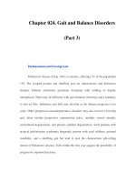
Fig. 1a, b Comparative schematic structures and relatedness of plant cytosine DNA
methyltransferases. a DNA methyltransferase structures. The size of each protein is
indicated in amino acid numbers; conserved motifs in the catalytic region are indicated
by closed boxes with numbers. Specific regions in the regulatory region are indicated
by shaded boxes with appropriate names. BAH, bromo-adjacent homology domain;
H
CD, chromodomain; Glu-rich, glutamine-rich acidic region; NLS, nuclear localization
signal; UBA, ubiquitin association domain. b Phylogenetic relationships among DNA
methyltransferases. (Figure is adapted from Wada et al. 2003)
2000). It was suggested that similarly to animals the aminoterminal domain
in METI is important for discrimination between hemimethylated and unmethylated DNA, giving the enzyme a strong preference for a hemimethylated
template to effectively accomplish maintenance methylation (Finnegan and
Kovac 2000). The expression of MET1 is associated with DNA replication: In
DNA Methylation in Plants
81
maize the transcripts of MET1 exclusively accumulate in actively proliferating cells of the meristems in mesocotyls and root apices (Steward at al. 2000).
METI antisense decreased methylation of cytosine residues in CG and CCG
but not in CAG or CTG sequences (Finnegan et al. 1996). A cDNA encoding
a DNA methyltransferase, with a predicted polypeptide of 1,556 amino acid
residues containing all motifs conserved in this enzyme family, was isolated
from tobacco plants, and the corresponding gene was designated as NtMET1.
Similarly to MET1 the NtMET1 transcripts accumulate in dividing tobacco
cells and are localized exclusively in actively proliferating tissues around axillary apical meristem. Methylation levels of genomic DNA from transgenic
plants with NtMET1 antisense significantly decreased in comparison with
wild-type levels, and distinct phenotypic changes including small leaves,
short internodes and abnormal flower morphology were noted (Nakano et
al. 2000). METI and chromatin remodelling protein DDM1 are required for
maintenance of global cytosine methylation of genome in plants (Bartee and
Bender 2001).
A second class of methyltransferases—chromomethylases (CMT family)—
found in Arabidopsis (Henikoff and Comai 1998; Genger et al. 1999) and other
plants is characterized by insertion of a chromodomain between conserved
motifs II and IV of the methyltransferase domain. Chromomethylases seem
to be involved in modifying DNA in heterochromatin, and they are responsible for maintenance of cytosine methylation at CNG sites, particularly in
retrotransposons (Lindroth et al. 2001; Tompa et al. 2002). In Arabidopsis,
CMT3 takes part in methylation of the SUPERMAN gene and is responsible
for maintaining epigenetic gene silencing; cmt3 mutants display a wild-type
morphology but exhibit decreased CNG methylation of the SUPERMAN gene
and of other sequences throughout the genome; they also show reactivated
expression of endogenous retrotransposon sequences (Lindroth et al. 2001).
Conserved motifs in CMT are relatively (up to 70%) homologous to that of
METI; but the length of the aminoterminal domain in CMT proteins is variable, and this domain has no similarity to that of the METI family (Genger
et al. 1999). A cytosine DNA methyltransferase containing a chromodomain,
Zea methyltransferase 2 (ZMET2), was recently cloned from maize. The sequence of ZMET2 is similar to that of the Arabidopsis chromomethylases
CMT1 and CMT3, and the enzyme is required for in vivo methylation of CNG
sequences (Papa et al. 2001). Arabidopsis cmt3 chromomethylase mutations
block non-CG methylation and silencing of an endogenous reporter gene
and reduce CNG methylation at repetitive centromeric sequences (Bartee et
al. 2001). CMT methyltransferases seem to be unique to plants because no
methyltransferases of this class have been identified in species from other
kingdoms (Genger et al. 1999).
82
B. F. Vanyushin
The third class of methyltransferase genes—composed of DRM1 and
DRM2—has catalytic domains with a sequence homologous to those of
mammalian Dnmt3 methyltransferases. In a plant (Arabidopsis) genome,
the sequences homologous to de novo methyltransferases Masc1 from Ascobolus and Dnmt3 from mouse are observed (Finnegan and Kovac 2000).
The DRM loci in plants are required for asymmetric DNA methylation. At
some loci, drm1drm2 double mutants eliminated all asymmetric methylation, but at the SUPERMAN locus this methylation was completely eliminated only in the drm1drm2cmt3 triple mutant plants. DRM and CMT3
methylate the same asymmetrical sites that follow cytosine residue (Cao and
Jacobsen 2002; Cao et al. 2003). It is interesting that neither drm1drm2 double mutants nor the cmt3 single mutants show morphological defects, the
pleiotropic defects in plant development (development and growth retardations, partial sterility) were observed only in drm1drm2cmt3 triple mutants,
probably due to distortions in RNA-directed DNA methylation (Cao et al.
2003). In animal cells, a novel gene, Dnmt3L, encodes a protein that acts
as a regulator of DNA methylation rather than as a DNA methylation enzyme; the protein functions as a transcriptional repressor through its ability
(like Dnmt3a and Dnmt3b) to associate with histone deacetylase activity
(Deplus et al. 2002). It cannot be ruled out that a similar situation with
some Dnmt3 genes may take place in plant cells also. In tobacco cells the
DRM NtDRM1 was described; the enzyme de novo methylates cytosines in
non-CG sequences (Wada et al. 2003). NtDRM1 is constitutively expressed
through the cell cycle and in all tobacco plant tissues. As a constitutive part
of multiple protein complexes, the enzyme may take part in modulation of
chromatin structure and thereby methylate particular DNA regions (Wada
et al. 2003). DRM enzymes from Arabidopsis, maize and tobacco contain the
conservative ubiquitin association (UBA) domains (Cao et al. 2000; Wada
et al. 2003), which suggests a link between DNA methylation and ubiquitin/proteasome pathways. It is assumed that plant DRMs are controlled in
a cell cycle by ubiquitin-mediated protein degradation or (and) the ubiquitinization may alter the cellular localization of the DRM proteins due to
respective external signals, the cell cycle or transposon or retroviral activity.
UBA domains are found neither in other classes of plant DNA methyltransferases nor in mammalian Dnmt3 proteins; therefore, ubiquitin-associated
pathway may be restricted to Dnmt3-like methylases in plants (Cao et al.
2000).
DNA Methylation in Plants
83
2.5
Methyl-DNA-Binding Proteins and Mutual Controls Between DNA Methylation
and Histone Modifications
It has been well known that DNA methylation influences essentially the interaction of DNA in chromatin with various proteins, including different
regulatory factors, histones and others. It may diminish or even prevent specific protein binding to target DNA (Staiger et al. 1989; Inamdar et al. 1991;
Ehrlich et al. 1992; Ashapkin et al. 1993; Fisscher et al. 1996; Galweiler et al.
2000; Sturaro and Viotti 2001) or vice versa, an obligatory element for such
a binding. In animals, DNA methylation can lead to the recruitment of specific
m5 C-binding proteins taking part in formation of unique gene silencing complexes (Bird and Wolffe 1999; Hendrich and Bird 2000; Ballestar and Wolffe
2001; Jaenisch and Bird 2003; Kimura and Shiota 2003; Kriaucionis and Bird
2004).
Genes for the m5 CG-binding-domain proteins are found in plants also; they
are transcriptionally active and crucial for normal plant development (Berg et
al. 2003). The Arabidopsis genome contains 12 putative genes for such proteins.
These putative proteins were identified and classified into seven subclasses
(Zemach and Grafi 2003). AtMBD7 (subclass VI), a unique protein containing a double MBD motif, as well as AtMBD5 and AtMBD6 (subclass IV),
specifically bind the symmetrically methylated CG sites (Scebba et al. 2003;
Zemach and Grafi 2003); the MBD motif derived from AtMBD6, but not from
AtMBD2, was sufficient for binding methylated CG dinucleotides. AtMBD6
precipitated histone deacetylase activity from the leaf nuclear extract. The
examined AtMBD proteins neither bound methylated CNG sequences nor did
they display DNA demethylase activity. It is suggested that AtMBD5, AtMBD6
and AtMBD7 are likely to function in Arabidopsis plants as mediators of the CG
methylation, linking DNA methylation-induced gene silencing with histone
deacetylation (Zemach and Grafi 2003). On the other hand, it was mentioned
that MBD5 and MBD6, despite their high homology, can be differentiated by
their ability to recognize methylated asymmetrical sites (Scebba et al. 2003).
Ten members of the Arabidopsis gene family encoding methyl-CG-binding domain proteins are transcriptionally active, differentially expressed in diverse
tissues and at least one, AtMBD11, is crucial for normal development (Berg et
al. 2003). This protein showed a strong affinity for DNA independently from
the level of methylation (Scebba et al. 2003). Transformed Arabidopsis plants
with a construct aimed at RNA interference with expression of the AtMBD11
gene, normally active in most tissues, displayed the phenotypic effects such
as aerial rosettes, serrated leaves, abnormal position of flowers, fertility problems and late flowering. Arabidopsis lines with reduced expression of genes
84
B. F. Vanyushin
involved in chromatin remodelling and transgene silencing show similar phenotypes (Berg et al. 2003). These data along with others suggest an important
role for AtMBD proteins in plant development.
The methyl-DNA-binding proteins were found in pea (Zhang et al. 1989;
Ehrlich 1993), maize (Rossi et al. 1997; Sturaro and Viotti 2001) and carrot
(Pitto et al. 2000) cells. The Opaque-2 (O2) protein from the maize endosperm
cell extracts binds in vitro to the cytosine-methylated target sequence of the
maize O2 promoter with different affinities depending on the methylation status of DNA (CG-methylated, hemimethylated, partially methylated and fully
methylated target DNA). Thus, it was hypothesized that DNA methylation
modulates, in vivo, the response of the promoter to the cognate transcription
factors (Rossi et al. 1997). The dcMBP1 protein from carrot protoplasts binds
to symmetrically methylated sequences with high affinity and displays binding properties similar to mammalian MeCP2; protein dcMBP2 has unique
binding properties, it binds specifically to m5 C in unconventional CNN and
symmetrical CNG sequences and seems to be specific for plants (Pitto et al.
2000).
There is no doubt that a peculiar cross-talk between DNA methylation and
histone modifications does exist in eukaryotes. In Neurospora the methylation of lysine 9 in histone H3 is critical for cytosine DNA methylation, normal
growth and fertility of fungus (Tamaru and Selker 2001). Histones there may
be a type of the signal transducers for DNA methylation. On the other hand,
in Arabidopsis the maintenance CG methylation precedes and directs the histone H3 lysine 9 methylation in heterochromatin (Soppe et al. 2002). It is
suggested that DDM1, MET1, H3K9-specific histone methylase and histone
deacetylase (H4K16) play an essential role in the formation of heterochromatin directly after replication, and the CG methylation is performed when
newly formed nucleosomes are still accessible due to acetylated H4K16. H3K9
methylation directed by methylated DNA seems to complete heterochromatin
assembly (Soppe et al. 2002). Complete removal of CG methylation in an Arabidopsis mutant null for maintenance methyltransferase (homozygous for
met1 mutant) results in a clear loss of histone H3 methylation at lysine 9
in heterochromatin and heterochromatic loci that remains transcriptionally
silent; the loss of both CG methylation and H3K9 methylation at condensed
heterochromatic centromers had no effect on their structure (Tariq et al.
2003). This provides additional evidence that methylation of H3K9 is directed
by CG DNA methylation, and the process seems to be transcriptionally independent. In a mutant used with completely erased CG methylation, the
methylation at the CNG and CNN sites was reduced only to 57.6% and 73%,
respectively (Saze et al. 2003). In kyp mutants defective in histone H3 lysine 9 methyltransferase, the DNA methylation is affected only at CNG and
DNA Methylation in Plants
85
CNN sites, which suggests that non-CG methylation is controlled by histone
methylation (Jackson et al. 2002). Loss-of-function kryptonite alleles resemble mutants in the DNA methyltransferase gene CMT3; CMT3 interacts with
an Arabidopsis homologue of HP1, which in turn interacts with methylated
histones (Jackson et al. 2002).
The product of the ddm1 gene is one of the ATP-dependent chromatin
remodelling factors that is required to maintain histone H3 methylation patterns and control the DNA methylation level. The gene is responsible for
transposon and transgene silencing. Thus, transposon methylation in plants
may be guided by histone H3 methylation (Gendrel et al. 2002). As the H3mK9dependent DNA methylation is carried out by chromomethylase CMT3 that
binds histone methylase via an HP-1-like protein, the loss of DNA methylation
in ddm1 may be due to a reduced association of heterochromatin with H3mK9
(Gendrel et al. 2002).
Histone and DNA methylations are under the control of ARGONAUTE proteins involved in post-transcriptional RNA-mediated gene-silencing systems
and in transcriptional gene silencing in various eukaryotes. In the Arabidopsis
ago4-1 mutant, the silent SUPERMAN gene was reactivated and the CNG and
asymmetric DNA methylations, as well as histone H3 lysine 9 methylation,
were decreased. In addition, the accumulation of 25-nucleotide siRNAs that
correspond to the retroelement AtSN1 was observed. Thus, ago4 and long
siRNAs direct chromatin modifications, including histone methylation and
non-CG DNA methylation (Zilberman et al. 2003). Histone and DNA methylations in plant cells are well co-ordinated and seem to be interdependent.
It was shown that rRNA gene dosage control and nucleolar dominance
utilize a common mechanism. Central to the mechanism is an epigenetic
switch in which concerted changes in promoter cytosine methylation and
specific histone modifications dictate the on and off states of the rRNA genes
(Lawrence et al. 2004). A key component of the off switch is HDT1, a plantspecific histone deacetylase that localizes to the nucleolus and is required
for H3 lysine 9 deacetylation and subsequent H3 lysine 9 methylation. It is
assumed that cytosine methylation and histone deacetylation seem to be each
upstream of one another in a self-reinforcing repression cycle (Lawrence et
al. 2004).
Thus, like in animal cells (Nan et al. 1998; Jones et al. 1998; Deplus at al.
2002), the close connection between DNA methylation and histone deacetylation does exist in plants (Aufsatz et al. 2002b). Transgenic plants treated
with propionic or butyric acid (inhibitors of histone deacetylases) display
increased level of DNA methylation and epigenetic variegation (ten Lohus
et al. 1995a). Growth of Brassica seedlings in the presence of inhibitor of
DNA methylation 5-aza-2 -deoxycytidine or histone deacetylase inhibitors