DNA Methylation: Basic Mechanisms - Part 8 pdf
Bạn đang xem bản rút gọn của tài liệu. Xem và tải ngay bản đầy đủ của tài liệu tại đây (727.68 KB, 33 trang )
This page intentionally left blank
Part V
Epigenetic Phenomena
This page intentionally left blank
CTMI (2006) 301:229–241
c Springer-Verlag Berlin Heidelberg 2006
Familial Hydatidiform Molar Pregnancy:
The Germline Imprinting Defect Hypothesis?
O. El-Maarri1 (u) · R. Slim2
1 Institute of Experimental
Hematology and Transfusion Medicine,
Sigmund-Freud Str 25, 53127 Bonn, Germany
2 McGill University Health Centre, Montreal QC, Canada
1
Introduction: The Life Cycle of an Imprint . . . . . . . . . . . . . . . . . . . . . . 230
2
2.1
2.2
2.3
2.4
2.5
2.6
Familial Hydatidiform Molar Pregnancy . . . . . . . . . . . . . . .
Diagnosis and Clinical Manifestations of Molar Pregnancies . .
Epidemiology and Genetics of Molar Pregnancies . . . . . . . . .
Methylation Analysis in Molar Tissues . . . . . . . . . . . . . . . . .
Imprinted Gene Expression Analysis . . . . . . . . . . . . . . . . . .
Hypothesis of a Germline Imprinting Reprogramming Defect .
Variability of Phenotype . . . . . . . . . . . . . . . . . . . . . . . . . . .
3
Concluding Remarks . . . . . . . . . . . . . . . . . . . . . . . . . . . . . . . . . . . . . 237
.
.
.
.
.
.
.
.
.
.
.
.
.
.
.
.
.
.
.
.
.
.
.
.
.
.
.
.
.
.
.
.
.
.
.
.
.
.
.
.
.
.
.
.
.
.
.
.
.
.
.
.
.
.
.
.
231
231
232
232
235
236
237
References . . . . . . . . . . . . . . . . . . . . . . . . . . . . . . . . . . . . . . . . . . . . . . . . . . 237
Abstract
Imprinting is the uniparental expression of a set of genes. Somatic cells carry two
haploid sets of chromosomes, one maternal and one paternal, while germ cells contain
only one of the two forms of chromosomes, male or female. This implies that during
early embryogenesis the cells committed for developing the future germ cell lineage,
the primordial germ cells, which are diploid, have to undergo a total chromosome
reprogramming process. This process is delicately controlled during gametogenesis to
ensure that males and females have only their respective form of gametes. The machinery involved in this process is yet poorly defined. Familial hydatidiform molar (HM)
pregnancy is an abnormal form of pregnancy characterized by hydropic degeneration
of placental villi and abnormal, or absence of, embryonic development. To date, the
molecular defect causing this condition is unknown. However, in a few studied cases,
the presence of paternal methylation patterns on the maternal chromosomes was observed. In this chapter, we summarize what is known about methylation aberrations
in HMs and examine more closely the proposed hypothesis of a maternal germline
imprinting defect.
230
O. El-Maarri · R. Slim
1
Introduction: The Life Cycle of an Imprint
In the process of fertilization, both male and female gametes contribute equal
amounts of genetic material to the newly formed zygote; however, the two
haploid genomes (in the gametes) are not functionally equal (Walter and
Reik 2001; Ferguson-Smith and Surani 2001). A set of genes is marked for
silencing of transcription in one of the gametes but transcribed from the other.
These sets of marked genes are said to be imprinted. Imprinting in somatic
tissues is defined as mono-allelic transcription of a given gene depending
on the parental origin of the chromosome. The imprinting process defines
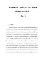
A diagram showing the cycle of reprogramming of parental chromosomes
during gametogenesis with respect to CpG methylation marks. Maternal alleles are
shown in light gray while paternal alleles are in dark gray. Open and filled circles on
the alleles represent unmethylated and methylated sites, respectively
Familial Hydatidiform Molar Pregnancy
231
the asymmetry between the two gametes and implies that the primordial
germ cells, which are still diploid and carrying both maternal and paternal
chromosomes in both sexes, have to undergo a reprogramming process to
reflect the sex of the newly formed embryo (Hajkova et al. 2002, Li E 2002,
Yamazaki et al. 2003; Fig. 1).
One unique example in humans for a disease that is manifested, or caused,
by an imprinting defect is recurrent familial hydatidiform moles (HMs)
(OMIM 231090). HMs mimic uni-parental mouse embryos (Barton et al. 1984)
where androgenotes develop normal extra-embryonic tissues but there is no
or little embryonic development, while parthenogenotes, on the other hand,
give rise to the opposite phenomenon, normal embryonic development with
poor development of extra-embryonic tissues. The exact molecular mechanism leading to familial HM is currently unknown. In this chapter, we will discuss the reasons that led investigators to suggest that it is a maternal germline
defect in establishing the maternal imprinting marks and the validity of this
hypothesis.
2
Familial Hydatidiform Molar Pregnancy
2.1
Diagnosis and Clinical Manifestations of Molar Pregnancies
HM is an abnormal form of human pregnancy characterized by hydropic
degeneration of placental villi with the absence of, or abnormal, embryonic
development. Based on the histology of the evacuated molar tissues, HMs are
divided into two types: complete hydatidiform moles (CHMs) and partial hydatidiform moles (PHMs). CHMs are characterized by hydropic degeneration
of all villi and absence of embryo, cord, and amniotic membranes. All the villi
are (1) enlarged with cisternae, (2) avascular, and (3) surrounded by areas
of trophoblastic proliferation. PHMs are characterized by focal trophoblastic proliferation with a mixture of normal-sized villi and edematous villi.
The trophoblastic proliferation is less pronounced than in complete moles.
An embryo, cord, and amniotic membranes are usually present in partial
moles (Copeland 1993; Bonilla-Musoles 1993). This subdivision is supported
by karyotype data, which show that most complete moles are diploids while
partial moles are triploids. We note that moles are not always easily divisible
into partial and complete moles; in a minority of cases, embryonic tissues
are found in complete moles evacuated at early stages (Zaragoza et al. 1997;
Fukunaga 2000) and some partial moles are found diploid with biparental
origin.
232
O. El-Maarri · R. Slim
2.2
Epidemiology and Genetics of Molar Pregnancies
The most recent reports estimate that 80% of CHMs have a diploid genome
and are androgenetic. Among those, 60% are monospermic and 20% are
dispermic (Kovacs et al. 1991; Lindor et al. 1992). The remaining 20% have
a biparental genomic contribution to their genome. Most reported cases of
HMs are sporadic and not recurrent. Occasionally, recurrent cases have been
reported in one family member (Patek and Johnson 1978; Neumann 1980;
Thavarasah and Kanagalingam 1988; Narayan et al. 1992; Tuncer et al. 1999;
Ozalp et al. 2001; Fisher et al. 2000) and in a few cases, in at least two related
women (familial cases) (Ambani et al. 1980; La Vecchia 1981, Parazzini et
al. 1984, Seoud et al. 1995; Kircheisen and Schroeder-Kurth 1991; Sensi et al.
2000; Judson et al. 2002; Fisher et al. 2002; Al-Hussaini et al. 2003; Hodges et al.
2003; Fallahian et al. 2003; Agarwal et al. 2004; for review see Fisher et al. 2004).
In several of these cases, women with recurrent moles had also abortions at
various gestational stages and some achieved normal pregnancies and gave
birth to healthy babies (Ambani et al. 1980; Seoud et al. 1995; Fallahian et al.
2003).
Consanguineous marriages were observed in many of these families, and
in all of them the segregation of the defect is compatible with an autosomal
recessive mode of transmission, with the women having recurrent moles being
homozygous for the defective locus.
One group characterized the parental contribution to familial moles and
demonstrated, using homozygosity mapping, that a maternal locus mapping
to 19q13.4 between markers D19S924 and D19S890 is responsible for this
condition (Moglabey et al. 1999). This locus was confirmed by other groups
and on several families that allowed narrowing down the candidate region to
1.1 Mb flanked by markers D19S418 and AAAT11138 (Sensi et al. 2000; Hodges
et al. 2003). However, not all families show linkage to 19q13.4, indicating the
genetic heterogeneity of this disease (Judson et al. 2002; Slim et al. 2005),
which could also reflect heterogeneity in the molecular mechanisms leading
to familial moles.
2.3
Methylation Analysis in Molar Tissues
Methylation of DNA at the cytosines’ fifth carbon is the most abundant modification of DNA in the human genome. This fifth base (5-methyl-cytosine:
5mC) occurs at a frequency of about 3%–4% of total cytosines. Most 5mCs
occur at clusters called CpG islands. These are found in the promoter region
of about one-third of human genes. These CpG islands play an important
Familial Hydatidiform Molar Pregnancy
233
role in the regulation of gene activity and expression of the nearby genes. Together with other epigenetic signals such as histone acetylation/methylation,
they impose an open or closed chromatin structure that is associated with
expressed (on) or repressed (off) gene expression. Regions that are actively
transcribed (euchromatin) have promoter regions with mostly unmethylated
CpG sites, acetylated histone tails and methylated lysine 4 on H3 histone subunits, while transcriptionally silent regions (heterochromatin) have mostly
methylated CpG sites, deacetylated histones and methylated lysine 9 on H3
subunits (Fournier et al. 2002; Tamaru and Selker 2001). Imprinted genes that
make the asymmetry in gene expression between the two sets of male and female gametes, and thus the two parental sets of chromosomes, are associated
with differentially methylated regions (DMRs). These DMRs are CpG-rich
regions that are heavily methylated on the non-expressed (repressed) allele
and nearly devoid of methylation on the expressed allele.
The importance of correct methylation settings in the gametes and early
embryogenesis is illustrated by the facts that aborted cloned animals (following nuclear transfer) show irregular methylation patterns (Kang et al. 2001;
Beaujean 2004; Chen et al. 2004; Jaenisch 2004). Low methylation levels in
sperm were also found to give lower rate of pregnancy in assisted reproductive techniques (Benchaib et al. 2005). Molar pregnancies—whether androgenetic or biparental (sporadic or familial)—are identical at the histopathological level; the only known functional difference between the maternal and
the paternal genome is in the expression of imprinted genes. This has led to
a common belief that imprinted genes play an important role in the pathology
of moles and that a defective gene causing their deregulation could underlie
the etiology of familial biparental molar pregnancies.
The above hypothesis was first tested by Judson et al. (2002) who studied
a single biparental molar tissue from a family with recurrent HM. In this
study, the authors made a detailed analysis of a well-characterized set of
DMRs associated with H19, KCNQ1OT1 (LIT1) SNRPN, PEG1, PEG3, and
N
four with the GNAS1 locus. They showed that seven of the nine analyzed
maternally methylated DMRs (at KCNQ1OT1, SNRPN, PEG1, PEG3, and
N
two of the four GNAS) were not methylated. For the paternally methylated
DMRs, again, not all of them behaved similarly; the H19 DMR had a normal
methylation level while the NESP55 DMR (at the GNAS1 locus) was completely
hypermethylated, indicating that the maternal allele behaved like a paternal
allele. In the above study, no DNA polymorphisms were used to track the
parental origin of the abnormally methylated alleles in the molar tissue.
Indeed, this is needed to identify the parental alleles and see whether
abnormal methylation is affecting both of them or only one. Abnormal
methylation at both parental alleles would indicate epigenetic changes
234
O. El-Maarri · R. Slim
during the proliferation of the trophoblast; while an abnormal methylation
exclusively on the maternal alleles may indicate a primary defect that could
be traced in origin to the maternal defect leading to familial moles.
We also analyzed the methylation of four DMRs, two paternally methylated,
H19 and NESP55, and two maternally methylated, SNRPN and PEG3, in two
molar samples from one family (El-Maarri et al. 2003). Using a quantitative
method (El-Maarri et al. 2002, 2004), we found similar trends of abnormal
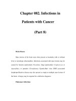
methylation like the ones reported by Judson et al. (Fig. 2). The studied paternal methylation (at H19 and NESP55) in the two molar samples [biparental
complete hydatidiform moles (BiCHM) 9 and 16] were lower than that of
androgenetic complete hydatidiform moles (AnCHMs) and higher than that
of normal chorionic villi and total blood DNAs, while the maternal methylation (SNRPN and PEG3) were decreased. This suggests that portions of the
maternal chromosomes are assuming a paternal methylation patterns.
To investigate whether the two parental alleles are affected by the abnormal
DNA methylation, we looked for single nucleotide polymorphisms (SNPs) and
identified informative ones in a number of DMRs in one or two molar tissues
Fig. 2 The sum of methylation levels obtained at four imprinted genes in two molar
tissues from two sisters (BiCHM9, BiCHM16) and a normal healthy daughter (Helwani
et al. 1999). Analyzed samples include biparental sporadic and androgenetic cases,
controls of normal sperms, chorionic villi, and total blood. The lower two groups
represent paternal methylation; while the upper two represent maternal methylation.
Data are reconstructed from El-Maarri et al. (2003)
Familial Hydatidiform Molar Pregnancy
235
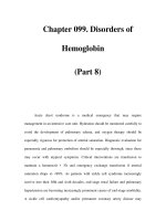
A detailed methylation analysis by bisulfite sequencing from one molar tissue
from family MoLb1 (sample Molb1–6). At both DMRs associated with imprinted genes,
we have a considerable percentage of the maternal clones that acquired the paternal
pattern of methylation (El-Maarri et al. 2003)
(SNRPN in BiCHM16; NESP55 in H19 in both BiCHM9 and BiCHM16). Bisulfite sequencing of individual clones at these DMRs (Fig. 3) showed paternal
methylation pattern most maternal chromosomes; H19 acquired methylation
marks while SNRPN did not show methylation as it should on the maternal
allele. This partial shift from the maternal to paternal patterns of methylation
is intriguing and deserves to be investigated on additional imprinted genes. In
case a similar shift is observed on all imprinted genes, this would indicate an
abnormality in the setting or maintaining of the correct maternal methylation
imprinting marks on the maternal chromosomes rather than a general failure
in the methylation machinery. This is further supported by the fact that the
two patients with recurrent HMs have both normal patterns and levels of
methylation at the same four imprinted loci in their blood (El-Maarri et al.
2005).
2.4
Imprinted Gene Expression Analysis
Transcription analysis of imprinted genes in sporadic androgenetic moles
showed abnormal imprinted gene expression and relaxation of imprinting in
some androgenetic moles (Ohlsson et al. 1999; Ariel et al. 2000; Kim et al. 2003).
These results are compatible with our data on androgenetic moles, in which
we observed at H19 a lower level of methylation than that observed in sperm
DNAs. In familial biparental moles, only one study addressed the expression of
one maternally expressed gene, p57KIP2 (CDNK1C; Fisher et al. 2002). The authors used mouse monoclonal antibody against the p57KIP2 protein on histological sections from familial and sporadic molar tissues. They demonstrated
that p57KIP2 , which is expressed in normal first trimester placenta, is not expressed in biparental moles (familial and sporadic) nor in androgenetic moles.
236
O. El-Maarri · R. Slim
2.5
Hypothesis of a Germline Imprinting Reprogramming Defect
Familial biparental HM pregnancy could be regarded as a disease of imprint
reprogramming that takes place in the affected females to produce female
gametes with paternal methylation imprints. However, to date there is no direct proof for such hypothesis mainly because of the impossibility of studying
germ cells from such patients. Hereon, we list the reasons/observations that
support such a hypothesis as well as reasons against it.
As indirect support for the germline imprinting defect hypothesis involving the maternal chromosomes we could list: (1) the fact that at both gross
morphology and histology levels both familial biparental moles and androgenic moles are undistinguishable; (2) the similarity in the pattern of growth
between biparental or androgenetic moles with that of experimentally created
mouse androgenotes with two male pronuclei; (3) methylation analysis of the
few available molar samples revealing that differentially methylated regions
associated with imprinted genes show variable degree of paternal methylation patterns only on the maternal alleles; (4) the fact that only methylation
at imprinted loci seem to be affected [the analysis of two X-linked genes
(Judson et al. 2002; El-Maarri et al. 2003) revealed that they are normally
methylated].
Reasons that could argue against a maternal germline imprinting defect
are: first, all performed studies on molar tissues were done on samples of
6–14 weeks of gestation in which several changes could have occurred since
fertilization, mainly because of the dynamic nature of early trophoblast and
the postzygotic methylation changes that take place between fertilization
and implantation; second, molar pregnancies are benign tumors of the trophoblast, and several studies have shown gain or loss of methylation marks at
several imprinted genes including PEG3, PEG1, SNRPN, and H19 in a variety
N
of tumors. In colorectal cancer and Wilms’ tumors, a similar shift from a maternal methylation pattern to a paternal one was observed at the H19-IGF2
imprinted region (Steenman et al. 1994; Moulton et al. 1994; Taniguchi et al.
1995; Maegawa et al. 2001; Cui et al. 2001; Nakagawa et al. 2001); third, studies
on sporadic, (androgenetic and biparental) moles demonstrated abnormal
methylation or/and expression of a number of non-imprinted genes including oncogenes, tumor suppressors, and genes involved in protein synthesis,
cell cycle, and intercellular communication (Olvera et al. 2001; Kato et al.
2002; for review see Li et al. 2002; Batorfi et al. 2003; Durand et al. 2003; Xue
et al. 2004). It would be expected that at least some of these genes are also
deregulated in familial biparental moles. The presence of normal methylation
levels on two X-linked genes in familial biparental moles does not allow reach-
Familial Hydatidiform Molar Pregnancy
237
ing a conclusion on the methylation status of non-imprinted genes. A more
comprehensive analysis of a large number of non-imprinted genes in molar
tissues is needed.
2.6
Variability of Phenotype
One important observation derived from the methylation analysis on MoLb1
is that the abnormalities in the level of methylation was not the same at all loci
and in all samples. This may also be true in other cases but could not be seen
since only one molar tissue was analyzed (Judson et al. 2002). This also may
be allelic and restricted to some families where variability in the phenotype of
the conceptuses of these patients ranged from complete moles to spontaneous
abortions at various developmental stages, and normal birth. This variability
could be explained by the contribution of other environmental or/and genetic
factors to the disease phenotype.
3
Concluding Remarks
Familial HM pregnancy is manifested by abnormal imprinting methylation
marks. This abnormal pregnancy reflects the importance of establishing and
maintaining the correct methylation marks for normal embryogenesis. The
gene defect underlying this disorder is still to be identified; when identified
it will increase our understanding of the protein machinery involved in the
setting and maintenance of imprinting during embryogenesis and will answer
the question as to when and how this abnormal paternal methylation was
acquired in these tissues.
Acknowledgements We thank Prof. Dr. Johannes Oldenburg for supporting this work
and Judith Schwalbach for technical support. R.S. is supported by the “Fonds de
Recherche en Santé du Québec” and by operating grants from the CIHR (MOP-67179
and OPD-73018).
References
Agarwal P, Bagga R, Jain V, Karla J, Gopalan S (2004) Familial recurrent molar pregnancy: a case report. Acta Obstet Gynecol Scand 83:218–219
Al-Hussaini TK, Abd el-Aal DM, Van den Veyver IB (2003) Recurrent pregnancy loss
due to familial and non-familial habitual molar pregnancy. Int J Gynaecol Obstet
83:179–186
238
O. El-Maarri · R. Slim
Ambani LM, Vaidya RA, Rao CS, Daftary SD, Motashaw ND (1980) Familial occurrence
of trophoblastic disease—report of recurrent molar pregnancies in sisters in three
families. Clin Genet 18:27–29
Ariel I, de Groot N, Hochberg A (2000) Imprinted H19 gene expression in embryogenesis and human cancer: the oncofetal connection. Am J Med Genet 91:46–50
Barton SC, Surani MA, Norris ML (1984) Role of paternal and maternal genomes in
mouse development. Nature 311:374–376
Batorfi J, Ye B, Mok SC, Cseh I, Berkowitz RS, Fulop V (2003) Protein profiling of
complete mole and normal placenta using ProteinChip analysis on laser capture
microdissected cells. Gynecol Oncol 88:424–428
Beaujean N, Taylor J, Gardner J, Wilmut I, Meehan R, Young L (2004) Effect of limited
DNA methylation reprogramming in the normal sheep embryo on somatic cell
nuclear transfer. Biol Reprod 71:185–193
Benchaib M, Braun V, Ressnikof D, Lornage J, Durand P, Niveleau A, Guerin JF (2005)
Influence of global sperm DNA methylation on IVF results. Hum Reprod 20:768–
773
Bonilla-Musoles F (1993) The diagnosis of gestational trophoblastic neoplasm by
ultrasonography. In: Chervenak FA, Campbell S (eds) Ultrasound in obstetrics
and gynecology, vol. 2. Little Brown and Company, Boston, pp 1665–1673
Chen T, Zhang YL, Jiang Y, Liu SZ, Schatten H, Chen DY, Sun QY (2004) The DNA
methylation events in normal and cloned rabbit embryos. FEBS Lett 578:69–72
Copeland LJ (1993) Gestational trophoblastic neoplasia. In: Copeland LJ (ed) Textbook
of gynecology. W.B. Saunders Company, Philadelphia, pp 1133–1151
Cui H, Niemitz EL, Ravenel JD, Onyango P, Brandenburg SA, Lobanenkov VV, Feinberg AP (2001) Loss of imprinting of insulin-like growth factor-II in Wilms’ tumor
commonly involves altered methylation but not mutation of CTCF or its binding
site. Cancer Res 61:4947–4950
Durand S, Abadie P, Angeletti S, Genti-Raimondi S (2003) Identification of multiple
differentially expressed messenger RNAs in normal and pathological trophoblast.
Placenta 24:209–218
El-Maarri O (2004) SIRPH analysis: SNuPE with IP-RP-HPLC for quantitative measurements of DNA methylation at specific CpG sites. Methods Mol Biol 287:195–205
El-Maarri O, Herbiniaux U, Walter J, Oldenburg J (2002) A rapid, quantitative, nonradioactive bisulfite-SNuPE- IP RP HPLC assay for methylation analysis at specific
CpG sites. Nucleic Acids Res 30:e25
El-Maarri O, Seoud M, Coullin P, Herbiniaux U, Oldenburg J, Rouleau G, Slim R (2003)
Maternal alleles acquiring paternal methylation patterns in biparental complete
hydatidiform moles. Hum Mol Genet 12:1405–1413
El-Maarri O, Seoud M, Riviere JB, Oldenburg J, Walter J, Rouleau G, Slim R (2005)
Patients with familial biparental hydatidiform moles have normal methylation at
imprinted genes. Eur J Hum Genet 13:486–490
Fallahian M (2003) Familial gestational trophoblastic disease. Placenta 24:797–799
Ferguson-Smith AC, Surani MA (2001) Imprinting and the epigenetic asymmetry
between parental genomes. Science 293:1086–1089
Fisher RA, Khatoon R, Paradinas FJ, Roberts AP, Newlands ES (2000) Repetitive complete hydatidiform mole can be biparental in origin and either male or female.
Hum Reprod 15:594–598
Familial Hydatidiform Molar Pregnancy
239
Fisher RA, Hodges MD, Rees HC, Sebire NJ, Seckl MJ, Newlands ES, Genest DR,
Castrillon DH (2002) The maternally transcribed gene p57(KIP2) (CDNK1C) is
abnormally expressed in both androgenetic and biparental complete hydatidiform
moles. Hum Mol Genet 11:3267–3272
Fisher RA, Hodges MD, Newlands ES (2004) Familial recurrent hydatidiform mole:
a review. J Reprod Med 49:595–601
Fournier C, Goto Y, Ballestar E, Delaval K, Hever AM, Esteller M, Feil R (2002) Allelespecific histone lysine methylation marks regulatory regions at imprinted mouse
genes. EMBO J 21:6560–6570
Fukunaga M (2000) Early partial hydatidiform mole: prevalence, histopathology, DNA
ploidy, and persistence rate. Virchows Arch 437:180–184
Hajkova P, Erhardt S, Lane N, Haaf T, El-Maarri O, Reik W, Walter J, Surani MA (2002)
Epigenetic reprogramming in mouse primordial germ cells. Mech Dev 117:15–23
Hodges MD, Rees HC, Seckl MJ, Newlands ES, Fisher RA (2003) Genetic refinement
and physical mapping of a biparental complete hydatidiform mole locus on chromosome 19q13.4. J Med Genet 40:e95
Jaenisch R (2004) Human cloning—the science and ethics of nuclear transplantation.
N Engl J Med 351:2787–2791
Judson H, Hayward BE, Sheridan E, Bonthron DT (2002) A global disorder of imprinting in the human female germ line. Nature 416:539–542
Kang YK, Koo DB, Park JS, Choi YH, Chung AS, Lee KK, Han YM (2001) Aberrant
methylation of donor genome in cloned bovine embryos. Nat Genet 28:173–177
Kato HD, Terao Y, Ogawa M, Matsuda T, Arima T, Kato K, Yong Z, Wake N (2002)
Growth-associated gene expression profiles by microarray analysis of trophoblast
of molar pregnancies and normal villi. Int J Gynecol Pathol 21:255–260
Kim SJ, Park SE, Lee C, Lee SY, Kim IH, An HJ, Oh YK (2003) Altered imprinting,
promoter usage, and expression of insulin-like growth factor-II gene in gestational
trophoblastic diseases. Gynecol Oncol 88:411–418
Kircheisen R, Schroeder-Kurth T (1991) Familial hydatidiform mole syndrome and
genetic aspects of this disturbed trophoblast development. Geburtshilfe Frauenheilkd 51:569–571
Kovacs BW, Shahbahrami B, Tast DE, Curtin JP (1991) Molecular genetic analysis of
complete hydatidiform moles. Cancer Genet Cytogenet 54:143–152
La Vecchia C, Franceschi S, Fasoli M, Mangioni C (1982) Gestational trophoblastic
neoplasms in homozygous twins. Obstet Gynecol 60:250–252
Li E (2002) Chromatin modification and epigenetic reprogramming in mammalian
development. Nat Rev Genet 3:662–673
Li HW, Tsao SW, Cheung AN (2002) Current understandings of the molecular genetics
of gestational trophoblastic diseases. Placenta 23:20–31
Lindor NM, Ney JA, Gaffey TA, Jenkins RB, Thibodeau SN, Dewald GW (1992) A genetic
review of complete and partial hydatidiform moles and nonmolar triploidy. Mayo
Clin Proc 67:791–799
Maegawa S, Yoshioka H, Itaba N, Kubota N, Nishihara S, Shirayoshi Y, Nanba E,
Oshimura M (2001) Epigenetic silencing of PEG3 gene expression in human
glioma cell lines. Mol Carcinog 31:1–9
240
O. El-Maarri · R. Slim
Moglabey YB, Kircheisen R, Seoud M, El Mogharbel N, Van den Veyver I, Slim R (1999)
Genetic mapping of a maternal locus responsible for familial hydatidiform moles.
Hum Mol Genet 8:667–671
Moulton T, Crenshaw T, Hao Y, Moosikasuwan J, Lin N, Dembitzer F, Hensle T, Weiss L,
McMorrow L, Loew T, Kraus W, Gerald W, Tycko B (1994) Epigenetic lesions at
the H19 locus in Wilms’ tumour patients. Nat Genet 7:440–447
Nakagawa H, Chadwick RB, Peltomäki P, Plass C, Nakamura Y, de la Chapelle A (2001)
Loss of imprinting of the insulin-like growth factor II gene occurs by biallelic
methylation in a core region of H19-associated CTCF-binding sites on colorectal
cancer. Proc Natl Acad Sci USA 98:591–596
Narayan H, Mansour P, McDougall WW (1992) Recurrent consecutive partial molar
pregnancy. Gynecol Oncol 46:122–127
Neumann H (1980) Case report of recurring hydatidiform mole. Geburtshilfe Frauenheilkd 40:385–388
Ohlsson R, Flam F, Fisher R, Miller S, Cui H, Pfeifer S, Adam GI (1999) Random
monoallelic expression of the imprinted IGF2 and H19 genes in the absence of
discriminative parental marks. Dev Genes Evol 209:113–119
Olvera M, Harris S, Amezcua CA, McCourty A, Rezk S, Koo C, Felix JC, Brynes RK
(2001) Immunohistochemical expression of cell cycle proteins E2F-1, Cdk-2, Cyclin E, p27(kip1), and Ki-67 in normal placenta and gestational trophoblastic
disease. Mod Pathol 14:1036–1042
Ozalp S, Yalcin OT, Tanir HM, Etiz E (2001) Recurrent molar pregnancy: report of a case
with seven consecutive hydatidiform moles. Gynecol Obstet Invest 52:215–216
Parazzini F, La Vecchia C, Franceschi S, Mangili G (1984) Familial trophoblastic disease:
case report. Am J Obstet Gynecol 149:382–383
Patek E, Johnson P (1978) Recurrent hydatidiform mole. Report of a case with five
recurrences. Acta Obstet Gynecol Scand 57:381–383
Sensi A, Gualandi F, Pittalis MC, Calabrese O, Falciano F, Maestri I, Bovicelli L, Calzolari E (2000) Mole maker phenotype: possible narrowing of the candidate region.
Eur J Hum Genet 8:641–644
Seoud M, Khalil A, Frangieh A, Zahed L, Azar G, Nuwayri-Salti N (1995) Recurrent
molar pregnancies in a family with extensive intermarriage: report of a family
and review of the literature. Obstet Gynecol 86:692–695
Slim R, Fallahian M, Riviere JB, Zali MR (2005) Evidence of a genetic heterogeneity of
familial hydatidiform moles. Placenta 26:5–9
Steenman MJ, Rainier S, Dobry CJ, Grundy P, Horon IL, Feinberg AP (1994) Loss of
imprinting of IGF2 is linked to reduced expression and abnormal methylation of
H19 in Wilms’ tumor. Nat Genet 7:433–439
Tamaru H, Selker EU (2001) A histone H3 methyltransferase controls DNA methylation
in Neurospora crassa. Nature 414:277–283
Taniguchi T, Sullivan MJ, Ogawa O, Reeve AE (1995) Epigenetic changes encompassing
the IGF2/H19 locus associated with relaxation of IGF2 imprinting and silencing
of H19 in Wilms tumor. Proc Natl Acad Sci USA 93:2159–2163
Thavarasah AS, Kanagalingam S (1988) Recurrent hydatidiform mole: a report of
a patient with 7 consecutive moles. Aust N Z J Obstet Gynaecol 28:233–235
Tuncer ZS, Bernstein MR, Wang J, Goldstein DP, Berkowitz RS (1999) Repetitive
hydatidiform mole with different male partners. Gynecol Oncol 75:224–226
Familial Hydatidiform Molar Pregnancy
241
Walter J, Reik W (2001) Genomic imprinting: parental influence on the genome. Nat
Rev Genet 2:21–32
Xue WC, Chan KY, Feng HC, Chiu PM, Ngan HY, Tsao SW, Cheung AN (2004) Promoter
hypermethylation of multiple genes in hydatidiform mole and choriocarcinoma.
J Mol Diagn 6:326–334
Yamazaki Y, Mann MR, Lee SS, Marh J, McCarrey JR, Yanagimachi R, Bartolomei MS
(2003) Reprogramming of primordial germ cells begins before migration into
the genital ridge, making these cells inadequate donors for reproductive cloning.
Proc Natl Acad Sci U S A 100:12207–12212
Zaragoza MV, Keep D, Genest DR, Hassold T, Redline RW (1997) Early complete
hydatidiform moles contain inner cell mass derivatives. Am J Med Genet 70:273–
277
This page intentionally left blank
CTMI (2006) 301:243–256
c Springer-Verlag Berlin Heidelberg 2006
Dual Inheritance
R. Holliday (u)
12 Roma Court, West Pennant Hills, Sydney, NSO 2125, Australia
1
Introduction . . . . . . . . . . . . . . . . . . . . . . . . . . . . . . . . . . . . . . . . . . . 244
2
The Inheritance of DNA Methylation . . . . . . . . . . . . . . . . . . . . . . . . . . 245
3
Mutations and Epimutations . . . . . . . . . . . . . . . . . . . . . . . . . . . . . . . . 247
4
Epigenetic and Classical Inheritance . . . . . . . . . . . . . . . . . . . . . . . . . . 249
5
The Lamarckian Dimension . . . . . . . . . . . . . . . . . . . . . . . . . . . . . . . . 251
6
The Central Dogma of Molecular Biology Revisited . . . . . . . . . . . . . . . . 252
7
Conclusions . . . . . . . . . . . . . . . . . . . . . . . . . . . . . . . . . . . . . . . . . . . . 253
References . . . . . . . . . . . . . . . . . . . . . . . . . . . . . . . . . . . . . . . . . . . . . . . . . . 254
Abstract Genetic inheritance in higher organisms normally refers to the transmission
of information from one generation to the next. Nevertheless, there is also inheritance
in somatic cells, characterised by the phenotypic stability of differentiated cells that
divide (such as fibroblasts and lymphocytes), and also mitosis of stem line cells, which
gives rise to another stem line daughter cell, and one that will differentiate. Thus, there
is a dual inheritance systems in these organisms, one of which is genetic and the other
epigenetic. In the latter, heritable information is superimposed on DNA sequences, and
one well-known mechanism is heritable methylation of cytosine. Much information
will come from the human epigenome project that will reveal the patterns of DNA
methylation in distinct differentiated cells. There have also been innumerable studies
on the abnormal de novo methylation and silencing of tumour suppressor genes in
cancer cells.
This paper ist dedicated to the memory of the late John Maynard Smith.
244
R. Holliday
1
Introduction
In 1990 John Maynard Smith published a paper “Models of a dual inheritance
system”. In his Introduction he wrote:
In higher plants, animals and fungi there are two inheritance systems,
as follows:
1. The familiar system, depending on DNA sequence, used in transmitting information between sexual generations.
2. An epigenetic inheritance system (EIS), responsible for cellular inheritance during ontogeny—for example fibroblasts give rise to fibroblasts, epithelial cells to epithelial cells, and Drosophila wing discs
continue to be wing discs in serial transfer.
This paper was in response to the proposals by Jablonka and Lamb (1989)
that epigenetic changes might sometimes be transmitted by sexual reproduction, following earlier discussion of the same theme (Holliday 1987). These
proposals were further elaborated in their book Epigenetic Inheritance and
Evolution (Jablonka and Lamb 1995). It was characteristic of Maynard Smith
that he immediately recognised the significance of the epigenetic system, and
it was he who first coined the term “dual inheritance”.
In the standard literature it is not usual to categorise the division of the
specialised cells of higher organisms as an inheritance system. Instead, it is
simply stated that some differentiated cells (e.g. lymphocytes and fibroblasts)
are capable of mitotic division, whereas others (e.g. neurons and muscle cells)
are not. Traditionally, genetics and inheritance in multicellular organisms
refers only to sexual reproduction. However, in micro-organisms such as
yeasts and fungi, it is common to refer to asexual or vegetative reproduction,
and there may also be a parasexual cycle. Thus, in a microbial eukaryote an
induced mutation will be inherited through mitotic division to form a clone.
The same mutation can also be transmitted through meiosis, or segregate
from a diploid nucleus.
Historically, it was probably the work of Hadorn and his colleagues that
first demonstrated the importance of somatic cell inheritance (reviewed in
Ursprung and Nothiger 1972). In their experiments (included by Maynard
Smith under system ii), imaginal disc tissue of Drosophila is inherited in
a determined state. The cells are undifferentiated, but when the tissue is
treated with the hormone ecdysone they differentiate. Thus, leg disc tissue
differentiates into recognisable leg structures, wing tissue into wing and so
on. The determined but undifferentiated disc tissue can be propagated in the
Dual Inheritance
245
abdominal cavity of adult flies, in some cases for hundreds of generations.
Sometimes the determined state changes into that of a different tissue. This is
known as transdetermination, and it is not random, but follows certain rules,
such that determined state A can change to B or C, but not to D. However, B
or C might change to D. The frequencies of transdetermination also vary, and
are in fact an inherited property of the particular determined state.
There is nothing intrinsically different between the inheritance of determined states to the inheritance of differentiated states. The phenotype of
a fibroblast is the result of the specialised expression of one set of genes,
producing so-called luxury proteins, and the lack of expression of all those
genes that produce luxury proteins in other specialised cells. This phenotype
is stably inherited through serial subculture—and also in vivo—until the
cells become senescent and post-mitotic. It is well established that the phenotypes of specialised cells are very stable, and what would be the equivalent of
transdetermination does not occur. It is generally assumed that the change of
one specialised cell type into another never occurs, but one should be careful
not to make this a dogmatic assertion, because in developing or adult organisms it might happen in specific circumstances, for example, in limb or tail
regeneration.
2
The Inheritance of DNA Methylation
It is important to understand that the classical gene regulatory systems categorised in bacteria do not provide an inheritance system. If a gene is expressed
in response to a specific inducing chemical in the medium, the daughter cells
produced by division will only have the same phenotype provided the inducer
is present. If the inducer is removed then the cell reverts to its original state.
The phenotype of a differentiated eukaryotic cell is not dependent on extracellular signals. Cells such as fibroblasts require protein factors in serum in
the medium in order to grow, but these same factors are not necessary for the
cellular phenotype. Therefore, there are intrinsic mechanisms that maintain
the phenotype.
Recognising this, I proposed with my colleague John Pugh (Holliday and
Pugh 1975) specific mechanisms involving the modification of DNA. There
were four features in the development of higher organisms that were emphasised:
1. The modification of a controlling or regulatory region of DNA adjacent
to a gene can occur. The modification would silence a gene, and in the
absence of modification the gene would be active, or vice versa.
246
R. Holliday
2. The modified and non-modified forms of the gene would be stably inherited.
3. There would be switching between modified and non-modified states,
either during normal development, or in stem cell situations. In the latter
case, a stem cell would produce a daughter the same as itself and one that
was destined or determined to differentiate subsequently. In the former
case, a cell A could give rise to two B cells, with new properties, or to two
different cells, B and C.
4. There would be developmental clocks capable of counting cell divisions,
and at the end of a specified number of cell divisions a regulatory mechanism of some kind would be triggered.
We proposed that the modification could be based on the methylation of
cytosine in DNA to form 5-methyl cytosine, and also that the methylation
pattern could be inherited if there was a maintenance methylase that recognised hemi-methylated DNA at the replication fork and methylated the new
strand. This enzyme would not recognise non-methylated DNA. We also proposed, following Scarano (1971), that cytosine might be deaminated at specific
sites to form thymidine, or that adenine would be converted to guanine. In
the same year, Riggs (1975) proposed essentially the same DNA methylation
mechanism and applied it in the context of the inactivation of one X chromosome in female mammals. The two X chromosomes are in the same cellular
milieu, yet one is very stably maintained in an active form and the other
in an inactive form. His model also involved a rapid initial switch in which
one chromosome was marked by methylation, and this process immediately
inhibited the methylation of the other X chromosome. Also in 1975, Sager
and Kitchin suggested that the methylation of DNA may control the processes
of elimination or inactivation of chromosomes in various contexts. From the
many studies of bacterial methylation, they suggested that non-methylated
DNA might be lost through the activity of restriction enzymes, or that genes
might be inactivated. Our models for the events listed above were based on cell
lineages, which is probably incorrect, because many developmental events are
known to occur in groups of cells, which Crick and Lawrence (1975) dubbed
“polyclones” to describe the compartments in Drosophila development.
Subsequent to 1975, a vast amount of evidence has accumulated that differential inherited methylation does occur in higher organisms and that the
methylation of CpG islands near promoter sequences silences genes (Millar
et al. 2003; Beck and Olek 2003). On the other hand, evidence that there are
specific base changes in DNA has not been forthcoming, except in the context
of antibody gene variability (Neuberger et al. 2003; Pham et al. 2003). In 1987,
Dual Inheritance
247
I adopted Waddington’s use of the word epigenetic, which I took to mean the
totality of mechanisms that are required to unfold the genetic programme
for the development of a complex organism (Holliday 1987). In particular, I
discussed epigenetic defects that were changes in gene expression following
methylation or demethylation, and suggested that these might be an important contributor to ageing, and also, following an earlier proposal (Holliday
1979), that they might be responsible for changes in gene expression during
tumour progression. There is now a huge literature documenting the methylation and silencing of tumour suppressor genes in many types of cancer
(Jones and Baylin 2002). In contrast, the role of DNA methylation changes
in normal development is not well documented. Although there are many
suggestive observations (nine were listed in Holliday 1996), it would be true
to say that most developmental biologists do not regard DNA methylation
as a key mechanism in development. Nor is there widespread acceptance of
the proposal that the switching on or off of genes coding for luxury proteins is based on DNA methylation. It is more commonly believed that the
modification of the histones of chromatin—for example by acetylation, deacetylation or methylation—is a more likely mechanism. Although this may
provide a means whereby the formation of inactive heterochromatin at CpG
islands occurs in non-dividing cells, it does not by itself provide a mechanism for the strict heritability of a given cell phenotype. Those who work on
Drosophila cite the behaviour of the Polycomb group of proteins that appear
to provide a basis for heritability of given chromatin configurations, at least
over the fairly small number of generations that occur during fly development
(Grewal and Maozed. 2003; Lavigne et al. 2004). It is not known whether these
proteins could also explain the heritability of the determined state of imaginal
disc cells during long-term serial passaging.
3
Mutations and Epimutations
In the early days of mammalian somatic cell genetics there was a controversy
between those who believed the cells could be handled in much the same way
as micro-organisms, and those who believed that mammalian cells’ genetic
behaviour did not correspond to that of simple eukaryotes, and that their
variability was not just due to simple mutations. As is often the case in controversies, both viewpoints proved to be correct (Holliday 1991). Experiments
with CHO (Chinese hamster ovary) cells provided strong evidence that mutations could be induced in many housekeeping genes; these could be shown
to be recessive in hybrids and to reappear in segregants. It was proposed that
248
R. Holliday
CHO cells were functionally hemizygous, meaning that substantial parts of
the genome were haploid, and this facilitated the isolation of mutants. Later it
was shown that this haploidy was due in many cases to the presence of a silent
gene on the homologous chromosome. The silent gene was methylated and
could be reactivated by the powerful demethylating agent 5-azacytidine. In
some cases both genes were inactivated by methylation. In fact, it became clear
that if there was no selective disadvantage during the laboratory growth of
CHO cells, any gene might become inactivated through de novo methylation.
These studies provide clear evidence for a dual inheritance system, but not
one which involves sexual reproduction. Instead, we have classical mutation,
induced by mutagens, and involving a change in DNA base sequence, in which
there is great stability and rare back-mutation. In contrast, we have gene inactivation and activation due to alterations in DNA methylation. The new term
epimutation was coined. The main features of mutations and epimutations
are listed in Table 1.
CHO cells are transformed, and it is in such cells that uncontrolled changes
in DNA methylation can occur. It is extremely unlikely that similar changes
occur in normal diploid cells, except perhaps at very low frequency. It is
appropriate to state that tumour progression involves epimutations, and it
is probable that early steps in tumourigenesis result in both chromosomal
destabilisation and loss of normal methylation control. Thus, genetic and
epigenetic instability provides the variability on which cellular selection can
act, and this ultimately leads to malignancy. Epimutations play no part in
development, but they may well be important during ageing. This would introduce “random noise” in the normal controls of gene regulation, which
Table 1 Differences between normal gene mutations and epimutations
Mutation
1. Change in DNA sequence
2. Spontaneous frequency very low;
stimulated by a wide range of DNA
damaging agents. Unaffected by
other environmental influence
3. Altered gene product,
or regulatory sequence
4. Transmitted through meiosis
5. No Lamarckian inheritance
(follows from No. 2 above)
Epimutation
Heritable change in DNA modification
Arise by gain or loss of DNA methylation;
often at high frequency. May be subject
to environmental influence
Altered transcription;
no change in gene product
May be recognised
and repaired at meiosis
Lamarckian inheritance is conceivable
Dual Inheritance
249
would contribute to the overall processes of ageing. Insufficient information
is available to know whether this is more or less important than other likely
contributors to the senescent phenotype, such as mitochondrial deletions,
classical chromosomal mutations, chromosome abnormalities, the accumulation of defective proteins, membrane defects and so on. One promising
approach, which has not been exploited, would be to examine the frequency
of ectopic expression of a luxury protein in differentiated ageing cells that do
not normally synthesise that protein.
4
Epigenetic and Classical Inheritance
The successful study of classical genetics in any organism depends on the
variability in phenotype produced by gene mutations. In most cases the frequency of mutations is increased by the application of mutagens. The mutations, which may be dominant, recessive, intermediate or neutral, are due to
changes in the base sequence of DNA, whether base substitution, addition,
deletion, inversion or a small chromosomal change. These mutations are
normally very stable and are faithfully transmitted through meiosis. Larger
chromosome changes are not usually regarded as mutations, and they may be
unstable at meiosis if they disrupt homologous pairing and/or segregation.
Classical genetics is based on clones and lineages. The mammalian zygote
forms a clone in which all the cells have the same genotype, except in special
cases such as the DNA rearrangements in the assembly of antibody genes. Also,
in female mammals random X chromosome inactivation produces a mosaic
of two types of cell within the body. (Clonal gene expression may then be seen,
as in the tortoiseshell cat.) In the formation of the sperm and eggs, complete
haploid genomes are derived from the diploid germ cells, and recombination
with chromosome re-assortment ensures that every gamete is genetically
distinct. It is generally accepted that the influence of the environment on
these events is nil, or minimal.
Epigenetic inheritance is very different. The development of the zygote
soon leads to the segregation of gene activities, so the cells diverge in their
phenotypes. They can be said to acquire different “epigenotypes”. They all
have the same genes, but their pattern of activities becomes very different.
Development is not clonal, because groups of cells may follow the same developmental pathway and have the same developmental fate. Thus, certain
groups will form muscle, the central nervous system and so on. There are also
developmental signals from given cells that influence other cells. Both the
transmitting and the receiving cells are not behaving clonally because they
250
R. Holliday
may form part of a group. In a stem cell situation, epigenetic switches continually produce cells with new epigenotypes. Large populations of cells may all
be behaving the same way, but differently from other types of stem cells.
Genomic imprinting comprises a set of epigenetic signals, superimposed
on the DNA sequences, and DNA methylation is strongly implicated. The
imprints in the male and female gametes complement each other to produce
normal development. Although some imprints may be long lasting, it is quite
likely that some are lost during the growth of somatic tissues. This may be
the reason why the cloning of animals using somatic cell nuclei is usually
unsuccessful, or the cloned animal is defective. Imprints are erased during
gametogenesis, and new ones are imposed. It is likely that any other epigenetic
signals, acquired for instance in germ line cells, are also erased. The fate of
epigenetic defects is contentious. It was suggested that the loss of normal
methylation can be repaired by recombination at meiosis, as heteroduplex
DNA will be hemimethylated and recognised by the maintenance methylase
(Holliday 1987). In the fungus Ascobolus immersus, it has been possible to
follow the inheritance of a normal genetic marker and also a methylation
marker (Colot et al. 1996). This experiment was a tour de force, and it showed
that the absence of methylation at a given site could be repaired at meiosis:
The heterozygous methylation site could become a methylation homozygote.
Jablonka and Lamb (1995) review the evidence for the transmission of
epigenetic information through the germ line, following my earlier discussion.
There are many examples where the rules of normal genetic transmission and
segregation are not evident, but how the epigenetic information is actually
transmitted and processed is in most cases unknown. There are many human
phenotypes with a strong familial association but which are not inherited in
a normal Mendelian fashion. Such traits may be labelled “multifactorial, with
variable penetrance”, which means that we simply do not understand how
the condition is inherited.
There is now accumulating evidence that ionising radiation can induce
transgenerational effects, and that at least some of these can be due to heritable changes in DNA methylation (Dubrova et al. 2000; Dubrova 2003; Barber et
al. 2000; Morgan 2003; Pogribny et al. 2004). There may well be other environmental influences that have similar effects. Teratogens such as thalidomide
interfere with development at a sensitive stage, but it is not known how
they act. They presumably target normal cells, or possibly signalling between
cells. If teratogens also affect germ line cells, for example by altering DNA
methylation, then it is conceivable that their effect could be transmitted to
the following generation (Holliday 1998). Although remarkably little is known
about the epigenetic system, it is possible to make some comparisons between
it and the classical genetic system, and these are summarised in Table 2.