Kaplan anatomy coloring book - part 5 pptx
Bạn đang xem bản rút gọn của tài liệu. Xem và tải ngay bản đầy đủ của tài liệu tại đây (857.79 KB, 21 trang )
MIDDLE
EAR
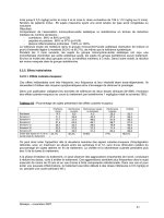
The middle ear consists of the
tympanic
cavity
and
structures in
that cavity. It is connected to the
nasopharynx by the
auditory
tube.
This tube allows for equalization
of
pressure from the middle ear and the
external environment. The three ear
ossicles transfer
sound
from the
tympanic membrane to the oval
window
of the inner ear. Label the
three ear ossicles, the malleus, incus,
and
stapes,
and
color each one a
different color. Color the oval
window where the stapes connects
and use lighter colors for the
auditory tube
and
the tympanic
cavity.
11\1
I'J
ER
EAR
The inner ear consists of the
cochlea, the vestibule,
and
the
semicircularducts. In Latin, the
name
cochlea
means snail shell and it
spirals like a snail. Its function is to
translate the mechanical vibrations
of
sound
into nerve impulses. The
cochlea has an oval
window
that
attaches to the stapes and a
round
window
that allows for changes in
pressure to occur in the inner ear.
Label the cochlea
and
color it in. The
vestibule has two parts, the
utricle
and the saccule. These are involved
in equilibrium. They determine
static equilibrium whereby a person
can determine the position of the
body at rest. They also register
acceleration. Color each of these
parts of the vestibule a different
color. The semicircular ducts
respond to angular acceleration.
There are three semicircular ducts,
the
posterior,
the
anterior,
and the
lateral
semicircular
ducts. Color
each of the semicircular ducts a
different color.
Answer Key: a.
Malleus,
b.
Incus,
e.
Stapes,
d. Ovalwindow, e.
Tympanic
membrane,f.
Tympanic
cavity,
g.
Auditory
(Eustachian)
tube,
h. Semicircular ducts,i. Anteriorduct,
J.
Posterior
duct, k.
Lateral
duct,
I.
Vestibule,
m.
Utricle,
n.
Saccule,
o.
Round
window, p.
Cochlea
Chapter
Six
I
IAPLAll
d
·· I
169
Sense Organs me lea
b
a. _
g._
e. _
f.
h.
1.
J.
k.
1.
d. _
c
0
p
Chapter Six
Sense Organs
I
mellical
171
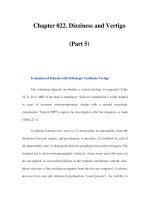
LABYRINTHS
OF THE
II\II\IER
EAR
The outer part
of
the inner ear
consists
of
the
bony
labyrinth,
an
outer encasement
of
bone. Inside
of
this is a fluid called
perilymph.
Inside of this is the
membranous
labyrinth.
It is filled with a fluid
called
endolymph.
Label these
structures and fluids.
a. _
b. _
f.
~~~""-'""
g._
h
_
1. _
J.
c. _
Cross Section of a
Semicircular Canal
Look at the cross section of a
semicircularduct. The outer
part
of
the canal is the
bony
labyrinth.
Perilymph
is the fluid between the
bonylabyrinth
and
the
membranous
labyrinth.
Inside the
membranous
labyrinth is a fluid
called
endolymph.
Label these
structures and fluids.
Answer Key: a. Membranous
labyrinth,
b.
Semicircular
ducts,
c.
Utricle,
d.
Saccule,
e.
Cochlear
duct,
f.
Perilymph,
g.
Endolymph,
h. Bony
labyrinth,
i.
Semicircular
canals,
J.
Vestibule,
k.
Cochlea,
I.
Vestibulocochlear
nerve
k. _
b
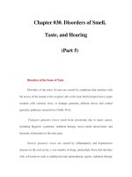
CROSS
SECTION
OF
COCHLEA
Look at the cross section of cochlea.
Each coil of the cochlea has three
chambers and three membranes.
The upper chamber in the
illustration is the scalavestibuli. It is
connected to the oval window. The
vestibular
membrane
is the tissue
that forms the
bottom
of the scala
vestibuli. Belowthis is the scala
media
that houses the spiral
organ
(or the
organ
of
Corti). The
bottom
chamber isthe scala
tympani.
Between the scala tympani and the
scala media is the basilar
membrane.
Label these features and
color each space (scala) a different
color.
Spiral
Organ
The scala media is the region of the
cochlea involved in hearing. It is
bounded
by the vestibular
membrane
on top and the
basilar
membrane
on the bottom. Attached
to the basilar membrane are the
hair
cells. These cellsare attached to the
tectorial
membrane
which vibrates
when sound impulses enter the
cochlea. The tectorial
membrane
tugs on the hair cellswhich converts
the sound impulse to a neural
impulse which travels by the
cochlear nerve to the brain where
hearing is interpreted. Label these
structures and color
them
in, each
with a different color.
b. _
e. _
Chapter
Six
I
KAPLA~.
I 173
Sense Organs meulCa
Vestibulocochlear nerve
Answer Key:a.
Scala
vestibuli,
b.
Vestibular
membrane, c.
Scala
media,
d.
Scala
tympani, e.
Basilar
membrane,
f.
Haircell,g.
Tectorial
membrane
Chapter
Seven:
Endocrine
System
175
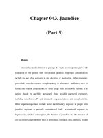
OVERVIEW
OFTHE
ENDOCRINE
SYSTEM
The endocrine system is a collection of glands and organs that secrete
hormones. This system is grouped according to the function that the
individual organs have. Some of these organs have two roles and are
called mixed organs. They secrete hormones and also perform
other
functions such as digestion or secretion. The pancreas is a good example
of this. It secretes hormones (an endocrine function) that regulate blood
sugar levelsand also secretes enzymes (exocrine secretions) that break
down material in the digestive tract. Hormones are released from
endocrine glands and typically travel through the body in blood vessels
and reach target areas that have cells receptive to the hormones. Locate
and label the
pineal
gland,
pituitary
gland,
thyroid
gland, pancreas,
adrenal
glands, testes, and ovaries. Color the organs in with different
colors for each organ.
d.
~
J
.~e._-/
\i
v
f. _
g._
Answer Key:a.
Pineal
gland,b.
Pituitary
gland,c.Thyroid
gland,
d.
Adrenal
glands,
e.
Pancreas,
f.
Ovary,
g.
Testis
\
I
y.,
U
/
Chapter Seven I
mKA
PeLA
N(I'
-Ical
171
Endocrine System
e. _
.~ ~.
c.
~-\
<_
~~
:\
g.
X::;
h. _
d. _
ORGANS
OF THE
HEAD
The
pineal
gland
is a small gland
located posterior to the
corpus
callosum in the brain. It has the
shape of a pine
nut
but
is a little bit
smaller.
It secretes the
hormone
melatonin; melatonin levels increase
during the night and decrease
during the day.
The
pituitary
gland, or hypophysis,
is suspended from the brain by a
stalk called the
infundibulum.
The
pituitary sits in the hypophyseal
fossa which is a depression in the
sphenoid
bone. The pituitary is a
complicated gland that has
numerous functions. The
adenohypophysis
or
anterior
pituitary
originates from the oral
cavity during development and
consists of epithelium. It produces
several hormones which will be
discussed later. The anterior
pituitary has cells that pick up
histological stain differently. These
are acidophiliccells and basophilic
cells. The
neurohypophysis
or
posterior
pituitary
is derived from
the brain during development and
does not make its own
hormones
but stores
hormones
produced in the
hypothalamus. Label the pineal
gland, the corpus callosum, and the
pituitary gland and color them in.
Label the parts of the pituitary and
use different colors for each part.
f.
Answer Key:a
Pituitary
gland
(hypophysis),
b.
Pineal
gland,
c.
Corpus
callosum,
d.
Hypophyseal
fossa,
e.
Adenohypophysis
(anteriorpituitary),
f.
Sphenoid
bone,g. Infundibulum,
h.
Neurohypophysis
(posterior
pituitary),
i.
Basophilic
cell
j.
Acidophilic
cell
e. _
Chapter Seven
Endocrine System
I
KAPLA~.
I
meulCa
179
HORMONES
SECRETED
BY
THE
PITUITARY
AND
THEIR
TARGET
ORGANS
The
adenohypophysis
produces
and
secretes many
hormones
that
have
diverse target areas.
Growth
hormone
(GH) is released by the
pituitary and causes growth and
division of cells
throughout
the
body.
Prolactin
is
more
specific in
its function. Prolactin stimulates the
mammary
glands to become
functional in milk production.
Follicle
stimulating
hormone
(FSH) and
luteinizing
hormone
(LH) are gonadotropins that cause
the ovaries and testes to release
hormones.
Thyroid
stimulating
hormone
(TSH) causes the thyroid
gland to secrete
hormones
and
adrenocorticotropic
hormone
(ACTH) has an influence on the
adrenal cortex.
The posterior pituitary, or
neurohypophysis,
stores
and
secretes a
hormone
called oxytocin.
This
hormone
has many functions.
It causes milk letdown
during
nursing
and
has multiple functions
as a neurotransmitter in the brain. It
issecreted during orgasm in the
female and is also released when the
infant is nursing. Oxytocin also has
an effect on kidney water balance.
The other
hormone
stored in the
neurohypophysis is
antidiuretic
hormone
or ADH. It is also
known
as vasopressin. It causes absorption
of water from the collecting tubules
of the kidney decreasing the volume
of water in urine.
Answer
Key: a. Adenohypophysis,
b. Thyroid stimulating hormone,
c. Prolactin, d. Growth hormone,
e. Adrenocorticotropic hormone,
f.Luteinizing hormone, g. Follicle
stimulating hormone,
h. Neurohypophysis, i. Oxytocin,
J.
Antidiuretic hormone (vasopressin)
h. _
J
e. _
f. _
g._
e. _
THYROID GLAND
The thyroid gland is just inferior to
the thyroid cartilage of the larynx.
It
has two main lobes and a small
connection between them called the
isthmus.
The histology of the
thyroid is very distinctive. There are
cells called follicular cells forming a
sphere and these make up the
follicle. Inside the follicle is the
colloid where thyroid
hormones
are
stored. The
parafollicular
cells are
between the follicles. Label the main
parts of the thyroid gland, the
follicular cells, the parafollicular cells
and the colloid and color them in.
a
e. _
b. _
Chapter Seven I
KAPLA!I_
I 181
Endocrine System meulCa
__
.,,
Hyoid bone
IS.! fh+-
Thyroid cartilage
c. _
d. _
Trachea
Answer Key:a.
Thyroid
gland,
b. Right
lobe,
c
Isthmus,
d. Leftlobe, e.
Colloid,
f.
Follicular
cells,
g.
Parafollicular
cells
g._
PARATHYROID
GLANDS
There are typically four glands on
the posterior of the thyroid gland
and these are known as the
parathyroid
glands. Theysecrete a
hormone
called
parathormone
which regulates calcium balance in
the blood. Parathormone increases
blood calcium levels by causing
more absorption of calcium from
the digestive tract, increased
osteoclast activity in the bones, and
reabsorption of calcium from the
kidney. The
principal
or chiefcells
secrete parathyroid hormone. The
oxyphilic cells are less
common
and
their function is poorly understood.
Label the parathyroids on the
posterior thyroid gland and color
them in.
Answer Key:a.
Thyroid
gland,
b.
Parathyroid
glands,
c.
Principal
(chief)
cells,
d.
Oxyphilic
cells
Chapter
Seven
I
KAPLA~.
I 183
Endocrine System meulCa
0 , Hyoid bone
/
.~~
~
Inferior constrictor muscle
Esophagus
PANCREAS
The
pancreas
is inferior to
the
stomach and has several digestive
functions. These exocrine secretions
are initiated by the
acinar
cells. The
endocrine function of the pancreas
consists of the secretion of insulin,
glucagon,
and
somatostatin from the
pancreatic
islets. These islets are
microscopic collections of cells
that
have specialized cells for the
secretion of hormones. Insulin
lowers blood glucose levels while
glucagon does the reverse.
Somatostatin moderates
some
of the
pancreatic cells that have a role in
digestion. Label and color in the
pancreas and make the pancreatic
islets lighter
than
the acinar cells of
the pancreas.
Answer
Key: a. Pancreas, b. Pancreatic
islets,
c. Acinar cells (exocrine)
Chapter Seven I
mKAPeLANd'
-Ical
185
Endocrine System
b. _
c. _
Chapter Seven
Endocrine System
I
UPLANd'· I
me lea
187
ADRENAL
GLANDS
The
adrenal
glands
are positioned
superior to the kidneys and are
divided into the adrenal
cortex
and
the medulla. The cortex has three
layers. The most superficiallayer is
the
zona
glornerulosa, which is deep
to the adrenal capsule
and
responsible for the secretion of
mineralocorticoid hormones. The
next layer is the
zona
fasciculata
which mainly secretes
glucocorticoids,
hormones
responsible for the breakdown of
proteins
and
lipids
and
the synthesis
of glucose. The
zona
reticularis
is
the deepest layer of the cortex and it
secretes androgens (male sex
hormones)
and
small
amounts
of
estrogens (female sex
hormones)
in
both
sexes. The most prevalent male
hormone
is DHEA
(dehydroepiandrosterone) which is
responsible for the development of
the sex drive, pubic hair, and axillary
hair. The effects of DHEA are
minimized in males as the testes
secrete greater
amounts
of
testosterone. The adrenal medulla is
the deepest part of the adrenal gland
and
it secretes epinephrine
and
norepinephrine. Label and color the
adrenal glands
and
use a different
color for each layer of the cortex
and
another for the medulla.
Answer Key: a.
Adrenal
glands,
b.
Cortex,
c. Medulla,d.
Capsule,
e. Zona glomerulosa,
f.
Zona
fasciculata,
g.Zona
reticularis,
h. Medulla
1
~
~
~.
d. _
e. _
f.
rc-~~?
i
h. _
c. _
Chapter Seven
Endocrine System
I
meilical
189
d. _
GONADS
The ovaries are a mixed gland
because they produce the oocytes
(egg cells) and also have an
endocrine function by producing
estrogens. Estrogens are a class of
female sex
hormones
that include
estradiol and progesterone. Estradiol
is produced in the
granulosa
cells of
the
ovarian
follicles. These follicles
surround
the oocytes. Progesterone
is produced by the
corpus
luteum
after the oocyte has been ovulated.
The testes are also mixed glands. As
exocrine glands they produce
sperm
cells
and
as endocrine glands the
interstitial
cells
prod
uce
testosterone. Label
and
color the
interstitial cells
and
seminiferous
tubules
in the microscopic view of
the testes.
Answer
Key: a Ovary, b. Granulosa
cells,
c. Ovarian follicles, d. Ova,
e. Corpus luteum,
f. Interstitial cells,
g. Testis, h. Seminiferous
tubules
b~
_
e. _
f. _
h. _
Chapter
Eight:
Cardiovascular
System
191
OVERVIEW
OF THE
CARDIOVASCULAR
SYSTEM
The cardiovascular system consists
of the heart as a
pump,
blood vessels
that take blood away from the heart
(arteries), and blood vessels that take
blood back to the heart (veins).
Locate the
heart
on the illustration
and color it in purple. Label the
common
carotid
artery
and color it
in red. Arteries are typically colored
in red and veins are colored blue.
Label and color in the
internal
jugular
vein too. The internal
jugular vein takes blood to the
superior
vena cava which takes
blood to the heart. Label and color
the
aortic
arch
red and find the
continuation of the
aorta
that
travels down the left side of the
body, splits and takes blood to the
femoral artery. The vessel parallel to
the femoral artery is the femoral
vein and it should be colored blue.
The femoral vein takes blood to the
inferior
vena cava before it goes to
the heart. Blood travels to the arm by
the
brachial
artery
and
deoxygenated (color it blue) blood
travels to the lungs in the
pulmonary
trunk.
Answer Key: a.Internaljugularvein,
b.
Cornman carotid
artery,
c. Superior
vena
cava,
d.
Brachial
artery,
e. Infenor
vena
cava,
f.
Aortic
arch,
g. Pulmonary
trunk,h.
Heart,
i.
Aorta,
j.
Femoral
artery,
k.
Femoral
vem.
a. _
b. _
c. _
d. _
e
\
p/Cr
H
'
Chapter
Eight
I
KAPLA~.
I 193
Cardiovascular System meulca
CIRCULATION
m.
<===-
oxygen-rich blood
~
oxygen-poor blood
D oxygen-poor blood
Doxygen-rich blood
f. circulation:
internal organs
and
legs
f.
circulation:
head and arms
1.
_
J.
d. _
k. _
a. _
1. _
There are two major circulations in
the body.
One
goes to the lungs
and
this is called the
pulmonary
circulation. Deoxygenated blood
leaves the right ventricle of the heart
and
travels through the
pulmonary
artery
(blue) to the lungs where the
blood is oxygenated. Blood
returns
from the lungs to the left
atrium
of
the heart by the
pulmonary
veins
(red). The
other
main circulation in
the body is called the systemic
circulation
where
blood
travels from
the left ventricle of the heart
and
goes to the
other
regions of the body.
Arteries are vascular tubes that take
blood away from the heart while
veins are vessels
that
return
blood to
the heart. Most arteries carry
oxygenated
blood
and most veins
carry deoxygenated blood
but
there
are a few exceptions.
The first vessel
that
leaves the heart
is the
aorta
which is
part
of the
arterial system. Color it red.
Arteries
receive blood from the
aorta
and
take blood
throughout
the body.
They branch and become smaller
until they become arterioles. The
arterioles are the structures that
control blood pressure in the body.
Asthey get smaller they become
capillaries. The capillaries are the
site of exchange with the cells of the
body. Label
and
then
color the
capillaries purple. Purple is a good
choice because the capillaries are the
interchange between the arteries
(red) and the veins (blue). On the
return flow the capillaries enlarge
and
turn
into venules, which take
blood to the veins. Color the venules
and remainingveins of the body
blue. Blood from the inferior
portion
of the heart returns to the
heart by the
inferior
vena
cava.
The heart has four chambers
including the superior atria
and
the
inferior ventricles. There is a typical
coloring
pattern
for the
cardiovascular system. Vesselsor
chambers that
carry
deoxygenated
blood are colored in blue while
vessels that
carry
oxygenated blood
are colored red. Label and color the
right
atrium
(blue),
right
ventricle
(blue), left
atrium
(red)
and
left
ventricle (red). Remember the heart
is in anatomical position so the right
atrium
is on the left in the
illustration.
Answer Key: a.
Right
atrium, b.
Aorta,
c. Leftatrium, d. Right
ventricle,
e. Leftventricle, f.
Systemic,
g.
Pulmonary,
h. Pulmonary
artery,
i. Pulmonaryvein,j. Infenorvena
cava,
k.
Vein,
I.
Venule,
m.
Capillary,
n.Arteriole, o. Artery
Chapter Eight
Cardiovascular System
I
meclical
195
Agranular
g,
h.
Answer
Key: a. Erythrocyte, b. thrombocyte,
c. leukocytes, d. basophil, e. eosinophil,
f. neutrophil, g. lymphocyte, h. monocyte
b.
@
f
,
".:
::
-,
',.
c.
Granular
a.
f.
e.
d.
There
are
about
5 million erythrocytes per
cubic millimeter
of
blood.
The
erythrocytes
do
not
have a nucleus
and
they
appear
like
a
donut
with
a
thin
spot
instead of the
donut
hole. About a third
of
the weight
of
a
red blood cell is
due
to
hemoglobin
which
makes the cells red. Color in the surface
view
and
cross section
of
the red
blood
cell.
Note also the size
of
the
thrombocyte.
There are
about
7
thousand
leukocytes
per
cubic millimeter
of
blood.
There
are two
main
types of leukocytes;
granular
leukocytes
and
agranular
leukocytes.
The
granular leukocytes have cytoplasmic
granules
that
either stain
pink,
dark
purple
or do not stain
much
at all.
The
granular
leukocytes that do
not
stain
much
at all are
called
neutrophils
because the granules are
neutral to the stains. They are the
most
numerous
of the leukocytes
making
up 60-
70% of the leukocytes. Neutrophils have a
three to five lobed nucleus.
Color
in the
cells by shading the cytoplasm light bl
ue
and
coloring in the nucleus purple.
The
eosinophils
are
granular
leukocytes
that have
pink
or orange staining granules.
The
nucleus isgenerally two-lobed. Color
in the eosinophil by first coloring in the
purple
nucleus and
then
adding
orange
to
the cytoplasm. Eosinophils
make
up
about
3 percent
of
the white blood cells.
Basophils
are a rare
granular
leukocyte in
that they
make
up less
than
one
percent
of
the white
blood
cells.
The
nucleus isS-
shaped
but
it is frequently difficult to see
because it is
obscured
by the
dark
staining
cytoplasmic granules. Label the basophil
and color in the granules a
dark
purple.
The
two kinds of agranular leukocytes are
the
lymphocytes
and
the
monocytes.
The
lymphocytes can be large or small
and
they
make
up 20-30% of the leukocytes.
The
cytoplasm is light blue
and
the nucleus is
purple.
The
nucleus
of
the
lymphocyte
is
dented or flattened. Lymphocytes
come
in
two kinds. B cells secrete
antibodies
(antibody-mediated
immunity)
and
T cells
which are involved in cell-mediated
immunity.
Label
and
color the
lymphocytes.
The
monocytes
are large cells
(about
3
times the size of a red
blood
cell)
and
they
have a strongly lobed nucleus.
Some
people
say this looks like a kidney bean or a
horseshoe.
They
represent only
about
5%
of the leukocvtes.
Color
in the nucleus with
a
purple
and 'the cytoplasm a light blue.
Blood consists of
plasma
and
formed
elements.
The
plasma is the tluid
portion
of the
blood
and
consists of water, proteins,
and
dissolved materials such as oxygen,
carbon
dioxide, electrolytes (ionic
particles)
and
other
materials. Plasma
makes up
about
55% of the
blood
volume.
Formed
elements
make
up
about
45%
of
the
blood
volume
and
consist of
erythrocytes
(red
blood
cells),
leukocytes
(white
blood
cells)
and
thrombocytes
(platelets). Label
and
color in the red
blood
cells
with
a light red color. Label the
white
blood cells
and
color in
the
nucleus
with
purple
and
the cytoplasm a light blue. Label
and color the
thrombocvtes
purple.
There
are
about
200,00-450,00 thrornbocytes per
cubic millimeter of blood.
They
assist
the
body in clotting to prevent blood from
tlowing
out
of
small
ruptures
in
blood
vessels.
BLOOD
ANTERIOR
SURFACE
VIEW OF
HEART
The
apex
of
the
heart
is
inferior
and
the
base
is
superior.
Label each
chamber
of
the
heart
and
color
them
each a
different
color. Locate
the
coronary
arteries
and
their
branches
and
color
them
in red.
The
right
coronary
artery
leads to
the
right
marginal
artery.
The
left
coronary
q.
p.
o.
n. _
m.
1.
Chapter
Eight
I
UPLANd'·
I 197
Cardiovascular System me lea
artery
takes
blood
to
the
anterior
interventricular
branch
and
the
circumflex
branch.
The
cardiac
veins
can
also be seen on
the
anterior
side.
The
great
cardiac
vein
runs
in
the
interventricular
sulcus on
the
anterior
side. Label all
of
the
major
vessels
entering
and
exiting
the
heart.
c. _
1.
Answer Key: a. Aorticarch, b. Pulmonary trunk, c. Base of heart, d. Leftatrium, e. Circumflexbranch, f.Anterior interventricular branch, g. Leftcoronary artery,
h. Great cardiac vein,
I. Left ventricle, j.Apex of heart, k. Descending aorta,
I.
Inferiorvena cava, rn. Rightventricle, n. Rightmarginal artery, o. Rightatrium,
p. Rightcoronary artery, q. Superior vena cava
POSTERIOR
SURFACE
OF
HEART
On the posterior side of the heart are additional arteries and veins. The
posterior
interventricular
artery
occurs between the ventricles on the
posterior surface.
It
receivesblood from the
right
coronary
artery. The
middle
cardiacvein runs the opposite direction and takes blood into the
coronary
sinus. The small cardiac vein is also found on the posterior
a.
_
J.
1.
1.
h _
g.
c.
Chapter
Eight
I UPLANd'· I 199
Cardiovascular System me lea
surface of the heart and enters the coronary sinus from the opposite
direction. Label the posterior features of the heart and color the arteries
in red (except for the
pulmonary
arteries
that carry deoxygenated
blood-they
should be colored in blue). Color the veins in blue (except
for the
pulmonary
veins which should be colored in red).
:e
e.
Tb.~
Answer
Key: a. Aortic arch, b. Inferior vena cava, c. Right ventricle, d. Right atrium, e. Right coronary artery,
f.
Superior vena cava,
g. Posterior interventricular artery, h. Middle cardiac vein, i. Coronary sinus,
J.
Pulmonary veins, k. Pulmonary arteries,
I.
Small cardiac vein
Chapter
Eight
I KAPLANd'. I 201
Cardiovascular System me lea
CORONAL
SECTION
OF
HEART
The
heart
is located in a
tough,
fibrous sac
known
as the
parietal
pericardium
which has an
outer
fibrous
layer
and
an
inner
serous
layer.
If this sac is
opened
you
can see a space called the pericardial cavity.
The
heart
is in this cavity.
The
outer
surface
of
the
heart
is called the visceral
pericardium
or the
epicardium.
Inside
of
this is the
main
portion
of the
heart wall called the
myocardium
(made
of
cardiac muscle)
and
the
innermost
layer
of
the
heart
is the
endocardium.
Deoxygenated
blood
enters the
right
atrium
of
the
heart
by three
vessels: the
superior
vena
cava, the
inferior
vena
cava
and
the
coronary
sinus.
The walls
of
the right
atrium
are thin-walled as they
only
have to
pump
blood
to the
right
ventricle. The
blood
in the right
atrium
is in
contact
with
the fossa ovalis which is a
thin
spot
in the interatrial
septum.
This
thin
spot
is a
remnant
of
a hole in the fetal
heart
know
as
the
foramen
ovale. Blood in the right
atrium
flows
through
the cusps of
the
tricuspid
or
right
atrioventricular
valve into the
right
ventricle.
The
tricuspid valve is
made
of
the three cusps, the
chordae
tendineae
and
the
papillary
muscles
that
hold
the
chordae
tendineac to the
ventricle wall. The ventricle wall is lined with
trabeculae
carneae
that
act as struts
along
the edge
of
the wall. The wall between the ventricles is
known
as the
interventricular
septum.
From the right ventricle, blood passes
through
the
pulmonary
semilunar
valve and into the
pulmonary
trunk
where the
blood
goes to
the lungs. In the lungs the
blood
is oxygenated. From the lungs the
blood
returns
to the left
atrium
of
the heart. Blood in the left
atrium
moves to
the
left
ventricle
through
the left
atrioventricular
valve or the
biscuspid
valve. This valve has two cusps,
chordae
tendineae
and
papillary muscles.
When
the left ventricle contracts, the
blood
moves
through
the
aortic
semilunar
valve
and
into
the
ascending
aorta.
1.
m.
y._
1.
J.
k
e.
From
lungs
To lungs
c.
White arrows = oxygen-rich blood
/ 1 Black arrows = oygen-poor blood
j D = veins D = arteries
~
w.
v.
u.
t.
s.
r.
q.
p.
x.
From
lungs
To lungs
n.
o. _
Answer
Key:
a. Pulmonarytrunk,b. Pulmonarysemilunar
valve,
c.
Left
atrium,e.
Left
atrioventricular
valve,
f.
Aortric
semilunar
valve,
g.
Left
ventricle,
h. Endocardium,
i.
Epicardium,
j.
Myocardium,
k.
Parietal
pericardium,
I.
Fibrous
layer,
m. Serous
layer,
n. Interventricular septum, o. Trabeculaecarneae, p.
Inferior
vena
cava,
q.
Papillary
muscle,
r.
Right
ventricle,
s. Chordae tendineae, t.
Right
atrioventricular
valve,
u. Opening of coronarysinus,v.Fossa
ovalis,
w.
Right
atrium,x.Superiorvena
cava,
y.
Aorta
SUPERIOR
ASPECT
OF THE
HEART
This view of the heart is seen as if the atria
and
the major vessels have
been removed. You should be able to see all of the major valves of the
heart. The most anterior valve isthe
pulmonary
semilunar
valve that
occurs between the right ventricle and the
pulmonary
trunk. Label
and
color this valve blue. Posterior to this is the
aortic
semilunar
valve. It
occurs between the left ventricle and the aorta. Label this valve and color
it in red. Both of these valves prevent blood from returning to the
a. _
c. _
ECG-CONDUCTION
PATHWAY
The heart has specialized cells that initiate an electrical impulse
that
radiates
throughout
the heart. The cells are clustered in a particular area
known
as the
sinoatrial
node
or the pacemaker. These cells produce a
depolarization
that
travels across the atria which depolarize
and
then
contract. Depolarization is an electrical event while
contraction
is a
mechanical event. Between the wall of the right
atrium
and
the right
Chapter
Eight
I
IAPLAN
d
··
I
203
Cardiovascular System me lea
ventricles once they have finished contracting. On the right side of the
illustration (and on the right side of the heart) is the
right
atrioventricular
(or
tricuspid)
valve, so
named
because it has three flaps
or cusps. This valve occurs hetween the right atrium and the right
ventricle.
It prevents the blood from returning to the right atrium during
ventricular contraction. Label this valve and color it blue. On the left side
of the heart is the left
atrioventricular
(bicuspid) valve. It prevents blood
from moving back to the left atrium when the left ventricle contracts.
d. _
ventricle is a
lump
of tissue
known
as the
atrioventricular
(AV)node.
Once the impulse reaches this area the AVnode pauses a
moment
before
sending the impulse to the
atrioventricular
bundle.
This
bundle
divides
into the
bundle
branches
and
then
the impulse travels to the
conduction
(Purkinje)
fibers. These fibers reach the muscle of the
ventricles and stimulate
them
to contract. Color each of the components
of the conduction pathway a different color.
h. _
e.
g.
1
_
Answer
Key: a. Pulmonary semilunar valve, b. Aortic semilunar valve, c. Left atrioventricular valve, d. Right atrioventricular valve, e. Sinoatrial node, f. Atrioventricular
bundle, g. Atrioventricular node, h. Bundle branches,
I. Purkinje fibers
Chapter
Eight
I
IAPLAN
d
··
I
205
Cardiovascular System me lea
Closed
n. _
Open
m.
a ~c.
\C~:·
\
f. _
Veins are thinner walled
than
arteries and they do
not
have the
same elastic fibers in the tunica
media as arteries. Color the tunica
media of the veins red
and
select the
same colors as you did for the
arteries for the tunica externa
and
the tunica interna. The tunica
interna of veins is folded into valves
that allow for a one-way flow
of
blood
through
veins.
Capillaries are different from both
arteries and veins in
that
they are
composed of only simple
squamous
epithelium (called
endothelium).
The thin nature
of
capillaries allows
them to exchange nutrients, water,
carbon
dioxide and oxygen with the
cells. Color in the endothelium
of
the capillary with the same color
that you selected for the tunica
interna.
Answer Key:a.
Vein,
b.
Artery,
c.
Lumen,d.
Tunica
intima, e.
Tunica
media,f.
Tunica
externa,
g.
Lamina
elastica
mterna,
h. Lamina
elastica
externa,
i. Smooth muscle,
J.
Venule,
k.
Endothelium,
I.
Arteriole,
m.
Capillary,
n.
Venous
valve
The blood vessels have different
thickness due to the differences in
pressure that occur in
them
or their
function with respect to exchanging
nutrients with the cells.
Arteries
have thick walls due to the higher
pressure found in them. Just as high
pressure hoses have thick walls so do
arteries. The
outer
layer of the artery
is the
tunica
extcrna
(tunica
adventitia). Youshould locate the
tunica externa and color it in.
The
middle layer of the artery, the
tunica
media
is the thickest layer and it is
made of
smooth
muscle
and elastic
fibers. Color the tunica media red.
The
innermost
layer
of
the artery is
the
tunica
intima
(tunica
interna)
and
it has a special elastic layer called
the
lamina
elastic
interna.
Color
this layer.The area in the artery
where the blood flows is called the
lumen.
VESSELS
OVERVI
EW
ARTERY
OVERVI
EW
One of the ways to study arteries is to
draw
them
as if you were making a
street map. Begin with the heart and
draw the blood vesselsthat occur as
you take blood to the fingers, toes or
to a particular organ of the body.
Arteries are typically colored red and
you should select that color for this
illustration. Use the following artery
list and label the appropriate arteries
and color them in red. The
abbreviation for artery is
a.
Ascending
aorta
Aortic arch
Thoracic
aorta
Abdominal
aorta
Brachiocephalic
trunk
Common
carotid
artery
Subclavian
artery
Axillary
artery
Brachial
artery
Radial
artery
Ulnar
artery
Common
iliac
artery
Femoral
artery
Anterior
tibial
artery
Fibular
artery
Answer Key: a.Common carotid a.,
b. Brachiocephalic trunk,
c.Ascending
aorta,d.
Brachial
a.,e. Ulnar a, f.
Radial
a.,
g.
Subclavian
a.,h. Aorticarch,
i.
Axillary
a, J
Thoracic
aorta, k.
Abdominal aorta,
I.
Common iliac
a.,
m.
Femoral
a.,
n. Anteriortibial
a.,
o.
Fibular
a.
b.
e. _
f.
__
Chapter
Eight
I KAPLAN
d·.
I
207
CardiovascularSystem me lea
1. _
f-+
o. _
HEAD
AND
AORTIC
ARTERIES
Blood from the heart exits the
brachiocephalic
artery
and takes
two main pathways to the right side
of the head. One of these isthe
right
common
carotid
artery
which exits
the brachiocephalic artery and then
splits into the external
carotid
artery
and the
internal
carotid
artery. The external carotid artery
has severalbranches, among them
the facial artery, the superficial
temporal artery, the maxillary
artery, and the occipital artery. The
internal carotid artery takes blood
through the carotid canal of the skull
and into the brain. The other main
pathway of blood to the right side of
the head isthe vertebral
artery
which arises from the subclavian
artery. The left side of the head has a
similar pathway except that the left
common
carotid
artery
and the left
subclavian
artery
arise from the
aortic arch and not from the
brachiocephalic artery. Label these
vesselsand color them in red.
Answer Key: a.Superficial temporal
a.,
b. Occipital
a.,
c.Internal carotid
a.,
d.
Vertebral
a.,
e.
Subclavian
a.,
f. Brachiocephalic a.,g.
Thoracic
aorta,
h.
Facial
a.,
i. Maxillary
a.,
j.
External
carotid
a.,
k.Common carotid
a.,
I.Aortic arch,m. Ascendingaorta
a
c
__
d. _
e. _
f.
g
k
d
e. _
f
1.
_
m. _
Chapter
Eight
I UPLANd'· I
209
Cardiovascular System me lea
/