BASIC HUMAN ANATOMY - PART 4 pdf
Bạn đang xem bản rút gọn của tài liệu. Xem và tải ngay bản đầy đủ của tài liệu tại đây (240.89 KB, 25 trang )
MD0006 4-22
Figure 4-8. The human scapula and clavicle (pectoral girdle).
MD0006 4-23
Figure 4-9. The humerus, radius, and ulna.
MD0006 4-24
Figure 4-10. The human hand.
Figure 4-11. The bony pelvis (two pelvic bones and sacrum).
MD0006 4-25
Figure 4-12. The femur, tibia, and fibula (anterior views).
MD0006 4-26
Figure 4-13. The human foot.
MD0006 4-27
EXERCISES, LESSON 4
REQUIREMENT. The following exercises are to be answered by completing the
incomplete statement or by writing the answer in the space provided at the end of the
question.
After you have completed all the exercises, turn to "Solutions to Exercises," at the
end of the lesson and check your answers.
1. What is a skeleton?
2. What are four functions of the human skeleton?
a. .
b. .
c. .
d. .
3. An individual bone consists of the outer and the inner .
4. The two types of bone marrow are and bone marrow.
Yellow bone marrow is mostly yellow tissue. Red bone marrow is the only site
in adults for the formation of .
5. The parts and portions of an individual long bone are the s (d s)
and the (e s). The shaft is the portion of the long
bone. The ends are made up mainly of c (s ) bone tissue. An
articular cartilage covers each area where a bone s another bone.
6. The periosteum is a covering of bone surface area not covered by
. The innermost layer is the o (b -f ) layer. The
outermost layer is an layer.
MD0006 4-28
7. In the early fetus, bones are preformed as s bones and
e bones which have the shape and location of the t bones.
Developing long bones have growing masses of actual bone called centers.
These centers are located in the and in each . Preparing material
surrounding these centers is destroyed and replaced with tissue. A
bone grows in width through the activity of the layer of the .
8. What are four types of bones according to shape?
a. .
b. .
c. .
d. .
9. What is a syndesmosis?
10. What is a suture?
11. What is a synosteosis?
12. What is a synchondrosis?
13. What is a symphysis?
14. What is a synovial joint?
MD0006 4-29
15. What are the major parts of a "typical" synovial joint?
a. B .
b. Articular c .
c. (1) Synovial m .
(2) Synovial s .
(3) Synovial f .
d. C .
e. L .
f. M .
16. Name and describe three classifications of synovial joints. Along with each,
name common subclassifications.
a. U .
(1) Hi .
(2) Pi .
b. B .
Sa .
c. M .
(1) Ba .
(2) Pl .
MD0006 4-30
17. Name and define the two major subdivisions of the skeleton.
a. skeleton the of the human
body including the , column, and .
b. skeleton skeletal elements of the upper and
.
18. Name and describe the two major parts of a typical vertebra.
a. Vertebral -shape cylinder. Its function is to
.
b. arch arch over posterior of the . The
arch has several . The are sites of attachment of
and act as for trunk motions.
19. Name the regions of the vertebral column and give the number of vertebrae
in each region.
a. (neck) region, .
b. (chest) region, .
c. (low back) region, .
d. , fusion of .
e. ("tail"), - together.
20. Describe the two ways that vertebrae are held together.
a. Intervertebral discs:
b. Ligaments:
MD0006 4-31
21. The thoracic cage consists of the s (m ,
b , and x p ), 12 pairs of , and
12 t v . The thoracic cage provides p for
v o within the t . It also allows the m of
breathing.
22. What are the two major subdivisions of the skull and with which organs or
systems is each subdivision involved?
a. : Encases and protects .
b. : Involved with beginning of ;
encases and protects the .
23. In the first column below, name a general segment or part of a member. In
the second column, name bones or bone groups which are found in each segment of
the upper member. In the third column, name bones or bone groups which are found in
each segment of the lower member.
PART UPPER MEMBER LOWER MEMBER
a. G GIRDLE GIRDLE
b. P SEGMENT S R
c. M SEGMENT S, A,
A A
d. D SEGMENT C , T ,
M , M ,
P P _____________
Check Your Answers on Next Page
MD0006 4-32
SOLUTIONS TO EXERCISES, LESSON 4
1. The skeleton is a combination of bones joined together that serves as a support or
framework of the human body. (para 4-1)
2. The four functions of the human skeleton are:
a. Bodily support.
b. Protection.
c. Motion.
d. Formation of blood cells. (para 4-2)
3. An individual bone consists of the outer cortex and the inner medulla. (para 4-4a)
4. The two types of bone marrow are red bone marrow and yellow bone marrow.
Yellow bone marrow is mostly yellow fat tissue. Red bone marrow is the only site in
adults for the formation of red blood cells. (para 4-4b)
5. The parts or portions of an individual long bone are the shaft (diaphysis) and the
ends (epiphyses). The shaft is the central portion of the long bone. The ends are made
up mainly of cancellous (spongy) bone tissue. An articular or cartilage covers each
area where a bone contacts another bone. (para 4-4c)
6. The periosteum is a covering of bone surface area not covered by articular
cartilage. The innermost layer is the osteogenic (bone-forming) layer. The outermost
layer is an FCT layer. (para 4-4d)
7. In the early fetus, bones are "preformed" as membranous bones and cartilage
bones which have the shape and location of the adult bones. Developing long bones
have growing masses of actual bone called ossification centers. These centers are
located in the shaft and in each end. Preforming material surrounding these centers is
destroyed and replaced with bony tissue. A bone grows in width through the activity of
the osteogenic layer of the periosteum. (paras 4-5a, c, d)
8. Four types of bones according to shape are:
a. Long bones.
b. Short bones.
c. Flat bones.
d. Irregular bones. (para 4-6)
9. A syndesmosis is a joint in which the bones are held together by FCT (fibrous
connective tissue). (para 4-8a(1))
10. A suture is a joint in which the bones are very close together with a minimum of
FCT. (para 4-8a(2))
MD0006 4-33
11.A synosteosis is a joint in which the bones are united by bony material. (para 4-8b)
12. A synchondrosis is a joint in which the bones are held together by hyaline
cartilage. (para 4-8c(1))
13. A symphysis is a joint in which the bones are held together by a disc of
fibrocartilage. (para 4-8c(2))
14. A synovial joint is a joint in which the bones are able to move freely upon one
another. (para 4-8d)
15. The major parts of a synovial joint are:
a. Bones.
b. Articular cartilages.
c. (1) Synovial membrane.
(2) Synovial space.
(3) Synovial fluid.
d. Capsule.
e. Ligaments.
f. Muscles. (para 4-9)
16. Synovial joints may be classified as follows:
a. Uni-axial motion in one plane.
(1) HInge joint.
(2) Pivot joint.
b. Bi-axial motion in two planes.
Saddle joint.
c. Multi-axial motion in all three planes.
(1) Ball-and-socket joint.
(2) Plane joint. (para 4-10)
17. The major subdivisions of the skeleton are the:
a. Axial skeleton the central framework of the human body including the skull,
vertebral column, and thoracic cage.
b. Appendicular skeleton skeletal elements of the upper and lower members.
(paras 4-12, 4-13, 4-14)
18. The two major parts of a typical vertebra are the:
a. Vertebral body drum-shape cylinder. Its function is to bear weight.
b. Neural arch arch over posterior of the spinal cord. The neural arch has
several processes. The processes are sites for attachment of trunk muscles and act as
levers for trunk motions. (para 4-13a(1))
MD0006 4-34
19. The regions of the vertebral column and the number of vertebrae in each are as
follows:
a. Cervical (neck) region, 7.
b. Thoracic (chest) region, 12.
c. Lumbar (low back) region, 5.
d. Sacrum, fusion of 5.
e. Coccyx ("tail"), 3-4 together. (para 4-13a(2))
20. a. Intervertebral discs hold the bodies of adjacent vertebrae together, are fibrous
rings with soft centers, allow adjacent vertebral bodies to move on one another, and are
part of plane-type joints between vertebrae.
b. Ligaments are dense FCT structures extending from bone to bone (along the
vertebral column from the base of the skull to the coccyx). (para 4-13a(3))
21. The thoracic (rib) cage consists of the sternum (manubrium, body, and xiphoid
process), 12 pairs of ribs, and 12 thoracic vertebrae. The thoracic cage provides
protection for vital organs within the thorax. It also allows the movements of breathing.
(para 4-13b)
22. The two major subdivisions of the skull are as follows:
a. Cranium: Encases and protects brain.
b. Facial skeleton: Involved with beginning of digestive and respiratory tracts;
encases and protects the special sense organs (eyes, ears, etc.). (para 4-13c)
23. PART UPPER MEMBER LOWER MEMBER
a. GIRDLE PECTORAL GIRDLE PELVIC GIRDLE
b. PROXIMAL SEGMENT HUMERUS FEMUR
c. MIDDLE SEGMENT RADIUS, TIBIA,
ULNA FIBULA
d. DISTAL SEGMENT CARPUS, TARSUS,
METACARPALS, METATARSALS,
PHALANGES PHALANGES
(table 4-2)
End of Lesson 4
MD0006 5-1
LESSON ASSIGNMENT
LESSON 5 The Human Muscular System.
TEXT ASSIGNMENT Paragraphs 5-1 through 5-8.
LESSON OBJECTIVES After completing this lesson, you should be able to:
5-1. Describe the general features of the skeletal
muscles.
5-2. Describe the general arrangement of the trunk
and limb musculature.
5-3. Given a sample drawing, identify the class of
lever.
5-4. Name the components of a skeleto-muscular
unit. Given a description of a muscle's role in a
motion, name that role.
SUGGESTION After completing the assignment, complete the
exercises at the end of this lesson. These exercises
will help you to achieve the lesson objectives.
MD0006 5-2
LESSON 5
THE HUMAN MUSCULAR SYSTEM
Section I. THE SKELETAL MUSCLE
5-1. MUSCLE TISSUES
The cellular elements of muscle tissues are specialized to produce motion by
contraction. They also produce body heat. (See paragraphs 2-14 and 2-15 of lesson 2
for a discussion of muscle tissues.)
a. Smooth muscle tissue is utilized to make up the muscular portion of the
various visceral organs (stomach, blood vessels, etc.).
b. Cardiac muscle tissue makes up the muscular wall of the heart the
myocardium.
c. Striated muscle tissue is used in the makeup of several types of muscles.
The main type of muscle is the skeletal muscle. Other types of muscles made with
striated muscle tissue are the facial or integumentary muscles and muscles of the jaw
apparatus.
5-2. THE SKELETAL MUSCLE
Each skeletal muscle is an individual organ of the human body. Each is made up
of several types of tissues mainly, striated muscle fibers and FCT (fibrous connective
tissue). Each is attached to and moves bones. Bones are parts of the skeleton serving
as levers.
a. General Construction of a Skeletal Muscle. The large portion of a muscle
is known as its belly or fleshy belly. This muscle is attached to bones by tendons or
aponeuroses. Tendons and aponeuroses are similar to each other. However, tendons
are cord-like and aponeuroses are broad and flat. The fleshy portion may be directly
connected to the bone. If so, it is called a "fleshy attachment."
b. Muscular NAVL (Nerves, Arteries, Veins, Lymphatics).
(1) From the main NAVL (nerve, artery, vein, lymphatic), there are branches
going to each muscle. These muscular branches are bound together by an FCT sheath
to form a neurovascular bundle.
(2) The motor point is that specific location on the surface of the muscle
where the neurovascular bundle enters.
MD0006 5-3
(3) A motor unit is the single motor neuron and the number of striated muscle
fibers activated by it (innervation). The importance of the motor unit is that its fibers
work in unison. Either all fibers within a unit contract or none contract. When a certain
amount of force is needed, one unit after another is recruited until just enough units are
available to produce the desired action.
5-3. NAMING SKELETAL MUSCLES
The name of a muscle may appear with the abbreviation M., meaning Musculus
or muscle. We abbreviate muscles (plural) with the symbol Mm. Skeletal muscles are
named according to their physical attributes (shape, size, length, etc.), their location, or
their function. For example:
SHAPE: deltoid M.
DELTA = ∆ , Greek letter D
biceps M.
BICEPS = two-head
BI = two CEPS = head
SIZE: adductor magnus M.
MAGNUS = great, large
LENGTH: adductor longus M.
LONGUS = long
LOCATION: biceps brachii M.
BRACHII = of the arm
biceps femoris M.
FEMORIS = of the thigh
FUNCTION: rotatores Mm.
ROTATORES = rotators
(They turn/rotate the vertebral column.)
5-4. ARRANGEMENT OF HUMAN SKELETAL MUSCLES
See figures 5-1 and 5-2 for some of the skeletal muscles.
MD0006 5-4
Figure 5-1. Skeletal and facial muscles, anterior view.
MD0006 5-5
Figure 5-2. Skeletal and facial muscles, posterior view.
MD0006 5-6
a. Trunk Musculature. The trunk musculature is arranged in two
ways longitudinal muscles and oblique muscles. Together, they:
(1) Maintain trunk posture.
(2) Move the parts of the trunk.
(3) Adjust the internal pressures of the trunk to perform certain functions
such as breathing.
b. Limb Musculature. The limb musculature is arranged around the joints to
produce the appropriate motions of the limbs. Elementary mechanics are described in
the next section to help you to understand typical arrangements of limb musculature.
Section II. SOME ELEMENTARY SKELETO-MUSCULAR MECHANICS
5-5. GENERAL
Muscles and bones together work like machines within the laws of physics and
chemistry. Lever and pulley systems are examples of simple machines found
commonly in the human body.
5-6. LEVER SYSTEMS
See figure 5-3 for an illustration of the three classes of levers.
a. First Class. In a first class lever, the weight to be moved is at one end of the
lever, the applied force is at the other end, and the fulcrum (the pivot or turning point) is
between the two.
b. Second Class. In a second class lever, the weight to be moved is between
the applied force and the fulcrum. This type of lever enables a weight to be moved with
less force than would be required without a lever. (Many feel that there are no second
class levers in the human body.)
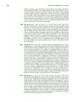
c. Third Class. In a third class lever, the weight to be moved is at one end of
the lever, the fulcrum is at the other end, and the applied force is between the weight
and the fulcrum. This type of lever provides speed, but a greater amount of force is
required for a given weight. This is the most common type of lever in the human body.
MD0006 5-7
Figure 5-3. Types of lever systems.
5-7. SIMPLE PULLEY SYSTEM
a. In the human body when the tendon of a skeletal muscle slides over a round
bony surface, the "system" acts like a simple pulley (figure 5-4). A simple pulley
provides a change in the direction of the force or muscle pull. There is no change in the
amount of force produced by the muscle. For example, the knee acts as a simple pulley
by which the quadriceps femoris M. extends the leg.
Figure 5-4. A simple pulley (the human knee mechanism).
MD0006 5-8
b. Sesamoid bones, such as the patella (kneecap), develop in tendons where
pressure is applied to the tendon.
5-8. THE SKELETO-MUSCULAR UNIT
The skeleto-muscular unit (figure 5-5) is a working concept of muscle and
skeleton producing motion. The components of an S-M unit are bones, a joint, and
skeletal muscle(s).
Figure 5-5. The skeleto-muscular unit (arm-forearm flexion
(3rd class lever system)).
a. Bones. Bones act as levers and as attachment sites for skeletal muscles.
b. Joint (Articulation). The joint is the center, fulcrum, point, or axis of motion.
c. Skeletal Muscle(s). Skeletal muscles apply the forces for motion. Any given
motion utilizes a group of muscles working together. A skeletal muscle may serve only
one of the three following major roles during a particular motion:
(1) Prime mover. The muscle which makes the main effort for a given motion
is called the prime mover, or agonist.
(2) Synergist. A synergist is a muscle which assists the prime mover.
SYN = together
ERG = unit of effort
MD0006 5-9
(3) Antagonist. An antagonist applies a force opposite to that of the prime
mover.
(a) By opposing the prime mover, the antagonist helps control the
motion.
(b) The antagonist also brings the limb or other part back to its original
position.
Continue with Exercises
MD0006 5-11
5. Label the drawings below according to class of lever.
6. The components of a skeleto-muscular unit are:
a. .
b. .
c. .
7. The muscle which makes the main effort for a given motion is called the
. A muscle which assists the first is called a . A muscle
which applies a force opposite to that of the first is called an .
Check Your Answers on Next Page
MD0006 5-12
SOLUTIONS TO EXERCISES, LESSON 5
1. The main types of tissues in skeletal muscles are striated muscle fibers and
fibrous connective tissue. (para 5-2)
2. The large portion of skeletal muscle is known as its belly or fleshy belly.
Generally, a skeletal muscle is attached to bone by a tendon or aponeurosis. If
the fleshy portion is directly connected to the bone, it is called a fleshy attachment.
(para 5-2a)
3. A neurovascular bundle is a branch from the main NAVL, sheathed in fibrous
connective tissue. The motor point is the specific location on the surface of the
muscle where the neurovascular bundle enters. A motor unit is a single motor
neuron and the striated muscle fibers activated by the neuron. All fibers of a motor
unit contract or none contract. (para 5-2b)
4. The trunk musculature is arranged in two ways longitudinal muscles and oblique
muscles. The limb musculature is arranged around the joints to produce the
appropriate motions of the limbs. (para 5-4)
5. a. Third class.
b. First class.
c. Second class. (para 5-6; figure 5-3)
6. The components of a skeleto-muscular unit are:
a. Bones.
b. Joint (articulation).
c. Skeletal muscles. (para 5-8)
7. The muscle which makes the main effort for a given motion is called the prime
mover (agonist). A muscle which assists the first is called a synergist. A muscle
which applies a force opposite to that of the first is called an antagonist.
(para 5-8c)
End of Lesson 5
MD0006 6-1
LESSON ASSIGNMENT
LESSON 6 The Human Digestive System.
TEXT ASSIGNMENT Paragraphs 6-1 through 6-16.
LESSON OBJECTIVES After completing this lesson, you should be able to:
6-1. Define the human digestive system.
6-2. Name six major organs of the human digestive
system.
6-3. Name and describe six structures of the oral
complex.
6-4. Describe the pharynx, the esophagus, the
stomach, the small intestines, the liver and
gallbladder, the pancreas, and the large
intestines.
SUGGESTION After completing the assignment, complete the
exercises at the end of this lesson. These exercises
will help you to achieve the lesson objectives.