USMLE ROAD MAP BIOCHEMISTRY – PART 9 ppt
Bạn đang xem bản rút gọn của tài liệu. Xem và tải ngay bản đầy đủ của tài liệu tại đây (464.94 KB, 24 trang )
180 USMLE Road Map: Biochemistry
N
c. Silent mutations, which often occur in the 3′ base of a codon, do not alter
codon specificity, so there is no effect on the protein’s sequence.
B. Insertions and deletions may cause production of altered proteins that have ei-
ther subtle or drastic functional changes.
1. If the DNA sequence is altered by deletion or insertion of 3n nucleotides, then
the mutant protein will lack or gain n amino acid(s), but its sequence will oth-
erwise be normal.
2. Insertion or deletion of a number of nucleotides that is not divisible by three
will cause a frameshift such that different, garbled protein sequence will be
synthesized downstream of the mutation.
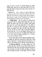
Single-base substitutions
Insertions and deletions
Splicing error
Silent
AUG-UCA-AGA-CCG-AAU-GGC-UAC-UUC-GAU-CUA-AUA
Met-Ser-Arg-Pro-Asn-Gly-Tyr-Phe-Asp-Leu-Ile
Normal
AUG-UCA-AGA-CCG-AAC-GGC-UAC-UUC-GAU-CUA-AUA
Met-Ser-Arg-Pro-Asn-Gly-Tyr-Phe-Asp-Leu-Ile
Insert (+1 nt)
Garbled protein sequence
AUG-UGC-AAG-ACC-GAA-CGG-CUA-CUU-CGA-UCU-AAU-A
Met-Cys-Lys-Thr-Glu-Arg-Leu-Leu-Arg-Ser-Asn-
Nonsense
Truncated protein
AUG-UCA-AGA-CCG-AAC-GGC-UAG-UUC-GAU-CUA-AUA
Met-Ser-Arg-Pro-Asn-Gly-STOP
Delete (—1 nt)
Garbled sequence, truncated protein
AUG-UCA-AGA-cCGA-ACG-GCU-ACU-UCG-AUC-UAA-UA
Met-Ser-Arg Arg-Thr-Ala-Thr-Ser-Ile-STOP
Garbled sequence
AUG-UCA-AGA-CCG-AAC-GGC-AGA-AGC-UGU-GUC-AAA
Met-Ser-Arg-Pro-Asn-Gly-Arg-Ser-Cys-Val-Lys
Missense
AUG-UCA-AGA-CCG-AAC-GUC-UAC-UUC-GAU-CUA-AUA
Met-Ser-Arg-Pro-Asn-Val-Tyr-Phe-Asp-Leu-Ile
Figure 12–6. Mutations of gene sequences that may affect protein function and
cause disease.
Chapter 12: Gene Expression 181
N
C. Splicing errors alter the critical sequence around an intron-exon splice junction.
1. This may be caused by single-base substitutions, insertions, or deletions.
2. Creation of an abnormal splicing site or destruction of the normal site may re-
sult in incorporation of an intron into a “finished” mRNA.
3. Translation of the intron region of the mutant mRNA produces a garbled pro-
tein sequence until an in-frame stop codon causes termination of the trun-
cated, mutant protein.
CLINICAL PROBLEMS
A 9-year-old boy is referred for evaluation of his hearing. A note from his school principal
explains that he is inattentive in class. Initial physical examination indicates that he is at
the 10th percentile for height, has coarse facial features, and is somewhat macrocephalic;
however, the remainder of the examination is within normal limits. Audiometry results
confirm partial bilateral deafness, which is sensorineural in etiology. An IQ examination
shows that he is in the 60th percentile for intelligence. Family history of mucopolysaccha-
ridoses prompts specialty testing, which indicates elevated levels of dermatan sulfate and
heparan sulfate in both a skin biopsy and urine sample.
1. Biochemical analysis of a skin biopsy from this patient would most likely indicate a de-
ficiency of which of the following enzymes?
A. β-Galactosidase
B. α-L-Iduronidase
C. Iduronate sulfatase
D. N-Acetylgalactosamine sulfatase
E. β-Glucuronidase
The sickled shape of erythrocytes in patients with sickle cell anemia occurs because of the
tendency for HbS to polymerize. HbS differs from HbA by substitution of a solvent-
exposed glutamate by valine in β-globin, which forms a “sticky” patch that promotes
aggregation and polymerization of the protein.
2. The genetic change that produced the mutant hemoglobin in sickle cell anemia can be
classified as which type of mutation?
A. Silent
B. Missense
C. Nonsense
D. Insertion
E. Deletion
Infections by the ulcer-causing bacterium Helicobacter pylori can be treated effectively with
a prolonged course of doxycycline or another of the tetracycline family of antibiotics, po-
tent inhibitors of prokaryotic protein synthesis.
182 USMLE Road Map: Biochemistry
N
3. Which of the following explains why tetracycline is selective for prokaryotes and mini-
mally toxic to humans?
A. It is ineffective against the 70S ribosomes.
B. It is ineffective against the mitochondrial ribosomes.
C. It only inhibits prokaryotic peptidyl transferase.
D. It cannot pass across eukaryotic membranes.
E. It blocks the A site only of prokaryotic ribosomes.
Some patients with familial hypercholesterolemia produce a truncated form of the LDL
receptor, termed the “Lebanese” allele, which lacks three of the five domains of the protein
and causes it to be retained in the endoplasmic reticulum. Analysis of the mutant gene in-
dicated that the sequence of the protein was normal up to the point where it terminated.
4. The genetic change that produced the mutant LDL receptor in these cases can be clas-
sified as which type of mutation?
A. Silent
B. Missense
C. Nonsense
D. Insertion
E. Deletion
A 2-year-old boy in whom Down syndrome was diagnosed when he was an infant comes
in for a check-up. Although he is developmentally delayed indicating potential mental re-
tardation, he is exhibiting some clinical features that are inconsistent with Down syn-
drome. These features include coarse facial features, small stature, radiographic evidence of
kyphoscoliosis, widening of the ribs, and malformed vertebrae.
5. Microscopic examination of skin or muscle biopsy specimens from this patient would
be likely to reveal dense inclusions corresponding with which organelles?
A. Mitochondria
B. Nuclei
C. Golgi apparatus
D. Lysosomes
E. Peroxisomes
A 17-year-old woman with cystic fibrosis is evaluated for knee pain. On review of systems,
she also notes persistent bleeding from cuts in her skin and bleeding of her gums after
brushing her teeth. Physical examination is remarkable for an obviously swollen right knee
that is tender with limited range of motion. Fluid drained from the knee is bloody
(hemarthrosis). Her complete blood count is normal, but prothrombin time is elevated.
6. This patient appears to be suffering from a deficiency of which of the following vita-
mins?
A. Vitamin A
B. Vitamin B
12
C. Vitamin C
D. Vitamin D
E. Vitamin K
ANSWERS
1. The answer is C. This patient’s clinical presentation is consistent with one of the mu-
copolysaccharidoses, but it can be difficult to determine which type given the wide
variability of expression of these disorders. One clue is provided by the hearing loss, a
characteristic feature of MPS-II, Hunter syndrome. In addition, his above-average in-
telligence for his age group and the absence of scoliosis distinguish MPS-II from MPS
I, the Hurler-Scheie syndromes. The latter are characterized by mental retardation to
varying degrees. The patient appears to have a severe form of Hunter syndrome, so the
cells of his tissues should be deficient in the lysosomal enzyme iduronate sulfatase.
2. The answer is B. A missense mutation results from a change in codon specificity from
one amino acid to another. This alters the protein sequence and may affect the pro-
tein’s structure and function. By definition, substitution of valine for glutamic acid in
the β-globin molecule represents a missense mutation at the level of the gene. Sickle
cell anemia illustrates how important even a single amino acid in a large protein can be
to the function of the protein and the physiology of the cell. However, it is more com-
mon to find that missense mutations have less dramatic effects than in this case.
3. The answer is D. Tetracycline antibiotics operate by blocking the aminoacyl binding
site of 30S ribosomes found both in prokayotes and in the mitochondria of eukaryotes.
However, the drug may be used as a selective antibiotic with minimal toxicity to pa-
tients because it cannot pass through the plasma membranes of human cells. If it could
do so, the drug would be cytotoxic because it would interfere with mitochondrial func-
tion by inhibiting protein synthesis on the 70S ribosomes of the organelles.
4. The answer is C. Production of a truncated protein indicates that a mutation has oc-
curred, but this phenomenon may have arisen from a frameshift mutation (insertion or
deletion) or by a nonsense mutation. The most likely possibility is a nonsense mutation
because sequence analysis of the truncated protein showed that it had normal (wild-
type) sequence. Insertion and deletion events often produce a stretch of garbled or ab-
normal protein sequence at the C-terminal end of the truncated protein arising from
out-of-frame translation of the mRNA downstream of the mutation until a stop codon
is encountered.
5. The answer is D. As this patient ages, a variety of skeletal defects and short stature that
are consistent with a lysosomal storage disease (mucolipidosis), either I-cell disease or
pseudo-Hurler polydystrophy, are developing. Both diseases arise from a deficiency of
an enzyme involved in synthesis of the Man-6-P marker on lysosomal enzymes. Such
“misaddressed” proteins are secreted rather than trafficked to the lysosomes. The
degradative function of lysosomes is impaired as a result and the organelles tend to ac-
cumulate waste products (hence, the term “storage disease”). It is these inclusion bodies
or dense structures that would be visible by microscopic examination of the patient’s
cells in a biopsy specimen.
Chapter 12: Gene Expression 183
N
6. The answer is E. The patient’s symptoms and prolonged prothrombin time suggest
that she has a mild coagulation disorder potentially due to vitamin K deficiency. Sev-
eral coagulation factors including prothrombin require carboxylation on glutamic acid
residues for optimal function. These proteins are carboxylated in vitamin K–dependent
reactions. Vitamin K deficiency may occur in people suffering from cystic fibrosis,
which causes gastrointestinal complications due to pancreatic insufficiency. Secretion
of pancreatic enzymes such as lipase, which releases fatty acids from triglycerides to fa-
cilitate absorption from the gut, is impaired in cystic fibrosis patients. This fat malab-
sorption condition has manifested itself in this patient’s case as a deficiency in the
fat-soluble vitamin K. Although bleeding gums are one characteristic of scurvy, other
manifestations of vitamin C deficiency, eg, loose teeth, are absent in this case.
184 USMLE Road Map: Biochemistry
N
I. Overview of Mendelian Inheritance
A. A gene is defined as a unit of DNA that encodes an RNA product.
1. The RNA product may encode transfer RNAs (tRNAs), ribosomal RNAs
(rRNAs), or small nuclear RNAs (snRNAs) that have end point functions in
the cell.
2. If the RNA product is a messenger RNA (mRNA), then it must be translated
into a protein to complete expression of the gene.
3. Variants of a gene that differ in DNA sequence among individuals in the popu-
lation are called alleles.
a. The single most prevalent version of the gene in the population is referred to
as the wild-type (“normal”) allele.
b. If there is more than one common version of the gene in the population,
these are called polymorphisms.
c. Mutant alleles are versions of the gene that differ in sequence from the wild-
type allele and that produce defective products.
d. The chromosomal location of a gene is its locus.
B. The set of alleles that make up the genetic composition of a person is called the
genotype, which may refer either to all genes or to a specific gene or locus.
1. The diploid content of human cells is 46 chromosomes—22 autosomal pairs
and 2 sex chromosomes (XX in females, XY in males).
2. For genes located on the autosomes, the genotype at a locus is formed from
two alleles.
3. Each parent contributes one allele through random segregation of chromo-
somes during meiosis.
4. If both alleles at a locus are identical, the person is said to be homozygous for
that gene.
5. In the case where the two alleles are different, the person is heterozygous for
that gene.
6. Since males have only one X chromosome, they usually have only a single allele
and are thus hemizygous for all X-linked genes.
C. The measurable expression of the genotype as a molecular, clinical, or biochemical
trait is the phenotype.
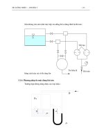
D. Pedigree analysis evaluates transmission of a single-gene disorder within a family
or kindred (Figure 13–1).
N
CHAPTER 13
CHAPTER 13
HUMAN GENETICS
185
Copyright © 2007 by The McGraw-Hill Companies, Inc. Click here for terms of use.
II. Modes of Inheritance in Single-Gene Disorders
A. In autosomal recessive inheritance, the condition is expressed only in persons
who have two copies of (ie, are homozygous for) the mutant allele (Figure 13–2).
1. Autosomal recessive inheritance is often observed with enzyme deficiencies,
where heterozygotes express 50% of normal activity.
a. However, 50% of normal enzyme activity in these cases permits normal
physiologic function because expression of enzyme from the normal allele is
sufficient to provide for the needs of the cell.
b. This phenomenon is often called the margin of safety effect.
2. Both parents of an affected person for an autosomal recessive disorder must
have one normal and one mutant allele, making them obligate carriers barring
very rare new mutations.
3. The likelihood of a person being homozygous for an autosomal recessive trait
increases in consanguineous matings because of the existence of a common
ancestor.
4. Rare autosomal recessive diseases also occur with high frequency among genet-
ically isolated populations due to inbreeding.
TAYS-SACHS DISEASE IN A GENETICALLY ISOLATED POPULATION
• The biochemical defect in Tay-Sachs disease is an inherited deficiency of -hexosaminidase, a lysoso-
mal enzyme responsible for hydrolysis of GM
2
ganglioside, which accumulates abnormally in the lyso-
somes.
• Children with Tay-Sachs disease exhibit hypotonia (poor muscle tone) and progressive neurologic
symptoms, including blindness and mental retardation.
186 USMLE Road Map: Biochemistry
N
Male
Female
Affected
Unaffected
Deceased
Obligate carrier
Marriage or mating
Consanguinity
Proband
1234
12345
12 3
123
4567
I
II
III
IV
,
,
,
,
Figure 13–1. Definitions of symbols used to evaluate inheritance patterns for
pedigree analysis and relationships within kindreds. Generations are assigned
Roman numerals and individuals within each generation are indicated by Arabic
numerals. The arrow points at the proband, the person in whom the genetic
disorder was first diagnosed.
CLINICAL
CORRELATION
• Most patients are diagnosed at 5–6 months and do not live beyond their second year.
• This autosomal recessive disease occurs in Ashkenazi Jews, the Pennsylvania Amish, and several
other populations with an incidence of 1 in 3600, 100 times higher than the overall population; the car-
rier frequency in these populations is about 3%.
5. Pedigree charts for an autosomal recessive disorder may show the following:
a. The disease phenotype is expressed by siblings but not by their parents or
offspring.
b. Equal occurrence in males and females.
c. Recurrence risk for each sibling is 25%.
d. Possible consanguinity.
Chapter 13: Human Genetics 187
N
A
I
II
III
IV
B
I
II
III
IV
C
I
II
III
IV
Figure 13–2. Pedigrees illustrating autosomal inheritance patterns. Recessive in-
heritance is shown in pedigrees A and B. Note that consanguinity in pedigree B re-
inforces the hypothesis of an autosomal recessive disorder. Dominant inheritance is
shown in pedigree C, in which every affected person has an affected parent.
B. In autosomal dominant inheritance, the condition is expressed even if a single
mutant allele is present, ie, in the heterozygous state (Figure 13–2).
1. Following are at least four possible situations by which having one normal copy
of a gene is insufficient to prevent disease, leading to a dominant phenotype
(Table 13–1):
a. When the presence of 50% normal activity (ordinarily the margin of safety)
is not generous enough to allow normal physiologic function, a condition
called haploinsufficiency.
b. When the defective allele produces a malfunctioning protein product that
binds to and interferes with function of the normal gene product—the
dominant negative effect.
c. When the mutant protein has an enhanced function that overrides normal
controls or is cytotoxic.
d. When the phenotype appears as dominant inheritance even though the ac-
tual allele is recessive at the level of function in individual cells.
2. The homozygous mutant state usually produces a more severe clinical con-
dition than the heterozygous condition in autosomal dominant diseases.
3. Pedigree charts for an autosomal dominant disorder may show the following
features:
a. The disease phenotype appears in all generations, with each affected person
having an affected parent.
b. There is an equal occurrence in males and females, except in cases when ex-
pression of the trait is influenced by the person’s sex (ie, sex-limited).
c. Risk of transmission of the mutant allele is 50%, but because there usually
are so few persons in a family, there may be deviations from this expectation.
d. Potential for some cases to be due to a new mutation, which is more likely
for a dominant condition because disease symptoms would be expressed in
heterozygotes.
188 USMLE Road Map: Biochemistry
N
Table 13–1. Molecular phenotypes of autosomal dominant disease.
a
Molecular Explanation Disease and Gene
Haploinsufficiency, or when 50% of normal gene α-Thalassemia trait and the α-globin gene
activity is inadequate β-Thalassemia trait and the β-globin gene
Dominant negative effect, when the mutant Osteogenesis imperfecta and the collagen 1A
protein interferes with function of the normal gene (COL1A1)
protein Marfan syndrome and the fibrillin-1 gene (FBN1)
Cytotoxic effect due to a dysregulated, mutant Huntington disease and the huntingtin gene (HD)
protein
Dominant effect at the cellular level of a Retinoblastoma and RB1
recessively inherited loss-of-function mutant Li-Fraumeni syndrome and TP53
of a tumor suppressor gene (see Chapter 14)
a
These genetic diseases are examples of the various molecular explanations for dominant inheritance.
FIBRILLIN DEFECTS IN MARFAN SYNDROME
• Marfan syndrome is a connective tissue disorder with manifestations in many organs, but especially
the skeleton, blood vessels, eyes, and lungs.
• Many tissues, such as lung, blood vessels, and skin, require elasticity for proper function; this prop-
erty is fulfilled by the matrix elastic fibers, which are composed of the proteins elastin and fibrillin.
– Marfan syndrome arises from a mutation in the gene encoding fibrillin-1 (FBN1).
– The pattern of inheritance of Marfan syndrome is autosomal dominant due to the failure of elastic
fibers to assemble properly upon interaction of mutant fibrillin with normal elastin.
• The disease is usually diagnosed by adolescence, and patients exhibit tall stature and a variety of
skeletal deformities, including very long, thin bones of the digits and limbs; flat feet; scoliosis; and
breastbone deformation.
– Joint hypermobility and a positive wrist/thumb sign are evident.
– The upper segment is the distance from the top of the head to the top of the pubic symphysis; the
lower segment is the distance from the top of the pubic symphysis to the floor. The upper segment
to lower segment ratio in persons with Marfan syndrome is low (< 0.9) because the arms and legs
are long relative to the torso.
• Characteristic ocular features of Marfan syndrome, such as ectopia lentis (upward lens dislocation in-
stead of downward dislocation as in homocystinuria) and myopia, arise from the effects of defective
fibrillin in the elastic fibers of the lens.
• The major cardiovascular manifestations are mitral valve prolapse and loss of elasticity of the aortic
root, which may lead to progressive aneurysm and potentially fatal aortic dissection.
C. Most X-linked diseases show a recessive inheritance pattern (Figure 13–3).
Chapter 13: Human Genetics 189
N
CLINICAL
CORRELATION
A
I
II
III
IV
B
I
II
III
Figure 13–3. Pedigrees illustrating X-linked recessive (A) and dominant (B) inheri-
tance patterns. Note the absence of male-to-male transmission in both pedigrees
and the predominance of affected males over females in the X-linked recessive
pedigree.
1. A distinguishing feature of these diseases is that there can be no male-to-male
transmission because the sex of male offspring is determined by contribution
of a Y chromosome from the father.
2. Because they have only one X chromosome, the sons of heterozygous mothers
have a 50% chance of being affected.
3. Pedigree charts for an X-linked recessive disorder may show the following
features:
a. Incidence of disease is higher in males than in females.
b. Female heterozygotes are usually unaffected carriers.
c. Affected men transmit the gene to all daughters, but never to sons.
d. New mutations cause a significant number of isolated cases in males due to
unopposed expression of the mutant allele.
D. X-linked dominant diseases are relatively rare (Figure 13–3).
1.
Such genes may be transmitted either to sons or daughters by an affected
mother but only to daughters by an affected father.
a. The mechanisms at the molecular and cellular levels that produce a domi-
nant phenotype are the same as in autosomal dominant disorders.
b. Only a few such disorders are known, including the Xg blood group and
vitamin D–resistant rickets.
2. Pedigree charts for an X-linked dominant disorder may show the following
features:
a. All daughters of affected men are affected but never their sons, which may
lead to prevalence of affected females over affected males.
b. Recurrence risk is 50% for both male and female offspring of an affected
female.
c. Absence of affected males in several generations may suggest prenatal lethal-
ity for the hemizygous state.
E. Incompletely dominant disorders occur in cases where the heterozygous geno-
type produces a different phenotype from that seen in the homozygous genotype.
1. The effect is often of intermediate severity between the unaffected and fully
affected phenotypes.
2. For example, in sickle cell anemia, the normal allele is incompletely dominant
in heterozygotes.
a. At the molecular level, the presence of some abnormal hemoglobin distorts
some RBCs.
b. This causes some sickling and the mild anemia characteristic of the sickle
trait.
F. Mitochondrial disorders exhibit a maternal inheritance pattern.
1. Mitochondria each have at least one and often several chromosomes that have
genes important for function of the organelle.
a. The mitochondrial chromosome (mtDNA) is a 16.5 kb circular plasmid.
b. The mtDNA bears 37 genes encoding rRNAs, tRNAs, and some genes for
proteins involved in oxidative phosphorylation.
2. Mitochondrial disorders are maternally transmitted because the ovum pro-
vides all mitochondria to the fertilized embryo (Figure 13–4).
3. In these disorders, affected cells usually have a mixture of mitochondria, some
with mutant mtDNA and others with wild-type mtDNA, a condition called
heteroplasmy.
190 USMLE Road Map: Biochemistry
N
a. Segregation of mitochondria during cell division is not as tightly controlled
as for nuclear chromosomes, leading to random distribution of mitochon-
dria carrying normal and mutant mtDNA to ova.
b. This contributes to variable expression and reduced penetrance of the
phenotype among persons within kindreds with mitochondrial disorders.
MITOCHONDRIAL MYOPATHY AND NEUROPATHY
• Mitochondrial diseases are caused by mutations in various mtDNA-encoded genes, most of which re-
sult in defective mitochondrial protein synthesis.
• The pathology is due to decreased mitochondrial function, eg, impaired oxidative phosphorylation,
and thus manifests in energy-intensive tissues, such as muscles and nerves.
• Microscopic examination of a muscle biopsy specimen may show ragged red fibers (distorted, dysfunc-
tional mitochondria).
• Mitochondrial diseases may be manifest as ready fatigability; elevated lactic acid levels in blood; in-
creased muscle enzymes in serum; ataxia; and a variety of neurosensory deficits, including blindness
and deafness.
– MERRF (myoclonic epilepsy with ragged red fibers) is characterized by weakness on exertion, ataxia,
and associated deafness and is due to mutation of the mitochondrial tRNA
Lys
gene.
– MELAS (mitochondrial encephalomyopathy with lactic acidosis and stroke-like episodes) results
from a point mutation in the mitochondrial tRNA
Leu
gene.
Chapter 13: Human Genetics 191
N
A
I
II
III
IV
B
I
II
III
62
54 43 48
48 35 30 23 29
Figure 13–4. Pedigrees illustrating inheritance of (A) a mitochondrial disorder and
(B) an autosomal dominant disorder exhibiting anticipation. In pedigree A, note the
similarity to the X-linked dominant inheritance pattern (Figure 13-3A), but incom-
plete penetrance as exemplified by individuals II-4 and III-4. In pedigree B, the age of
onset, indicated next to the symbols for affected individuals, becomes progressively
earlier with each generation.
CLINICAL
CORRELATION
– LHON (Leber’s hereditary optic neuropathy) causes blindness arising from mutations in the ND1
gene encoding complex I of the electron transport chain (see Chapter 7).
III. Major Concepts in Human Genetics
A. When similar phenotypes or disease conditions can be caused by different geno-
types, this may produce heterogeneity.
1. Allelic heterogeneity occurs when different alleles of the same gene produce
clinically similar conditions.
a. Many patients categorized as having autosomal recessive disorders actually
have two different mutant alleles of the disease gene and are therefore com-
pound heterozygotes.
b. Allelic heterogeneity may account for phenotypic variability in some families
with genetic disease.
2. Locus heterogeneity refers to the condition when mutations of more than one
gene or locus can produce similar disease states.
a. This genotypic variability is responsible for different inheritance patterns of
some disorders.
b. For example, Ehlers-Danlos syndrome (see Chapter 2) may be caused by
mutations at more than 10 known loci, producing inheritance patterns
ranging from autosomal recessive or dominant to X-linked.
B. Variable expression arises when the nature and severity of the phenotype for a ge-
netic condition varies from one person to another.
VARIABLE EXPRESSION IN NEUROFIBROMATOSIS TYPE I
• The biochemical defect in type I neurofibromatosis (NF) involves loss-of-function mutations of the NF1
tumor suppressor gene.
• Approximately 50% of cases involve new mutations and even in cases of inherited type I NF, variability
of expression in kindreds makes genetic counseling very difficult.
• Type I NF is an autosomal dominant disorder characterized by a wide range of clinical presentations by
patients.
– Among the symptoms seen in patients with type I NF are hyperpigmented skin lesions (café-au-lait
spots), benign skin tumors (neurofibromas), dysplasia of the sphenoid bone, and benign tumors of
the iris (hamartomas or Lisch nodules).
– Patients also may suffer from mental retardation, but the main concern is a high risk of potentially
fatal CNS tumors.
C. Pleiotropy refers to a condition in which a mutant allele may have different phe-
notypic effects in various organ systems in an affected person.
D. Genomic imprinting is a complex phenomenon by which expression of an allele
differs depending on whether it is inherited from the mother or the father.
1. A gene that is shut off when inherited from the mother is maternally im-
printed.
2. A gene that is silenced when inherited from the father is paternally imprinted.
3. Imprinting involves an epigenetic mechanism, ie, an alteration in phenotype
that does not result from a change in the genotype.
a. Expression of the imprinted genes is silenced or shut off by methylation of
certain chromatin regions after DNA replication during gametogenesis.
b. The imprint is reversible upon passage through gametogenesis in the next
generation.
192 USMLE Road Map: Biochemistry
N
CLINICAL
CORRELATION
DISORDERS THAT EXHIBIT IMPRINTING
• Two clinically distinguishable conditions arise from deletion of the same region of chromosome 15
(15q11–q13) depending on whether the defective chromosome is inherited maternally or paternally,
which indicates an imprinting phenomenon for one or more genes in the region.
• Prader-Willi syndrome arises when deleted chromosome 15 is paternally inherited.
– The precise biochemical defect for Prader-Willi syndrome is unknown, but this region of chromo-
some 15 encompasses an imprinting control center and at least 6 maternally imprinted genes.
– Patients with Prader-Willi syndrome exhibit failure to thrive and short stature initially, which converts
to a tendency toward excessive eating, obesity, mild-to-moderate mental retardation, hypogo-
nadism, and characteristic facial dysmorphology.
• Angelman syndrome occurs when chromosome 15 with the deletion is maternally inherited.
– This disorder appears to involve paternal imprinting of UBE3A, encoding a ubiquitin-protein ligase
so that this gene product must be produced from the maternal chromosome.
– Angelman syndrome is a devastating neurologic disorder featuring severe mental retardation, a
“happy puppet” demeanor, seizures, ataxic gait, and aphasia.
E. Certain inherited disorders exhibit increased severity of phenotype or decreased
age of onset as the disease gene is passed from one generation to the next, an effect
known as anticipation (Figure 13–4).
1. Examples of genetic diseases that show anticipation are Huntington disease,
Fragile X syndrome, and other disorders that arise from trinucleotide repeat
expansion (see Chapter 11).
2. Disease symptoms occur only when the length of the trinucleotide repeat re-
gion exceeds a threshold.
F. Mosaicism is defined as the presence of cells in the body that are genetically different.
1. In somatic mosaicism, mutation of a gene occurs in a non-germline (somatic)
cell at some point during early development of the person, and all cells de-
scendent from that progenitor are genetically distinct.
2. In germline mosaicism, a mutation that occurred in the parent’s gonadal
make-up is transmitted through some gametes, but not all.
3. All females are technically mosaic for the genes of their X chromosomes due
to inactivation of one or the other X chromosomes early in development, a
phenomenon termed the Lyon hypothesis or lyonization.
a. Because X inactivation is random, this phenomenon accounts for variable
expression of some X-linked disorders, depending on whether the disease
allele or wild-type allele was inactivated.
b. Distribution of cells from the early embryo to the tissues may be imbalanced,
so that expression of the disease phenotype is not uniform among the organs.
4. Up to 25% of patients with Turner syndrome exhibit a mosaic karyotype, in
which only some cells have the 45,X karyotype classically associated with the
condition.
G. Uniparental disomy refers to a condition in which one or more cells of the body
have two identical chromosomes derived from a single parent, which increases
the likelihood of expression of recessive alleles inherited from that parent.
UNIPARENTAL DISOMY IN BECKWITH-WIEDEMANN SYNDROME
• Children afflicted with Beckwith-Wiedemann syndrome (BWS) show an overgrowth condition from
birth as well as macroglossia (enlarged tongue).
Chapter 13: Human Genetics 193
N
CLINICAL
CORRELATION
CLINICAL
CORRELATION
• BWS is associated with severe hypoglycemia that may become life-threatening in addition to an en-
hanced tendency to develop cancers of the liver, kidney, and adrenal glands.
• The gene for BWS has been mapped to chromosome 11 (11p15), a region encompassing the gene for
insulin-like growth factor II (IGF2), which is maternally imprinted and thus is expressed only when pa-
ternally inherited.
• Uniparental disomy may contribute to BWS in that excess paternal or decreased maternal contribu-
tions of chromosome 11 have been observed in some patients.
IV. Population Genetics: The Hardy-Weinberg Law
A. The Hardy-Weinberg Law allows calculation of genotypes based on the allele fre-
quencies in a population for a given genetic disorder.
1. If there are two alternative alleles for a gene, A and a, in a population, then we
can define p as the frequency of allele A and q as the frequency of allele a,
where p + q = 1.
2. The following binomial expression governs the frequencies of the various
genotypes in the population.
(p + q)
2
= p
2
+ 2 pq + q
2
3. Thus, the chances of occurrence of AA and aa homozygotes are p
2
and q
2
, re-
spectively, whereas the chances of finding an Aa heterozygote are 2pq.
B. The Hardy-Weinberg Law is especially useful during genetic counseling for au-
tosomal recessive disorders, in which heterozygotes cannot be distinguished in
phenotype from normal homozygotes (Figure 13–5).
1. For example, in the United States, the incidence of live births of babies of
Northern European descent who have cystic fibrosis (q
2
, for the aa genotype)
can be estimated as approximately 1 in 2500 (0.04%).
2. The Hardy-Weinberg Law can then be used to calculate the gene frequencies
for a and A (q and p):
3. The frequency of heterozygotes having the pq genotype (unaffected carriers)
can be estimated as ~4% using Hardy-Weinberg principles based on these same
data.
2pq = 2(0.02)(0.98) = 0.0392 or 3.9%
q =
A
1
2500
= 20.0004 = 0.02, and: p = 1- q = 1- 0.02 = 0.98
194 USMLE Road Map: Biochemistry
N
Males
A
(p)
A
(p)
a
(q)
a
(q)
AA
(p
2
)
Aa
(pq)
A
(pq)
aa
(q
2
)
Females
Figure 13–5. Punnett square showing application of the
Hardy-Weinberg Law. The allele frequencies p and q are
assumed to be equal for males and females within the
population.
C. The Hardy-Weinberg Law is based on several important assumptions about the
population and mating dynamics within it.
1. Matings are assumed to be random among persons within the population.
2. Allele frequencies are assumed to be relatively constant for the gene in ques-
tion because of the following:
a. The mutational rate at the locus is minimal.
b. Persons with all genotypes are capable of passing on their alleles.
c. The immigration rate for the population is low.
D. The Hardy-Weinberg equilibrium for particular alleles within a population may
be disturbed by several factors that violate the following assumptions:
1. Stratified or isolated populations that tend to mate within the group.
2. Assortative mating, by which persons tend to choose mates who resemble
themselves.
3. Consanguinity.
4. Preservation of mutations as alleles in the population.
5. Selection for or against certain genotypes.
a. Dominant mutations may be subject to negative selection because they
are expressed in the heterozygous state.
b. Selection against homozygous affected persons in an autosomal recessive
disorder usually has little effect on Hardy-Weinberg equilibrium because so
few of the disease alleles are found in the homozygous state.
c. Several diseases exhibit positive selection for the heterozygous state (het-
erozygote advantage), including sickle cell anemia and the thalassemias.
CLINICAL PROBLEMS
A couple undergoes genetic counseling for consultation on the possibility that they may
have a child with Friedrich’s ataxia, an autosomal recessive neuromuscular disorder. The
disorder has been diagnosed in the wife’s sister, and they are concerned that she is a carrier.
The incidence of the disorder in the population is 1 in 25,000 live births.
1. What is the probability that this couple’s first child will have Friedrich’s ataxia?
[Assume that the couple is not consanguineous].
A. 1/300
B. 1/500
C. 1/750
D. 1/3000
E. 1/6000
A 6-year-old boy shows signs of significant developmental delay. After interviewing the
boy’s parents, a family history of varying degrees of cognitive impairment becomes appar-
ent; specifically, the patient’s father, maternal grandfather, aunt, uncle, and several cousins
are affected.
2. Based on these findings, what is the most likely mode of inheritance for the disorder?
Chapter 13: Human Genetics 195
N
A. Autosomal recessive
B. Autosomal dominant
C. X-linked recessive
D. X-linked dominant
E. Mitochondrial
3. The incidence of sickle cell anemia among blacks is 1 in 400. Calculate the frequency
of heterozygotes for sickle cell gene, encoding β
S
-globin, in this population.
A. 1 in 6
B. 1 in 11
C. 1 in 20
D. 1 in 22
E. 1 in 44
The following pedigree shows the pattern of inheritance of a neurologic disorder in a large
Central American kindred. Note that the numbers above symbols representing affected
persons in this pedigree indicate age of diagnosis.
196 USMLE Road Map: Biochemistry
N
I
II
III
67
61 37 51 57
54 44 43 38
4. What type of molecular phenomenon is most likely responsible for this disorder?
A. Trinucleotide repeat expansion
B. Parental imprinting
C. Reduced penetrance
D. Multifactorial inheritance
E. Haploinsufficiency
Several members of a family have type I neurofibromatosis in which DNA analysis indi-
cates that a son and daughter have inherited the disease from their father. The 16-year-old
daughter has a pelvic neurofibroma, her brother exhibits only café-au-lait spots and a
hamartoma, while the father has no detectable symptoms.
5. Type I neurofibromatosis in this family exhibits which of the following phenomena?
A. Mosaicism
B. Anticipation
C. Silencing
D. Variable expression
E. Uniparental disomy
You have assembled a pedigree for the occurrence of hemophilia A in a family of Greek-
American heritage.
Chapter 13: Human Genetics 197
N
I
II
III
12
123 45 67 89
12
12
345678910111213
6. Based on these data, what is the most likely mode of inheritance for this disorder?
A. Autosomal recessive
B. Autosomal dominant
C. X-linked recessive
D. X-linked dominant
E. Mitochondrial
7. What is the genotype at the hemophilia A locus of individual II-7 in the pedigree
shown above?
A. Homozygous—AA
B. Homozygous—aa
C. Hemizygous—Ao
D. Hemizygous—ao
E. Heterozygous—Aa
A 5-year-old boy complains of “being tired all the time.” His mother insists that he eats
well but that he sleeps 11–14 hours per day and “can’t keep up with his friends on the ball
fields.” Physical examination reveals that he is at the 30th percentile for height and weight,
with mild bilateral deafness and ataxic gait. After interviewing his mother, it becomes ap-
parent that several other members of this family were deaf by their mid-twenties and “a
muscle problem” was diagnosed in an uncle.
8. A pedigree for this family would likely reveal which of the following features?
A. Predominant occurrence in females
B. Absence of male-to-male transmission
C. Lack of expression in heterozygous females
D. Occurrence in siblings but not in their parents
E. Tendency for the disease to skip generations
9. Angelman and Prader-Willi syndromes are related disorders affecting genes on chromo-
some 15 by which of the following epigenetic mechanisms?
A. Mosaicism
B. Histone acetylation
C. Haploinsufficiency
D. Imprinting
E. Viral infection
ANSWERS
1. The answer is D. The probability that the wife has inherited the disease gene from one
of her parents and is a carrier is 2/3. This couple is not consanguineous, so the likeli-
hood that he carries the disease allele is equivalent to the gene frequency in the popula-
tion. The probability that either of them would pass the allele to their offspring is 1/2.
Thus, the calculated overall probability of having an affected child is:
2. The answer is B. The presence of many affected persons in multiple generations of this
family suggests autosomal dominant inheritance. Male-to-male transmission of the
condition in several generations would rule out both X-linked and mitochondrial disor-
ders. Each affected person has an affected parent and there are multiple affected per-
sons in several successive generations.
3. The answer is B. The incidence of the disease among blacks, 1/400, can be used to de-
termine q and p:
The frequency of heterozygotes is then calculated using Hardy-Weinberg Law:
Frequency =2 pq = 2(0.95)(0.05) = 0.095 or ∼1 in 11
4. The answer is A. The pedigree demonstrates that the age of disease onset decreases in
each successive generation, suggesting anticipation. This phenomenon occurs in dis-
eases caused by expansion of a trinucleotide repeat in the disease-causing gene. In such
disorders as Fragile X syndrome and Huntington disease, the disease occurs with
greater severity or earlier age of onset in persons once a threshold length of the trinu-
cleotide repeat tract in the gene is exceeded.
5. The answer is D. Neurofibromatosis is representative of many genetic diseases that ex-
hibit pronounced variability of expression in individuals in families. This phenomenon
is not understood at the molecular level but may involve the influence of environment
q =
A
1
400
= 1/20 = 0.05 and p = 1- q = 1- 0.05 = 0.95
P =
¢
1
2
≤¢
1
2
≤¢
2
3
≤
A
1
25,000
=
¢
1
6
≤¢
1
500
≤
=
1
3000
198 USMLE Road Map: Biochemistry
N
or other genes. Anticipation would have been a reasonable answer if the family history
showed affected persons in multiple generations.
6. The answer is C. Several features of this pedigree suggest an X-linked mode of inheri-
tance. All the affected persons are male, the disease tends to skip a generation, and there
is no male-to-male transmission evident. Individuals III-2 and III-5, daughters of an af-
fected father, are unaffected. This establishes that the condition is not dominant but
recessive.
7. The answer is E. Individual II-7 is the mother of an affected son, so barring the occur-
rence of a new mutation in his case, she is an obligate carrier, ie, heterozygous for the
condition.
8. The answer is B. The clinical symptoms in this case strongly suggest a mitochondrial
disorder affecting both neurologic and musculoskeletal functions. In such cases, no
male-to-male transmission is possible because the mother’s ovum provides all the cyto-
plasmic components, including the mitochondria, for the fertilized egg. A pedigree for
this family would resemble the inheritance pattern of an X-linked disorder with the
likelihood of variable expression.
9. The answer is D. These syndromes involve reciprocal imprinting of one or more genes
in a region of the long arm of chromosome 15. Although both diseases manifest neuro-
logic impairment, Angelman syndrome is by far the more severe of the two disorders.
Chapter 13: Human Genetics 199
N
I. General Principles of Cellular Signaling
A. Signaling is a process by which information received from one cell is converted
into a response by another cell.
1. The signaling cell produces and ordinarily secretes a ligand, which travels to
the target cell.
2. Receptors displayed by the target cell bind the ligand and transduce the sig-
nal into a second messenger or a series of biochemical events that mediate the
response by the target cell.
a. The second messengers produced in response to these changes include
cyclic AMP, diacylglycerol (DAG), inositol trisphosphate (IP
3
), and cal-
cium.
b. Intermediate steps in the response involve alterations in the activity of en-
zymes, eg, kinases that phosphorylate cellular substrates.
c. The ultimate physiologic response of the cell often involves changes in gene
expression.
3. The signaling cell may be located far away from or adjacent to the target cell; in
some cases, a cell signals itself.
B. The events set in motion by ligand binding to the receptor represent the signaling
pathway.
1. In many cases, the signal is transferred or internalized from outside to inside
the cell via the action of the receptor.
2. The signal may be amplified; in other words, activation of an enzyme at one
step leads to multiple events as the signal is propagated through the pathway.
3. The steps of the signaling pathway also distribute the signal to several response
pathways in the cell (eg, glucagon action to increase glucose production by the
liver is mediated by increases in both gluconeogenesis and glycogenolysis).
4. The signal is often modulated by integration with input from other signaling
pathways (eg, the insulin/glucagon ratio is important for balancing carbohy-
drate metabolism in the liver).
C. There is a variety of signaling modes used for intercellular communication.
1. In endocrine signaling, a hormone moves from a gland to a distant target cell
by direct secretion into the bloodstream.
2. During neurotransmission, the axon of one neuron sends a chemical signal (a
neurotransmitter) to receptors on the dendrite of another neuron across the
synaptic cleft.
N
CHAPTER 14
CHAPTER 14
CELLULAR SIGNALING
AND CANCER BIOLOGY
200
Copyright © 2007 by The McGraw-Hill Companies, Inc. Click here for terms of use.
a. An important example of such neurotransmission via synaptic signaling is
represented by the action of acetylcholine in cholinergic neurons.
b. A closely related mechanism operates in neuroendocrine secretions (eg, argi-
nine vasopressin and oxytocin release from hypothalamic neurons whose
axons terminate at blood vessels within the posterior pituitary).
3. Paracrine signaling involves diffusion of a ligand from one cell to another lo-
cally within tissues.
a. This signaling mode is commonly used by solid-tumor growth factors, such
as epidermal growth factor (EGF) and the insulin-like growth factors IGF-I
and IGF-II, for self-stimulation of continued cell division.
b. Signaling by nitric oxide to regulate relaxation of smooth muscle cells also
operates by the paracrine mode.
4. Juxtacrine signaling is a special case of the paracrine mode in which a growth
factor displayed on the surface of one cell binds to a receptor on a neighbor-
ing cell.
5. Autocrine signaling occurs when a growth factor produced by a cell binds to a
receptor on that same cell.
a. This signaling mode is frequently used as a strategy for growth self-stimula-
tion.
b. Inappropriate autocrine growth loops resulting in uncontrolled cell growth
are observed in many cancer cells, often involving EGF or IGF-II.
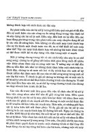
II. Signaling by G Protein-Coupled Receptors
A. Some cell-surface receptors having a characteristic structure with seven mem-
brane-spanning domains couple to heterotrimeric G proteins for purposes of
signal transduction (Figure 14–1).
B. Signaling is initiated by binding of an extracellular ligand, which produces a con-
formational change in the receptor that allows it to bind to the heterotrimeric G
protein.
1. Binding of the ligand-receptor complex initiates a conformational change in
the G protein that allows exchange of GDP for GTP on its α subunit, lead-
ing to dissociation of the ␣ and ␥ subunits of the G protein (Figure 14–1).
a. The heterotrimeric G proteins reside in the plasma membrane and are com-
posed of α, β, and γ subunits.
b. Interactions between the α subunit and the βγ complex are regulated by
binding of ligand-receptor complex within the plane of the membrane.
2. Both α and βγ can bind to and regulate the activity of various effectors of
signaling.
a. Regulation of the effectors may either be positive (activation) or negative
(inhibition).
b. Examples of some effectors are adenylate cyclase, ion channels, and phos-
pholipase C (PLC), each of which regulates a different response pathway
within the cell.
3. The signal is shut off by the action of the intrinsic GTPase activity of the α
subunit, which catalyzes hydrolysis of the bound GTP to GDP and P
i
.
a. GTP hydrolysis to GDP + P
i
causes a conformational change in the α sub-
unit that returns it to the inactive state.
b. The inactive α subunit dissociates from the effector and then binds a βγ
complex to be ready to undergo a new round of activation.
Chapter 14: Cellular Signaling and Cancer Biology 201
N
4. The family of heterotrimeric G proteins that mediate signals from various cell-
surface receptors is large (Table 14–1).
a. Each G protein can be activated by one or more types of receptors.
b. The G proteins are specific for a more limited range of effectors, which may
be activated or inhibited by binding of the G protein α subunit.
c. G protein signaling specificity and ligand binding functions reside in the α
subunit; the βγ subunits for all members of the family are derived from a
common pool in each cell.
202 USMLE Road Map: Biochemistry
N
Ligand
OFF
ON
Ligand
binding
GDP
GDP
GDP
GTP
Receptor
Effectors
Activated
Gα
G protein
α
β
γ
GTP
α
β
γ
Activated
βγ complex
Effectors
Figure 14–1. Signaling via G protein-coupled receptors. Ligand binding to its cell-
surface receptor initiates interaction of the receptor with the heterotrimeric G
protein for which it is specific. A conformational change in the G protein brought
about by binding of the ligand-receptor complex promotes exchange of GDP for
GTP. The activated G
α
-GTP dissociates from the G
βγ
complex and both can interact
with effectors, which carry on the signal to the mechanism that implements the cel-
lular response.
C. Second messengers mediate activation of signaling pathways downstream of the G
protein–coupled receptors.
1. Adenylate cyclase regulates synthesis of cyclic AMP, a second messenger that
carries the signal through various cellular pathways to end points of response
within the cell (Figure 14–2).
a. Activation of adenylate cyclase stimulates synthesis of cyclic AMP from
ATP.
b. Cyclic AMP binds to protein kinase A (PKA), which dissociates into the
active catalytic subunits and the regulatory subunits.
c. The free catalytic subunits of PKA phosphorylate cellular substrates to
change their activities and implement the cellular response.
(1)
Phosphorylation of cellular substrates by PKA occurs on serine and thre-
onine residues.
(2)
Some examples of PKA substrates are glycogen synthase (see Chapter 6)
and CREB (see Chapter 12).
d. In order to shut off the signal mediated by elevated cellular cyclic AMP,
phosphodiesterase hydrolyzes cyclic AMP to AMP (Figure 14–2).
METHYLXANTHINES
• Several methylxanthines produced by plants are inhibitors of cyclic AMP phosphodiesterase and
thereby produce an elevation in cyclic AMP levels in cells throughout the body.
• Effects of the methylxanthines are particularly pronounced in cardiovascular and nervous systems
as increased heart rate, smooth muscle relaxation, and heightened activity of some neurons (percep-
tion of enhanced alertness).
• Methylxanthines include caffeine (present in coffee and tea), theobromine (tea and chocolate), and
theophylline (tea).
• Due to its ability to stimulate relaxation of bronchial smooth muscle, theophylline is useful for treat-
ing the bronchoconstriction of asthma.
Chapter 14: Cellular Signaling and Cancer Biology 203
N
Table 14–1. Heterotrimeric G proteins.
α Subunit Effector and Directional Change
G
s
(stimulatory) ↑ Adenylate cyclase and Ca
2+
channels
G
i
(inhibitory) ↓ Adenylate cyclase
G
o
↓ Ca
2+
channels
G
q
↑ Phospholipase C
G
11
, G
14
, G
15/16
↑ Phospholipase C
G
t
(transducin) ↑ Cyclic GMP phosphodiesterase
G
Olf
(Olfactory receptors) ↑ Adenylate cyclase
CLINICAL
CORRELATION