USMLE ROAD MAP BIOCHEMISTRY – PART 10 docx
Bạn đang xem bản rút gọn của tài liệu. Xem và tải ngay bản đầy đủ của tài liệu tại đây (423.06 KB, 20 trang )
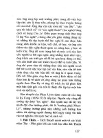
2. Calcium and DAG are second messengers that mediate some responses initi-
ated by signaling from G protein-coupled receptors (Figure 14–3).
a. Activation of PLC by binding of a G protein α subunit activates the en-
zyme.
b. PLC hydrolyzes a membrane-bound inositol phospholipid, phosphatidyl-
inositol 4,5-bisphosphate (PIP
2
), into the active products IP
3
and DAG.
c. DAG forms a binding site for protein kinase C (PKC) and thereby re-
cruits it to the plasma membrane, which partially activates the enzyme.
d. IP
3
binds to the endoplasmic reticulum to release Ca
2+
stores.
e. Ca
2+
binds to PKC and further activates it.
f. PKC phosphorylates multiple substrates to alter gene expression in the cell.
TUMOR-PROMOTING PHORBOL ESTERS
• Extracts from the croton plant (croton oil) are not themselves carcinogenic but enhance tumor forma-
tion if administered after initial exposure to a carcinogen.
Chapter 14: Cellular Signaling and Cancer Biology 205
N
α
GTP
PLC
P
P
P
P
P
P
PIP
2
IP
3
Ca
2
+
DAG
Endoplasmic
reticulum
Activated
PKC
Phosphorylation of
cellular substrates
Figure 14–3. Signaling through protein kinase C (PKC). Activated phospholipase
C cleaves the inositol phospholipid PIP
2
to form both soluble (IP
3
) and membrane-
associated (DAG) second messengers. DAG recruits PKC to the membrane, where
binding of calcium ions to PKC fully activates it. To accomplish this, IP
3
promotes a
transient increase of intracellular Ca
2+
concentration by binding to a receptor on
the endoplasmic reticulum, which opens a channel allowing release of stored cal-
cium ions. PIP
2
, phosphatidylinositol 4,5-bisphosphate; DAG, diacylglycerol; PLC,
phospholipase C; IP
3
, inositol trisphosphate.
CLINICAL
CORRELATION
• The active agents in croton oil are phorbol esters, specifically 12-O-tetradecanoyl phorbol-13-acetate
(TPA) or phorbol myristate acetate (PMA), which are structural analogs of DAG.
• Both TPA and PMA enhance carcinogenesis by binding to the DAG binding site and activating PKC,
which bypasses normal cell cycle regulation and stimulates cell division to produce its “tumor pro-
moting” effect.
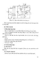
III. Receptor Tyrosine Kinases
A. Some cell-surface receptors transduce their signals by means of a kinase cascade
initiated by their protein tyrosine kinase activity (Figure 14–4).
206 USMLE Road Map: Biochemistry
N
P P
PGDP
GDP
GTP
GTP
Activated
tyrosine
kinase
GRB2
SOS
Raf
Ras Ras
Ligand
Receptor
Raf
MEK
Protein
kinases
Transcription
factors
Other
proteins
P
ERK
P
1
2
3
Figure 14–4. Receptor tyrosine kinase signaling mediated by the Ras-MEK-ERK
pathway. Binding of a growth factor (ligand) to its cell-surface receptor promotes
dimerization of the receptor with subsequent autophosphorylation mediated by ac-
tivation of the intrinsic tyrosine kinase of the receptor’s cytoplasmic domain. Dock-
ing of the adaptor GRB2-SOS complex promotes activation of Ras by GDP-GTP
exchange. Ras recruits the first serine/threonine kinase of the signaling pathway, Raf.
Raf then phosphorylates itself as well as the downstream kinase (MEK), which in
turn phosphorylates ERK (also called MAP kinase). Activated ERK is capable of dis-
tributing the signal by phosphorylation of multiple substrates leading to the cell’s
pleiotropic response to the growth factor. Reactions of the kinase cascade are de-
noted by the numbers in diamonds.
1. These signal transducers have a large extracellular domain with its ligand-
binding site, a single transmembrane domain and an intracellular domain with
intrinsic tyrosine kinase activity.
2. Ligand binding to the receptor’s extracellular domain activates signaling by
causing the receptors to form dimers and cross-phosphorylate (autophospho-
rylate) their intracellular domains on tyrosine sites.
B. The signaling pathway downstream of the activated receptors is composed of a se-
ries of kinases, a kinase cascade.
1. The phosphotyrosine sites on the receptor act as docking points for adap-
tors and effectors, which couple the signal to the kinase cascade.
2. One of the major adaptors is the GRB2-SOS complex, which upon docking to
the phosphorylated receptor, binds the small G protein Ras and activates it by
GDP-GTP exchange in a manner analogous to the heterotrimeric G proteins.
3. Activated Ras recruits the first kinase in the cascade, Raf-1, to the plasma
membrane, where it becomes active.
4. The signal is then transferred from one kinase to the other by sequential phos-
phorylation and activation, ie, the kinase cascade.
5. The signal ultimately is sent into the nucleus, where transcription factors such
as Elk-1 are activated by phosphorylation.
CLINICAL APPLICATIONS OF MONOCLONAL ANTIBODIES
THAT TARGET LIGANDS AND RECEPTORS
• By 2005, 18 monoclonal antibodies had been approved for treatment of several diseases, especially for
various cancers as well as infectious and inflammatory conditions, with many more under devel-
opment.
• Some of these agents are targeted to ligands or their receptors, and they work by preventing binding
and subsequent signal transduction, as illustrated in the following examples.
– HER2, a member of the EGF receptor family, drives growth of breast cancers that overexpress the re-
ceptor. Trastuzumab, which binds HER2 and prevents receptor activation, has been shown to be ef-
fective in reducing tumor growth and metastasis in such cases.
– Interleukin-2 (IL-2) signaling is important in the immune response that can lead to rejection in solid
organ transplantation. Basiliximab binds the α subunit of the IL-2 receptor to prevent IL-2 binding
and provide an immunosuppressive effect to inhibit renal transplant rejection.
– Tumor necrosis factor-␣ (TNF-␣) is a critical mediator of inflammation in autoimmune diseases
like Crohn’s disease and rheumatoid arthritis. Infliximab binds TNF-α and prevents its binding to the
TNF receptor for treatment of these diseases.
– Many cancers depend on vascular endothelial growth factor (VEGF) for formation of a blood sup-
ply to allow tumor growth and metastasis. Bevacizumab binds VEGF, which prevents its binding to
the VEGF receptor and thereby inhibits tumor vascularization (angiogenesis) in combination therapy
with 5-fluorouracil for treatment of metastatic cancers, particularly colorectal cancer.
IV. The Nuclear Receptor Superfamily
A. Many hormones diffuse into the cell and initiate signaling by binding to soluble
intracellular receptors that act as transcription factors.
1. This mechanism is used by steroid hormones (Table 14–2), thyroid hormone,
vitamin D
3
, and retinoic acid.
a. These ligands for the nuclear receptor superfamily are capable of dissolving
in water at low concentrations but are mainly lipophilic, capable of passing
through the lipid bilayer into the cell by diffusion.
Chapter 14: Cellular Signaling and Cancer Biology 207
N
CLINICAL
CORRELATION
b. Some of these molecules require metabolism or modification to be able to
bind their receptors.
(1)
Dihydrotestosterone is the preferred (high affinity) ligand for the an-
drogen receptor and is formed by reduction of testosterone catalyzed
by the enzyme steroid 5α-reductase.
(2)
The form of thyroid hormone active in binding its receptor is tri-
iodothyronine (T
3
) rather than thyroxine (T
4
).
2. The receptors may be located in the nucleus or cytoplasm of the cell, but they
are collectively called the “nuclear receptor superfamily” because the nucleus
is their main site of action.
3. The receptors in this family have a similar overall structure with a ligand-
binding domain specific for the hormone or vitamin, a DNA-binding
domain, and a variable domain that differs among the receptors.
B. Binding of ligand activates the receptor so that it can bind specific DNA se-
quences in regulatory regions of target genes that have hormone-response ele-
ments (HREs) (Figure 14–5).
1. After formation of the initial ligand-receptor complex, other partner proteins
are recruited that complete the active complex.
2. Binding of a co-activator confers on the complex the ability to activate tran-
scription when it binds to the target gene.
3. Conversely, transcription of a target gene may be inhibited by binding of a
complex formed when a co-repressor binds to the ligand-receptor.
208 USMLE Road Map: Biochemistry
N
Table 14–2. Ligands of the nuclear receptor superfamily.
Hormone or Ligand Family Name Major Ligands in Humans
Glucocorticoids Cortisol
Mineralocorticoids Aldosterone
Progestins Progesterone
Estrogens Estradiol
Estriol
Estrone
Androgens Testosterone
Dihydrotestosterone (DHT)
Dehydroepiandrosterone (DHEA)
Vitamin D compounds 1,25-Dihydroxycholecalciferol or
1,25-Dihydroxy vitamin D
3
Retinoids (vitamin A compounds) All-trans retinoic acid
Thyroid hormones Thyroxine (T
4
)
Triiodothyronine (T
3
)
DISORDERS OF ANDROGEN ACTION PRODUCE FEMINIZATION IN MALES
• Steroid 5␣-reductase deficiency is an autosomal recessive disorder that causes decreased conver-
sion of testosterone to dihydrotestosterone and decreased androgen action that is particularly critical
during sexual development.
Chapter 14: Cellular Signaling and Cancer Biology 209
N
HRE
Receptor
Activated
hormone-receptor
complex
Coactivator
Steroid
hormone
Cytoplasm
Nucleus
RNA
Polymerase
Gene transcription
RESPONSE
Figure 14–5. Regulation of gene transcription by members of the nuclear recep-
tor superfamily. Binding of a steroid hormone to its receptor promotes a confor-
mational change that causes dissociation of proteins that associate with the
inactivated receptor, including several heat shock proteins. In this example, the re-
ceptor is localized in the cytoplasm in its inactive state. In such a case, the acti-
vated hormone-receptor complex undergoes a conformational change that
exposes a nuclear localization signal. Within the nucleus, the receptor binds a
coactivator protein and the complete complex mediates transcriptional activation
of target genes having the appropriate hormone-response element (HRE).
CLINICAL
CORRELATION
• External genitalia of men deficient in steroid 5α-reductase are female in character rather than male.
• Several inherited disorders that produce defective androgen receptors (androgen resistance) also
cause disruption of sexual development that may culminate in infertility or testicular feminization.
• Testicular feminization is characterized by expression of a female external phenotype despite a nor-
mal blood level of testosterone and standard male karyotype (46,XY).
V. Overview of Cancer Biology
A. Cancer is considered a genetic disease in that mutations of various genes cause
disease by dysregulation of cellular mechanisms that control proliferation, sur-
vival, and death.
1. Once a cell has become “transformed,” ie, capable of autonomous prolifera-
tion through mutation of some of its genes, these characteristics are heritable
from cell to cell.
2. Dominant, gain-of-function mutations that activate oncogenes confer a
rapid-growth phenotype on cells.
3. Recessive, loss-of-function mutations that delete or inactivate tumor sup-
pressor genes alleviate controls on cell proliferation and survival.
4. Activated oncogenes are rarely passed through the germline.
5. Mutated, inactivated tumor suppressor genes can be inherited through the
germline from one person to another.
a. These cancer susceptibility genes usually have an autosomal dominant ex-
pression pattern.
b. Examples of such conditions are the genes for familial colorectal cancer
(eg, HNPCC or APC) and the familial breast cancer genes BRCA1 and
BRCA2.
B. Development of cancer or neoplastic transformation requires an accumulation
of mutations in the same cell.
1. The first mutation in a tumor suppressor gene such as BRCA1 may be either
inherited via the germline or sporadic (due to a random event in that person)
and then the normal allele is somehow inactivated (see loss of heterozygosity
below).
2. Multiple mutations that activate oncogenes or inactivate tumor suppressor
genes accumulate due to progressive loss of DNA repair mechanisms and cell
cycle control.
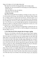
3. An important example of how a progression of somatic mutations leads to can-
cer is in hereditary colorectal cancer (Figure 14–6).
VI. Oncogenes and Tumor Suppressor Genes
A. Oncogene activation by overexpression, mutation, or chromosomal rearrange-
ment can lead to rapid proliferation of cells and cancer.
1. Oncogenes are the mutant, out-of-control versions of normal cellular genes,
the proto-oncogenes, which regulate a variety of critical cellular processes such
as signaling, cell cycle control, and transcription.
2. The mutations that have converted the proto-oncogenes to their oncogene
forms are gain-of-function or activating mutations.
RAS MUTATIONS OCCUR IN MANY HUMAN CANCERS
• Over 30% of all human cancers have activating mutations of the gene encoding the small G protein
Ras.
210 USMLE Road Map: Biochemistry
N
CLINICAL
CORRELATION
• Several missense mutations (ie, at codons 12, 13, or 61) render the mutant protein incapable of hy-
drolyzing bound GTP to GDP.
• These mutant forms of Ras thus persist in the ON state, which provides continuous activation of the ki-
nase cascade downstream of Ras and stimulates the cell to keep dividing even in the absence of appro-
priate signals from cell-surface receptors.
3. Tumor viruses carry activated versions of important cellular genes that regu-
late cell cycle and transcription.
a. The virus that causes Kaposi’s sarcoma, Kaposi’s sarcoma–associated her-
pesvirus, induces transformation of infected cells by up-regulating expres-
sion of the cellular form of the Kit oncogene, among others.
b. Human papillomavirus (HPV) causes a variety of epithelial cancers, espe-
cially of the alimentary canal and the cervix, by means of two associated
oncogenes, E6 and E7.
4. Overexpression or deregulated expression of cell cycle-dependent transcription
factors such as Myc and Fos may stimulate continued cell division.
Chapter 14: Cellular Signaling and Cancer Biology 211
N
Normal colon
epithelial cell
Loss of tumor
suppressor gene
APC
Increased epithelial
proliferation
Activation of
oncogene by mutation
RAS
Benign tumor
(adenoma)
Loss of tumor
suppressor gene
DCC
Large adenoma
Loss of tumor
suppressor gene
TP53
Aggressive, invasive
tumor (carcinoma)
Accumulation of
many mutations
Many
genes
Metastic tumors
Figure 14–6. Accumulation of mutations
leads to progressive development of familial
colorectal cancer. Development of cancer
does not require that these steps occur in
the particular sequence shown.
5. Activation of an oncogene may occur by chromosomal rearrangement creat-
ing a dysregulated fusion protein.
THE PHILADELPHIA CHROMOSOME IN CHRONIC MYELOGENOUS LEUKEMIA
• Cytogenetic analysis of patients with chronic myelogenous leukemia (CML) reveals an unusual translo-
cation between chromosomes 9 and 22 termed the “Philadelphia chromosome.”
• The translocation moves the c-ABL gene that encodes a tyrosine kinase from chromosome 9 to the
breakpoint cluster region (BCR) of chromosome 22.
• The resultant gene, BCR-ABL, encodes a constitutively active kinase that stimulates cell division and
leads to the transformed phenotype of the cells.
• Patients with CML experience weakness, fatigue, excessive sweating, low-grade fever, enlarged spleen,
and elevated WBC count.
• Imatinib, a drug that inhibits the kinase activity of the Bcr-Abl fusion protein, has been successfully
used for treatment of CML.
B. Loss or inactivation of tumor suppressor genes may lead to cancer.
1. Tumor suppressors are genes that encode a diverse array of proteins that con-
trol cellular growth and death.
2. Loss or mutation that inactivates one copy of the gene can be tolerated because
there is no functional deficit in the heterozygous condition.
3. Loss of heterozygosity (LOH) that deletes the only available functional copy
of the gene can contribute to unregulated proliferation of those cells (Figure
14–7).
212 USMLE Road Map: Biochemistry
N
Loss of normal
chromosome
Loss and reduplication
Somatic recombination
or mitotic crossing over
Independent mutation
N
Constitutional
genotype
M
M
M M
M M
M M
Figure 14–7. Possible mech-
anisms for loss of heterozy-
gosity at a tumor suppressor
locus. All these mechanisms
have been observed in
retinoblastoma involving the
RB1 gene on chromosome 13.
CLINICAL
CORRELATION
Chapter 14: Cellular Signaling and Cancer Biology 213
N
LOH IN RETINOBLASTOMA
• Retinoblastoma produces childhood neoplasms arising from neural precursor cells of the retina
(retinoblasts) at an incidence of 1 in 20,000 live births.
• The biochemical defect is mutation or loss of the tumor suppressor gene, RB1, encoding the protein
pRb.
– pRb binds to and inactivates members of the E2F transcription complex, which normally prevents
cells from entering S phase of the cell cycle.
– Loss of E2F regulation by pRb impairs cell cycle control, and unregulated proliferation (clonal ex-
pansion) may lead to a tumor derived from that cell.
• Most cases are inherited and multiple tumors arise bilaterally in heterozygotes when the normal RB1
allele undergoes mutation or loss due to LOH.
• Retinoblastoma shows an apparently autosomal dominant phenotype due to the high probability of
LOH during the ~10
6
cell divisions of retinoblasts and despite the recessive nature at the cellular level.
4. TP53 is an important tumor suppressor gene that encodes the p53 transcrip-
tion factor that is up-regulated when the cellular DNA is damaged.
a. High levels of p53 up-regulate transcription of the WAF1/CIP1 gene, whose
protein product, p21, blocks entry into S phase of the cell cycle by a mecha-
nism called checkpoint control.
b. TP53 is the most commonly mutated gene in human cancer, occurring in
over 50% of tumors examined.
LI-FRAUMENI SYNDROME
• Patients with Li-Fraumeni syndrome have increased susceptibility to a variety of cancers, including
bone and soft-tissue sarcomas, breast tumors, brain cancers, leukemia, and adrenocortical carcinoma,
all arising at an early age (often before 30 years).
• The biochemical defect in families exhibiting this syndrome is a loss-of-function mutation of the
tumor suppressor gene, TP53, encoding p53.
• The incidence of Li-Fraumeni syndrome has not been calculated because it is so rare.
• Inheritance is apparently autosomal dominant with high penetrance but with variable expression
(family members may have a wide range of tumor types and ages of onset).
VII. Apoptosis
A. Apoptosis, or programmed cell death, is a complex, highly regulated process by
which a cell self-destructs in an organized manner.
1. The mechanism of death in apoptosis contrasts with that occurring when a cell
breaks open or lyses producing a necrosis.
2. Necrosis allows the contents of the cell to spill over the local area, causing an
inflammatory response that leads to damage to nearby cells within the tissue.
3. By contrast, cells undergoing apoptosis do not lyse, so there is no associated in-
flammatory response.
B. Major changes that occur in the cell during apoptosis include the following:
1. Chromatin condensation.
2. Disintegration of the nuclear envelope.
3. Fragmentation of DNA between the nucleosomes.
4. Blebbing of the cell membrane.
5. Recruitment of macrophages, which ultimately engulf the dead cells.
C. Both extrinsic and intrinsic pathways can lead to programmed cell death (Figure
14–8).
CLINICAL
CORRELATION
CLINICAL
CORRELATION
214 USMLE Road Map: Biochemistry
N
1. The extrinsic pathway involves response to an external signal.
a. The external signal of death ligands, such as FasL and tumor necrosis
factor–related apoptosis-inducing ligand (TRAIL), is transduced by cell-
surface death receptors, such as FADD.
b. Activation of an array of proteases called caspases (the caspase cascade)
mediates the response within the cell, which involves initiator caspases that
cleave and activate effector caspases.
Caspase 8
(active)
Caspase 8
(inactive)
Caspase 9
(active)
Caspase 9
(inactive)
Caspase 3
(inactive)
Caspase 3
(active)
Death ligands
Death receptor
Extrinsic
pathway
Intrinsic
pathway
Cytochrome c
Mitochondrion
Stress
Death
Trail or TNF
+
+
Apaf-1
Figure 14–8. Overview of pathways that regulate programmed cell death. Apoptosis
may occur in response to signaling through either the extrinsic pathway or the intrinsic
pathway. In each case, proteolytic cleavage activates an initiator caspase, caspase 8 or
9, either of which can cleave an effector caspase such as caspase 3. Apaf-1 is part of a
large complex called the apoptosome that mediates the intrinsic pathway. Binding of an
extracellular death ligand to its cell-surface receptor activates the extrinsic pathway.
Chapter 14: Cellular Signaling and Cancer Biology 215
N
c. Effector caspases in turn degrade key cellular proteins and activate an en-
donuclease that digests the DNA.
2. The intrinsic pathway responds to stress, usually resulting in the cell’s inabil-
ity to repair extensive DNA damage, sparking a decision to commit suicide.
a. Activation of pro-apoptotic (death-causing) factors may occur in response
to the DNA damage, which causes increased mitochondrial permeability.
b. Leakage of cytochrome c, among other proteins, from the intermembrane
space of the mitochondria causes activation of the caspase cascade.
CLINICAL PROBLEMS
A 19-year-old woman has been referred to an endocrinologist by her gynecologist because
of delay in the initiation of her menstrual periods. Physical examination reveals underde-
veloped breasts, an enlarged clitoris (rudimentary penis), and the presence of small masses
within the labia majora. Blood testosterone is in the normal range for males and a chromo-
some spread indicates a karyotype of 46,XY.
1. This patient most likely has a defect in signaling through a pathway involving which of
the following?
A. Cyclic AMP–dependent protein kinase (PKA)
B. Protein kinase C (PKC)
C. A cell-surface tyrosine kinase receptor
D. A nuclear receptor
E. A heterotrimeric G protein
In order for a solid tumor to grow beyond a certain size, it must develop a blood supply by
elaborating factors such as vascular endothelial growth factor (VEGF). VEGF secreted by
the tumor cells diffuses to nearby endothelial cells, which respond by dividing and migrat-
ing toward the tumor to eventually develop into blood vessels and vascularize the tumor.
2. Which of the following modes of intercellular signaling is operative in the case of VEGF?
A. Endocrine
B. Paracrine
C. Autocrine
D. Juxtacrine
E. Synaptic
Many of the drugs used in the treatment of hypertension and cardiovascular disease are de-
signed to interfere with the action of cell-surface receptors that couple to heterotrimeric G
proteins.
3. In order for these drugs to operate in a specific manner so that cellular responses to
only one type of receptor are affected, the drug would need to be targeted toward
which element of the pathway?
216 USMLE Road Map: Biochemistry
N
A. The ligand binding site of the receptor
B. The βγ complex of the G protein
C. The α subunit of the G protein
D. Adenylate cyclase
E. Phospholipase C
It is estimated that mutations of RAS occur in over 30% of human cancers. In most of
these cases, the mutations interfere with the intrinsic GTPase activity of Ras so that the
protein becomes constitutively or continuously active, irrespective of whether growth fac-
tors are present.
4. Constitutively activated Ras has become insensitive to which of the following elements
of the growth factor signaling pathway?
A. Raf-1
B. MEK
C. MAP kinase
D. Ras-GAP
E. Elk-1
Patients with retinoblastoma suffer from a high incidence of tumors arising from clonal
outgrowth of some retinal precursor cells due to mutation of the tumor suppressor gene
RB1. Analysis of cells from these tumors indicates that both copies of the RB1 gene are mu-
tated or lost, whereas the surrounding retinal cells have at least one functional RB1 allele.
5. Which of the following terms best describes the genetic phenomenon that leads to
tumor development in retinoblastoma patients?
A. Loss of imprinting
B. Deregulated expression
C. Incomplete penetrance
D. Gain of function
E. Loss of heterozygosity
Osteosarcoma has recently been diagnosed in a 12-year-old girl. Family history indicates
that her paternal aunt died of breast cancer at age 29 after having survived treatment for an
adrenocortical carcinoma. An uncle died of a brain tumor at age 38 and the patient’s fa-
ther, age 35, has leukemia.
6. An analysis of this patient’s DNA would most likely reveal a mutation in which of the
following genes?
A. RB1
B. RAS
C. TP53
D. c-ABL
E. PKC
Chapter 14: Cellular Signaling and Cancer Biology 217
N
ANSWERS
1. The answer is D. The patient’s ambiguous secondary sex characteristics and lack of
menstrual activity suggest the possibility of an androgen resistance syndrome. The male
karyotype and blood testosterone levels confirm this. This clinical condition might
have arisen as a result of steroid 5α-reductase deficiency or inherited defects in the an-
drogen receptor (testicular feminization).
2. The answer is B. Paracrine signaling involves diffusion of a substance locally from one
cell to another via the interstitial space rather than through blood vessels. Endocrine sig-
naling would require that VEGF travel through the blood to reach the endothelial target
cells. Autocrine signaling requires that the same cell both send the signal and respond to
it. Juxtacrine signaling requires that the VEGF be displayed from the surface of one cell
and bound by a receptor on another. Synaptic signaling is reserved for neurons. None of
these other signaling modes fit the description for the mechanism of action of VEGF.
3. The answer is A. Most of the drugs that target specific types of G protein-coupled re-
ceptors are either agonists that bind to the ligand-binding site and stimulate receptor
activity or are antagonists that bind to the receptor and prevent ligand binding. The G
protein α and βγ subunits, adenylate cyclase, and phospholipase C are all elements
shared among many types of receptors.
4. The answer is D. In response to binding of a growth factor to its cell-surface receptor, the
receptor forms a dimer that stimulates its intrinsic kinase activity to phosphorylate tyro-
sine residues on the cytoplasmic region. These phosphotyrosine sites allow docking of the
adaptor complex GRB2-SOS, which binds and thereby activates Ras through GDP to
GTP exchange. Constitutively activated Ras is unable to hydrolyze bound GTP and thus
cannot respond to the binding of Ras-GAP. Raf-1, MEK, MAP kinase, and Elk-1 all are
downstream elements of the signaling pathway that depend on the activity of Ras.
5. The answer is E. At the cellular level, the RB1 gene is recessive because loss of function
affecting both alleles must occur to produce disease. This patient has inherited a defec-
tive RB1 allele from her father and is thus heterozygous at the RB1 locus. Most of her
retinal precursor cells have one functional RB1 allele and those cells proliferate under
normal growth restraints. However, these cells are susceptible to mutations affecting
pRb function or an error leading to loss of the remaining functional RB1 allele. These
mutations occur by chance during cell division and lead to a tumor by clonal out-
growth. The process by which the sole functional allele is lost or mutated is referred to
as loss of heterozygosity (LOH).
6. The answer is C. The occurrence of a variety of cancers at fairly early ages in this fam-
ily, particularly the finding of osteosarcoma in such a young girl, suggests the possibil-
ity of an inherited disorder of a tumor suppressor gene. Since the tumors are not
associated with the eye, RB1 is unlikely as the cause. The spectrum of cancers in the
family is consistent with the Li-Fraumeni syndrome, which involves inheritance of a
loss-of-function mutant form of the tumor suppressor gene, TP53, encoding p53.
218
INDEX
Note: Page numbers followed by f or t indicate figures or tables, respectively.
Acetyl CoA biosynthesis,
90–91, 91f
Acetylcholinesterase, suicide
inhibitors of, 32
Acid sphingomyelinase
deficiency, 24–25
Acidic amino acids, 9
Acids and bases, physiologic
chemistry of, 2
Adipose, 61, 63–64
Albinism, 128
Alcohol. See also Ethanol
effects on membrane
fluidity, 41
Alkaptonuria, 24
Amides of carboxylic amino
acids, 9
Amino acids
biosynthesis, 129, 130f
catabolism of, 126–129,
127f
charge characteristics of,
10–11, 10f
groupings of, 9
structural classification, 9
Amphipathic lipids, 37–39.
See also Cholesterol;
Glycerophospholipids
(phosphoglycerides);
Sphingolipids
Amphipathic molecules, physi-
ologic chemistry of, 6
Anabolism, 52
anabolic pathways, 54
Androgen action disorders,
feminization in males,
209–210
Anesthetic, effects on
membrane fluidity, 41
Angelman syndrome, 193
Antibiotics, inhibitors of
protein synthesis, 173
clinical problems/solutions,
215–217
neoplastic transformation,
210
oncogenes, 210–212
overview, 210
tumor suppressors,
212–213, 212f
tumor viruses, 211
Carbohydrate metabolism. See
also G6PD deficiency;
Lactic acidosis; Pyruvate
kinase deficiency
clinical problems/solutions,
87–89
digestion and absorption
of dietary carbohydrates,
70
enzymes regulating glucose
metabolism rate-limiting
steps, 78, 78t
glycogen metabolism,
78–80
glycolysis, 70, 71f, 72–73
pentose phosphate pathway
(PPP), 76–77, 77f
regeneration of NAD
+
,
73–76, 75f, 76t
Carbohydrates
dietary, 53–54, 70
as membrane components,
42, 43f, 44
Carbonic acid-bicarbonate
system, 4, 5f
Cardiolipin, 37
Carnitine, 109–110
CPT-I/-II deficiency,
110
primary deficiency, 109
secondary deficiency to
other conditions, 110
shuttle, 109, 109f
inhibitors of topoiso-
merases, 156–157
Antibodies/immunoglobulins
(Ig), 19
Anticancer agents, 156
Anticipation, 193
Antiviral agents, 32, 156
Apoptosis, 213–215, 214f
Aromatic amino acids, 9
Arsenic toxicity, 94
Atherosclerosis and trans fats,
41
ATP
generation inhibitors,
97–98
stoichiometry of ATP
generation, 98
Autocrine signaling, 201
Basic amino acids, 9
Beckwith-Wiedemann
syndrome (BWS),
uniparental disomy
in, 193–194
Bilirubin metabolism, 133–134
BPG response to hypoxemic
conditions, 19
Brittle bone disease, 14–15
Buffers
metabolic acidosis, 5
metabolic alkalosis, 5
physiologic chemistry of,
3–5, 4f
Cancer
as a genetic disease, 210
chemical carcinogenesis, 159
susceptibility genes, 210
and telomerase activity, 158
Cancer biology. See also
Cellular signaling
apoptosis, 213–215, 214f
Copyright © 2007 by The McGraw-Hill Companies, Inc. Click here for terms of use.
Index 219
N
Catabolism, 52
catabolic pathways, 54
Catecholamines, 56
Celiac disease, 104
Cell membranes. See also
Atherosclerosis; Cystin-
uria; Hartnup disorder;
Krabbe disease; Schindler
disease
amphipathic lipids
(membrane component),
37–39
carbohydrate component
of, 42, 43f, 44
clinical problems/solutions,
48–51
glycerophospholipids,
37–38, 38f
lipid bilayer organization,
39–41, 40f, 41f
membrane fluidity,
40–41
protein component of, 41f,
42
structure and function
overview, 37
transmembrane transport,
44–47, 46f, 47f
uptake of particles and large
molecules, 117
Cellular signaling, 200
clinical problems/solutions,
215–217
by G protein-coupled
receptors, 201–203,
202f, 203t
nuclear receptor super-
family, 207–208
nuclear receptor super-
family/ligands, 208t
nuclear receptor super-
family/regulation of
gene transcription, 209f
paracrine/juxtacrine/
autcrine signaling, 201
receptor tyrosine kinase,
206–207, 206f
replication inhibitors
(anticancer/antiviral
agents), 156
Drug absorption, in digestive
tract, 3
dTMP inhibitors, 145
Dyslipedemia, 61
Ehlers-Danlos syndrome
(EDS), 14, 192
Electrolytes, physiologic
chemistry of, 1–2
Electron transport chain,
96–97, 96f, 98t
energy capture, 97
energy yield of oxidative
phosphorylation, 97
inhibitors of ATP
generation, 97–98
Endocytosis, 117
Energy diagram, 26, 27f
Enzyme-catalyzed reactions
deficiency in enzyme
activity, 23
enzyme replacement ther-
apy for inborn errors
of metabolism, 25, 24t
kinetics of, 29–30, 29f
substrate binding, 23
Enzymes
allosteric regulation of,
33–34
catalysis mechanisms,
27–28, 28f
catalytic of reactions by,
26–27, 27f
clinical problems/solutions,
34–36
classification, 25–26, 26t
coenzymes and cofactors,
32, 33t
covalent modification of,
54–55
in glucose metabolism
(rate-limiting steps), 78,
78f
inhibitors, 30–32
signaling modes, 200–201
signaling pathway, 200
Cholera toxin, 204
Cholesterol, 39
gallstone disease, 116–117
metabolism, 115–116, 116f
CK (creatine kinase), and
heart attack/muscle
damage diagnosis, 25–26
Coenzymes and cofactors, 32,
33t
Collagen, protein structure
and function, 13–14,
13f
Competitive enzyme
inhibitors, 30–31
Crohn’s disease and lipid
malasorption disorders,
104
Cyclic AMP
mechanisms of action, 203,
204f
phosphodiesterase
inhibitors, 203
Cystathionine β-synthase
deficiency, 25
Cystic fibrosis, and lipid
malabsorption disorders,
104
Cystic fibrosis (CF), 12–13
Cystinuria, 48
Diabetes mellitus. See Type1/2
diabetes mellitus
Diabetic ketoacidosis, 115
Diet and nutritional needs,
52–54
Digestion, 70
drug absorption factors
in
digestive tract, 3
DNA, 151
chromosomal (structure),
152–154, 153f
mutations, 158
repair, 159
replication, 154–158,
155f
Enzymes (cont.)
low-K
m
and ethanol
sensitivity, 30
physiological roles of/
clinical problems and
solutions, 34–36
snake venom, 28–29
as therapeutic agents, 29
Enzyme replacement therapy
(ERT),25
Ethanol sensitivity (low-K
m
enzyme), 30
Fabry disease, enzyme replace-
ment therapy for, 24
Familial breast cancer genes
(BRCA1/2), 210
Familial colorectal cancer
genes (HNPCC or FAP),
210, 211f
Fanconi anemia, 160
Farnesylation inhibitors (as
anti-cancer/antiparasitic
agents), 174–175
Fatty acids, 6
oxidation, 109–113
synthesis, 106–109, 107f,
108f
Fetal hemoglobin (HbF), 16
Folic acid deficiency, 142
Fragile X syndrome, 157–158
example of anticipation, 193
Fructose metabolism, 86
disorders of, 86
G protein functions,
interference by bacterial
toxins, 204
G6PD deficiency, 77–78
Galactose metabolism, 85–86
galactosemia, 86
Gaucher disease, enzyme
replacement therapy
for, 24
Gene, 151, 185. See also
Human genetics;
Population genetics
cancer susceptibility genes,
210
220 Index
N
factors disturbing balance
for alleles within a
population, 195
use in genetic counseling,
194, 194f
Hartnup disorder, 47
Heart attack and muscle
damage diagnosis,
physiologic role of
enzymes in, 25–26
Heinz bodies, 78
Heme biosynthesis, disorders,
133–134
Hemoglobin, 15, 15f
heterotetramer, 16
Hemolytic anemia, 16
Henderson-Hasselbalch
equation, 3–4
Hepatobiliary disease, and
lipid malabsorption
disorder, 104
HER2, 207
Heterogeneity/allelic and
locus, 192
Hexose monophosphate shunt.
See Pentose phosphate
pathway (PPP)
High altitude conditions, BPG
response to, 19
Homocystinuria, 25, 130, 131f
Homogentisate oxidase defi-
ciency (alkaptonuria), 24
Hormonal control, 569
Human genetics. See also Gene
anticipation, 193
clinical problems/solutions,
195
inheritance mode/single-
gene disorders, 186–192
kindreds, 185, 186f
major concepts in, 192–194
Mendelian inheritance
overview, 185
mosaicism, 193
uniparental disomy, 193
variable expression, 192
Human papillomavirus
(HPV), 211
Hunter syndrome, 176
expression/clinical problems/
solutions, 181–184
gene therapy, 23
genetic code, 168
genetic code/post-trans-
lational protein modifi-
cations, 173–176
genetic code/translation
steps, 168–173, 171f,
172f
mutations, 179–181, 180f
oncogenes, 210
regulation of gene expres-
sion, 55, 176–178, 177f
transcription, 161–164,
162f, 163f
Genomic imprinting, 192
disorders (examples), 193
Genotype, 185
Glucagon, 56
mechanism of actin, 56
regulatin of blood glucose
by, 56–64
Gluconeogenesis, 82–85
Glucose homeostasis, 56–58,
57f
Glycerophospholipids
(phosphoglycerides),
37–38, 38f
distinguishing structures, 37
fatty acids, 38, 38f
Glycogen metabolism, 55,
78–82
glycogen storage disease,
80
glycogenesis, 79–80, 79f
glycogenolysis, 80, 81f
hormonal regulation of, 80,
82, 83f
Glycosaminoglycan
accumulation, 176
Glycolysis, 70, 71f, 72
Gout, 146
Hardy-Weinberg Law,
194–195
assumptions about pop-
ulation and mating
dynamics, 195
Index 221
N
Huntington disease, 157
example of anticipation, 193
Hurler syndrome, 176
Hydrogen bonds, 1
Hydrolases that produce toxic
effects, 28–29
Hydrophilic substances, 1
Hydrophobic substances, 1
Hydroxyl groups (amino
acids), 9
Hyperammonemia, 123–126
acquired, 123–124
hereditary, 125–126
Hypercholesterolemia
(familial), defective
LDL receptor, 118
Hypoxemic conditions, BPG
response to, 19
I-cell disease, 174
Immunodeficiency (severe
combined), 146
Immunoglobulins (Ig)/
antibodies, 19
Inborn errors of metabolism,
23
enzyme replaement therapy
for, 25
Inheritance mode/single gene
disorders
autosomal dominant, 188,
188t, 190
autosomal recessive,
186–187
incompletely dominant,
190
mitochondrial disorders,
190–191, 191f
X-linked dominant, 189f,
190
Insulin, 56
mechanism of actin, 56
regulation of blood glucose
by, 56–64
resistance and type 2
diabetes, 66
secretion in type 1 diabetes,
65
Interleukin-2, 207
Lung surfactant, 6
and respiratory distress
syndrome, 6
Lysosomal enzymes
deficiencies, 25
localization disorders (mu-
colipidoss), 174
transport, 174
Maple syrup urine disease,
126–127
Marasmus, 53
Marfan syndrome fibrillin
defects, 189
MCAD (medium-chain fatty
acyl CoA dehydrogenase)
deficiency, 112
Melanin production, disorder
(albinism), 128
MELAS (mitochondrial
encephalomyopathy), 191
Mendelian inheritance, 185
MERRF (myoclonic epilepsy
with ragged red fibers),
191
Metabolic acidosis, 5, 75
Metabolic alkalosis, 5
Metabolic interrelationships/
regulation. See also
Obesity (dysregulation
of fat metabolism);
Protein-calorie mal-
nutrition; Type 1
diabetes mellitus; Type 2
diabetes mellitus
clinical problems/solutions,
66–69
diet and nutritional needs,
52–54
glucose homeostasis, 56–58,
57f
metabolism (fasting state),
61–63, 62f
metabolism (fed state),
58–61, 59f, 60f
metabolism (starvation),
63–64, 64f
regulation of metabolic
pathways, 54–56, 55f
Irreversible enzyme inhibitors,
31–32
Isozymes, 25
Jaundice, 134–135
Juxtacrine signaling, 201
Kanzaki disease, 39
Karposi’s sarcoma–associated
herpesvirus, 211
Ketone body metabolism,
113–115, 114f
Krabbe disease, 45
Krebs cycle. See TCA
(tricarboxylic acid) cycle
Kwashiorkor, 53
Lactic acidosis, 74–75, 96
Lead poisoning, and heme
biosynthesis, 133
Leber’s hereditary optic
neuropathy (LHON),
99, 192
Lesch-Nyhan syndrome, 147
Leukodystrophies, 45
Li-Fraumeni syndrome, 213
Lineweaver-Burk equation, 30
Lipid bilayer of biologic
membranes, 37, 38f
organization, 39–40
lipid domains/rafts, 40
Lipid metabolism. See also
Cholesterol metabolism
clinical problems/solutions,
118–121
digestion and absorption
of
dietary fats, 103
fatty acid oxidation,
109–113
fatty acid synthesis,
106–109, 107f, 108f
functions of fatty acids, 105
lipid malabsorption
disorders, 104
lipoproteins/processing
and transport, 104–105
Lipids, dietary, 54
Loss of heterozygosity (LOH),
212
Metabolic responses
(long-term), 55–56
Methemoglobinemia, 17–18
Methylxanthines, 203
Micelles, 6
Michaelis-Menten equation,
30
Mitochondrial myopathy and
neuropathy, 191–192
Monoclonal antibodies
targeting ligands and
receptors, clinical
applications, 207
Mosaicism, 193
Mucolipidoses, 174
Mucopolysaccharidoses, 176
Mushroom toxin, 163
Mutations, 179–181, 180f
Myelogenous leukemia
(chronic), 212
Myoglobin, 15, 17f
Neurofibromatosis Type I,
variable expression, 192
Niemann-Pick disease, 24–25
Nitrogen metabolism
amino acid biosynthesis,
129, 130f, 131f
amino acid catabolism,
126–129, 127f
ammonia metabolism, 123,
124f
clinical problems/solutions,
135–138, 148–150
dietary protein digestion,
122–123
porphyrin metabolism,
131–132
purine and pyrimidine nu-
cleotides degradation,
146
salvage pathways, 147
urea cycle, 124–125, 125f
Noncompetitive enzyme
inhibitors, 31
Nonpolar (hydrophobic)
amino acids, 9
Nucleic acid, clinical prob-
lems/solutions, 164–167
222 Index
N
adult hemoglobin (HbA),
15, 15f
hemoglobin heterotetramer,
16
myoglobin, 15, 17f
Paracrine signaling, 201
PDH deficiency, 91–92
Pentose phosphate pathway
(PPP), 76–77, 77f
Peptidyl transferase activity, 25
Pertussis toxin, 204
pH
effect on drug absorption
in digestive tract, 3
physiologic chemistry of,
2–3
Phagocytosis, 117
Phenotype, 185
Phenylketonuria (PKU),
130–131
Philadelphia chromosome, 212
Phosphoglycerides. See
Glycerophospholipids
Physiologic chemistry, clinical
problems and solutions,
6–8
Pleiotropy, 192
Pompe disease, enzyme
replacement therapy
for, 24
Population genetics, 194–195
Porphyrias, 133–134
Porphyrin metabolism,
131–132f, 134f
Prader-Willi syndrome, 193
Protein (dietary), 53
protein-calorie
malnutrition, 53
Protein synthesis inhibitors, as
antibiotics, 173
Proteins. See also Amino acids;
Antibodies; Collagen;
Oxygen binding proteins
charge characteristics of,
10–11, 10f
clinical problems and
solutions (structure
and function), 19–22
Nucleic acid metabolism
nucleotide structures/
functions, 139
purine biosynthesis,
139–142, 140f, 141f,
142f
pyrimidine biosynthesis,
142–144, 143f
Nucleic acid/structure and
function
chromosomal DNA
structure, 152–154, 153f
DNA repair, 159
functional overview,
151–152
mutations, 158
replication, 154–158, 155f
RNA structure, 160–161
transcription, 161–164,
162f, 163f
Nutritional needs and diet,
52–54
dietary carbohydrates,
53–54
dietary proteins, 53
metabolism of nutrients, 52
nutritional balance, 52
Obesity (dysregulation of fat
metabolism), 61
Oncogenes, 210–212
Organophosphorous
pesticides, as suicide
inhibitors of acetyl-
cholinesterase, 32
Orotic aciduria, 144
Osteogenesis imperfecta (OI),
14–15
Oxaloacetate synthesis from
pyruvate, 95
Oxidative damage of RBCs, 77
Oxidative phosphorylation, 90.
See also Tricarboxylic
acid (TCA) cycle and ox-
idative phosphorylation
energy yield of, 97
Oxygen binding proteins,
15–19, 15f, 17f
fetal hemoglobin (HbF), 16
Index 223
N
farnesylatin, 174
function, 13–19
membrane component, 41f,
42
structure, 11–13, 12f
synthesis, 168–173, 169f,
171f, 172f
Pseudo-Hurler polydystrophy,
174
Purine biosynthesis, 139–142,
140f, 141f, 142f
Pyrimidines biosynthesis,
142–144, 143f
Pyruvate
conversion to PEP, 82–83
pyruvate carboxylase
deficiency, 96
pyruvate dehydrogenase
(PDH) complex, 90,
91f
pyruvate kinase deficiency,
73
synthesis of oxaloacetate
from, 95–96
Ras mutations, 210–211
Respiratory distress syndrome
and lung surfactant, 6
Retinoblastoma, 213
Ribozymes, 25
RNA, 151–152
polymerase II inhibition by
mushroom toxin, 163
processing (splicing),
163–164, 163f
structure, 160–161
transcription, 161–162,
162f
example of anticipation,
193
Tumor necrosis factor-α
(TNF-α), 207
Tumor-promoting phorbol
esters, 205–206
Tumor suppressors, 212–213,
212f
loss of heterozygosity
(LOH), 212
Tumor viruses, 211
Turner syndrome, 193
Type 1 diabetes mellitus, 65
Type 2 diabetes mellitus, 66
Uniparental disomy, 193
Urea cycle, 124–125, 125f
Vascular endothelial growth
factor (VEGF), 207
Vitamin B
6
deficiency, 123
Vitamin C deficiency, 14
Vitamin K deficiency,
175–176
von Gierke disease, 80
Water
hydrogen bonding,
1
physiologic solvent, 1
special properties for
sustaining life, 1
X-linked adrenoleukodystro-
phy (X-ALD), 113
Xeroderma pigmentosum, 159
Zellweger syndrome, 113
Schindler disease, 39
Severe combined immunodefi-
ciency (SCID), 146
Sickle cell anemia, 18
Snake venom enzymes, 28–29
Sphingolipids, 39
Steroid 5α-reductase 2
deficiency, 209
Sugars, 41–42, 43f, 44
Sulfur-containing amino acids,
9
Tay-Sachs disease, 186–187
TCA (tricarboxylic acid) cycle,
90
acetyl CoA biosynthesis,
90–91, 91f
clinical problems/solutions,
99–102
electron transport chain,
96–99, 96f
oxaloacetate synthesis from
pyruvate, 95–96
PDH deficiency, 91–92
regulation of, 93f, 94
role in metabolic reactions,
94–95, 95f
steps of, 92–93, 93f
Telomerase activity, 158
Thalassemias, 16–17
Theophylline, 203
Thiamine deficiency, 94
Topoisomerase inhibitors,
156–157
Trans fats and atherosclerosis,
41
Trinucleotide repeat disorders,
157–158