Báo cáo y học: "Tracheobronchopathia osteochondroplastica: A rare cause of chronic cough with haemoptysis" pptx
Bạn đang xem bản rút gọn của tài liệu. Xem và tải ngay bản đầy đủ của tài liệu tại đây (504.85 KB, 4 trang )
BioMed Central
Page 1 of 4
(page number not for citation purposes)
Cough
Open Access
Case report
Tracheobronchopathia osteochondroplastica: A rare cause of
chronic cough with haemoptysis
Hinrich Willms
1
, Volker Wiechmann
2
, Ulrich Sack
3
and Adrian Gillissen*
1
Address:
1
Robert-Koch-Hospital, St. George Medical Center, Nikolai-Rumjanzew-Str. 100, D-4207 Leipzig, Germany,
2
Institute of Pathology and
Tumour Diagnostic, St. George Medical Center, Delitzscher-Str. 141, D-04129 Leipzig, Germany and
3
Institute of Clinical Immunology and
Transfusion Medicine, Medical Faculty of the University, Johannisallee 30, D-04103 Leipzig, Germany
Email: Hinrich Willms - ; Volker Wiechmann - ;
Ulrich Sack - ; Adrian Gillissen* -
* Corresponding author
Abstract
A case of tracheobronchopathia osteochondroplastic (TPO) was diagnosed in a 69-year old male
with prolonged cough. TPO is a rare condition of unknown cause and only sporadic cases have
been reported. The condition is benign, characterized by submucosal nodules growing from the
submucosal layer of the airways, protruding into the bronchial lumen. The bronchscopic view
together with bronchial cartilage with abnormal distributed mineralization of the histologic
examination of theses nodules leads to the correct diagnosis. Mild cases are treated
symptomatically, whereas we tried an inhaled corticosteroid. Prominent protrusions in the trachea
or the bronchi must be removed. In most cases the disease is stable over years but progressive
forms have been reported. TPO may cause chronic refractory cough, which eventually is the only
prominent symptom of this disease.
Background
Tracheobronchopathia osteochondroplastica (TPO) is a
rare benign disorder of the lower part of the trachea and
the upper part of the main bronchi [1-3]. It was first
described in the middle of the 19
th
century and since than,
approximately 300 cases have been reported. A higher
incidence of TPO was seen in northern Europe countries,
especially in Finland [4]. Because many cases are asymp-
tomatic TPO is mainly diagnosed post mortem. Symp-
toms can range from productive or non-productive cough,
haemoptysis, dyspnoea, dryness of the throat, recurrent
pulmonary infections (e.g. retention pneumonia) or oza-
ena [4-7]. In severe cases the diagnosis is made during a
difficult intubation [1,8]. The characteristic broncho-
scopic finding is described as beaded, speculated, rock
garden, cobble stoned or stalactite grotto appearance [9].
The diagnosis is confirmed by the typical histological
appearance.
Case presentation
A 69-year-old male presented to our pulmonary and criti-
cal care center suffering from chronic dry cough since sev-
eral months and haemoptysis since about 4 weeks.
Because of the cough and an assumed respiratory infec-
tion, he was treated with cefuroxim and moxifloxacine.
Because lacking any apparent success, he finally was
admitted to our center where he complaint about inter-
mittent sweating at night, fever up to 39°C, and weight
loss about 6 kg during the last 2 months. Total cigarette
consumption was about 30 pack-years but he stopped
smoking 25 years ago. History revealed no dust exposure.
Allergies were unknown.
Published: 30 June 2008
Cough 2008, 4:4 doi:10.1186/1745-9974-4-4
Received: 19 March 2008
Accepted: 30 June 2008
This article is available from: />© 2008 Willms et al; licensee BioMed Central Ltd.
This is an Open Access article distributed under the terms of the Creative Commons Attribution License ( />),
which permits unrestricted use, distribution, and reproduction in any medium, provided the original work is properly cited.
Cough 2008, 4:4 />Page 2 of 4
(page number not for citation purposes)
Apart from fine crackles over the lower part of both lungs
physical examination was normal. Blood tests showed ele-
vated c-reactive protein (41.0 mg/l), and slight anemia
(erythrocytes: 4.05 Tpt/l; hemoglobin: 8.4 mmol/l) was
apparent. Both chest X-ray and lung function tests were
normal (VC 3.4 liters [76% predicted], FEV
1
2,5 liters
[76% predicted], FEV
1
/FVC 74%). Further, diffusing
capacity and arterial blood gas values did not reveal any
abnormalities. We first performed a gastroscopy, which
turned out to be normal as well. Due to haemoptysis the
patient underwent flexible fiber-optic bronchoscopy
where we found in the middle of the trachea up to the
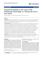
main carina multiple tubercular nodules (Fig. 1). From
these, various biopsies were taken, because we initially
expect them to be malignant. In contrast, histological
examination revealed bronchial cartilage and lamellar
bone with little marrow (Fig. 2), a clear evidence for TPO.
The mucous membrane of the trachea was lumpy, stiff
and bled easily. Secretion was copious. Cytologic brushes
of the trachea wall revealed bronchus epithelium with an
accumulation of neutrophils. Smears and cultures for
Mycobacterium tuberculosis were all negative. To reduce
the inflammatory process of the trachea, and thus treating
the cough [10], the patient was treated with inhaled
budesonide (2 × 200 μg/day), and he eventually was dis-
missed from the hospital.
Discussion
Sometimes TPO is diagnosed in a routine bronchoscopy,
or it is seen coincidently in CT-scan or MRI [11-13,8].
Until now, approximately 300 cases worldwide have been
reported. In our center with ca. 2 500 bronchoscopies/
year, it was the first case in 10 years. There seems to be a
higher prevalence in northern Europe, especially in Fin-
land from which about 25% of all cases have been
reported [4]. Cold-air-related hyperreactivity of the airway
epithelium, high incidence of respiratory infection due to
the cold climate together with a predisposing genetic fac-
tor or simply higher awareness by the doctors were dis-
cussed to be contribute factors [14]. But other
contributing factors may be possible, because an associa-
tion of habitually isolated M. ozaenae indicate that
chronic infections with this bacterium and/or other germs
may have a promoting effect although the exact mecha-
nism is unknown [5,15]. Reduction of mucociliary trans-
port, metaplasia of the connective tissue, exostosis arising
in the cartilaginous ring, chronic inflammation with a
possible link to amyloidosis of the lung are currently the
most frequent hypothesis how TPO develops on the cellu-
lar level [9,2,4]. Once the disease is rare, it seems impos-
sible to prove these hypotheses in a controlled trial. No
gender predominance has been reported. Although most
patients are older than 50 years, TPO is also found in chil-
dren [16].
In the bronchoscopic view TPO appears as whitish, hard
spicules projecting into the tracheal lumen from the ante-
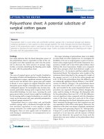
Bronchial cartilage with abnormal and unevenly distributed mineralization leads the diagnosis tracheobronchopathia osteochondroplastica: Tubercular nodule (haematoxylin-eosin x 50), normal bronchial epithelium (1), new cartilage (2) in abnormal submucous position with metaplastic ossifica-tion (3).Figure 2
Bronchial cartilage with abnormal and unevenly distributed
mineralization leads the diagnosis tracheobronchopathia
osteochondroplastica: Tubercular nodule (haematoxylin-
eosin x 50), normal bronchial epithelium (1), new cartilage
(2) in abnormal submucous position with metaplastic ossifica-
tion (3).
Bronchoscopic view of the trachea. Multiple tubercular nod-ules are seen (arrow).Figure 1
Bronchoscopic view of the trachea. Multiple tubercular nod-
ules are seen (arrow).
Cough 2008, 4:4 />Page 3 of 4
(page number not for citation purposes)
rior and lateral walls, with sparing of the posterior wall.
Also the larynx and the main bronchi could be involved
[17,6]. The diagnosis TPO is confirmed by typical histo-
logical findings, usually from biopsies or post mortem
analysis. In severe cases CT scan reveals spicules in the tra-
chea when they are big [11]. Our case was comparably
mild because the small whitish nodules occurred mainly
in the distal two thirds of the trachea which did not
obstruct the lumen. Consequently, our patient did not
suffer from dyspnea or asthma like symptoms like sever
cases reported in the literature. The chronic cough is most
likely caused be TPO because we did not find other causes
although the patient underwent rigorous diagnostic pro-
cedures.
Besides of TPO nodules may also be caused by endobron-
chial sarcoidosis, calcificating lesions of tuberculosis, pap-
illomatosis, malignant lesions and tracheobronchial
calcinosis [4,9]. Some patients were initially thought to
have asthma [18] or bronchial/trachea tumors like in our
case or a middle lobe syndrome [11].
Because typical symptoms are absent, TPO is most likely
under diagnosed. Only severe cases suffer from wheezing
and dyspnoea caused by the obstruction of the airway
lumen. Sometimes TPO causes difficulties in endotracheal
intubation [12,17]. In most cases the disease progresses
very slowly although progression have been reported
eventually leading to respiratory insufficiency [8,19].
Once no specific therapy is available treatment is only
symptomatic, which includes antibiotics in case of bacte-
rial infections, mechanical measures to remove obstruc-
tion nodules using either cryotherapy, laser excision,
external beam irradiation, radiotherapy, stent insertion or
surgical resection therapy [20,12,3].
In conclusion, patients with chronic cough must undergo
bronchoscopy at some time in order to uncover the
underlying cause which may be a rare disorder [13,21].
Consent
Written informed consent was obtained from the patient
for publication of this case report and any accompanying
images. A copy of the written consent is available for
review by the Editor-in-Chef of this journal
Competing interests
The authors declare that they have no competing interests.
Authors' contributions
HW worked with the patient and did all the clinical work
for diagnostics and therapy. Further, he wrote the first
draft of the manuscript, VW evaluated the biopsies taken
from our patient and prepared the histologic figure, US
was involved in drafting the manuscript. He suggested
sending it to "Cough", and he revised every manuscript
version meticulously, AG wrote the manuscript based on
the first version of HW. He further did all revisions of the
manuscript, including the numerous suggestions made by
US
Acknowledgements
Dr. Katleen Gutjahr is acknowledged for doing the bronchoscopy, for tak-
ing the picture of the tracheal nodules (fig. 1), and for obtaining the biopsies
of the trachea within her daily routine in our institution.
References
1. Vilkman S, Keistinen T: Tracheobronchopathia osteochondro-
plastica. Report of a young man with severe diseases and ret-
ropsective review of 18 cases. Respiration 1995, 62:151-154.
2. Karlikaya C, Yüksel M, Kilicli S, Candan L: Tracheobronchopathia
osteochondroplastica. Respirology 2000, 5:377-380.
3. Jabbardarjani HR, Radpey B, Kharabian S, Masjedi MR: Tracheo-
bronchopathia osteochondroplastica: Presentation of ten
cases and review of the literature. Lung 2008 in press.
4. Prakash UB: Tracheobronchopathia osteochondroplastica.
Semin Respir Crit Care Med 2002, 23:167-175.
5. Kart L, Kiraz K, Büyükoglan H, Ozesmi M, Sentürk Z, Gülmez I, Demir
R, Oymak FS: Tracheobronchopathia osteochondroplastica:
two cases and review of the literature. Tuberk Toraks 2004,
52:268-271.
6. Neumann A, Kasper D, Schultz-Coulon HJ: Clinical aspects of tra-
cheopathia osteoplastica. HNO 2001, 49:41-47.
7. Sutor GC, Glaab T, Eschenbruch C, Fabel H: Tracheobroncho-
pathia osteochondroplastica: an uncommon cause of reten-
tion pneumonia in an elderly patient. Pneumologie 2001,
55:563-567.
8. Hantous-Zannad S, Sebai L, Zidi A, Ben Khelil J, Mestiri I, Besbes M,
Hamzaoui A, Ben Miled-M'rad K: Tracheobronchopathia osteo-
chondroplastica presenting as a respiratory insufficiency:
diagnosis by bronchoscopy and MRI. Eur J Radiol 2003,
45:113-116.
9. Meyer CN, Dossing M, Broholm H: Tracheobronchopathia oste-
ochondroplastica. Respir Med 1997, 91:499-502.
10. Gillissen A, Richter A, Oster H: Clinical efficacy of short-term
treatment with extra-fine HFA beclomethasone dipropion-
ate in patients with post-infectious persistent cough. J Physiol
Pharmacol 2007, 58:223-232.
11. Restrepo S, Pandit M, Villamil MA, Rojas IC, Perez JM, Gascue A: Tra-
cheobronchopathia osteochondroplastica: helical CT find-
ings in 4 cases. J Thorac Imaging 2004,
19:112-116.
12. Khan M, Shim C, Simmons N, Chung V, Alterman DD, Haramati LB,
Berman AR: Tracheobronchopathia osteochondroplatica: a
rare cause of tracheal stenosis - "TPO stenosis". J Thorac Car-
diovasc Surg 2006, 132:714-716.
13. Decalmer S, Woodcock A, Greaves M, Howe M, Smith J: Airway
abnormalities at flexible bronchoscopy in patients with
chronic cough. Eur Respir J 2007, 30:1138-1142.
14. Lundgren R, Stjernberg NJ: Tracheobronchopathia osteochon-
droplastica. A clinical bronchoscopic ansspirometric study.
Chest 1981, 80:706-709.
15. Baugnee PE, Delaunois LM: Mycobacterium avium-intracellu-
lare associated with tracheobronchopathia osteochondro-
plastica. Eur Respir J 1995, 8:180-182.
16. Simsek PO, Ozcelik U, Demirkazik F, Unal OF, Orhan D, Aslan AT,
Dogru D: Tracheobronchopathia osteochondroplastica in a 9-
year-old girl. Pediatr Pulmonol 2006, 41:95-97.
17. Coetmeur D, Bovyn G, Leroux P, Niel-Duriez M: Tracheobroncho-
pathia osteochondroplastica presenting at the time of a dif-
ficult intubation. Respir Med 1997, 91:496-498.
18. Hayes DJ: Tracheopathia osteochondroplastica misdiagnosed
as asthma. J Asthma 2007, 44:253-255.
19. Molloy AR, McMahon JN: Rapid progression of tracheal stenosis
associated with tracheopathia osteochondroplastica. Inten-
sive Care Med 1988, 15:60-62.
20. Shigematsu Y, Sugio K, Yasuda M, Ono K, Takenoyama M, Hanagiri T,
Yasumoto K: Tracheobronchopathia osteochondroplastica
Publish with BioMed Central and every
scientist can read your work free of charge
"BioMed Central will be the most significant development for
disseminating the results of biomedical research in our lifetime."
Sir Paul Nurse, Cancer Research UK
Your research papers will be:
available free of charge to the entire biomedical community
peer reviewed and published immediately upon acceptance
cited in PubMed and archived on PubMed Central
yours — you keep the copyright
Submit your manuscript here:
/>BioMedcentral
Cough 2008, 4:4 />Page 4 of 4
(page number not for citation purposes)
occuring in subsegmental bronchus and causing obstructive
pneumonia. Ann Thorac Surg 2005, 80:1936-1938.
21. McCool FD: Global physiology and pathophysiology of cough:
ACCP evidence-based clinical practice guidelines. Chest 2006,
129:48S-53S.