Báo cáo y học: "A comparison of high-mobility group-box 1 protein, lipopolysaccharide-binding protein and procalcitonin in severe community-acquired infections and bacteraemia: a prospective study" ppt
Bạn đang xem bản rút gọn của tài liệu. Xem và tải ngay bản đầy đủ của tài liệu tại đây (301.91 KB, 10 trang )
Open Access
Available online />Page 1 of 10
(page number not for citation purposes)
Vol 11 No 4
Research
A comparison of high-mobility group-box 1 protein,
lipopolysaccharide-binding protein and procalcitonin in severe
community-acquired infections and bacteraemia: a prospective
study
Shahin Gaïni
1
, Ole G Koldkjær
2
, Holger J Møller
3
, Court Pedersen
1
and Svend S Pedersen
1
1
Department of Infectious Diseases, Odense University Hospital, Søndre Boulevard 29, DK-5000 Odense C, Denmark
2
Department of Clinical Biochemistry, Sønderborg Hospital, Sønderborg, Denmark
3
Department of Clinical Biochemistry, AS-NBG Aarhus University Hospital, Aarhus, Denmark
Corresponding author: Shahin Gaïni,
Received: 27 Apr 2007 Revisions requested: 31 May 2007 Revisions received: 22 Jun 2007 Accepted: 11 Jul 2007 Published: 11 Jul 2007
Critical Care 2007, 11:R76 (doi:10.1186/cc5967)
This article is online at: />© 2007 Gaïni et al., licensee BioMed Central Ltd.
This is an open access article distributed under the terms of the Creative Commons Attribution License ( />),
which permits unrestricted use, distribution, and reproduction in any medium, provided the original work is properly cited.
Abstract
Introduction High-mobility group box-1 protein (HMGB1) has
been known as a chromosomal protein for many years. HMGB1
has recently been shown to be a proinflammatory cytokine with
a role in the immunopathogenesis of sepsis.
Lipopolysaccharide-binding protein (LBP) has a central role in
the innate immune response when the host is challenged by
bacterial pathogens. Procalcitonin (PCT) has been suggested
as a marker of severe bacterial infections and sepsis. The aim of
the present study was to investigate levels of HMGB1, LBP and
PCT in a well-characterised sepsis cohort. The study plan
included analysis of the levels of the inflammatory markers in
relation to the severity of infection, to the prognosis and to the
ability to identify patients with bacteraemia.
Methods Patients suspected of having severe infections and
admitted to a department of internal medicine were included in
a prospective manner. Demographic data, comorbidity, routine
biochemistry, microbiological data, infection focus, severity
score and mortality on day 28 were recorded. Plasma and serum
were sampled within 24 hours after admission. Levels of all
studied markers (HMGB1, LBP, PCT, IL-6, C-reactive protein,
white blood cell count and neutrophils) were measured with
commercially available laboratory techniques.
Results A total of 185 adult patients were included in the study;
154 patients fulfilled our definition of infection. Levels of
HMGB1, LBP and PCT were higher in infected patients
compared with a healthy control group (P < 0.0001). Levels of
HMGB1, LBP and PCT were higher in the severe sepsis group
compared with the sepsis group (P < 0.01). No differences
were observed in levels of the inflammatory markers in fatal
cases compared with survivors. Levels of all studied markers
were higher in bacteraemic patients compared with
nonbacteraemic patients (P < 0.05). PCT performed best in a
receiver–operator curve analysis discriminating between
bacteraemic and nonbacteraemic patients (P < 0.05). HMGB1
correlated to LBP, IL-6, C-reactive protein, white blood cell
count and neutrophils (P < 0.001). LBP correlated to PCT, IL-6
and C-reactive protein (P < 0.001).
Conclusion Levels of HMGB1, PCT and LBP were higher in
infected patients compared with those in healthy controls, and
levels were higher in severe sepsis patients compared with
those in sepsis patients. Levels of all studied inflammatory
markers (HMGB1, LBP, PCT, IL-6) and infection markers (C-
reactive protein, white blood cell count, neutrophils) were
elevated among bacteraemic patients. PCT performed best as a
diagnostic test marker for bacteraemia.
Introduction
Sepsis is a serious clinical condition with a considerable mor-
bidity and mortality [1]. Clinicians are in need of good diagnos-
tic and prognostic markers to identify infected patients who
could benefit from prompt empirical antibiotic therapy and
other supportive therapy as early as possible. An increased
AUC = area under the curve; CRP = C-reactive protein; ELISA = enzyme-linked immunosorbent assay; FiO
2
= fraction of inspired oxygen; HMGB1
= high-mobility group box-1 protein; IL = interleukin; LBP = lipopolysaccharide-binding protein; PaO
2
= partial pressure of arterial oxygen; PCR =
polymerase chain reaction; PCT = procalcitonin; ROC = receiver–operator characteristic; SIRS = systemic inflammatory response syndrome; TNF
= tumour necrosis factor.
Critical Care Vol 11 No 4 Gaïni et al.
Page 2 of 10
(page number not for citation purposes)
knowledge of the immunopathogenesis of sepsis could have
the potential of generating new diagnostic and treatment
modalities for this serious condition.
High-mobility group-box 1 protein (HMGB1) is a nuclear chro-
mosomal protein [2,3]. A new role for HMGB1 has been
explored in recent years. HMGB1 has been suggested to have
an important role as a 'late-onset' proinflammatory cytokine
[4,5]. HMGB1 was rediscovered in this role when cultures of
macrophages were exposed to endotoxin [4]. Animal models
confirmed these observations, and there has been considera-
ble attention on this protein especially in relation to sepsis and
rheumatoid arthritis [4]. Lipopolysaccharide-binding protein
(LBP) is an acute-phase protein with an important role in the
innate immune system [6,7]. For the past 15 years attention
has been pointed at the inflammatory marker procalcitonin
(PCT) [8,9], which has been associated with severe bacterial
infections among adults and children [9].
The present study purpose was to examine levels of HMGB1,
LBP and PCT in patients with sepsis of different severity, in
bacteraemic patients and in relation to the outcome of the
patients. Another purpose was to examine the diagnostic test
abilities of HMGB1, LBP and PCT to predict bacteraemia.
Finally, correlations between the examined markers were
explored.
Methods
Patients
Patients were included in a prospective manner in the period
January 2003–June 2005. The setting was a large department
of internal medicine at Odense University Hospital. The hospi-
tal serves a local population of approximately 185,000 inhab-
itants. Inclusion criteria for the study were suspicion of sepsis
by the doctor in charge, initiation of empirical treatment with
antibiotics and, finally, blood sampling should be possible
within 24 hours after admission. Exclusion criteria were age
<18 years, earlier participation in the study or prior hospitali-
sation within 7 days before admission. Plasma and serum were
sampled from the included patients within 24 hours after
admission. The samples were processed and frozen at -80°C
within 1.5 hours. The patients received a standard of care
according to departmental guidelines. The project protocol
was approved by the Ethics Committee of Fyns and Vejle
Counties. Informed consent was obtained from all patients or
from their close relatives.
The patients' baseline characteristics, demographic data, bio-
chemical parameters, systemic inflammatory response syn-
drome (SIRS) criteria and severity score were obtained at the
time of inclusion. Severity was assessed with the Sepsis-
related Organ Failure Assessment Score [10]. Comorbidity
was assessed with the Charlson index [11].
Patients were classified at the time of inclusion according to
the SIRS criteria [12]. Severe sepsis was defined as the pres-
ence of sepsis and one or several of the following indices of
organ dysfunction: Glasgow coma scale ≤ 14, PaO
2
≤ 9.75
kPa, oxygen saturation ≤ 92%, PaO
2
/FiO
2
≤ 250, systolic
blood pressure ≤ 90 mmHg, systolic blood pressure fall ≥ 40
mmHg from baseline, pH ≤ 7.3, lactate ≥ 2.5 mmol/l, creatinine
≥ 177 μmol/l, doubling of creatinine in patients with known kid-
ney disease, oliguria ≤ 30 ml/hour for >3 hours or ≤ 0.7 l/24
hours, prothrombin time ≤ 0.6 s (reference 0.70–1.30 s),
platelets ≤ 100 × 10
9
/l, bilirubin ≥ 43 μmol/l, and paralytic
ileus. Septic shock was defined as hypotension persisting
despite adequate fluid resuscitation for at least 1 hour. If a
patient had any comorbidity that could more probably explain
one or more of the criteria for organ dysfunction stated above,
then the patient could not be categorised as having severe
sepsis.
Infection was categorised according to the following defini-
tions: culture/microscopy of a pathogen from a clinical focus;
positive urine dip test in the presence of dysuria symptoms;
chest X-ray-verified pneumonia; infection documented with
another imaging technique; obvious clinical infection (that is,
erysipelas, wound infection); and identification of a pathogen
by serology or by PCR. The classification of the status of infec-
tion was made by only one physician, who was blinded to all
biochemical results.
Laboratory assays
HMGB1 was measured in serum with a commercially available
ELISA (HMGB1 ELISA kit; Shino-Test Corporation, Tokyo,
Japan). The measuring range was 0.6–93.8 ng/ml. The range
could be broadened by dilution of high samples. The coeffi-
cient of variation was 5% for samples larger than 10 ng/ml and
was 10% for samples between 2 and 5 ng/ml. Recovery of
HMGB1 in this ELISA has been reported to be 92–111%
[13]. The detection limit of HMGB1 was 0.6 ng/ml.
PCT was measured in plasma with a time-resolved amplified
cryptate emission technology assay (Kryptor PCT
®
; BRAHMS
Aktiengesellschaft, Hennigsdorf, Germany). The functional
detection limit was 0.06 ng/ml. LBP and IL-6 were measured
in plasma with a chemiluminiscent immunometric assay (Immu-
lite-1000
®
; DPC, Los Angeles, CA, USA). The detection limit
of LBP was 0.2 μg/ml and the detection limit of IL-6 was 2 pg/
ml.
C-reactive protein (CRP) was measured with an immunoturbi-
dimetric principle (Modular P
®
; Roche, Mannheim, Germany).
White blood cells and neutrophils were counted on a Sysmex
SE 9000
®
(TOA Corporation, Kobe, Japan).
Levels of HMGB1, PCT, LBP and IL-6 were previously meas-
ured in a control group consisting of 32 healthy hospital work-
ers [14].
Available online />Page 3 of 10
(page number not for citation purposes)
Statistical analyses
Data are presented as the median and interquartile range or as
the mean ± standard deviation. Significance testing was car-
ried out using the Kruskal–Wallis test and Wilcoxon's two-
sample test. A two-tailed P value < 0.05 was considered sta-
tistically significant.
Receiver–operator characteristic (ROC) curves and the area
under the curve (AUC) were determined for HMGB1, LBP and
PCT. The AUC values are reported with the 95% confidence
interval. The method described by DeLong and colleagues
was used as the significance test for ROC and AUC compar-
ison [15]. We compared diagnostic test performance by com-
paring the AUCs and by comparing the specificities when the
sensitivity was approximately 80%. The Spearman rank corre-
lation test was used to determine correlations. HMGB1 levels
below 0.6 ng/ml were assigned a value of 0.6 ng/ml for calcu-
lations. IL-6 measurements below 2 pg/ml were assigned a
value of 2 pg/ml for calculations. All statistical calculations
were performed in the STATA 8
®
statistical software package
(STATA Corporation, College Station, TX, USA).
Results
Patient characteristics
One hundred and eighty-five adult patients were initiated on
empirical antibiotic sepsis treatment and were included in our
study. One hundred and fifty-four of the patients fulfilled our
definitions for infection. Thirty-one patients were excluded
from analyses (no infection present n = 9, uncertain diagnosis
n = 22). Patients included in this study were elderly with a bur-
den of comorbidity.
The patients were divided into the following groups for analy-
ses: infections without SIRS (n = 20), sepsis (n = 56), severe
sepsis (n = 67) and septic shock (n = 11). They were also
divided according to the outcome (survivors n = 138, fatal
cases n = 16). Finally the patients were divided according to
the presence of bacteraemia (infections without bacteraemia
n = 120, bacteraemia n = 34). Pneumonia and urinary tract
infections were the most common infections.
The baseline characteristics/outcome and infectious charac-
teristics are presented in Tables 1 and 2.
Levels of HMGB1, LBP and PCT related to the severity of
infection
HMGB1 levels were significantly higher among infected
patients without SIRS compared with those in the healthy con-
trol group, and were significantly higher among severe sepsis
patients compared with sepsis patients (P < 0.0001) (Figure
1 and Table 3). LBP levels were significantly higher among
infected patients without SIRS compared with the healthy
control group, were significantly higher among sepsis patients
compared with infected patients without SIRS and, finally,
were significantly higher among severe sepsis patients com-
pared with sepsis patients (P < 0.05) (Table 3). PCT levels
were significantly higher among infected patients without
SIRS compared with the healthy control group, were signifi-
cantly higher among severe sepsis patients compared with
sepsis patients and, finally, were significantly higher among
septic shock patients compared with severe sepsis patients (P
< 0.05) (Table 3).
Table 1
Baseline characteristics and outcome of the patients
Variable Infection without systemic
inflammatory response
syndrome (n = 20)
Sepsis (n = 56) Severe sepsis (n = 67) Septic shock (n = 11)
Male 7 31 37 2
Female 13 25 30 9
Age (years) 56.8 ± 22.9 56.9 ± 16.8 61.9 ± 17.5 67.3 ± 12.8
Hospitalisation (days) 5.9 ± 2.9 10.4 ± 9.2 14.3 ± 11.1 26.7 ± 22.9
Mortality on day 28 1 (5) 3 (5.4) 9 (13.4) 3 (27.3)
Sepsis-related Organ Failure Assessment score 1.4 ± 1.5 1.5 ± 0.9 3.4 ± 2.1 5.2 ± 2.7
Charlson index 0.7 ± 0.9 1.4 ± 1.9 1.3 ± 1.6 2.7 ± 1.5
Haemoglobin (mmol/l) 7.9 ± 0.9 8.2 ± 1.4 8.3 ± 1.4 7.7 ± 2.2
Platelet count (× 10
9
/l) 309.3 ± 152.3 299.6 ± 177.2 247.9 ± 142.8 270.6 ± 178.6
Bilirubin (μmol/l) 13.8 ± 15 11.2 ± 4.9 19.2 ± 14.9 15.1 ± 11.3
Prothrombin time (s) 0.8 ± 0.2 0.8 ± 0.2 0.7 ± 0.3 0.8 ± 0.2
Creatinine (μmol/l) 101.9 ± 47.9 98.7 ± 28.9 165.7 ± 118.8 239 ± 92.8
Data presented as the absolute number (%) or the mean ± standard deviation.
Critical Care Vol 11 No 4 Gaïni et al.
Page 4 of 10
(page number not for citation purposes)
Levels of HMGB1, LBP and PCT in survivors and in fatal
cases
There were no statistically significantly differences in the levels
of the examined inflammatory markers in surviving patients
compared with those in fatal cases (Table 4). The IL-6 levels
were marginally significantly higher among fatal cases (P =
0.06).
Levels of HMGB1, LBP and PCT in nonbacteraemic
patients and in bacteraemic patients
The HMGB1, LBP and PCT levels were significantly higher
among patients with bacteraemia compared with the non-
bacteraemic patients (P < 0.05) (Table 5).
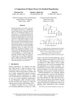
Diagnostic test abilities of HMGB1, LBP and PCT in
diagnosing bacteraemia
PCT had a sensitivity of 80.7% and a specificity of 67.8% in
diagnosing bacteraemia, with a cut-off level of 2.19 ng/ml
(Table 6). In a ROC analysis examining the abilities to identify
patients with bacteraemia, PCT performed best with an AUC
of 0.79 (95% confidence interval: 0.73–0.88) (Figure 2).
HMGB1 performed with an AUC of 0.62 (95% confidence
interval: 0.51–0.73) in the analysis, and LBP presented an
AUC of 0.74 (95% confidence interval: 0.65–0.85) (Figure 2).
Correlations between the examined markers
HMGB1 correlated weakly to IL-6 and CRP, and correlated
moderately to LBP, white blood cells and neutrophils (Table
7). LBP correlated weakly to IL-6, and correlated moderately
to PCT and CRP (Table 7).
Discussion
HMGB1 has been known for many years as a chromosomal
protein. In recent years there has been interest in HMGB1's
role as a proinflammatory cytokine [4,5]. Animal models have
shown that HMGB1 has an important role in immunopatho-
genesis in sepsis [4]. Administration of exogenous HMGB1 to
septic animals increased mortality, and administration of anti-
bodies against HMGB1 ameliorated the clinical outcome of
septic animals [4]. HMGB1 has been characterised as a 'late-
onset' proinflammatory cytokine involved in the late phases of
the septic process, after the early induction of 'early-onset'
proinflammatory cytokines such as TNFα and IL-1 [4,5]. Dis-
appointing results in trials trying to suppress early proinflam-
matory pathways in sepsis have made HMGB1 an interesting
target molecule in sepsis [4,5,16].
HMGB1 levels have been measured in several clinical sepsis
cohorts [4,14,17-20]. Three of these studies used blotting
methods [4,17,20] and three of the studies used ELISA tech-
niques [14,18,19]. In the study by Wang and colleagues,
patients with fatal sepsis had median HMGB1 levels of 84 ng/
ml and surviving sepsis patients had median HMGB1 levels of
25 ng/ml [4]. In the study by Sunden-Cullberg and colleagues,
the HMGB1 levels in critically ill patients remained elevated for
up to 1 week, with mean levels of HMGB1 over 340 ng/ml
Table 2
Microbiological and infection characteristics of the patients
Variable Infection without systemic
inflammatory response
syndrome (n = 20)
Sepsis (n = 56) Severe sepsis (n = 67) Septic shock (n = 11)
Bacteraemia
Gram-positive bacteria 0 3 17 2
Gram-negative bacteria 1 2 5 3
>1 pathogen involved 0 0 1 0
Focus of infection
Meningitis 1 2 9 0
Pneumonia 5 18 32 6
Endocarditis 0 1 4 0
Pyelonephritis 2 6 4 1
Cystitis 4 6 10 2
Cholecystitis/cholangitis 1 1 3 0
Gastroenteritis 0 1 0 0
Skin/soft tissue infection 6 9 2 1
Bone/joint infection 0 3 1 0
Other 1 9 2 1
Data presented as the absolute number.
Available online />Page 5 of 10
(page number not for citation purposes)
after a 144-hour observation period [17]. In a study of commu-
nity-acquired pneumonia by Angus and colleagues, median
HMGB1 levels of 190 ng/ml were observed [20]. Much lower
levels were seen in the three studies using HMGB1 ELISA
techniques [14,18,19]. In the study by Hatada and colleagues,
infected patients had median HMGB1 levels of 4.54 ng/ml
[18]; Yasuda and colleagues, studying infected patients with
severe acute pancreatitis, observed mean HMGB1 levels of
7.8 ng/ml [19]; and, finally, in a study performed by our group,
the median HMGB1 level in mild sepsis was 2.14 ng/ml [14].
In the present study the HMGB1 levels were comparable with
the latter three aforementioned studies using ELISA for
HMGB1 measurements [14,18,19]. HMGB1 levels in the
present study were higher in bacteraemic patients compared
with those in nonbacteraemic patients and HMGB1 correlated
to several proinflammatory markers (LBP, CRP, white blood
cells and neutrophils). These correlations seem to confirm a
proinflammatory role for HMGB1 in human sepsis. HMGB1
did not perform well in a ROC analysis examining its ability to
identify bacteraemic patients, with an AUC of only 0.62. As
Table 3
Inflammatory markers related to the severity of infection
Variable Healthy controls
(n = 32)
Infection without
SIRS (n = 20)
Sepsis (n = 56) Severe sepsis
(n = 67)
Septic shock
(n = 11)
P value
a
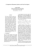
HMGB1 (ng/ml) <0.001
Median 0.77 3.4 4.3 6.7 4.8
IQR 0.6–1.5 1.8–5.4 2.9–7.1 4.1–11.1 4.1–9.2
P value
b
<0.0001 NS < 0.01 NS
Lipopolysaccharide-binding protein (μg/ml) <0.001
Median 12.7 46.3 63.3 88.7 73.3
IQR 9.8–16.8 23.9–64.7 44.8–87.9 61.3–129 62.3–91.8
P value
b
<0.0001 <0.05 <0.01 NS
Procalcitonin (ng/ml) <0.001
Median 0.05 0.15 0.4 4.4 46.1
IQR 0.04–0.06 0.07–0.5 0.13–1.3 1.3–22.2 5.9–127.5
P value
b
<0.0001 NS <0.0001 <0.05
IL-6 (pg/ml) <0.001
Median 3.4 23.6 46.9 120 6117
IQR 3–3.7 12.3–46.1 13.9–102.9 35.9–661 110–10,212
P value
b
<0.0001 NS <0.001 <0.01
C-reactive protein (mg/l) <0.01
Median 71 181 205 197
IQR 28.5–199.5 120–255 126–306 146–270
P value
b
<0.01 NS NS
White blood cells (× 10
9
/l) <0.05
Median 10.4 11.2 14.8 16.8
IQR 7.2–13.9 8.5–16.8 10.5–18.5 7.4–25.3
P value
b
NS NS NS
Neutrophils (× 10
9
/l) <0.01
Median 7.8 8.9 12.4 15.5
IQR 5.5–11.7 6.5–14.7 7.9–16.3 6.4–21.8
P value
b
NS <0.05 NS
Data presented as median and interquartile range (IQR). HMGB1, high-mobility group box-1 protein; SIRS, systemic inflammatory response
syndrome.
a
Kruskal–Wallis test.
b
Compared with the previous group in the table (Wilcoxon's two-sample test); NS, not significant.
Critical Care Vol 11 No 4 Gaïni et al.
Page 6 of 10
(page number not for citation purposes)
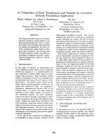
Figure 1
Boxplot of high-mobility group box-1 protein levels in healthy controls and infected patientsBoxplot of high-mobility group box-1 protein levels in healthy controls and infected patients. (Kruskal–Wallis, P < 0.001). NS, not significant.
Table 4
Inflammatory markers in survivors and in fatal cases
Variable Survivors (n = 138) Fatal cases (n = 16) P value
a
High-mobility group-box 1 protein (ng/ml) 4.9 (2.9–9.1) 5.6 (3.4–14.2) NS
Lipopolysaccharide-binding protein (μg/ml) 70.7 (45.6–112.3) 70.6 (57.1–89.7) NS
Procalcitonin (ng/ml) 1.3 (0.17–8.9) 1.7 (0.4–12.2) NS
IL-6 (pg/ml) 66.5 (21.2–174.5) 193.5 (47.9–589) NS
b
C-reactive protein (mg/l) 185 (109–263) 198 (130.5–274) NS
White blood cells (× 10
9
/l) 13.2 (8.5–17.3) 14.7 (11.5–20.9) NS
Neutrophils (× 10
9
/l) 11.2 (6.8–15.5) 12.7 (8.7–18.9) NS
Data presented as median and interquartile range. NS, not significant.
a
Wilcoxon's two sample test.
b
P = 0.06.
Table 5
Inflammatory markers in nonbacteraemic patients and in bacteraemic patients
Variable Infections without bacteraemia (n = 120) Bacteraemia (n = 34) P value
a
High-mobility group-box 1 protein (ng/ml) 4.6 (2.9–8.3) 7.3 (4.4–10.7) <0.05
Lipopolysaccharide-binding protein (μg/ml) 65.3 (42.8–91.4) 101.4 (65.2–165.5) <0.0001
Procalcitonin (ng/ml) 0.6 (0.15–3.9) 14.1 (2.9–31) <0.0001
IL-6 (pg/ml) 50.3 (18.9–140) 211 (102–1833) <0.0001
C-reactive protein (mg/l) 164 (90–245) 243 (172–306) <0.001
White blood cells (× 10
9
/l) 12.7 (8.6–16.8) 15.8 (11–21.5) <0.05
Neutrophils (× 10
9
/l) 10.5 (6.9–15.2) 13.6 (10.5–19.4) <0.05
Data presented as median and interquartile range.
a
Wilcoxon's two sample test.
Available online />Page 7 of 10
(page number not for citation purposes)
mentioned earlier, levels of HMGB1 were much lower than lev-
els reported in studies using blotting techniques. The reason
for this is not clear. One possibility is that our patients who
were recruited from an ordinary department of internal medi-
cine were less ill compared with studies conducted on inten-
sive care units. Another possibility is that we sampled patients
in the early phase of disease (within 24 hours after admission),
which perhaps could explain the low levels of a 'late-onset'
proinflammatory cytokine such as HMGB1. Finally, the chosen
laboratory technique might explain the low levels. The pres-
ence of interfering inhibitory factors/autoantibodies to
HMGB1 in human serum could affect results of HMGB1
measurements with ELISA techniques [21]. It is still unknown
whether the currently used assays detect biologically active
HMGB1. This is an important issue for future studies focusing
on HMGB1 levels and disease activity.
LBP is a protein with a central role in the innate immune
response in both Gram-negative and Gram-positive infection
when the host is challenged by an invading pathogen [6,7]. In
Gram-negative infection, LBP carries the endotoxin lipopoly-
saccharide to the CD14 receptors on the monocyte-macro-
phage cell lineage [22,23]. CD14 receptors then interact with
the Toll-like receptor 4, initiating cytokine production [22,23].
The lipotheichoic acid from pneumococci and staphylococci
activates a cellular response through Toll-like receptor 2 [24].
This response can be enhanced by LBP and CD14 [7].
Several clinical studies have examined the levels of LBP in
infected patients [25-29], in which the median levels of LBP
were between 21.1 μg/ml and 59.7 μg/ml. Only one previous
study has examined LBP's diagnostic test abilities in diagnos-
ing Gram-negative bacteraemia [29]. The authors found a sen-
sitivity of 100% and a specificity of 92% with a high cut-off
level (46.3 μg/ml) for LBP. The study only included four
patients with Gram-negative bacteraemia [29]. In the present
study, the median levels of LBP were high compared with the
previous studies. LBP levels in the present study were higher
in bacteraemic patients compared with nonbacteraemic
patients, and LBP correlated to several proinflammatory mark-
ers (HMGB1, PCT and CRP). LBP correlated to the severity
of infection. LBP did not perform well in a ROC analysis exam-
ining its ability to identify bacteraemic patients, with an AUC of
0.74.
PCT is a protein involved in the immunopathogenesis of sep-
sis. Many different parenchymal cells are able to produce PCT
when the host is challenged by a pathogen [30]. Animal mod-
els have shown that administration of exogenous PCT to sep-
tic animals increased mortality and administration of
antibodies against PCT to septic animals protected against
fatal outcome [31,32]. Elevated levels of PCT have been asso-
ciated with several conditions, such as toxic shock syndrome,
bacterial sepsis, postoperative infectious complications, men-
ingitis, cholangitis, pancreatitis with infection, malaria and fun-
gemia [33]. PCT has been shown to be a marker associated
with the severity of sepsis [34-38]. Several previous studies
have examined PCT's diagnostic test abilities in diagnosing
bacteraemia [39-44]. These studies found AUCs between
0.71 and 0.85 [39-44]. In the present study the PCT levels
increased with increasing severity of infection, with the highest
levels in severe sepsis (median 4.4 ng/ml) and in septic shock
(median 46.1 ng/ml). These data confirm findings from earlier
studies showing that PCT is a severity marker in sepsis.
Table 6
Specificity of the studied markers with cut-off levels corresponding to a sensitivity of approximately 80% in diagnosing bacteraemia
Variable Cut-off level Sensitivity (%) Specificity (%)
High-mobility group-box 1 protein 4.2 ng/ml 79.4 45.0
Lipopolysaccharide-binding protein 64.6 μg/ml 79.4 50.0
Procalcitonin 2.19 ng/ml 80.7 67.8
IL-6 94.6 pg/ml 79.4 67.5
C-reactive protein 169 mg/l 79.4 51.3
Figure 2
Receiver–operator characteristic curves comparing inflammatory markersReceiver–operator characteristic curves comparing inflammatory mark-
ers. discriminating capabilities between nonbacteraemic patients and
bacteraemic patients (P < 0.05).
Critical Care Vol 11 No 4 Gaïni et al.
Page 8 of 10
(page number not for citation purposes)
Our study data showed more than 20-fold higher PCT levels
in bacteraemic patients compared with nonbacteraemic
patients. The AUC of PCT in diagnosing bacteraemia was
0.79. This result regarding PCT's diagnostic test abilities in
diagnosing bacteraemia confirms the abovementioned find-
ings in previous studies. Diagnosis of bacteraemia at the
present time is a relatively slow process, requiring up to
several days of culturing/processing in the laboratory of micro-
biology. It is possible that a good biochemical marker for the
presence of bacteraemia could have a role in stratifying
patients to faster microbiological diagnosis with molecular
diagnostic techniques. A broad-range PCR could perhaps be
a possible strategy speeding up the species diagnosis in
bacteraemia. PCT may have a role in identifying patients that
could benefit from fast molecular diagnostics.
The strengths of the present study are its prospective design,
the relatively large sample size, well-characterised patients
and fast blood sampling after admission to the hospital. The
study focused on patients admitted to a general department of
internal medicine. Many previous studies examining
immunological, prognostic and diagnostic markers in sepsis
have focused upon critically ill patients on intensive care units.
Most patients with infections and sepsis are, however, at the
milder end of the sepsis spectrum and will be treated on gen-
eral departments of internal medicine or surgery. The risk of
work-up bias was reduced by classifying the infectious status
of the patients without access to the biochemical laboratory
results. The laboratory technicians were blinded from the clin-
ical data. The risk of spectrum bias was reduced by using rel-
atively broad inclusion criteria, including all age groups over
18 years, all kinds of infectious foci, different aetiology, differ-
ent severity and comorbidity.
A drawback of the study design was the risk of an imperfect
gold standard bias. Before inclusion of patients was begun,
the criteria for infection and sepsis severity were established
by the study group. These criteria were followed rigorously to
minimise the risk of an imperfect gold standard bias. Patients
with uncertain diagnosis were excluded for the same reason.
Drawbacks of the present study, as in many other clinical sep-
sis studies, were the heterogeneity among included patients,
a heavy burden of comorbidity, variable severity of disease,
variation in infectious focus, variation in microbiological aetiol-
ogy and different lengths of disease prior to hospitalisation.
Conclusion
This is the largest prospective study that has been conducted
regarding HMGB1 measurements in infections and sepsis.
Elevated levels of HMGB1, LBP and PCT were associated
with the presence of infection and with the presence of bacter-
aemia in patients with community-acquired infections. None of
the examined inflammatory markers had prognostic abilities in
identifying patients with fatal outcome. PCT had better diag-
nostic test abilities in diagnosing the presence of bacteraemia
compared with HMGB1 and LBP. PCT could have a future
role in identifying patients who would benefit from new faster
molecular diagnostic techniques for diagnosing bacteraemia.
Competing interests
The authors declare that they have no competing interests.
Authors' contributions
SG planned the study, wrote the protocol, collected and ana-
lysed the data, and wrote the report. OGK was responsible for
the PCT, LBP and IL-6 analyses. HJM was responsible for the
Table 7
Correlations between high-mobility group-box 1 protein (HMGB1)/lipopolysaccharide-binding protein (LBP) and the examined
inflammatory markers
HMGB1 versus marker Spearman's rP value LBP versus marker Spearman's rP value
LBP 0.3 <0.001 HMGB1 0.3 <0.001
Procalcitonin 0.15 NS Procalcitonin 0.45 <0.0001
IL-6 0.18 <0.05 IL-6 0.29 <0.001
C-reactive protein 0.27 <0.001 C-reactive protein 0.64 <0.0001
White blood cells 0.39 <0.0001 White blood cells 0.11 NS
Neutrophils 0.39 <0.0001 Neutrophils 0.11 NS
NS, not significant.
Key messages
• HMGB1 is a proinflammatory cytokine in severe infec-
tions and bacteraemia.
• LBP and PCT are severity markers in severe infections
and bacteraemia.
• PCT is a better diagnostic test marker for bacteraemia
compared with HMGB1 and LBP.
Available online />Page 9 of 10
(page number not for citation purposes)
HMGB1 analyses. CP and SSP were involved in planning the
study, in revising the manuscript and in practical clinical
aspects. All authors read and approved the final manuscript.
Acknowledgements
The study was supported by the University of Southern Denmark. The
authors thank the nurses at the Medical Department C7 for excellent
clinical assistance, and also the study nurses Lene Hergens, Anita
Nymark, Nete Bỹlow and Helle Mứller for excellent clinical assistance.
They also thank Joan Clausen, Hanne Madsen and Kirsten Bank
Petersen for excellent technical assistance.
References
1. Wheeler AP, Bernard GR: Treating patients with severe sepsis.
N Engl J Med 1999, 340:207-214.
2. Wang H, Yang H, Czura CJ, Sama AE, Tracey KJ: HMGB1 as a
late mediator of lethal systemic inflammation. Am J Respir Crit
Care Med 2001, 164:1768-1773.
3. Bustin M: Regulation of DNA-dependent activities by the func-
tional motifs of the high-mobility-group chromosomal
proteins. Mol Cell Biol 1999, 19:5237-5246.
4. Wang H, Bloom O, Zhang M, Vishnubhakat JM, Ombrellino M, Che
J, Frasier A, Yang H, Ivanova S, Borovikova L, et al.: HMG-1 as a
late mediator of endotoxin lethality in mice. Science 1999,
285:248-251.
5. Andersson U, Tracey KJ: HMGB1 in sepsis. Scand J Infect Dis
2003, 35:577-584.
6. Schumann RR, Zweigner J: A novel acute-phase marker:
lipopolysaccharide binding protein (LBP). Clin Chem Lab Med
1999, 37:271-274.
7. Zweigner J, Schumann RR, Weber JR: The role of lipopolysac-
charide-binding protein in modulating the innate immune
response. Microbes Infect 2006, 8:946-952.
8. Gendrel D, Raymond J, Coste J, Moulin F, Lorrot M, Guộrin S,
Ravilly S, Lefộvre H, Royer C, Lacombe C, et al.: Comparison of
procalcitonin with C-reactive protein, interleukin 6 and inter-
feron-alpha for differentiation of bacterial vs. viral infections.
Pediatr Infect Dis J 1999, 18:875-881.
9. Assicot M, Gendrel D, Carsin H, Raymond J, Guilbaud J, Bohuon
C: High serum procalcitonin concentrations in patients with
sepsis and infection. Lancet 1993, 341:515-518.
10. Vincent JL, Moreno R, Takala J, Willatts S, De Mendonca A, Bruin-
ing H, Reinhart CK, Suter PM, Thijs LG: The SOFA (Sepsis-
related Organ Failure Assessment) score to describe organ
dysfunction/failure. On behalf of the Working Group on Sep-
sis-Related Problems of the European Society of Intensive
Care Medicine. Intensive Care Med 1996, 22:707-710.
11. Charlson ME, Pompei P, Ales KL, MacKenzie CR: A new method
of classifying prognostic comorbidity in longitudinal studies:
development and validation. J Chronic Dis 1987, 40:
373-383.
12. Bone RC, Sibbald WJ, Sprung CL: The ACCP-SCCM consensus
conference on sepsis and organ failure. Chest 1992,
101:1481-1483.
13. Yamada S, Yakabe K, Ishii J, Imaizumi H, Maruyama I: New high
mobility group box 1 assay system. Clin Chim Acta 2006,
372:173-178.
14. Gaùni S, Pedersen SS, Pedersen C, Koldkjổr OG, Mứller HJ: High
mobility group box-1 protein in patients with suspected com-
munity-acquired infections and sepsis: a prospective study.
Crit Care 2007, 11:R32.
15. DeLong ER, DeLong DM, Clarke-Pearson DL: Comparing the
areas under two or more correlated receiver operating charac-
teristic curves: a nonparametric approach. Biometrics 1988,
44:837-845.
16. Abraham E: Why immunomodulatory therapies have not
worked in sepsis. Intensive Care Med 1999, 25:556-566.
17. Sundộn-Cullberg J, Norrby-Teglund A, Rouhiainen A, Rauvala H,
Herman G, Tracey KJ, Lee ML, Andersson J, Tokics L, Treutiger CJ:
Persistent elevation of high mobility group box-1 protein
(HMGB1) in patients with severe sepsis and septic shock. Crit
Care Med 2005, 33:564-573.
18. Hatada T, Wada H, Nobori T, Okabayashi K, Maruyama K, Abe Y,
Uemoto S, Yamada S, Maruyama I: Plasma concentrations and
importance of High Mobility Group Box protein in the progno-
sis of organ failure in patients with disseminated intravascular
coagulation. Thromb Haemost 2005, 94:975-979.
19. Yasuda T, Ueda T, Takeyama Y, Shinzeki M, Sawa H, Nakajima T,
Ajiki T, Fujino Y, Suzuki Y, Kuroda Y: Significant increase of
serum high-mobility group box chromosomal protein 1 levels
in patients with severe acute pancreatitis. Pancreas 2006,
33:359-363.
20. Angus DC, Yang L, Kong L, Kellum JA, Delude RL, Tracey KJ,
Weissfeld L: Circulating high-mobility group box 1 (HMGB1)
concentrations are elevated in both uncomplicated pneumo-
nia and pneumonia with severe sepsis. Crit Care Med 2007,
35:1061-1067.
21. Urbonaviciute V, Fỹrnrohr BG, Weber C, Haslbeck M, Wilhelm S,
Herrmann M, Voll RE: Factors masking HMGB1 in human serum
and plasma. J Leukoc Biol 2007, 81:67-74.
22. Heumann D, Roger T: Initial responses to endotoxins and
Gram-negative bacteria.
Clin Chim Acta 2002, 323:59-72.
23. Pồlsson-McDermott EM, O'Neill LA: Signal transduction by the
lipopolysaccharide receptor, Toll-like receptor-4. Immunology
2004, 113:153-162.
24. Schrửder NW, Morath S, Alexander C, Hamann L, Hartung T,
Zọhringer U, Gửbel UB, Weber JR, Schumann RR: Lipoteichoic
acid (LTA) of Streptococcus pneumoniae and Staphylococcus
aureus activates immune cells via Toll-like receptor (TLR)-2,
lipopolysaccharide-binding protein (LBP), and CD14, whereas
TLR-4 and MD-2 are not involved. J Biol Chem 2003,
278:15587-15594.
25. Calvano SE, Thompson WA, Marra MN, Coyle SM, de Riesthal HF,
Trousdale RK, Barie PS, Scott RW, Moldawer LL, Lowry SF:
Changes in polymorphonuclear leukocyte surface and plasma
bactericidal/permeability-increasing protein and plasma
lipopolysaccharide binding protein during endotoxemia or
sepsis. Arch Surg 1994, 129:220-226.
26. Opal SM, Scannon PJ, Vincent JL, White M, Carroll SF, Palardy JE,
Parejo NA, Pribble JP, Lemke JH: Relationship between plasma
levels of lipopolysaccharide (LPS) and LPS-binding protein in
patients with severe sepsis and septic shock. J Infect Dis
1999, 180:1584-1589.
27. Blairon L, Wittebole X, Laterre PF: Lipopolysaccharide-binding
protein serum levels in patients with severe sepsis due to
gram-positive and fungal infections. J Infect Dis 2003,
187:287-291.
28. Prucha M, Herold I, Zazula R, Dubska L, Dostal M, Hildebrand T,
Hyanek J: Significance of lipopolysaccharide-binding protein
(an acute phase protein) in monitoring critically ill patients.
Crit Care 2003, 7:R154-R159.
29. Oude Nijhuis CS, Vellenga E, Daenen SM, van der Graaf WT, Gie-
tema JA, Groen HJ, Kamps WA, de Bont ES: Lipopolysaccha-
ride-binding protein: a possible diagnostic marker for Gram-
negative bacteremia in neutropenic cancer patients. Intensive
Care Med 2003, 29:2157-2161.
30. Christ-Crain M, Mỹller B: Procalcitonin in bacterial infections
hype, hope, more or less? Swiss Med Wkly 2005,
135:451-460.
31. Nylen ES, Whang KT, Snider RH Jr, Steinwald PM, White JC,
Becker KL: Mortality is increased by procalcitonin and
decreased by an antiserum reactive to procalcitonin in experi-
mental sepsis. Crit Care Med 1998, 26:1001-1006.
32. Martinez JM, Wagner KE, Snider RH, Nylen ES, Muller B, Sarani B,
Becker KL, White JC: Late immunoneutralization of procalci-
tonin arrests the progression of lethal porcine sepsis. Surg
Infect (Larchmt) 2001, 2:193-202.
33. Mỹller B, Becker KL: Procalcitonin: how a hormone became a
marker and mediator of sepsis. Swiss Med Wkly 2001,
131:595-602.
34. Ugarte H, Silva E, Mercan D, De Medonỗa A, Vincent JL: Procalci-
tonin used as a marker of infection in the intensive care unit.
Crit Care Med 1999, 27:498-504.
35. Meisner M, Tschaikowsky K, Palmaers T, Schmidt J: Comparison
of procalcitonin (PCT) and C-reactive protein (CRP) plasma
concentrations at different SOFA scores during the course of
sepsis and MODS. Crit Care (Lond) 1999, 3:45-50.
36. Cheval C, Timsit JF, Garrouste-Orgeas M, Assicot M, Jonghe BD,
Misset B, Bohuon C, Carlet J: Procalcitonin (PCT) is useful in
predicting the bacterial origin of an acute circulatory failure in
critically ill patients. Intensive Care Med 2000, 26:S153-S158.
Critical Care Vol 11 No 4 Gaïni et al.
Page 10 of 10
(page number not for citation purposes)
37. Chan YL, Tseng CP, Tsay PK, Chang SS, Chiu TF, Chen JC: Pro-
calcitonin as a marker of bacterial infection in the emergency
department: an observational study. Crit Care 2004,
8:R12-R20.
38. Gaïni S, Koldkjaer O, Pedersen C, Pedersen S: Procalcitonin,
lipopolysaccharide-binding protein, interleukin-6 and C-reac-
tive protein in community-acquired infections and sepsis: a
prospective study. Crit Care 2006, 10:R53.
39. Rintala EM, Aittoniemi J, Laine S, Nevalainen TJ, Nikoskelainen J:
Early identification of bacteremia by biochemical markers of
systemic inflammation. Scand J Clin Lab Invest 2001,
61:523-530.
40. Chirouze C, Schuhmacher H, Rabaud C, Gil H, Khayat N, Esta-
voyer JM, May T, Hoen B: Low serum procalcitonin level accu-
rately predicts the absence of bacteremia in adult patients
with acute fever. Clin Infect Dis 2002, 35:156-161.
41. Bell K, Wattie M, Byth K, Silvestrini R, Clark P, Stachowski E, Ben-
son EM: Procalcitonin: a marker of bacteraemia in SIRS.
Anaesth Intensive Care 2003, 31:629-636.
42. Aalto H, Takala A, Kautiainen H, Repo H: Laboratory markers of
systemic inflammation as predictors of bloodstream infection
in acutely ill patients admitted to hospital in medical
emergency. Eur J Clin Microbiol Infect Dis 2004, 23:699-704.
43. Persson L, Engervall P, Magnuson A, Vikerfors T, Söderquist B,
Hansson LO, Tidefelt U: Use of inflammatory markers for early
detection of bacteraemia in patients with febrile neutropenia.
Scand J Infect Dis 2004, 36:365-371.
44. von Lilienfeld-Toal M, Dietrich MP, Glasmacher A, Lehmann L,
Breig P, Hahn C, Schmidt-Wolf IG, Marklein G, Schroeder S, Stu-
ber F: Markers of bacteremia in febrile neutropenic patients
with hematological malignancies: procalcitonin and IL-6 are
more reliable than C-reactive protein. Eur J Clin Microbiol
Infect Dis 2004, 23:539-544.