Báo cáo y học: "The effect of different volumes and temperatures of saline on the bladder pressure measurement in critically ill patients" potx
Bạn đang xem bản rút gọn của tài liệu. Xem và tải ngay bản đầy đủ của tài liệu tại đây (180.09 KB, 7 trang )
Open Access
Available online />Page 1 of 7
(page number not for citation purposes)
Vol 11 No 4
Research
The effect of different volumes and temperatures of saline on the
bladder pressure measurement in critically ill patients
Davide Chiumello
1
, Federica Tallarini
2
, Monica Chierichetti
2
, Federico Polli
2
, Gianluigi Li Bassi
2
,
Giuliana Motta
2
, Serena Azzari
2
, Cristian Carsenzola
2
and Luciano Gattinoni
2
1
Dipartimento di Anestesia e Rianimazione, Fondazione IRCCS – 'Ospedale Maggiore Policlinico, Mangiagalli, Regina Elena', Via F Sforza 35, 20122
Milan, Italy
2
Istituto di Anestesia e Rianimazione Università degli Studi di Milano, 'Ospedale Maggiore Policlinico, Mangiagalli, Regina Elena', Via F Sforza 35,
20122 Milan, Italy
Corresponding author: Davide Chiumello,
Received: 9 Feb 2007 Revisions requested: 19 Mar 2007 Revisions received: 16 May 2007 Accepted: 26 Jul 2007 Published: 26 Jul 2007
Critical Care 2007, 11:R82 (doi:10.1186/cc6080)
This article is online at: />© 2007 Chiumello et al.; licensee BioMed Central Ltd.
This is an open access article distributed under the terms of the Creative Commons Attribution License ( />),
which permits unrestricted use, distribution, and reproduction in any medium, provided the original work is properly cited.
Abstract
Introduction Intra-abdominal hypertension is common in
critically ill patients and is associated with increased severity of
organ failure and mortality. The techniques most commonly used
to estimate intra-abdominal pressure are measurements of
bladder and gastric pressures. The bladder technique requires
that the bladder be infused with a certain amount of saline, to
ensure that there is a conductive fluid column between the
bladder and the transducer. The aim of this study was to
evaluate the effect of different volumes and temperatures of
infused saline on bladder pressure measurements in
comparison with gastric pressure.
Methods Thirteen mechanically ventilated critically ill patients
(11 male; body mass index 25.5 ± 4.6 kg/m
2
; arterial oxygen
tension/fractional inspired oxygen ratio 225 ± 48 mmHg) were
enrolled. Bladder pressure was measured using volumes of
saline from 50 to 200 ml at body temperature (35 to 37°C) and
room temperature (18 to 20°C).
Results Bladder pressure was no different between 50 ml and
100 ml saline (9.5 ± 3.7 mmHg and 13.7 ± 5.6 mmHg), but it
significantly increased with 150 and 200 ml (21.1 ± 10.4 mmHg
and 27.1 ± 15.5 mmHg). Infusion of saline at room temperature
caused a significantly greater bladder pressure compared with
saline at body temperature. The lowest difference between
bladder and gastric pressure was obtained with a volume of 50
ml.
Conclusion The bladder acts as a passive structure,
transmitting intra-abdominal pressure only with saline volumes
between 50 ml and 100 ml. Infusion of a saline at room
temperature caused a higher bladder pressure, probably
because of contraction of the detrusor bladder muscle.
Introduction
Intra-abdominal pressure (IAP) is the pressure generated
inside the abdominal cavity and depends on the degree of flex-
ibility of the diaphragm and abdominal wall, and on the density
of its contents [1]. Intra-abdominal hypertension (IAH), defined
as an abnormal increase in IAP, can be common in critically ill
patients, being present in 18% to 81% of the patients depend-
ing on the cut-off level used [2-8].
Several clinical conditions such as accumulation of blood,
ascites, retroperitoneal haematoma, bowel oedema, necrotiz-
ing pancreatitis, massive fluid resuscitation, packing after con-
trol laparotomy and closure of a swollen noncompliant
abdominal wall may induce IAH [3,9]. IAH has adverse effects
on several organs, causing reductions in cardiac output [10],
deterioration in gas exchange [11-13] and decreases in
splachnic-renal perfusion [14-16]. In surgical [17], trauma [2]
and medical [6] critically patients, the IAH was an independent
predictor factor of hospital mortality. Although surgical decom-
pression remains the only definitive therapy in the case of sub-
stantial IAH, and the IAP is lower after decompression,
mortality remains considerable [18,19].
Because the abdomen and its contents can be considered to
be relatively noncompressive and fluid in character, behaving
in accordance with Pascal's law, the IAP measured at one
IAH = intra-abdominal hypertension; IAP = intra-abdominal pressure; IBP = intra-bladder pressure; IGP = intra-gastric pressure.
Critical Care Vol 11 No 4 Chiumello et al.
Page 2 of 7
(page number not for citation purposes)
point is assumed to reflect the IAP throughout the abdomen
[4]. A variety of methods for measuring IAP have been pro-
posed, which are either indirect (by transduction of bladder,
gastric, or uterine pressure using a ballon catheter) or direct
(using a intraperitoneal catheter) [1,20]. However, among the
different methods, the intra-bladder pressure (IBP) technique
is the most commonly used because of its simplicity and low
cost [4,21].
The bladder technique, originally described by Kron and cow-
orkers [14], assumes that the bladder behaves like a passive
pressure membrane transducer when it is infused with a small
amount of saline [14]. However, various saline volumes for
bladder priming, 50 ml up to 250 ml, have been used to esti-
mate IBP [10,14,21-23]. Previous studies demonstrated that
a small volume of saline (10 to 25 ml) is required to prime the
bladder in order to avoid overestimating the IBP [22,24,25].
The International Abdominal Compartment Syndrome Con-
sensus Conference [1] suggested that a maximal instillation
volume of 25 ml of saline should be used. In addition the blad-
der – being a muscular organ – may change its elasticity in
response to various external stimuli, such as an infusion of
warm saline [26]. Thus the bladder may not always behave like
a passive elastic structure, leading to inaccurate estimation of
IAP.
The aim of this study was to evaluate IAP estimated by bladder
pressure, measured with the bladder infused with different vol-
umes of saline at room and body temperatures, in comparison
with intra-gastric pressure (IGP).
Materials and methods
Study population
Thirteen sedated, mechanically ventilated patients admitted to
the intensive care unit of Ospedale Policlinico were enrolled.
Exclusion criteria were contraindications to bladder pressure
measurement (a recent history of bladder surgery, haematuria,
trauma, or neurogenic bladder).
The study was approved by the institutional review board of
our hospital, and informed consent was obtained in accord-
ance with Italian national regulations.
Study protocol
The IBP was measured using a revision of the Cheatham's
original technique [21] with disposable pressure transducer
(Edward Lifesciences, Irvine, CA, USA). A 18-gauge needle
was inserted into the culture aspiration port of the Foley's cath-
eter and connected with a sterile tube to the pressure trans-
ducer using two three-way stopcocks. A standard infusion bag
of normal saline was attached to one stopcock and a 60 ml
syringe was connected to the second stopcock. Before taking
any measurements, the system was flushed with sterile saline
and the pubic symphysis was always used as zero reference
point with the subject in the complete supine position.
The IBP was measured at different volumes of saline infusion
(50, 100, 150 and 200 ml, with steps of 50 ml) at room tem-
perature (18 to 20°C). The sequence of measurements was
then repeated using saline infusion warmed to body tempera-
ture (35 to 37°C). At each volume of saline, the IBP was
recorded 5 to 10 s after the termination of saline infusion (early
recording) and 5 min later (late recording) by keeping the blad-
der catheter closed. After each measurement the bladder was
emptied.
Each patient was studied at an external positive end-expiratory
pressure of 10 cmH
2
O, with the other ventilatory parameters
(previously selected by the attending physician) unchanged
during the study. Thus, each patient underwent two rand-
omized series of measurements.
The IGP was measured using a radio-opaque balloon (Smart-
Cath; Bicore, Irvine, CA, USA) connected to a pressure trans-
ducer (Bentley Trantec; Bentley Laboratories, Irvine, CA, USA)
[27]. For measurement purposes, the gastric balloon was
inflated with 1.0 ml air.
The IBP and IGP were measured at end-expiration and the sig-
nals were recorded on a personal computer for subsequent
analysis (Colligo; Elekton, Milan, Italy).
The level of sedation before the study was evaluated using the
Ramsay scale [28]. The Simplified Acute Physiology Score II
was used to assess the severity of systemic illness at study
entry [29], whereas the Sepsis Related Organ Failure Assess-
ment score was computed on the day of the study by consid-
ering the worst value for each organ system (respiratory,
cardiovascular, renal, coagulation, liver and neurological) [30].
Statistical analysis
The effects of volume, temperature of saline infused and time
of recording were analyzed by two-way repeated measures
analysis of variance, followed by Student/Newman Keuls test
for multiple comparison (SigmaStat 2.03; SPSS Inc., Chi-
cago, IL, USA) [31]. P < 0.05 was considered statistically
significant.
The mean bias (bladder minus gastric pressure), precision
(standard deviation of the bias) and limits of agreement were
calculated using the Bland-Altman analysis [32]. The percent-
age error was calculated in accordance with the method pro-
posed by Crichley and coworkers [33].
All data are expressed as mean ± standard deviation.
Results
The main clinical characteristics are reported in Table 1. The
patients were studied after a mean of 6 ± 3.8 days from inten-
sive care admission.
Available online />Page 3 of 7
(page number not for citation purposes)
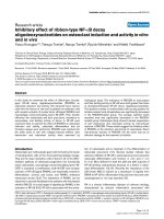
The IBP was no different with 50 and 100 ml volumes of saline
(9.5 ± 3.7 mmHg and 13.7 ± 5.6 mmHg; P = 0.071), but it
was significantly higher with 150 and 200 ml saline (21.1 ±
10.4 and 27.1 ± 15.5 mmHg; P < 0.001; Figure 1). Consider-
ing the IBP measured with 50 ml of saline infused as refer-
ence, we computed the agreements with the IBP measured
with 100, 150 and 200 ml of saline (Table 2).
Four patients (30.7% of the population) were classified as hav-
ing IAH (IAP >12 mmHg) when 50 ml saline was used. This
increased to eight patients (61.5% of the population) when
100 ml saline was used.
The IBP was significantly lower 5 min after saline infusion (late
recording) than just after the saline infusion (early recording),
but only with the bladder infused with 200 and 150 ml of saline
(21.1 ± 10.4 versus 16.2 ± 5.6 mmHg, and 27.1 ± 15.5 ver-
sus 19.3 ± 8.9 mmHg; P < 0.005; Figure 1). At each volume
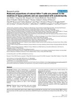
infused, the infusion of saline at body temperature resulted in
a significantly lower IBP than did infusion of saline at room
temperature (8.2 ± 4.4 versus 7.7 ± 3.7 mmHg with 50 ml
saline, 11.4 ± 5.9 versus 10.2 ± 3.8 mmHg with 100 ml saline,
15.4 ± 8.8 versus 13.3 ± 5.0 mmHg with 150 ml saline, and
25.7 ± 16.5 versus 22.8 ± 17.0 mmHg with 200 ml saline; P
< 0.001; Figure 2). The differences between the paired meas-
urements of IGP and IBP (bias) are given in Table 3. The low-
est bias was found for a 50 ml volume of saline, whereas the
bias increased with increasing the volume of saline infused in
the bladder.
Discussion
The major findings of this study were as follows. First, increas-
ing the volume of saline infused led to higher IBP. Second, the
IBP was significantly lower when measured after 5 min com-
pared with when it was measured just after the termination of
the volume infusion, but only with 150 and 200 ml saline.
Third, the IBP was significantly lower when measured with
infusion of saline at body temperature compared with saline at
room temperature. Finally, the lowest bias between the IBP
and IGP was obtained with the bladder infused with 50 ml
saline.
An increase in IAP is associated with various organ dysfunc-
tions (local and systemic), which in turn are associated with
significantly increased in morbidity and mortality [1]. Despite
these potential adverse clinical consequences, however, IAP
is commonly measured only when there is some clinical suspi-
cion; furthermore, there is currently no general consensus on
how frequently it should be measured [34]. Sugrue and cow-
orkers [35] found that clinical examination alone was not accu-
rate in estimating IAP, finding that the likelihood of physicians
correctly identifying IAH was lower than 50%. Thus, accurate
estimation of IAH is fundamental to appropriate and timely
patient management [36].
Table 1
Patient's characteristics
Patient Age
(years)
BMI
(kg/m
2
)
Sex SAPS II
score
SOFA PEEP
(cmH
2
O)
PaO
2
/FiO
2
(mmHg)
MAP
(mmHg)
Hourly
urine
output
(ml/hour)
Ramsay
score
Diagnosis Outcome
1 72 30.9 M 35 4 10 218 100 80 7 Sepsis D
2 83 26.3 M 48 8 10 285 67 100 5 Sepsis S
3 70 26.2 M 40 8 2 228 107 60 4 Sepsis S
4 72 26.0 M 32 8 6 208 100 60 5 Sepsis S
5 65 34.6 F 47 5 10 173 100 100 5 Sepsis S
6 55 20.2 F 36 7 13 230 68 100 6 Sepsis S
7 43 24.9 M 26 15 13 211 84 140 5 Sepsis S
8 87 27.8 M 41 3 17 280 100 80 7 Sepsis S
9 72 26.3 M 35 5 15 240 100 100 7 ALI post
surgery
S
10 77 16.4 M 43 12 15 170 100 80 7 ARDS D
11 56 19.6 M 27 2 8 288 100 200 6 Sepsis D
12 79 24.8 M 53 13 5 133 87 50 7 Sepsis D
13 74 27.8 M 46 9 5 195 80 110 7 Sepsis S
Total or mean ± SD 68 ± 13 25.5 ± 4.6 11 M/2 F 7.7 ± 3.8 9.5 ± 4.6 9.5 ± 4.6 225 ± 48 92 ± 13 96 ± 39 6 ± 1 4 D/9 S
The Simplified Acute Physiology Score (SAPS) II was used to assess the severity of systemic illness at study entry. The Sepsis-Related Organ
Failure Assessment (SOFA) was used to assess the organ failure at the day of the study. ALI, acute lung injury; ARDS, acute respiratory distress
syndrome; BMI, body mass index; D, dead; F, female; M, male; MAP, mean arterial pressure; PEEP, positive end-expiratory pressure; S, survived;
SD, standard deviation.
Critical Care Vol 11 No 4 Chiumello et al.
Page 4 of 7
(page number not for citation purposes)
The most widely used technique to measure the IAP is the
bladder pressure technique, as proposed by Kron and
coworkers [14]. In that study the authors found that the IBP
measured using saline volumes between 50 and 100 ml
through a Foley catheter correlated well with pressures meas-
ured using a peritoneal dialysis catheter during several infu-
sions of peritoneal dialysis solution. Iberti and colleagues [10],
in a canine model of increased IAP, estimated bladder pres-
sure with the bladder empty; they demonstrated that the IBP
accurately reflected the IAP. Fusco and coworkers [22], using
a human model in which IAP ranged between 0 and 25 mmHg
during laparoscopic surgery, found that the bladder emptied
(with a volume of 0 ml) yielded the most accurate estimation of
IAP. However, at an IAP of 25 mmHg the bladder volume
exhibiting the lowest bias was 50 ml.
In the present study, although we did not find any statistically
significant difference (there was only a trend) in IBP measured
using saline volumes of 50 and 100 ml, this difference could
lead to a patient being incorrectly identified as having IAH if a
100 ml rather than a 50 ml of volume were used. Similarly, De
Waele and colleagues [24] demonstrated that 12 patients
were categorized as suffering from IAH when a volume of 10
ml was used, increasing to 15 and 17 patients, respectively,
when 50 and 100 ml volumes were used. Previous studies
conducted in adult patients [22,24,25] found that the increase
in IBP was statistically significant with a small instillation vol-
ume, and two studies conducted in children and infants
[37,38] found that the IAP is most accurately measured by
instilling into the bladder 1 ml saline per kilogram of body
weight. Thus, it has been proposed that the appropriate
amount of volume is that required to create a fluid column with-
out interposed air [39].
Although these findings clearly indicate that the IBP can over-
estimate IAP when large volumes of saline are infused, the
possible mechanisms involved are still not clearly understood.
The bladder is a muscular membranous organ that is com-
posed of four layers, namely mucous, adventitia, serosa and
muscularis, and its elasticity decreases in response to a direct
mechanical increase in stress and strain on its structure (when
a large amount of saline is infused). In addition, the elasticity of
bladder can also be reduced by contraction of the detrusor
muscle, mediated by sensory receptors located in the bladder
wall, after a rapid infusion of saline or other fluid that is not at
body temperature [26].
A recording of bladder pressure 5 min after termination of the
infusion yielded a significantly lower IBP only with a volume of
saline up to 150 ml; this suggests that the bladder takes
longer to reach a stable condition only when it is infused with
large volumes. However, this is not relevant in current clinical
practice, because the IAP is usually measured with volumes of
saline lower than 150 ml.
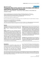
Figure 1
IBPs measured at different volumes of saline and IGP: early versus lateIBPs measured at different volumes of saline and IGP: early versus late.
Shown are the intra-bladder pressures (IBPs) measured at different vol-
umes of saline (black circle indicates early recording, and white circle
indicates late recording) and intra-gastric pressure (IGP; black square)
at 10 cmH
2
O of positive end-expiratory pressure. ^P < 0.05 versus 50
and 100 ml saline; *P < 0.05 versus 50, 100, and 150 ml saline; °P <
0.05 versus late recording;
†
P < 0.05 versus intra-bladder pressure.
Table 2
Agreement analysis between bladder pressure and bladder pressure
Volumes of saline
(ml)
Mean (mmHg) Bias (mmHg) Precision (mmHg) Lower limits of
agreement
(mmHg)
Upper limits of
agreement
(mmHg)
Percentage error
100 13.7 4.2 2.9 -1.4 (-4.5 to +1.6) 9.9 (6.9 to 13.0) ± 41%
150 21.1 11.2 9.8 -8.0 (-18.7 to +2.8) 30.4 (19.6 to 41.2) ± 91%
200 27.1 17.6 14.7 -11.1 (-26.4 to +4.3) 46.4 (31.0 to 61.7) ± 106%
Shown is an agreement analysis between bladder pressure measured with 50 ml saline (as reference) and that bladder pressure measured with
100, 150 and 200 ml saline. The bias, precision, limits of agreement and percentage error were computed considering intra-bladder pressure
(IBP) at 50 ml versus IBP at 100, 150 and 200 ml.
Available online />Page 5 of 7
(page number not for citation purposes)
We found that infusion of saline at body temperature, at each
volume infused, also resulted in a significantly lower IBP com-
pared with infusion of saline at room temperature. Rapid infu-
sion of saline at a temperature lower than body temperature
may activate contraction of the detrusor muscle (as mentioned
above) by a reflex loop through nociceptors with C afferent
fibres located in the bladder wall [26], causing a falsely ele-
vated IAP recording.
Another possible cause of reduced elasticity of the bladder
might be continued urine drainage through the catheter [40].
In critically ill patients, De Waele and coworkers [24] observed
a direct relationship between the duration of catheterization
and the difference in bladder pressure measured using vol-
umes of saline of 10 and 100 ml. This suggests that the blad-
der should be filled only minimally if an accurate measurement
of IAP is to be obtained, especially in patients with prolonged
catheterization.
In cases of bladder trauma, pelvic fractures or haematoma, or
neurogenic bladder, in which the bladder pressure technique
cannot be applied, the IGP technique is recommended [1].
Compared with the IBP technique, IGP measurements do not
interfere with urine output and avoid risk for infection [22]. In
critically ill patients and in patients undergoing laparoscopic
cholecystectomy with the abdominal cavity inflated at a pres-
sure of 20 mmHg, a clinically acceptable agreement between
IGP and IBP was observed [41,42]. Unexpectedly, we found
much greater limits of agreement, probably because of the
presence of gastric motor activity, which falsely increases
'true' estimation of IAP.
Conclusion
In clinical practice the IAP should be estimated using the IBP
technique, infusing the bladder with only a small amount of vol-
ume of saline at body temperature to avoid overestimating the
IAP. If this is not feasible, then the IGP should be measured.
Competing interests
The authors declare that they have no competing interests.
Figure 2
IBPs measures at different volumes of saline: saline at room tempera-ture versus body temperatureIBPs measures at different volumes of saline: saline at room tempera-
ture versus body temperature. The intra-bladder pressure (IBP) meas-
ured at the different volumes of saline (black circle indicates saline at
room temperature, and white circle indicates saline at body tempera-
ture). ^P < 0.05 versus 50 and 100 ml saline; *P < 0.05 versus saline
at room temperature.
Key messages
• In clinical practice, IAP should be estimated using the
IBP technique with the bladder infused with only a small
volume of saline.
• The saline infused should be at body temperature to
avoid overestimating the IAP.
• It is recommended that sufficient equilibration time be
allowed before the IAP is measured.
• IGP correlates with IBP only at low volumes of saline.
Table 3
Agreement analysis between bladder and gastric pressure
Volumes of saline
(ml)
Mean (mmHg) Bias (mmHg) Precision (mmHg) Lower limits of
agreement
(mmHg)
Upper limits of
agreement
(mmHg)
Percentage error
50 9.5 1.2 4.3 -7.2 (-11.7 to -2.7) 9.6 (5.1 to 14.1) ± 89%
100 13.7 -2.9 6.3 -15.3 (-21.9 to -8.7) 9.5 (2.8 to 16.1) ± 90%
150 21.1 -9.9 13.0 -35.4 (-49.8 to -21.1) 15.6 (1.3 to 30.0) ± 121%
200 27.1 -16.2 17.9 -51.2 (-69.9 to -32.5) 18.8 (0.1 to 37.5) ± 129%
Shown is an agreement analysis between bladder pressure (measured at different volumes of saline) and gastric pressure. The bias, precision,
limits of agreement and percentage error were computed considering intra-bladder pressure versus intra-gastric pressure at each volume of saline
infused.
Critical Care Vol 11 No 4 Chiumello et al.
Page 6 of 7
(page number not for citation purposes)
Authors' contributions
DC conceived of the study, participated in its design and coor-
dination, performed the measurements and wrote a first draft
of the manuscript. FT participated in the study design and
coordination, performed the measurements and to helped
draft the manuscript. MC participated in the study design and
coordination, and performed the measurements. FP performed
the statistical analysis and helped to draft the manuscript. GLB
participated in the study design and coordination, and
performed the measurements. GM participated in the study
design and coordination, and performed the measurements.
SA participated in the study design and coordination, and per-
formed the measurements. CC participated in the study
design and coordination, and performed the measurements.
LG conceived the study, participated in its design and
coordination, coordinated the final analysis of collected data
and revised the manuscript, writing its final version.
References
1. Malbrain ML, Cheatham ML, Kirkpatrick A, Sugrue M, Parr M, De
Waele W, Balogh Z, Leppäniemi A, Olvera C, Ivatury R, et al.:
Results from the International Conference of Experts on Intra-
abdominal Hypertension and Abdominal Compartment Syn-
drome. I. Definitions. Intensive Care Med 2006, 32:1722-1732.
2. Balogh Z, McKinley BA, Holcomb JB, Miller CC, Cocanour CS,
Kozar RA, Valdivia A, Ware DN, Moore FA: Both primary and sec-
ondary abdominal compartment syndrome can be predicted
early and are harbingers of multiple organ failure. J Trauma
2003, 54:848-859.
3. Malbrain MLNG: Abdominal pressure in the critically ill. Curr
Opin Crit Care 2000, 6:17-29.
4. Malbrain ML: Different techniques to measure intra-abdominal
pressure (IAP): time for a critical re-appraisal. Intensive Care
Med 2004, 30:357-371.
5. Malbrain ML, Chiumello D, Pelosi P, Wilmer A, Brienza N, Malcangi
V, Bihari D, Innes R, Cohen J, Singer P, et al.: Prevalence of intra-
abdominal hypertension in critically ill patients: a multicentre
epidemiological study. Intensive Care Med 2004, 30:822-829.
6. Malbrain ML, Chiumello D, Pelosi P, Bihari D, Innes R, Ranieri VM,
Del Turco M, Wilmer A, Brienza N, Malcangi V, et al.: Incidence
and prognosis of intraabdominal hypertension in a mixed pop-
ulation of critically ill patients: a multiple-center epidemiologi-
cal study. Crit Care Med 2005, 33:315-322.
7. Sugrue M, Buist MD, Hourihan F, Deane S, Bauman A, Hillman K:
Prospective study of intra-abdominal hypertension and renal
function after laparotomy. Br J Surg 1995, 82:235-238.
8. Sugrue M, Jones F, Janjua KJ, Deane SA, Bristow P, Hillman K:
Temporary abdominal closure: a prospective evaluation of its
effects on renal and respiratory physiology. J Trauma 1998,
45:914-921.
9. Morken J, West MA: Abdominal compartment syndrome in the
intensive care unit. Curr Opin Crit Care 2001, 7:268-274.
10. Iberti TJ, Kelly KM, Gentili DR, Hirsch S, Benjamin E: A simple
technique to accurately determine intra-abdominal pressure.
Crit Care Med 1987, 15:
1140-1142.
11. Cullen DJ, Coyle JP, Teplick R, Long MC: Cardiovascular, pulmo-
nary, and renal effects of massively increased intra-abdominal
pressure in critically ill patients. Crit Care Med 1989,
17:118-121.
12. Gattinoni L, Pelosi P, Suter PM, Pedoto A, Vercesi P, Lissoni A:
Acute respiratory distress syndrome caused by pulmonary
and extrapulmonary disease. Different syndromes? Am J
Respir Crit Care Med 1998, 158:3-11.
13. Ranieri VM, Brienza N, Santostasi S, Puntillo F, Mascia L, Vitale N,
Giuliani R, Memeo V, Bruno F, Fiore T, et al.: Impairment of lung
and chest wall mechanics in patients with acute respiratory
distress syndrome: role of abdominal distension. Am J Respir
Crit Care Med 1997, 156:1082-1091.
14. Kron IL, Harman PK, Nolan SP: The measurement of intra-
abdominal pressure as a criterion for abdominal re-explora-
tion. Ann Surg 1984, 199:28-30.
15. Diebel LN, Wilson RF, Dulchavsky SA, Saxe J: Effect of increased
intra-abdominal pressure on hepatic arterial, portal venous,
and hepatic microcirculatory blood flow. J Trauma 1992,
33:279-282.
16. Diebel LN, Dulchavsky SA, Wilson RF: Effect of increased intra-
abdominal pressure on mesenteric arterial and intestinal
mucosal blood flow. J Trauma 1992, 33:45-48.
17. Biancofiore G, Bindi ML, Romanelli AM, Boldrini A, Consani G,
Bisà M, Filipponi F, Vagelli A, Mosca F: Intra-abdominal pressure
monitoring in liver transplant recipients: a prospective study.
Intensive Care Med 2003, 29:30-36.
18. Cheatham ML, Malbrain ML, Kirkpatrick A, Sugrue M, Parr M, De
Waele J, Balogh Z, Leppäniemi A, Olvera C, Ivatury R, et al.:
Results from the International Conference of Experts on Intra-
abdominal Hypertension and Abdominal Compartment Syn-
drome. II. Recommendations. Intensive Care Med 2007,
33:951-962.
19. De Waele JJ, Hoste EA, Malbrain ML: Decompressive laparot-
omy for abdominal compartment syndrome: a critical analysis.
Crit Care 2006, 10:R51.
20. De Waele JJ, De laet I, Malbrain ML: Rational intraabdominal
pressure monitoring: how to do it? Acta Clin Belg Suppl 2007,
1:
16-25.
21. Cheatham ML, Safcsak K: Intraabdominal pressure: a revised
method for measurement. J Am Coll Surg 1998, 186:594-595.
22. Fusco MA, Martin RS, Chang MC: Estimation of intra-abdominal
pressure by bladder pressure measurement: validity and
methodology. J Trauma 2001, 50:297-302.
23. Yol S, Kartal A, Tavli S, Tatkan Y: Is urinary bladder pressure a
sensitive indicator of intra-abdominal pressure? Endoscopy
1998, 30:778-780.
24. De Waele J, Pletinckx P, Blot S, Hoste E: Saline volume in trans-
vesical intra-abdominal pressure measurement: enough is
enough. Intensive Care Med 2006, 32:455-459.
25. Malbrain ML, Deeren DH: Effect of bladder volume on meas-
ured intravesical pressure: a prospective cohort study. Crit
Care 2006, 10:R98.
26. Geirsson G, Lindström S, Fall M: The bladder cooling reflex and
the use of cooling as stimulus to the lower urinary tract. J Urol
1999, 162:1890-1896.
27. Chiumello D, Pelosi P, Taccone P, Slutsky A, Gattinoni L: Effect of
different inspiratory rise time and cycling off criteria during
pressure support ventilation in patients recovering from acute
lung injury. Crit Care Med 2003, 31:2604-2610.
28. Ramsay MA, Savege TM, Simpson BR, Goodwin R: Controlled
sedation with alphaxalone-alphadolone. Br Med J 1974,
2:656-659.
29. Le Gall JR, Lemeshow S, Saulnier F: A new Simplified Acute
Physiology Score (SAPS II) based on a European/North Amer-
ican multicenter study. JAMA 1993, 270:2957-2963.
30. Vincent JL, Moreno R, Takala J, Willatts S, De Mendonca A, Bruin-
ing H, Reinhart CK, Suter PM, Thijs LG: The SOFA (Sepsis-
related Organ Failure Assessment) score to describe organ
dysfunction/failure. On behalf of the Working Group on Sep-
sis-Related Problems of the European Society of Intensive
Care Medicine. Intensive Care Med 1996, 22:707-710.
31. Armitage P: Statistical Method in Medical Research Oxford, UK:
Blackwell Scientific; 1971.
32. Bland JM, Altman DG: Statistical methods for assessing agree-
ment between two methods of clinical measurement.
Lancet
1986, 1:307-310.
33. Critchley LA, Critchley JA: A meta-analysis of studies using bias
and precision statistics to compare cardiac output measure-
ment techniques. J Clin Monit Comput 1999, 15:85-91.
34. Ravishankar N, Hunter J: Measurement of intra-abdominal pres-
sure in intensive care units in the United Kingdom: a national
postal questionnaire study. Br J Anaesth 2005, 94:763-766.
35. Sugrue M, Bauman A, Jones F, Bishop G, Flabouris A, Parr M,
Stewart A, Hillman K, Deane SA: Clinical examination is an inac-
curate predictor of intraabdominal pressure. World J Surg
2002, 26:1428-1431.
36. Mayberry JC, Goldman RK, Mullins RJ, Brand DM, Crass RA, Trun-
key DD: Surveyed opinion of American trauma surgeons on
Available online />Page 7 of 7
(page number not for citation purposes)
the prevention of the abdominal compartment syndrome. J
Trauma 1999, 47:509-513.
37. Davis PJ, Koottayi S, Taylor A, Butt WW: Comparison of indirect
methods of measuring intra-abdominal pressure in children.
Intensive Care Med 2005, 31:471-475.
38. Suominen PK, Pakarinen MP, Rautiainen P, Mattila I, Sairanen H:
Comparison of direct and intravesical measurement of
intraabdominal pressure in children. J Pediatr Surg 2006,
41:1381-1385.
39. Ball CG, Kirkpatrick AW: 'Progression towards the minimum':
the importance of standardizing the priming volume during
the indirect measurement of intra-abdominal pressures. Crit
Care 2006, 10:153.
40. Hackler RH, Hall MK, Zampieri TA: Bladder hypocompliance in
the spinal cord injury population. J Urol 1989, 141:1390-1393.
41. Collee GG, Lomax DM, Ferguson C, Hanson GC: Bedside meas-
urement of intra-abdominal pressure (IAP) via an indwelling
naso-gastric tube: clinical validation of the technique. Inten-
sive Care Med 1993, 19:478-480.
42. Sugrue M, Buist MD, Lee A, Sanchez DJ, Hillman KM: Intra-
abdominal pressure measurement using a modified nasogas-
tric tube: description and validation of a new technique. Inten-
sive Care Med 1994, 20:588-590.