Báo cáo y học: "Clinical review: Prognostic value of magnetic resonance imaging in acute brain injury and coma" pot
Bạn đang xem bản rút gọn của tài liệu. Xem và tải ngay bản đầy đủ của tài liệu tại đây (450.35 KB, 12 trang )
Page 1 of 12
(page number not for citation purposes)
Available online />Abstract
Progress in management of critically ill neurological patients has
led to improved survival rates. However, severe residual neuro-
logical impairment, such as persistent coma, occurs in some
survivors. This raises concerns about whether it is ethically appro-
priate to apply aggressive care routinely, which is also associated
with burdensome long-term management costs. Adapting the
management approach based on long-term neurological prognosis
represents a major challenge to intensive care. Magnetic
resonance imaging (MRI) can show brain lesions that are not
visible by computed tomography, including early cytotoxic oedema
after ischaemic stroke, diffuse axonal injury after traumatic brain
injury and cortical laminar necrosis after cardiac arrest. Thus, MRI
increases the accuracy of neurological diagnosis in critically ill
patients. In addition, there is some evidence that MRI may have
potential in terms of predicting outcome. Following a brief
description of the sequences used, this review focuses on the
prognostic value of MRI in patients with traumatic brain injury,
anoxic/hypoxic encephalopathy and stroke. Finally, the roles played
by the main anatomical structures involved in arousal and aware-
ness are discussed and avenues for future research suggested.
Introduction
Severe brain impairment, most notably persistent coma, may
follow traumatic brain injury (TBI), anoxic/hypoxic encephalo-
pathy, or stroke. Although progress in the management of
critically ill neurological patients has led to improved survival
rates [1], some survivors remain in a persistent vegetative or
minimally conscious state. Up to 14% of patients with TBI
remain in a persistent vegetative state after 1 year [2-4], and
their medical cost has been estimated at US$1 to 7 billion
per year in the USA [5]. The possibility that aggressive
medical management may lead to survival with severe brain
impairment raises ethical issues. Adapting the level of medical
care to long-term neurological prognosis is a major challenge
for neurological intensive care. The first step in meeting this
challenge is validation of tools that accurately predict long-
term neurological outcome after severe cerebral insult.
Magnetic resonance imaging (MRI) is more sensitive than
computed tomography at detecting stroke in the early phase,
subtle abnormalities related to anoxic/hypoxic encephalo-
pathy, and diffuse axonal injury (DAI) in patients with TBI. MRI
provides valuable diagnostic information, although it is
cumbersome to perform in the acute phase in comatose
patients who are undergoing mechanical ventilation. Several
MRI sequences and techniques have been used to explore
the structures, metabolism and functions of the brain. The
data supplied by these methods could be used to predict
long-term neurological outcome.
In this review we briefly describe the MRI sequences and
techniques used in critically ill neurological patients, and then
we discuss their prognostic value in comatose patients with
TBI, anoxic/hypoxic encephalopathy, or stroke. Finally, we
discuss the prognostic influences of the main anatomical
structures that are involved in arousal and awareness, and we
suggest avenues for future research.
Review
Clinical review: Prognostic value of magnetic resonance imaging
in acute brain injury and coma
Nicolas Weiss
1
, Damien Galanaud
2
, Alexandre Carpentier
3
, Lionel Naccache
4
and Louis Puybasset
1
1
Department of Anesthesiology and Critical Care, Pitié-Salpêtrière Teaching Hospital, Assistance Publique - Hopitaux de Paris and Pierre et Marie
Curie University, Bd de l’hôpital, 75013, Paris, France
2
Department of Neuroradiology, Pitié-Salpêtrière Teaching Hospital, Assistance Publique - Hopitaux de Paris and Pierre et Marie Curie University,
Bd de l’hôpital, 75013, Paris, France
3
Department of Neurosurgery, Pitié-Salpêtrière Teaching Hospital, Assistance Publique - Hopitaux de Paris and Pierre et Marie Curie University,
Bd de l’hôpital, 75013, Paris, France
4
Department of Neurophysiology, Pitié-Salpêtrière Teaching Hospital, Assistance Publique - Hopitaux de Paris and Pierre et Marie Curie University,
Bd de l’hôpital, 75013, Paris, France
Corresponding author: Louis Puybasset,
Published: 18 October 2007 Critical Care 2007, 11:230 (doi:10.1186/cc6107)
This article is online at />© 2007 BioMed Central Ltd
ADC = apparent diffusion coefficient; ARAS = ascending reticular activating system; DAI = diffuse axonal injury; DTI = diffusion tensor imaging;
DWI = diffusion weighted imaging; FLAIR = fluid-attenuated inversion recovery; GOS = Glasgow Outcome Scale; MRI = magnetic resonance
imaging; MRS = magnetic resonance spectroscopy; NAA = N-acetyl-aspartate; TBI = traumatic brain injury.
Page 2 of 12
(page number not for citation purposes)
Critical Care Vol 11 No 5 Weiss et al.
Magnetic resonance imaging sequences and
techniques
Conventional magnetic resonance imaging
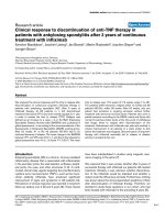
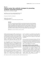
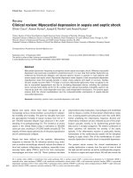
Conventional MRI relies chiefly on four sequences [6]. Fluid-
attenuated inversion recovery (FLAIR) is the primary
sequence used in neuroradiology (Figure 1). It detects brain
contusion, brain oedema and subarachnoid or intraventricular
haemorrhage, as well as the resulting ventricular dilatation or
herniation. The T2*-weighted sequence is more sensitive to
intraparenchymal blood than is FLAIR. This sequence can
also reveal haemorrhagic DAI [7,8]. The T2-weighted
sequence completes the FLAIR sequence and provides
greater detail on brainstem and central grey matter. Finally,
diffusion weighted imaging (DWI) is sensitive to random
movement of water molecules. This sequence shows cerebral
oedema and distinguishes cytotoxic from vasogenic oedema.
It is used chiefly in patients with ischaemic stroke.
Conventional MRI provides an initial evaluation of brain
lesions. However, when it is used alone it fails to predict
outcome accurately.
Magnetic resonance spectroscopy
This sequence is a noninvasive technique for assessing brain
metabolism in vivo. Proton-magnetic resonance spectro-
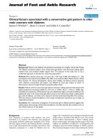
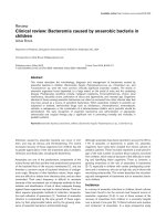
scopy (MRS) is most commonly used. Four main markers are
studied: the peak of N-acetyl-aspartate (NAA), an amino acid
present in neurones, which reflects the status of neuronal
tissue; creatine, found in glia and neurones, which serves as
a point of reference because its level is believed to be stable;
choline, a constitutive component of cell membranes, which
reflects glial proliferation or membrane breakdown [9]; and
lactate, a marker of anaerobic metabolism and therefore of
ischaemia [10]. As shown in Figure 2, three main pons
monovoxel profiles may be observed in patients with TBI.
Diffusion tensor magnetic resonance imaging
Diffusion tensor imaging (DTI), derived from DWI, measures
the degree and direction of water diffusion (anisotropy).
Water diffusion anisotropy reflects the integrity of white
matter tracts. Pathophysiological mechanisms that can alter
water diffusion anisotropy include DAI, effects of intracranial
hypertension and disconnection of white matter tracts.
Magnetization transfer imaging
This sequence is based on the principle that structure-bound
protons undergo T1 relaxation coupling with protons in the
aqueous phase. Saturated protons in macromolecules
exchange longitudinal magnetization with protons in the
aqueous phase, leading to a reduction in signal intensity.
Magnetization transfer imaging has been found to be
sensitive for detecting white matter lesions in several
neurological conditions [11,12].
Functional magnetic resonance imaging
Functional MRI may reveal foci of cerebral dysfunction in
regions that look structurally intact on conventional MRI.
Imaging is based on changes in the oxidative state of
haemoglobin, which reflects regional brain activation.
Functional MRI remains difficult to perform in critically ill
unstable patients and, consequently, few teams have
acquired the equipment and experience necessary to apply
this technique [13]. The few available studies conducted in
comatose patients with TBI showed a correlation between
prefrontal/cingulated cortical activation disturbation and
cognitive impairments [14,15]. However, functional MRI was
performed in these studies at a distance from the injury.
Magnetic resonance imaging findings in
specific critical neurological conditions
Traumatic brain injury
Conventional magnetic resonance imaging
MRI was first used to investigate patients with TBI in a 1986
study of 50 patients [16]. The three main findings, which have
since been confirmed, were as follows: MRI identified lesions
more frequently than did computed tomography; brain lesions
were common after TBI; and although patients who regained
consciousness rapidly had no lesions in fundamental deep
Figure 1
FLAIR and T2* sequences in a patient with an arteriovenous
malformation. (a) Axial fluid-attenuated inversion recovery (FLAIR)
sequence showing hypersignal in the left temporal lobe. (b) Axial T2*
sequence showing mild hyposignal in the same area suggestive of
bleeding. (c) Different section of the axial FLAIR sequence showing
hypersignal surrounded by hyposignal. Bleeding cannot be confirmed.
(d) Axial T2* sequence clearly showing hyposignal lateral to the left
putamen. The patient has bleeding from the arteriovenous
malformation.
Page 3 of 12
(page number not for citation purposes)
brain structures, some of them had severe cortical lesions.
Several descriptions of MRI lesions in TBI patients have been
reported since that initial study was published (Table 1)
[17-21], although few of them focused on the prognostic
value of MRI [17-20]. Conventional MRI findings that strongly
predicted outcome included DAI, total lesion burden and DAI
in the brainstem.
DAI is the most common primary lesion in TBI patients [22,23]
and may be the most common cause of poor outcome [22-24].
DAI may be ischaemic or haemorrhagic [7,8]. Ischaemic DAI is
seen as a hypersignal on DWI or FLAIR, with no abnormality on
the T2* sequence [25]. The hypersignal with DWI disappears
within about 2 weeks. Conversely, haemorrhagic DAI appears
as a hyposignal on the T2* sequence, with normal DWI
findings. It has been proposed [22] that DAI location could be
classified into the following stages: stage 1, frontal and
temporal white matter; stage 2, lobar white matter and
posterior part of corpus callosum; and stage 3, dorsolateral
midbrain and pons. With outcomes defined as Glasgow
Outcome Scale [26] scores of 2 to 3 versus 4 to 5, none of the
33 patients with good outcome in another study [27] had
haemorrhagic DAI (Table 1). DAI appears to be a major
determinant of poor outcomes, although its use as an outcome
predictor in the individual patient remains difficult. Whether the
correlation between DAI and outcome is due to the total lesion
burden or to DAI location remains debated.
In several prospective studies, lesion burden was associated
with outcome irrespective of DAI location (Table 1)
[17,19,28]. Among 40 prospectively enrolled patients with
severe TBI, lesions by FLAIR and T2*-weighted sequences
increased progressively with GOS score groups 1 to 2, 3,
and 4 to 5 [17]. Similar results were obtained in a study
comparing 42 patients with persistent vegetative state with
38 patients who recovered consciousness [19].
A number of studies have focused on the value of DAI
location in predicting outcome [19,29-31]. Brainstem lesions
in the pons and mesencephalon appear to be the most
potent markers of poor prognosis, most notably when they
are bilateral and symmetrical [18,19,29,31]. In a prospective
study conducted in 61 patients (Table 1) who were studied
within 7 days of TBI [18], all patients with bilateral pontine
lesions died as compared with 9% of patients with no
brainstem lesions. These results were confirmed by the same
group in a prospective study of 102 comatose patients [29]
using the following four-stage grading system: grade I,
lesions of the hemispheres only; grade II, unilateral lesions of
the brainstem at any level with or without supratentorial
lesions; grade III, bilateral lesions of the mesencephalon with
or without supratentorial lesions; and grade IV, bilateral
lesions of the pons with or without any of the lesions of lesser
grades. Mortality increased gradually from 14% with grade I
lesions to 100% with grade IV lesions. These findings were
corroborated by two independent studies [19,31] (Table 1).
We recently confirmed the prognostic value of brainstem
lesions in the upper pons and lower midbrain in a study of 73
patients [32]. Bilateral pontine lesions carry a high mortality
rate and predict poor neurological outcomes.
Three studies showed that corpus callosum lesions were
associated with poor outcomes [19,30,31] (Table 1). How-
ever, these lesions may merely represent markers for severe
initial injury. In addition to lesion burden, both total lesion
volume and frontal lobe lesion volume on FLAIR images
correlated significantly with clinical outcomes [30]. Never-
theless, evaluating DAI lesion volume is difficult (most notably
when the lesions are small), time consuming, cumbersome
and subject to inter-rater variability.
The presence of severe DAI and a heavy lesion burden are
associated with permanent neurological impairment.
However, these factors are difficult to use in the individual
patient, especially to distinguish GOS score 2 from GOS
score 3. In TBI patients, brainstem lesions are easily identified
by MRI. In our experience, they are associated with poor
outcomes, most notably when they are posterior and bilateral.
Available online />Figure 2
Magnetic resonance spectroscopy profile of the pons after traumatic brain injury. (a) Normal profile. The peak of N-acetyl-aspartate (NAA) is higher
than the peaks of choline (Cho) and creatine (Cr). (b) Neuronal loss profile. The NAA peak is decreased, nearly to the level of the Cr peak. The
NAA/Cr ratio is lower than in panel a. (c) Gliosis profile: increased Cho peak with no change in the Cr or NAA peak. Adapted from [17].
Critical Care Vol 11 No 5 Weiss et al.
Page 4 of 12
(page number not for citation purposes)
Table 1
Conventional magnetic resonance in traumatic brain injury
Authors (ref.)
Kampfl, 1998 Firsching, 1998 Pierallini, 2000 Yanagawa, 2000 Paterakis, 2000 Firsching, 2001 Firsching, 2002 Wedekind, 2002 Carpentier, 2006
[19] [18] [30] [28] [27] [29] [95] [31] [17]
Study design Case-control Prospective Prospective Prospective Prospective Prospective Prospective Retrospective Prospective
Sequences T1, T2 T1, T2 T1, T2, FLAIR T2, T2* T1, T2 T1, T2 T1, T2 T1, T2, T2* MRS, T2, T2*
Inclusion VS between Admission in GCS score Alive after Discrepancy Admission in GCS score GCS score Severe TBI
criteria 6 and 8 weeks coma (duration <8, coma 1 week between CT coma (duration <8 <8
>24 hours) >1 week, post- scan and >24 hours)
traumatic amnesia neurological
>4 weeks status
Number of 80 61 37 34 33 102 100 40
a
40
patients
Delay to MRI 6 to 8 weeks <7 days 60 to 90 days <3 weeks <48 hours <8 days <7 days 1 to 39 days 17.5 ± 6.4
Outcome GOS score Mortality Clinical GOS score at GOS score Mortality and Mortality at GOS score, GOS score
variable of (2 versus 3-5) assessment at 3 months (2-3 versus 4-5) outcome at 6 months DRS >6 months (1-2 versus 4-5)
interest at 2, 3, 6, 9 and 3, 6 and at 6 months 3 months to (mean delay: and DRS at
12 months 12 months 3 years
b
11.3 months) 18 months
Main results Independent Brainstem lesions: Volume of FLAIR Number of T2 DAI stages Bilateral pons Bilateral upper More lesions of Total burden of
factor of poor mortality rate of corpus callosum lesions correlated correlated with lesions: mortality pontine lesion corpus callosum, FLAIR and T2*
outcome on 44%. Bilateral lesions correlated with GOS score. outcome. No rate of 100%. predicts mortality basal ganglia and lesions correlated
multivariate brainstem lesions: with first clinical Number of T2* patient with good Outcome (para-)hippo- with DRS and GOS
analysis. mortality rate of evaluation. Volume lesions correlated outcome had correlated with campal lesions in score
Corpus callosum: 100% of FLAIR frontal with GOS score haemorrhagic DAI presence/absence patients with
OR 213.8 (95% lobe lesion and unilateral/ brainstem lesions
CI 14.2 to correlated with bilateral brainstem
3213.3). clinical outcome lesions
Brainstem lesions at 1 year
OR 6.9 (95% CI
1.1 to 42.9)
a
Twenty patients with brainstem lesions were matched to 20 patients without brainstem lesions.
b
At last examination. CI, confidence interval; DAI, diffuse axonal injury; DRS, disability rating
scale; FLAIR, fluid-attenuated inversion recovery; GCS, Glasgow Coma Scale; GOS, Glasgow Outcome Scale; MRI, magnetic resonance imaging; MRS, magnetic resonance spectroscopy;
NA, not applicable; OR, odds ratio; T2*, T2* weighted sequence; TBI, traumatic brain injury; VS, vegetative state.
Posterior brainstem lesions in the periaqueductal grey matter
are probably more relevant than anterior brainstem lesions as
predictors of poor outcomes in patients with brainstem stroke
[21] or TBI [19]. In clinical practice, treatment limitation may
deserve consideration in patients who have large bilateral
lesions in the posterior part of the pons after TBI.
Magnetic resonance spectroscopy
Several MRS studies have been conducted in TBI patients
(Table 2). Some of them were purely descriptive [33], others
assessed only the neuropsychological outcomes [34,35], and
yet others focused on global outcome as evaluated using the
GOS or Disability Rating Scale [17,36-42].
Compared with control individuals, TBI patients exhibited
decreased NAA levels, decreased NAA/creatine ratios and
increased choline levels (Table 2) in all brain regions
evaluated [35-39,41,42]. Increased lactate levels were
seldom found in TBI patients, contrary to patients with other
brain injuries [38]. The NAA/creatine ratio appeared to be the
best outcome predictor. Low NAA/creatine values correlated
with poor outcomes when they were located in the frontal
[37,39], frontoparietal [43], or occipitoparietal lobes [36,40];
the splenium of the corpus callosum [41]; the thalami [42];
the pons [17]; or a voxel including the corpus callosum, the
white matter, and part of the hemispheric cortex [38].
These studies are heterogeneous (Table 2) in terms of patient
selection, time from TBI to MRS, voxel location, method of
outcome assessment and timing of outcome assessment. For
instance, among studies of patients with TBI, one included
only patients in a vegetative state [42], another included
patients with severe TBI [17] and a third excluded patients
with early initial coma [36]. These differences in patient
selection may be associated with differences in severity of
brain oedema and in associated hypoxia and herniation,
thereby introducing bias into the interpretation of the results.
MRS findings vary greatly according to time since TBI. Four
phases may be distinguished: an acute phase, which lasts
24 hours after TBI; an early subacute phase, which spans
from the days 1 to 13; a late subacute phase, from days 14 to
20; and a chronic phase, which starts on day 21. Only two
studies included patients at the acute phase [38,40], and
only one of these included all patients before 72 hours [38].
Two studies were conducted from the early subacute phase
to the first month [17,37] and one began inclusion in the late
subacute phase but included patients up to 11 months after
TBI [43]. Four studies focused on the chronic phase; in two
of these studies, patients were included 3 weeks to 6 months
after TBI [36,39] and in the other two studies they were
included 2 months to 8 months after TBI [39,42].
Although NAA/creatine ratios were similar across studies, the
results should be interpreted with caution because experi-
mental in vitro and in vivo data suggest differences in the
underlying pathophysiological mechanisms and in the time
course of the lesions [44-46]. To interpret these results reliably,
information on NAA values over time are needed. Experiments
conducted in vitro [44] and in vivo [45,46] show an early NAA
decrease starting within a few minutes after TBI and reaching
the trough value within 48 hours. This finding explains why
spectroscopic disturbances may require 48 hours for
visualization [47]. NAA levels remain stable within the first
month after TBI, supporting the validity of MRS assessment
during the second or third week [48,49]. Later on, between
6 weeks and 1 year after TBI, NAA levels may decrease [9,37].
Partial recovery of NAA levels has been suggested and may
indicate recovery of mitochondrial function [41].
Another important factor that varied across studies was MRS
voxel location (Table 2). Voxels were located in the hemi-
sphere (the occipitoparietal, frontoparietal, or frontal lobes),
corpus callosum, thalamus, or brainstem (the pons). Because
whole brain analysis is time consuming, voxels are typically
restricted to the areas most affected by DAI, namely the lobar
white matter, corpus callosum and upper brainstem [50].
Estimation of NAA in the whole brain may improve the
prognostic value of MRS [41]. A good compromise may be a
voxel encompassing the corpus callosum, white matter and
part of the hemispheric cortex [38].
Studies also differed in their definitions of poor and good
GOS outcome groups: comparisons involved GOS score 1
to 2 versus GOS score 3 to 5 [39], GOS score 1 to 4 versus
GOS score 5 [41], or GOS score 1 to 2 versus GOS score
4 to 5 [17]. Finally, the time from TBI to outcome assessment
varied from 3 to 18 months (Table 2), further complicating
comparisons because neurological status may improve for up
to 1 year after TBI.
Although MRS has superseded conventional MRI, the combi-
nation of these two techniques may be useful [17]. Variations
in the NAA/creatine ratio over time have not been studied in a
large TBI patient population. The above-mentioned variability
in NAA levels constitutes the main limitation of this technique.
To overcome this limitation, repeated studies at intervals of 1
to 2 weeks are probably needed. In our experience, variations
in the NAA/creatine ratio are minimal in many patients. We
agree with Sinson and coworkers [41] that whole brain NAA
estimation might improve the prognostic value of MRS.
Absence of dysfunction by MRS is a valuable finding; in a
patient with normal results by both conventional MRI and
MRS, a poor outcome is unlikely. However, we have seen a
few patients with normal conventional MRI and MRS findings
who had poor outcomes, probably related to white matter
damage detected as DTI abnormalities.
Diffusion tensor magnetic resonance imaging
Initial reports of DTI in TBI patients suggest that this
technique may demonstrate alterations in white matter
connections that are missed by conventional MRI [51]. DTI
provides information on the physiological status of fibre
Available online />Page 5 of 12
(page number not for citation purposes)
Critical Care Vol 11 No 5 Weiss et al.
Page 6 of 12
(page number not for citation purposes)
Table 2
Outcome of traumatic brain injury by magnetic resonance spectroscopy
Authors (ref.)
Choe, 1995 Ricci, 1997 Ross, 1998 Friedman, 1999 Garnett, 2000 Sinson, 2001 Uzan, 2003 Carpentier, 2006 Marino, 2006
[43] [39] [40] [36] [37] [41] [42] [17] [38]
Study design Case-control Prospective Prospective Case-control Prospective Prospective Case-control Prospective Case-control
Delay 2 weeks to 1 to 90 months 1 to 74 days 45 ± 21 days/ 12 days (3-35)/ 41 days (median) 6 to 8 months 17.5 ± 6.4 days 48 to 72 hours
11 months 6 months 6.2 months
(2.9-50.6)
Number of 10 TBI patients 14 VS TBI 25 TBI patients 14 TBI patients 26 patients. Early 30 TBI patients 14 VS TBI 40 TBI patients 10 TBI patients
patients versus 10 control patients (12 children) versus 14 control study: 21. Late patients versus versus 10 control
individuals individuals study: 15. 5 control individuals
Both: 10 individuals
Grey matter NA NA Occipitoparietal Occipitoparietal Frontal NA Thalamus NA Mesial cortex
voxel location
White matter Frontoparietal Frontal Occipitoparietal Occipitoparietal Frontal Splenium of NA Pons Corpus callosum,
voxel location corpus callosum mostly white matter
Outcome GOS score after GOS score ROS at discharge GOS score and GOS score, GOS score at Aware versus not GOS score GOS score at
variable of MRI (1-2 versus 3-5) and follow up
b
neuropsycho- DRS at 6 months 3 months aware at (1-2 versus 4-5), 3 months
interest at follow up
a
logical (1-4 versus 5) >6 months DRS at
performance 18 months
Main results NAA/Cr ratio NAA/Cr ratio and NAA levels NAA levels in NAA/Cr ratio NAA/Cr ratio NAA/Cr ratio NAA/Cr ratio NAA/Cr and NAA/all
lower in TBI NAA/Cho ratio diminished. white matter lower lower in TBI lower. NAA/Cr lower in VS. correlated to metabolites ratios
patients. NAA/Cr lower, Cho/Cr NAA/Cr ratio in TBI patients. patients. Cho/Cr correlated with NAA/Cr ratio GOS score and lower. La/Cr and
ratio correlated ratio elevated, and correlated with Early NAA levels elevated in TBI GOS score lower in patients DRS. No La/all metabolites
with GOS score NAA/Cho lower in outcome in grey matter patients. NAA/Cr remained in VS correlation ratios increased in
GOS score 1-2 correlated with ratio correlated compared with between NAA/Cr TBI
versus GOS 3-5 GOS with GOS score patients who ratio and lesions
and DRS regained burden on FLAIR
awareness or T2*
a
No further information.
b
Up to 2 years, except for four out of 25 patients. Cho, choline; Cr, creatinine; DRS, disability rating scale; FLAIR, fluid-attenuated inversion recovery; GOS, Glasgow
Outcome Scale; La, lactate; MRI, magnetic resonance imaging; NA, not applicable; NAA, N-acetyl-aspartate; ROS, Rancho Los Amigos Medical Centre Outcome Score; T2*, T2* weighted
sequence; TBI, traumatic brain injury; VS, vegetative state.
bundles, thus complementing the metabolic and biochemical
information supplied by MRS. At present, little is known about
the prognostic value of DTI in patients with TBI. DTI findings
correlated with clinical status in patients with multiple
sclerosis or neurodegenerative disease [52,53]. In a mouse
model of TBI, DTI parameters were significantly reduced in
the injured brain, whereas conventional MRI showed no
significant changes [54]. Furthermore, changes in relative
anisotropy correlated significantly with the density of stained
axons on histological sections.
In a study comparing 20 TBI patients and 15 healthy control
individuals, fractional anisotropy was reduced in the internal
capsule and splenium of the corpus callosum and correlated
with Glasgow Coma Scale score and Rankin score at
discharge in the TBI patients [55]. Similar findings have been
reported in children [56]. Anecdotal case reports of DTI
abnormalities in TBI patients have been reported [57,58]. In
two patients who recovered partially after 6 years and
19 years, respectively, in a minimally conscious state, DTI
disclosed increased anisotropy within the midline cerebellar
white matter over an 18-month period [59]. This anisotropy
increase correlated with an increase in resting metabolism,
measured using positron emission tomography, which
suggests that axonal regrowth might underlie increases in
anisotropy. Larger studies of DTI variations over time are
needed. In our institution, comatose patients have been
included in a prospective DTI study for the past 3 years.
Patients with major connectivity abnormalities in both
hemispheres and the brainstem were at increased risk for
poor outcomes. A large multicentre prospective study is
ongoing in France to assess the usefulness of combining DTI
with MRS.
Magnetization transfer imaging
Magnetization transfer imaging is sensitive for detecting white
matter lesions in patients with multiple sclerosis, progressive
multifocal leukoencephalopathy, or wallerian degeneration
[11,12]. Preliminary results in TBI are promising [60,61]. The
magnetization transfer ratio was decreased in TBI patients
[60,61]. Out of 28 TBI patients, eight had abnormal
magnetization transfer ratios, and all eight had persistent
neurological deficits [62]. In another study, however, no
correlation was found between GOS score and abnormal
magnetization transfer ratio [41].
Anoxic/hypoxic encephalopathy
Anoxic/hypoxic encephalopathy is a devastating condition; its
development after prolonged cerebral hypoxia is often difficult
to predict on clinical grounds. No controlled studies of
routine MRI in large numbers of cardiac arrest patients have
been reported. Anecdotal case reports and small series are
available [63-67]. As with TBI, MRI findings in hypoxic/anoxic
encephalopathy go through four phases [66]: an acute
phase, which lasts 24 hours after anoxia or hypoxia; an early
subacute phase, from days 1 to 13; a late subacute phase,
from days 14 to 20; and a chronic phase, starting on day 21.
MRI findings in patients with hypoxic brain damage are
complex but distinctive. Brain swelling, cortical laminar
necrosis, hypersignal of basal ganglia, delayed white matter
degeneration and atrophy occur in succession, as shown in
Table 3 [63,66,67]. During the acute and early subacute
phases, DWI and T2-weighted sequence show hypersignals
in the cortex, thalamus and basal ganglia. DWI may be more
sensitive for detecting mild hypoxic/anoxic injury within the
first few hours, and the hypersignal may occur first in the
cerebral cortex and later in the basal ganglia. During the late
subacute phase the hypersignals previously seen by DWI
tend to fade, and diffuse white matter abnormalities denoting
delayed anoxic leukoencephalopathy may develop [68].
During the chronic phase diffuse atrophy and dilatation of the
ventricles are visible, whereas DWI is normal.
The three main series published to date included ten [66],
eight [67] and six [63] patients. Although the small numbers
of patients is a limitation, the succession of four phases was
confirmed in several case reports and supported by findings
of histological and animal studies [9,12,16,67], indicating far
greater vulnerability of grey matter to hypoxia as compared
with white matter. This difference in vulnerability may explain
why some brain regions are more susceptible than others to
diffuse insults such as hypoxia or anoxia [2,11,29,66].
A few studies recorded both MRI findings and long-term
outcomes in patients with hypoxic/anoxic encephalopathy
[64,67,69]. Diffuse cortical abnormalities by DWI in the acute
or early subacute phase appear to be of unfavourable
prognostic significance. Of six patients with hypoxic encepha-
lopathy investigated by sequential MRI, the only patient who
recovered a GOS score greater than 3 had hypersignals in
watershed zones in the parieto-occipito-temporal cortex
without cortical hypersignal by DWI. In a study of 10 patients
who had suffered a cardiac arrest, FLAIR and DWI showed
that eight patients had diffuse abnormalities in the
cerebellum, thalamus, frontal and parietal cortices, and
hippocampus [69]. None of the patients with cortical
structure abnormalities recovered beyond a severely disabled
state. In another prospective study, the prognostic value of
DWI was evaluated in 12 patients within 36 hours after global
cerebral hypoxia [64]. DWI findings correlated with clinical
outcomes after 6 months. The three patients with short resus-
citation times had a good recovery and normal DWI findings.
Of the remaining nine patients, all had DWI abnormalities and
developed a vegetative state. Thus, diffuse cortical
hypersignals by DWI appear to predict a poor outcome.
Conversely, several reports describe delayed anoxic
encephalopathy with a good final outcome and resolution of
MRI abnormalities. Therefore, finding diffuse hypersignals in
the white matter by either DWI or T2/FLAIR weighted
sequences should not lead to treatment limitation decisions.
In general, whether MRI findings can be used to guide
treatment limitation decisions remains unclear. In our unit,
Available online />Page 7 of 12
(page number not for citation purposes)
treatment limitation is considered in patients with diffuse cortical
hypersignals by DWI or cortical laminar necrosis images after
prolonged cardiac arrest, provided the MRI findings are
consonant with the clinical examination or electrophysiological
data. In contrast, a patient with normal MRI findings after anoxia
should probably be re-evaluated 1 or 2 weeks later by clinical
examination, electrophysiological testing and MRI.
Few data are available on MRS findings after anoxia [70,71].
No studies were specifically designed to assess the
prognostic value of DTI in patients with anoxic/hypoxic
encephalopathy. The unique ability of DTI to distinguish
between white matter and grey matter, allowing separate
quantitative assessment of these two tissues, should be of
particular interest in anoxic/hypoxic encephalopathy.
Severe hypoglycaemia has been likened to hypoxic encepha-
lopathy. Imaging study data in patients with hypoglycaemic
coma are scant [63,72,73]. Interestingly, DWI abnormalities
can mimic stroke in patients with hypoglycaemic coma
[74,75]. Rapid improvements in DWI and MRI abnormalities
after glucose infusion were recently reported [76].
Ischaemic stroke
Ischaemic stroke causes coma in two main settings, namely
malignant stroke and basilar artery occlusion. We focus on
these two situations, and we do not discuss the prognostic
value of MRI after stroke without coma.
In a study of 37 patients with acute middle cerebral artery
infarction, early quantitative DWI findings predicted
progression to malignant stroke, which occurred in 11
patients [77]. Factors that predicted malignant stroke were
as follows: size of the region with apparent diffusion
coefficient (ADC) < 80% greater than 82 ml; ADC in the core
of the stroke < 300 mm
2
/s; and relative ADC within the ADC
< 80% of the lesion under 0.62. Another study evaluated 28
patients, of whom 11 experienced malignant stroke [78]. The
best predictor of malignant stroke within 14 hours of stroke
onset was infarct volume by DWI greater than 145 cm
3
,
which was 100% sensitive and 94% specific. Regarding
brainstem stroke, a retrospective study of 47 patients
showed that coma, which was a feature in nine patients, was
associated with lesions in the posterior pons and lower
midbrain [21]. The patients who died had all bilateral
brainstem lesions in this area. None of the patients with
bilateral lesions survived. Although the number of patients
was small in the study, the results are consonant with clinical
experience that brainstem stroke with coma and large
brainstem lesions has a poor outcome and that some patients
who are initially comatose with limited anterior brainstem
infarction eventually experience good outcomes.
Critical Care Vol 11 No 5 Weiss et al.
Page 8 of 12
(page number not for citation purposes)
Table 3
Chronological magnetic resonance imaging findings in anoxic/hypoxic encephalopathy
Acute phase Early subacute phase Late subacute phase Chronic phase
(<24 hours) (24 hours to day 13) (days 14 to 20) (>21 days)
Characteristics Brain swelling Brain swelling Absence of brain swelling Diffuse atrophy and
dilatation of the ventricles
DWI Hypersignals in the cortex, Hypersignals in the cortex, Progressive disappearance Normal
in the thalamus and in the in the thalamus and in the of hypersignals found
basal ganglia basal ganglia previously
T2 Hypersignals in the cortex, Hypersignals in the cortex, Hypersignals of the cortex, Normal or possible
in the thalamus and in the in the thalamus and in the the thalamus, the basal ganglia hypersignals of the cortex,
basal ganglia basal ganglia. Possible and the pons the thalamus, the basal
subcortical hyposignals ganglia and the pons
T1 No abnormalities No abnormalities Possible spontaneous Can be normal
subcortical and basal ganglia
hypersignals
T1 with No abnormalities Possible subcortical Possible subcortical No abnormalities
gadolinium enhancement suggestive of enhancement suggestive of
enhancement cortical laminar necrosis cortical laminar necrosis
Comments DWI seems more sensitive Hypersignals on both DWI and In some cases, appearance of In some cases,
to mild hypoxic/anoxic injury T2 become more intense, diffuse white matter, hypersignals of the cortex
in the first hours, and the particularly in the thalamus and abnormalities of delayed anoxic and hyposignals in the
hypersignal in cerebral the basal ganglia leukoencephalopathy on both subcortical zone on both
cortex seems more DWI and T2 T2 and T1, suggestive of
precocious than in the basal cortical laminar necrosis
ganglia
DWI, diffusion weighted imaging; T1, T1 weighted sequence; T2, T2 weighted sequence. Adapted from [66,67].
DTI has been used to assess outcomes after stroke [79],
although we are not aware of studies of MRS or DTI to
predict outcomes after malignant or brainstem stroke. In a
study of 12 patients with subcortical infarcts involving the
posterior limb of the internal capsule, a decrease in fractional
anisotropy was detected by DTI, indicating secondary
degeneration of the fibre tract proximal and distal to the
primary ischaemic lesion [80]. Fibre tract degeneration
occurred gradually, which might have hampered functional
recovery. In patients with brainstem stroke or malignant
stroke, DTI may be of considerable value for assessing fibre
tract degeneration, thus predicting chances of recovery.
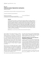
Ascending reticular activating system and
prognosis of brain injuries
Several brain areas involved in the prognosis of TBI or stroke
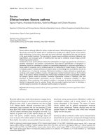
play a role in consciousness [17,19,21,81]. Figure 3 shows
the anatomical regions involved in arousal and conscious-
ness. Brainstem lesions have been shown to influence the
prognosis of patients with coma after TBI or stroke
[17,19,21,81]. Bilateral brainstem lesions were associated
with poorer outcomes [21,81], and the target area appeared
to be the posterior pons and lower midbrain, where the
ascending reticular activating system (ARAS) nuclei are
located. An MRI study of 88 patients in a vegetative state
after TBI confirmed the prognostic importance of lesions in
this area [19]. The ARAS projects in part to the basal fore-
brain through the hypothalamus by its ventral pathway, as
shown in Figure 3. Several pathological studies showed a
high rate of basal forebrain lesions in humans who died after
head injuries [82], and we found that hypothalamic and basal
forebrain lesions were associated with poor outcomes in TBI
patients [32]. Histological evidence of neuronal damage in
the nucleus basalis of Meynert (the main nucleus of the basal
forebrain) was found in most of the patients who died after
head injury [82]. The ARAS projects to the reticular thalamic
nuclei through its dorsal pathway (Figure 3). Focal damage to
the thalami was documented in pathological studies of
patients in vegetative state [83,84]. All three pathways lead
to cortical arousal. Widespread cortical damage (as
described in anoxic/hypoxic encephalopathy [83,85]) and
widespread white matter damage (as described in TBI
patients [86]) may result in inability to arouse cortical areas
(vegetative state). Clinical findings in patients with TBI
suggest that impairment in consciousness may correlate with
depth of the deepest lesion [20,87]. Although lesions to the
ARAS or its projections may correlate with severity of the
initial injury or the existence of herniation, another possibility
is that they directly contribute to the prognosis. Studies
involving multimodal investigations would provide valuable
insight in this area [88].
Avenues for research
Data from patients with TBI, stroke, or anoxic encephalopathy
suggest that specific MRI findings may hold promise for
outcome prediction. Large studies are not yet available, even
in patients with TBI. Given the major ethical, human and
economic issues involved, there is an urgent need for large
prospective multicentre studies. Only small numbers of
patients eligible for such studies are admitted to medical or
surgical intensive care units, and few neurosurgical or
neurological intensive care units exist; therefore, a multicentre
design is essential to ensure recruitment of a sufficiently large
population. In our institution, which is a neurosurgical
intensive care unit in a tertiary hospital, multimodal prospec-
tive imaging by conventional MRI, MRS and DTI is performed
routinely in all patients who are still comatose after 2 weeks.
A multicentre study funded by the French Ministry of Health is
under way.
Conclusion
Patients with severe brain injury, most notably those who
remain comatose, generate huge health care costs. Adapting
the level of medical care to the neurological outcome is a
major challenge currently faced by neurological intensive
care. Meeting this challenge will require the development of
tools that reliably predict long-term neurological outcomes.
Available online />Page 9 of 12
(page number not for citation purposes)
Figure 3
Anatomical substratum of arousal and awareness. Consciousness
involves two main components: arousal and awareness of oneself and
of the environment. Awareness is dependent on the integrity of specific
anatomical regions [89]. The ascending reticular activating system
(ARAS), the primary arousal structure, is located in the upper pons and
lower midbrain in the posterior part of the upper two-thirds of the
brainstem [90,91]. A ventral pathway (black solid arrows) projects to
the hypothalamus (hypo) and basal forebrain (Bfb); a dorsal pathway
(black dashed arrows) projects to the reticular nuclei of the thalamus
(thal); and a third pathway (light grey arrows) projects directly into the
cortical regions [90]. From the basal forebrain, two main bundles
project diffusely to several cortical areas [92]. The reticular nuclei of
the thalamus connect to other nuclei in the thalamus. They are involved
in a thalamo-cortical circuit [93] that controls cortical activity. Some
regions of the cerebral cortex may also make specific contributions to
consciousness [94].
Most MRI studies to date were conducted in patients with
TBI. By conventional imaging, presence of bilateral lesions in
the dorsolateral upper brainstem appears to be the factor of
greatest adverse prognostic significance. With MRS, low
NAA/creatine ratio in the hemispheres and in the pons
predicts a poor outcome. In anoxic/hypoxic encephalopathy,
the factor of greatest adverse significance appears to be the
presence of diffuse cortical abnormalities by DWI. However,
data are scarcer than in the field of TBI. Finally, regarding
brainstem stroke, posterior lesions appear to be associated
with poor outcome.
The prognostic value of imaging studies could be improved
by combining several techniques and sequences, for instance
by combining several MRI sequences or by combining MRI
with electrophysiological studies or clinical data. Complete
destruction of arousal structures is consistently associated
with poor outcome. Multimodal MRI is a promising technique
that can be expected to provide accurate prediction of
neurological outcome in the near future.
Competing interests
The authors declare that they have no competing interests.
References
1. Oddo M, Schaller MD, Feihl F, Ribordy V, Liaudet L: From evi-
dence to clinical practice: effective implementation of thera-
peutic hypothermia to improve patient outcome after cardiac
arrest. Crit Care Med 2006, 34:1865-1873.
2. Celesia GG: Persistent vegetative state. Neurology 1993, 43:
1457-1458.
3. Jennett B: Thirty years of the vegetative state: clinical, ethical
and legal problems. Prog Brain Res 2005, 150:537-543.
4. Payne K, Taylor RM, Stocking C, Sachs GA: Physicians’ atti-
tudes about the care of patients in the persistent vegetative
state: a national survey. Ann Intern Med 1996, 125:104-110.
5. Anderson CV, Wood DM, Bigler ED, Blatter DD: Lesion volume,
injury severity, and thalamic integrity following head injury. J
Neurotrauma 1996, 13:35-40.
6. Brandstack N, Kurki T, Tenovuo O, Isoniemi H: MR imaging of
head trauma: visibility of contusions and other intraparenchy-
mal injuries in early and late stage. Brain Inj 2006, 20:409-416.
7. Gerber DJ, Weintraub AH, Cusick CP, Ricci PE, Whiteneck GG:
Magnetic resonance imaging of traumatic brain injury: rela-
tionship of T2*SE and T2GE to clinical severity and outcome.
Brain Inj 2004, 18:1083-1097.
8. Scheid R, Preul C, Gruber O, Wiggins C, von Cramon DY:
Diffuse axonal injury associated with chronic traumatic brain
injury: evidence from T2*-weighted gradient-echo imaging at
3 T. AJNR Am J Neuroradiol 2003, 24:1049-1056.
9. Brooks WM, Friedman SD, Gasparovic C: Magnetic resonance
spectroscopy in traumatic brain injury. J Head Trauma Rehabil
2001, 16:149-164.
10. Garnett MR, Cadoux-Hudson TA, Styles P: How useful is mag-
netic resonance imaging in predicting severity and outcome
in traumatic brain injury? Curr Opin Neurol 2001, 14:753-757.
11. Filippi M, Rocca MA: Magnetization transfer magnetic reso-
nance imaging in the assessment of neurological diseases. J
Neuroimaging 2004, 14:303-313.
12. Horsfield Ma: Magnetization transfer imaging in multiple scle-
rosis. J Neuroimaging 2005, Suppl:58S-67S.
13. Pickard JD, Hutchinson PJ, Coles JP, Steiner LA, Johnston AJ,
Fryer TD, Coleman MR, Smielewski P, Chatfield DA, Aigbirhio F,
et al.: Imaging of cerebral blood flow and metabolism in brain
injury in the ICU. Acta Neurochir Suppl 2005, 95:459-464.
14. Azouvi P: Neuroimaging correlates of cognitive and functional
outcome after traumatic brain injury. Curr Opin Neurol 2000,
13:665-669.
15. Fontaine A, Azouvi P, Remy P, Bussel B, Samson Y: Functional
anatomy of neuropsychological deficits after severe traumatic
brain injury. Neurology 1999, 53:1963-1968.
16. Jenkins A, Teasdale G, Hadley MD, Macpherson P, Rowan JO:
Brain lesions detected by magnetic resonance imaging in
mild and severe head injuries. Lancet 1986, 2:445-446.
17. Carpentier A, Galanaud D, Puybasset L, Muller JC, Lescot T,
Boch AL, Riedl V, Cornu P, Coriat P, Dormont D, et al.: Early
morphologic and spectroscopic magnetic resonance in
severe traumatic brain injuries can detect ‘invisible brain stem
damage’ and predict ‘vegetative states’. J Neurotrauma 2006,
23:674-685.
18. Firsching R, Woischneck D, Diedrich M, Klein S, Ruckert A, Wittig
H, Dohring W: Early magnetic resonance imaging of brainstem
lesions after severe head injury. J Neurosurg 1998, 89:707-
712.
19. Kampfl A, Schmutzhard E, Franz G, Pfausler B, Haring HP, Ulmer
H, Felber S, Golaszewski S, Aichner F: Prediction of recovery
from post-traumatic vegetative state with cerebral magnetic-
resonance imaging. Lancet 1998, 351:1763-1767.
20. Levin HS, Mendelsohn D, Lilly MA, Yeakley J, Song J, Scheibel
RS, Harward H, Fletcher JM, Kufera JA, Davidson KC, Bruce D:
Magnetic resonance imaging in relation to functional outcome
of pediatric closed head injury: a test of the Ommaya-
Gennarelli model. Neurosurgery 1997, 40:432-440; discussion
440-441.
21. ParviziJ, Damasio AR: Neuroanatomical correlates of brainstem
coma. Brain 2003, 126:1524-1536.
22. Gentry LR: Imaging of closed head injury. Radiology 1994, 191:
1-17.
23. Parizel PM, Ozsarlak, Van Goethem JW, van den Hauwe L, Dillen
C, Verlooy J, Cosyns P, De Schepper AM: Imaging findings in
diffuse axonal injury after closed head trauma. Eur Radiol
1998, 8:960-965.
24. Wilberger JE Jr, Deeb Z, Rothfus W: Magnetic resonance
imaging in cases of severe head injury. Neurosurgery 1987,
20:571-576.
25. Huisman TA: Diffusion-weighted imaging: basic concepts and
application in cerebral stroke and head trauma. Eur Radiol
2003, 13:2283-2297.
26. Jennett B, Bond M: Assessment of outcome after severe brain
damage. Lancet 1975, 1:480-484.
27. Paterakis K, Karantanas AH, Komnos A, Volikas Z: Outcome of
patients with diffuse axonal injury: the significance and prog-
nostic value of MRI in the acute phase. J Trauma 2000, 49:
1071-1075.
28. Yanagawa Y, Tsushima Y, Tokumaru A, Un-no Y, Sakamoto T,
Okada Y, Nawashiro H, Shima K: A quantitative analysis of
head injury using T2*-weighted gradient-echo imaging. J
Trauma 2000, 49:272-277.
29. Firsching R, Woischneck D, Klein S, Reissberg S, Dohring W,
Peters B: Classification of severe head injury based on magnetic
resonance imaging. Acta Neurochir (Wien) 2001, 143:263-271.
30. Pierallini A, Pantano P, Fantozzi LM, Bonamini M, Vichi R, Zylber-
man R, Pisarri F, Colonnese C, Bozzao L: Correlation between
MRI findings and long-term outcome in patients with severe
brain trauma. Neuroradiology 2000, 42:860-867.
31. Wedekind C, Hesselmann V, Lippert-Gruner M, Ebel M: Trauma
to the pontomesencephalic brainstem: a major clue to the
prognosis of severe traumatic brain injury. Br J Neurosurg
2002, 16:256-260.
32. Weiss N, Galanaud D, Carpentier A, Tezenas de Montcel S, Nac-
cache L, Coriat P, Puybasset L: A combined clinical and MRI
approach for outcome assessment of traumatic head injured
comatose patients. J Neurol 2007, in press.
33. Cecil KM, Hills EC, Sandel ME, Smith DH, McIntosh TK, Mannon
LJ, Sinson GP, Bagley LJ, Grossman RI, Lenkinski RE: Proton
magnetic resonance spectroscopy for detection of axonal
injury in the splenium of the corpus callosum of brain-injured
patients. J Neurosurg 1998, 88:795-801.
34. Brooks WM, Stidley CA, Petropoulos H, Jung RE, Weers DC,
Friedman SD, Barlow MA, Sibbitt WL Jr, Yeo RA: Metabolic and
cognitive response to human traumatic brain injury: a quanti-
tative proton magnetic resonance study. J Neurotrauma 2000,
17:629-640.
35. Friedman SD, Brooks WM, Jung RE, Hart BL, Yeo RA: Proton MR
spectroscopic findings correspond to neuropsychological
Critical Care Vol 11 No 5 Weiss et al.
Page 10 of 12
(page number not for citation purposes)
function in traumatic brain injury. AJNR Am J Neuroradiol
1998, 19:1879-1885.
36. Friedman SD, Brooks WM, Jung RE, Chiulli SJ, Sloan JH, Montoya
BT, Hart BL, Yeo RA: Quantitative proton MRS predicts
outcome after traumatic brain injury. Neurology 1999, 52:
1384-1391.
37. Garnett MR, Blamire AM, Corkill RG, Cadoux-Hudson TA,
Rajagopalan B, Styles P: Early proton magnetic resonance
spectroscopy in normal-appearing brain correlates with
outcome in patients following traumatic brain injury. Brain
2000, 123:2046-2054.
38. Marino S, Zei E, Battaglini M, Vittori C, Buscalferri A, Bramanti P,
Federico A, De Stefano N: Acute metabolic brain changes fol-
lowing traumatic brain injury and their relevance to clinical
severity and outcome. J Neurol Neurosurg Psychiatry 2007, 78:
501-507.
39. Ricci R, Barbarella G, Musi P, Boldrini P, Trevisan C, Basaglia N:
Localised proton MR spectroscopy of brain metabolism
changes in vegetative patients. Neuroradiology 1997, 39:313-
319.
40. Ross BD, Ernst T, Kreis R, Haseler LJ, Bayer S, Danielsen E,
Bluml S, Shonk T, Mandigo JC, Caton W, et al.: 1H MRS in acute
traumatic brain injury. J Magn Reson Imaging 1998, 8:829-840.
41. Sinson G, Bagley LJ, Cecil KM, Torchia M, McGowan JC, Lenkin-
ski RE, McIntosh TK, Grossman RI: Magnetization transfer
imaging and proton MR spectroscopy in the evaluation of
axonal injury: correlation with clinical outcome after traumatic
brain injury. AJNR Am J Neuroradiol 2001, 22:143-151.
42. Uzan M, Albayram S, Dashti SG, Aydin S, Hanci M, Kuday C:
Thalamic proton magnetic resonance spectroscopy in vegeta-
tive state induced by traumatic brain injury. J Neurol Neurosurg
Psychiatry 2003, 74:33-38.
43. Choe BY, Suh TS, Choi KH, Shinn KS, Park CK, Kang JK: Neu-
ronal dysfunction in patients with closed head injury evalu-
ated by in vivo 1H magnetic resonance spectroscopy. Invest
Radiol 1995, 30:502-506.
44. Signoretti S, Marmarou A, Tavazzi B, Lazzarino G, Beaumont A,
Vagnozzi R: N-Acetylaspartate reduction as a measure of
injury severity and mitochondrial dysfunction following diffuse
traumatic brain injury. J Neurotrauma 2001, 18:977-991.
45. Cecil KM, Lenkinski RE, Meaney DF, McIntosh TK, Smith DH:
High-field proton magnetic resonance spectroscopy of a
swine model for axonal injury. J Neurochem 1998, 70:2038-
2044.
46. Rubin Y, Cecil K, Wehrli S, McIntosh TK, Lenkinski RE, Smith DH:
High-resolution 1H NMR spectroscopy following experimental
brain trauma. J Neurotrauma 1997, 14:441-449.
47. Alessandri B, al-Samsam R, Corwin F, Fatouros P, Young HF,
Bullock RM: Acute and late changes in N-acetyl-aspartate fol-
lowing diffuse axonal injury in rats: an MRI spectroscopy and
microdialysis study. Neurol Res 2000, 22:705-712.
48. Holshouser BA, Tong KA, Ashwal S, Oyoyo U, Ghamsary M,
Saunders D, Shutter L: Prospective longitudinal proton mag-
netic resonance spectroscopic imaging in adult traumatic
brain injury. J Magn Reson Imaging 2006, 24:33-40.
49. Signoretti S, Marmarou A, Fatouros P, Hoyle R, Beaumont A,
Sawauchi S, Bullock R, Young H: Application of chemical shift
imaging for measurement of NAA in head injured patients.
Acta Neurochir Suppl 2002, 81:373-375.
50. Adams JH, Graham DI, Murray LS, Scott G: Diffuse axonal injury
due to nonmissile head injury in humans: an analysis of 45
cases. Ann Neurol 1982, 12:557-563.
51. Levin HS: Neuroplasticity following non-penetrating traumatic
brain injury. Brain Inj 2003, 17:665-674.
52. Catani M: Diffusion tensor magnetic resonance imaging trac-
tography in cognitive disorders. Curr Opin Neurol 2006, 19:
599-606.
53. Reich DS, Smith SA, Jones CK, Zackowski KM, van Zijl PC, Cal-
abresi PA, Mori S: Quantitative characterization of the corti-
cospinal tract at 3T. AJNR Am J Neuroradiol 2006, 27:2168-2178.
54. Mac Donald CL, Dikranian K, Song SK, Bayly PV, Holtzman DM,
Brody DL: Detection of traumatic axonal injury with diffusion
tensor imaging in a mouse model of traumatic brain injury.
Exp Neurol 2007, 205:116-131.
55. Huisman TA, Schwamm LH, Schaefer PW, Koroshetz WJ, Shetty-
Alva N, Ozsunar Y, Wu O, Sorensen AG: Diffusion tensor
imaging as potential biomarker of white matter injury in
diffuse axonal injury. AJNR Am J Neuroradiol 2004, 25:370-
376.
56. Wilde EA, Chu Z, Bigler ED, Hunter JV, Fearing MA, Hanten G,
Newsome MR, Scheibel RS, Li X, Levin HS: Diffusion tensor
imaging in the corpus callosum in children after moderate to
severe traumatic brain injury. J Neurotrauma 2006, 23:1412-
1426.
57. Ewing-Cobbs L, Hasan KM, Prasad MR, Kramer L, Bachevalier J:
Corpus callosum diffusion anisotropy correlates with neu-
ropsychological outcomes in twins disconcordant for trau-
matic brain injury. AJNR Am J Neuroradiol 2006, 27:879-881.
58. Naganawa S, Sato C, Ishihra S, Kumada H, Ishigaki T, Miura S,
Watanabe M, Maruyama K, Takizawa O: Serial evaluation of dif-
fusion tensor brain fiber tracking in a patient with severe
diffuse axonal injury. AJNR Am J Neuroradiol 2004, 25:1553-
1556.
59. Voss HU, Uluc AM, Dyke JP, Watts R, Kobylarz EJ, McCandliss
BD, Heier LA, Beattie BJ, Hamacher KA, Vallabhajosula S, et al.:
Possible axonal regrowth in late recovery from the minimally
conscious state. J Clin Invest 2006, 116:2005-2011.
60. Kimura H, Meaney DF, McGowan JC, Grossman RI, Lenkinski RE,
Ross DT, McIntosh TK, Gennarelli TA, Smith DH: Magnetization
transfer imaging of diffuse axonal injury following experimen-
tal brain injury in the pig: characterization by magnetization
transfer ratio with histopathologic correlation. J Comput Assist
Tomogr 1996, 20:540-546.
61. McGowan JC, McCormack TM, Grossman RI, Mendonca R, Chen
XH, Berlin JA, Meaney DF, Xu BN, Cecil KM, McIntosh TK, et al.:
Diffuse axonal pathology detected with magnetization trans-
fer imaging following brain injury in the pig. Magn Reson Med
1999, 41:727-733.
62. Bagley LJ, McGowan JC, Grossman RI, Sinson G, Kotapka M,
Lexa FJ, Berlin JA, McIntosh TK: Magnetization transfer
imaging of traumatic brain injury. J Magn Reson Imaging
2000, 11:1-8.
63. Fujioka M, Okuchi K, Sakaki T, Hiramatsu K, Miyamoto S, Iwasaki
S: Specific changes in human brain following reperfusion
after cardiac arrest. Stroke 1994, 25:2091-2095.
64. Els T, Kassubek J, Kubalek R, Klisch J: Diffusion-weighted MRI
during early global cerebral hypoxia: a predictor for clinical
outcome? Acta Neurol Scand 2004, 110:361-367.
65. Torbey MT, Bhardwaj A: MR imaging in comatose survivors of
cardiac resuscitation. AJNR Am J Neuroradiol 2002, 23:738.
66. Arbelaez A, Castillo M, Mukherji SK: Diffusion-weighted MR
imaging of global cerebral anoxia. AJNR Am J Neuroradiol
1999, 20:999-1007.
67. Takahashi S, Higano S, Ishii K, Matsumoto K, Sakamoto K, Iwasaki
Y, Suzuki M: Hypoxic brain damage: cortical laminar necrosis
and delayed changes in white matter at sequential MR
imaging. Radiology 1993, 189:449-456.
68. Kim HY, Kim BJ, Moon SY, Kwon JC, Shon YM, Na DG, Lee KH,
Na DL: Serial diffusion-weighted MR Imaging in delayed post-
anoxic encephalopathy. A case study. J Neuroradiol 2002, 29:
211-215.
69. Wijdicks EF, Campeau NG, Miller GM: MR imaging in comatose
survivors of cardiac resuscitation. AJNR Am J Neuroradiol
2001, 22:1561-1565.
70. Wartenberg KE, Patsalides A, Yepes MS: Is magnetic reso-
nance spectroscopy superior to conventional diagnostic tools
in hypoxic-ischemic encephalopathy? J Neuroimaging 2004,
14:180-186.
71. Kucharczyk J, Moseley M, Kurhanewicz J, Norman D: MRS of
ischemic/hypoxic brain disease. Invest Radiol 1989, 24:951-
954.
72. Lo L, Tan AC, Umapathi T, Lim CC: Diffusion-weighted MR
imaging in early diagnosis and prognosis of hypoglycemia.
AJNR Am J Neuroradiol 2006, 27:1222-1224.
73. Yanagawa Y, Isoi N, Tokumaru AM, Sakamoto T, Okada Y: Diffu-
sion-weighted MRI predicts prognosis in severe hypo-
glycemic encephalopathy. J Clin Neurosci 2006, 13:696-699.
74. Bottcher J, Kunze A, Kurrat C, Schmidt P, Hagemann G, Witte
OW, Kaiser WA: Localized reversible reduction of apparent
diffusion coefficient in transient hypoglycemia-induced hemi-
paresis. Stroke 2005, 36:e20-e22.
75. Cordonnier C, Oppenheim C, Lamy C, Meder JF, Mas JL: Serial
diffusion and perfusion-weighted MR in transient hypo-
glycemia. Neurology 2005, 65:175.
Available online />Page 11 of 12
(page number not for citation purposes)
76. Maruya J, Endoh H, Watanabe H, Motoyama H, Abe H: Rapid
improvement of diffusion-weighted imaging abnormalities
after glucose infusion in hypoglycaemic coma. J Neurol Neuro-
surg Psychiatry 2007, 78:102-103.
77. Thomalla GJ, Kucinski T, Schoder V, Fiehler J, Knab R, Zeumer H,
Weiller C, Rother J: Prediction of malignant middle cerebral
artery infarction by early perfusion- and diffusion-weighted
magnetic resonance imaging. Stroke 2003, 34:1892-1899.
78. Oppenheim C, Samson Y, Manai R, Lalam T, Vandamme X,
Crozier S, Srour A, Cornu P, Dormont D, Rancurel G, et al.: Pre-
diction of malignant middle cerebral artery infarction by diffu-
sion-weighted imaging. Stroke 2000, 31:2175-2181.
79. Konishi J, Yamada K, Kizu O, Ito H, Sugimura K, Yoshikawa K,
Nakagawa M, Nishimura T: MR tractography for the evaluation
of functional recovery from lenticulostriate infarcts. Neurology
2005, 64:108-113.
80. Liang Z, Zeng J, Liu S, Ling X, Xu A, Yu J, Ling L: A prospective
study of secondary degeneration following subcortical infarc-
tion using diffusion tensor imaging. J Neurol Neurosurg Psy-
chiatry 2007, 78:581-586.
81. Firsching R, Woischneck D, Klein S, Ludwig K, Dohring W: Brain
stem lesions after head injury. Neurol Res 2002, 24:145-146.
82. Murdoch I, Nicoll JA, Graham DI, Dewar D: Nucleus basalis of
Meynert pathology in the human brain after fatal head injury. J
Neurotrauma 2002, 19:279-284.
83. Kinney HC, Samuels MA: Neuropathology of the persistent
vegetative state. A review. J Neuropathol Exp Neurol 1994, 53:
548-558.
84. Graham DI, Maxwell WL, Adams JH, Jennett B: Novel aspects of
the neuropathology of the vegetative state after blunt head
injury. Prog Brain Res 2005, 150:445-455.
85. Adams JH, Connor RC: The shocked head injury. Lancet 1966,
1:263-264.
86. Adams JH, Doyle D, Ford I, Gennarelli TA, Graham DI, McLellan
DR: Diffuse axonal injury in head injury: definition, diagnosis
and grading. Histopathology 1989, 15:49-59.
87. Ommaya AK, Gennarelli TA: Cerebral concussion and traumatic
unconsciousness. Correlation of experimental and clinical
observations of blunt head injuries. Brain 1974, 97:633-654.
88. Laureys S, Giacino JT, Schiff ND, Schabus M, Owen AM: How
should functional imaging of patients with disorders of con-
sciousness contribute to their clinical rehabilitation needs?
Curr Opin Neurol 2006, 19:520-527.
89. Laureys S, Owen AM, Schiff ND: Brain function in coma, vege-
tative state, and related disorders. Lancet Neurol 2004, 3:537-
546.
90. Parvizi J, Damasio A: Consciousness and the brainstem. Cogni-
tion 2001, 79:135-160.
91. Plum F, Posner JB: The Diagnosis of Stupor and Coma, 3rd ed.
Oxford, UK: Oxford University Press; 1980.
92. Selden NR, Gitelman DR, Salamon-Murayama N, Parrish TB,
Mesulam MM: Trajectories of cholinergic pathways within the
cerebral hemispheres of the human brain. Brain 1998, 121:
2249-2257.
93. Steriade M: Central core modulation of spontaneous oscilla-
tions and sensory transmission in thalamocortical systems.
Curr Opin Neurobiol 1993, 3:619-625.
94. Laureys S, Goldman S, Phillips C, Van Bogaert P, Aerts J, Luxen
A, Franck G, Maquet P: Impaired effective cortical connectivity
in vegetative state: preliminary investigation using PET. Neu-
roimage 1999, 9:377-382.
95. Firsching R, Woischneck D, Klein S, Ludwig K, Döhring W: Brain
stem lesions after head injury. Neurol Res 2002, 24:145-146.
Critical Care Vol 11 No 5 Weiss et al.
Page 12 of 12
(page number not for citation purposes)