Báo cáo y học: " The predominance of Human Immunodeficiency Virus type 1 (HIV-1) circulating recombinant form 02 (CRF02_AG) in West Central Africa may be related to its replicative fitness" pps
Bạn đang xem bản rút gọn của tài liệu. Xem và tải ngay bản đầy đủ của tài liệu tại đây (568.13 KB, 11 trang )
BioMed Central
Page 1 of 11
(page number not for citation purposes)
Retrovirology
Open Access
Research
The predominance of Human Immunodeficiency Virus type 1
(HIV-1) circulating recombinant form 02 (CRF02_AG) in West
Central Africa may be related to its replicative fitness
Harr F Njai*
1
, Youssef Gali
1
, Guido Vanham
1,2
, Claude Clybergh
1
,
Wim Jennes
3
, Nicole Vidal
4
, Christelle Butel
4
, Eitel Mpoudi-Ngolle
5
,
Martine Peeters
4,5
and Kevin K Ariën
1
Address:
1
HIV and Retrovirology Research Unit, Department of Microbiology, Institute of Tropical Medicine, 155 Nationalestraat, B-2000
Antwerp, Belgium,
2
Department of Biomedical Sciences, Faculty of Pharmaceutical, Veterinary and Biomedical Sciences, University of Antwerp,
Universiteitsplein 1, 2610 Antwerpen, Belgium,
3
Immunology Unit, Department of Microbiology, Institute of Tropical Medicine, 155
Nationalestraat, B-2000 Antwerp, Belgium,
4
Institut de Recherche pour le Développement (IRD-UR 36) and Department of International Health,
University of Montpellier, Montpellier, France and
5
Projet Presica, Hopital Militaire de Yaounde, BP 906, Yaounde, Cameroon
Email: Harr F Njai* - ; Youssef Gali - ; Guido Vanham - ; Claude Clybergh - ;
Wim Jennes - ; Nicole Vidal - ; Christelle Butel - ; Eitel Mpoudi-
Ngolle - ; Martine Peeters - ; Kevin K Ariën -
* Corresponding author
Abstract
Background: CRF02_AG is the predominant HIV strain circulating in West and West Central
Africa. The aim of this study was to test whether this predominance is associated with a higher in
vitro replicative fitness relative to parental subtype A and G viruses. Primary HIV-1 isolates (10
CRF02_AG, 5 subtype A and 5 subtype G) were obtained from a well-described Cameroonian
cohort. Growth competition experiments were carried out at equal multiplicity of infection in
activated T cells and monocyte-derived dendritic cells (MO-DC) in parallel.
Results: Dual infection/competition experiments in activated T cells clearly indicated that
CRF02_AG isolates had a significant replication advantage over the subtype A and subtype G
viruses. The higher fitness of CRF02_AG was evident for isolates from patients with CD4+ T cell
counts >200 cells/μL (non-AIDS) or CD4+ T cell counts <200 cells/μL (AIDS), and was
independent of the co-receptor tropism. In MO-DC cultures, CRF02_AG isolates showed a slightly
but not significantly higher replication advantage compared to subtype A or G isolates.
Conclusion: We observed a higher ex vivo replicative fitness of CRF02_AG isolates compared to
subtype A and G viruses from the same geographic region and showed that this was independent
of the co-receptor tropism and irrespective of high or low CD4+ T cell count. This advantage in
replicative fitness may contribute to the dominant spread of CRF02_AG over A and G subtypes in
West and West Central Africa.
Published: 03 July 2006
Retrovirology 2006, 3:40 doi:10.1186/1742-4690-3-40
Received: 10 May 2006
Accepted: 03 July 2006
This article is available from: />© 2006 Njai et al; licensee BioMed Central Ltd.
This is an Open Access article distributed under the terms of the Creative Commons Attribution License ( />),
which permits unrestricted use, distribution, and reproduction in any medium, provided the original work is properly cited.
Retrovirology 2006, 3:40 />Page 2 of 11
(page number not for citation purposes)
Background
Mutation and recombination are important mechanisms
by which HIV evades host immune responses and antiret-
roviral drug pressure [1]. Recombinant strains of HIV-1
have been found worldwide [2-8]. To date, sixteen Circu-
lating Recombinant Forms (CRFs) have been character-
ized according to the Los Alamos HIV sequence database
and at least two are of major epidemiological importance.
CRF01_AE [2,3,5] and CRF02_AG [6,7] are causing heter-
osexual epidemics in Asia and West and West Central
Africa, respectively. CRF02_AG caused approximately
5.3% of all new HIV-infections globally between 1998
and 2000, but is responsible for nearly 31% of new infec-
tions in West Africa and about 6.7% in Central Africa [8–
10, UNAIDS]. Earlier studies with smaller numbers of
samples and originating from various African countries
consistently showed that CRF02_AG is more prevalent
than HIV-1 subtypes A and G in West and Central Africa
[10-15]. In the mean time, CRF02_AG viruses have been
introduced in Europe and, to a minor extent, in the US
and Puerto Rico [16,17].
In West and West Central Africa HIV types (1 and 2), HIV-
1 groups (M, O, N) and many subtypes co-circulate
[18,19]. Cameroon, a country in West Central Africa, has
the genetically most diverse HIV epidemic in the world
and the wide variety of co-circulating HIV groups and sub-
types are a major source for intersubtype recombinants
(ISRs) and CRFs [20]. Interestingly, prevalence rates for
CRF02_AG seem to increase more rapidly than prevalence
rates of other subtypes in West Africa and suggest that,
particularly in Cameroon, CRF02_AG may spread more
rapidly than other clades [21-23]. The emergence of
CRF02_AG as the predominant strain causing HIV infec-
tions in West Africa may simply be the result of a founder
effect. However, theoretically genetic recombination and
selection may combine the best characteristics of two (or
more) viruses and as such provide an advantage to the
recombinant over other strains. This raises concern that
CRF02_AG may be favored, in terms of a superior replica-
tive fitness and/or transmission efficiency, over other co-
circulating strains.
Several studies relate the differential spread of HIV-1
group M, group O and HIV-2 in the human population
(i.e. in vivo fitness) to differences in transmission [24,25]
and pathogenesis [26]. Recent findings on the in vitro rep-
licative fitness of diverse human immunodeficiency
viruses support the hypothesis that the relative replicative
fitness and the prevalence of viral types and subtypes are
related. It was shown that HIV-1 group O and HIV-2 pri-
mary isolates had a reduced fitness in activated T cells and
in dendritic cells as compared to HIV-1 group M primary
isolates of subtypes A, B, C, D and CRF01_AE, corroborat-
ing with the much higher prevalence of group M, as com-
pared to group O and HIV-2 in the pandemic [27].
Furthermore, lower replicative fitness of HIV-2 isolates
compared to HIV-1 group M viruses could be related to
the delayed disease progression observed with HIV-2
infections [27].
In the present study, we tested whether the ex vivo replica-
tive fitness of CRF02_AG may be related to its predomi-
nance in West Central Africa. Therefore, we performed
pair-wise competitions using a number of primary
CRF02_AG strains and primary subtype A and G viruses,
all sampled in Cameroon. In order to mimic two relevant
micro-environments, we performed viral competitions in
activated T cells and in dendritic cells (DC). Activated T
cells are the major source of circulating HIV in vivo. For in
vitro testing, activated T cells can easily be generated by
mitogen stimulation of peripheral blood mononuclear
cells (PBMC). Although primary DC are more difficult to
obtain, monocyte-derived dendritic cells (MO-DC) can be
generated abundantly and have an interstitial-like pheno-
type (i.e. DC-SIGN+, CCR5+, high T cell stimulatory
capacity) which makes them a representative model for
DC in the genital mucosae. These cells are thought to have
a crucial role in the early events of heterosexual HIV trans-
mission [28,29].
Results
Characterization of primary HIV-1 isolates
Twenty HIV-1 isolates were obtained from a patient
cohort in Cameroon, previously described by Laurent et
al. [14]. Fifteen isolates were found to use only CCR5,
while three viruses could use only CXCR4 and two others
were able to use both CCR5 and CXCR4 as entry co-recep-
tor (Table 1). Sequencing and subsequent phylogenetic
analysis of the complete env and pol regions, gag p24 and
p17 regions, and accessory genes (tat, rev, nef, vpu)
revealed that ten isolates were CRF02_AG, five were sub-
type A and five were subtype G (Table 1, Figure 1). CD4+
T cell counts in this patient cohort showed a wide varia-
tion (from 0 to >1000 cells/μl blood). We subdivided the
patients according to their CD4+ T cell count, i.e. twelve
samples with >200 cells/μl and eight samples with <200
cells/μL (AIDS) (Table 1). Plasma viral load was measured
for each patient at the time of virus isolation. In concord-
ance with recent observations by Fischetti et al. [21] and
Sarr et al. [23], we observed an overall trend to slightly
higher viral load in a random sample of individuals
infected with CRF02_AG (average VL
(CRF02_AG)}
= 5.13
Log10 RNA copies/mL), compared to those infected with
a subtype A or G isolate (average VL
(subtype A and G)
= 4.58
Log10 RNA copies/mL) (Table 1), although this difference
was not statistically significant (P = 0.213, t-test). Further-
more, individuals infected with CRF02_AG appeared to
have reduced peripheral CD4+ T cell counts compared to
subjects infected with a subtype A or G virus (average
Retrovirology 2006, 3:40 />Page 3 of 11
(page number not for citation purposes)
CD4+ T cell count
(CRF02_AG)
= 226 cells/μL and average
CD4+ T cell count
(Subtype A and G)
= 334 cells/μL), but again
not statistically significant (P = 0.379, t-test). The samples
were randomly selected from an African patient cohort
and there was no data available on the duration of the
infection, or on the precise clinical condition, but obvi-
ously there may be considerable difference in the stage of
disease at which these viruses were isolated. Interestingly,
previous studies have shown that the replicative fitness of
HIV-1 correlates with disease progression [30,31]. There-
Table 1: Virus characteristics. Virus and patient characteristics of primary HIV-1 isolates obtained from Cameroon. Subtyping was
based on complete pol and complete env, gag p24, gag p17, tat, rev, nef, and vpu nucleotide sequences. CD4+ T cell counts and viral
load were determined at the time of virus isolation. Co-receptor usage was tested on U87.CD4 cells expressing either CCR5 or
CXCR4.
Virus
Isolate
Subtype Year of
isolation
Country CD4+ T
cell count
9
Viral
load
10
Co-
receptor
tropism
env
1
pol
2
gag
3
other
4
MP569 AG AG AG
5
- 1997 CM 1029 2.95 R5
MP538 AG AG AG
5
- 1996 CM 350 4.49 R5
MP573 AG AG AG
5
- 1997 CM 277 5.67 R5
MP568 AG AG AG
5
- 1997 CM 266 4.91 R5
MP570 AG AG AG
5
- 1997 CM 213 5.41 R5
Average 427 4.69
MP642 AG AG AG AG
7
1997 CM 104 5.71 R5
MP578 AG AG AG AG
7
1997 CM 8 5.41 R5X4
MP581 AG AG AG
5
- 1997 CM 8 5.80 X4
MP522 AG AG AG
5
- 1996 CM 2 5.10 X4
MP1378AGAGAG
5
AG
8
1999 CM 0 5.89 R5
Average 24 5.58
MP801 G G G
6
G
8
1997 CM 731 3.85 R5
MP582 A A A
5
A
7
1997 CM 521 3.78 R5
MP1370 A A - - 1999 CM 477 4.31 R5
MP1033 G G G
6
G
8
1998 CM 394 2.39 R5
MP1416 G G G G
7
1999 CM 368 5.59 R5
MP1433 A A - - 1999 CM 321 4.93 R5
MP812 A A A
5
A
8
1997 CM 310 4.52 X4
Average 446 4.20
MP1411 A A - - 1999 CM 105 5.18 R5
MP1287 G G - - 1999 CM 91 5.83 R5
MP1416 G G G G
7
1999 CM 23 5.38 R5X4
Average 73 5.46
1
Complete env nucleotide sequence
2
Complete pol nucleotide sequence
3
Gag p24 and gag p17 nucleotide sequence
4
Accessory gene nucleotide sequence (tat, rev, nef, vpu)
5
Gag p24 nucleotide sequence only
6
Gag p17 nucleotide sequence only
7
Tat, rev, nef nucleotide sequence
8
Vpu nucleotide sequence
9
Cells/μl blood
10
Log10 RNA copies/ml plasma
Retrovirology 2006, 3:40 />Page 4 of 11
(page number not for citation purposes)
fore, we have analyzed the relative viral fitness of samples
from infected subjects with CD4+ T cell counts above and
below 200 cells per microliter, separately.
Replicative fitness of CRF02_AG in activated human T-
cells
Ten CRF02_AG were competed in duplicate against five
subtype A and five subtype G isolates. CRF02_AG isolates
won 68 out of 100 competitions (68%), resulting in a
median relative fitness (W) of 1.50 (p25 = 0.96, p75 =
1.82). Subtype A isolates and subtype G viruses had a
median relative fitness of 0.50 (p25 = 0.18, p75 = 1.03)
and 0.66 (p25 = 0.22, p75 = 1.13), respectively. The
median relative fitness values for CRF02_AG viruses were
significantly higher than 1.0 (P < 0.001, t-test), and the
relative fitness of both subtype A and G isolates were sig-
nificantly lower than 1.0 (P < 0.001, t-test) (Figure 2)
(with W = 1.0 being equal replicative fitness).
Earlier experiments showed that HIV-1 replicative fitness
correlates directly with viral load and inversely with CD4+
T cell count [30,31]. Since, the CD4+ T cell counts and
viral loads tended to differ between CRF02_AG and non-
CRF02_AG infected subjects in our study population, we
re-analyzed competitions of viral isolates obtained from
CRF02_AG patients with CD4+ T cell counts <200 cells/μl
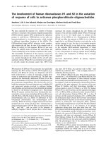
Virus phylogenyFigure 1
Virus phylogeny. The complete env and pol coding regions were sequenced for each virus isolate (EMBL accession numbers:
env; AM279343
–AM279369 and pol; AM279370–AM279396). Subsequently, NJ-trees were constructed and tree topology was
assessed by bootstrap analysis. The SIV
cpzGAB
sequence was used to root the tree. Ten isolates were found to group with the
CRF02_AG reference strains, five were subtype A and five were subtype G.
Retrovirology 2006, 3:40 />Page 5 of 11
(page number not for citation purposes)
and CD4+T cell counts >200 cells/μl against the entire set
of subtype A and G viruses, irrespective of the CD4+ T cell
counts in the patients from whom these viruses were iso-
lated. In the group with CD4+ T cells <200 cells/μl, the
CRF02_AG isolates won 35 out of 50 competitions
(70%), with a median relative fitness of 1.52 (p25 = 1.07,
p75 = 1.83). In the group with CD4+ T cells >200 cells/μl,
the CRF02_AG isolates won 34 out of 50 (68.0%) of the
competitions with a median relative fitness of 1.40 (p25 =
0.84, p75 = 1.79) (Figure 2). These observations suggest
that the difference in replicative fitness between these
viruses is not merely associated with the differences in
CD4+ T cell counts and VL.
There is evidence that the co-receptor tropism may influ-
ence HIV-1 replication in T cells [30,32] and that syncy-
tium-inducing (SI)/X4 viruses tend to be more virulent
than NSI/R5 strains. Since our cohort consisted of both X4
and R5 tropic isolates, we analyzed the data correcting for
viral co-receptor tropism. The majority of viruses were R5-
tropic, three viruses used CXCR4 (X4), and two others
were found to be dual-tropic (R5X4). The X4 CRF02_AG
isolates won 6 out of 6 (100%) competitions against X4
subtype A and G viruses. Similarly, X4 CRF02_AG strains
won 17 out of 24 (70.8%) competitions against R5 sub-
type A and G viruses. Interestingly, R5 CRF02_AG viruses
also out competed most of the X4 subtype A and G strains
(11 out of 14 competitions or 78.6%). Finally, R5
CRF02_AG won 62.5% (35 out of 56) competitions
against the R5 subtype A and R5 subtype G.
These results suggest that the increased fitness of
CRF02_AG in competitions with subtype A and G viruses
is not caused by differential co-receptor tropism.
Replicative fitness of CRF02_AG in dendritic cells
Since mucosal dendritic cells are thought to play an
important role in the early phase of sexual transmission
[28,29], assessing the replicative capacity of CRF02_AG
and subtype A and G viruses in a suitable model of
mucosa-like DC, such as the monocyte-derived DC,
would allow us to study the replication efficiency of pri-
mary HIV isolates in the context of virus transmission.
Because R5 viruses are consistently found early after trans-
mission, we restricted our analysis to isolates of this phe-
notype (i.e. four CRF02_AG, one subtype A and three
subtype G isolates).
In MO-DC, CRF02_AG isolates won 62.5% (10 out of 16)
of the competions and showed a median relative fitness of
1.48 (p25 = 0.68, p75 = 1.55, Figure 3). When comparing
fitness data obtained in T-cells and DC, it was obvious
that most of the CRF02_AG isolates that were able to out
compete subtype A and G viruses in DC also out com-
peted subtype A and G strains in T cells. In conclusion, we
found that the replicative fitness of CRF02_AG and sub-
type A and G viruses is significantly different when meas-
ured in activated T cells (P = 0.024, t-test) and also tends
to be different in DC, without reaching statistical signifi-
cance (P = 0.229, t-test) (Figure 3).
Discussion
The fact that CRF02_AG seems largely predominant over
other circulating HIV strains in an African area with
extremely high HIV genetic diversity may have several
explanations. First, the recombinant form may have some
biological advantage over the parental strains, including a
possibly higher replicative fitness and/or transmission
capacity. Second, the recombinant strain could have been
introduced first in that particular area and consequently
get established in a population before other subtypes
entered the scene (founder-effect) [31]. In the case of the
epidemiological spread of CRF02_AG in West Central
Africa, the founder hypothesis is probably a less likely
explanation. Several studies on the prevalence of HIV-1
Relative replicative fitness (W) in activated T cellsFigure 2
Relative replicative fitness (W) in activated T cells.
Dot plots represent the results of growth competitions in
PHA/IL-2 activated PBMC (10 CRF02_AG, 5 subtype A and 5
subtype G). Red dots represent competitions between
CRF02_AG and subtype A viruses; green dots show compe-
titions between CRF02_AG and subtype G. An open symbol
indicates that the CRF02_AG virus is X4-tropic, whereas
solid symbols represent competitions with R5-tropic
CRF02_AG viruses (irrespective of the coreceptor tropism
of the subtype A and G isolates). The competitions with
CRF02_AG viruses from patients with AIDS (CD4+ cells
<200 cells/μl) are shown at the left hand side and the compe-
titions with CRF02_AG viruses from non-AIDS patients at
the right hand side, again irrespective of the CD4+ T cell
counts in the patients from which the competing A or G
virus was derived.
Retrovirology 2006, 3:40 />Page 6 of 11
(page number not for citation purposes)
subtypes in the Democratic Republic of Congo (DRC)
have shown that subtypes A and G are relatively prevalent
in this area [34-37]. Moreover, it is likely that at least a
limited spread of subtype A and G viruses must have pre-
ceded the creation and spread of CRF02_AG in West Cen-
tral Africa [35].
In the present study, we explored whether the replicative
fitness of CRF02_AG was related to the epidemiological
spread of this virus in extended areas of West and Central
Africa. We showed that CRF02_AG primary isolates had a
higher replicative fitness compared to subtype A and G
isolates, in a cellular model for HIV pathogenesis (i.e. acti-
vated T-cells) and HIV transmission (i.e. MO-DC). The
higher relative replicative fitness of CRF02_AG viruses was
evident for isolates from patients with low (<200 cells/μL)
and with higher (>200 cells/μL) CD4+ T cell counts, and
it was found to be independent of the viral co-receptor
use. An independent study investigating the same hypoth-
esis was published recently and also showed an increased
replicative capacity of CRF02_AG viruses compared to
subtype A and G isolates, using basic virus growth kinetics
as a measure of replication capacity [38]. In contrast to
our study, Konings et al. [38] studied only thirteen HIV-1
isolates and presented limited data on viral load and
CD4+ T cell counts. Furthermore, the growth competition
assays used in our study are able to discriminate minor
differences in replication capacity and also provide the
internal control lacking in monoinfection kinetic assays,
as used by Konings et al. [38-40].
The viral load in the donor and the integrity of mucosal
tissues in the acceptor are amongst the most important
determinants upon HIV transmission [41,42]. Previous
observations by Fischetti et al. [21], showed significantly
higher viral loads in asymptomatic CRF02_AG infected
individuals compared to patients infected with non-
CRF02_AG strains. A direct correlation between viral load
and replicative capacity in activated T cells was repeatedly
shown [30,31]. Taken together with our observations,
these data suggest that patients infected with CRF02_AG
strains may more easily transmit virus, because of a higher
viraemia, which could be a consequence of the higher rep-
licative fitness in activated T cells. This interpretation is
consistent with the observation by Ariën et al. [27], who
previously showed that group M viruses in general have a
much higher in vitro replicative fitness than group O or
HIV-2 viruses, corresponding to the relative spread of
these viruses in the pandemic as a whole and in West
Africa (where they all co-circulate) in particular.
One could argue that the observed higher relative fitness
of CRF02_AG strains versus subtype A and G isolates in
the present study simply reflects a more advanced disease
stage of patients infected with CRF02_AG or a differential
viral co-receptor tropism. However, we have shown that
CRF02_AG with either X4 or R5 co-receptor tropism and
derived from patients with more or less advanced disease
(based on CD4+ T cell count) are on average more fit than
subtype A or G viruses (Figure 2). In addition, our data
suggests that the replicative fitness of CRF02_AG in MO-
DC was slightly, but not significantly higher than the
parental subtypes (A and G) (Figure 3). There is substan-
tial evidence that DC play an important role during HIV
transmission and it could be speculated that a slight
advantage in replicative fitness in dendritic cells may have
an important impact on transmission at the population
level. On the other hand, the number of competitions per-
formed in DC may just have been too low to result in a
significant difference.
Studies by Ball et al. [43] and Ariën et al. [27] showed that
viruses of subtypes B and C were equally fit in Langerhans'
dendritic cells, while subtype C isolates were out com-
peted by any other group M virus in activated PBMC. It is
not completely clear yet how HIV replicative fitness in T
cells and dendritic cells relate to transmission and epide-
miological spreading. It is also possible that the focus on
Relative replicative fitness (W) in monocyte-derived den-dritic cells (MO-DC)Figure 3
Relative replicative fitness (W) in monocyte-derived
dendritic cells (MO-DC): Dot plots represent growth
competitions in MO-DC and activated T cells using the same
R5-tropic viruses isolates (4 CRF02_AG, 1 subtype A and 3
subtype G isolates). Solid red squares indicate competitions
of a CRF02_AG against an A virus in MO-DC and solid green
squares indicate competitions of CRF02_AG against a G
virus in MO-DC. Solid red circles represent competitions of
a CRF02_AG against a subtype A virus in T cells and solid
green circles show competitions of a CRF02_AG against a
subtype G virus in T cells.
Retrovirology 2006, 3:40 />Page 7 of 11
(page number not for citation purposes)
replicative capacity in DC as a measure of transmission
efficiency may be too limited, since other cell types at the
mucosal interface are likely involved in transmitting HIV.
Hence a better model to study HIV transmission is desira-
ble and should include Female Genital Tract (FGT) epi-
thelia and other important target cells, such as T cells and
macrophages, in addition to DC [41,42]. We are currently
elaborating on such models in order to study early events
during HIV transmission and replicative fitness.
The CRF02_AG genome is a mosaic of subtype A (gag, vpr
and parts of pol, env and nef) and subtype G (LTR, rev, tat
and parts of pol, env, and nef). An important question that
needs to be answered is which part of the viral genome
may be responsible for the increased replicative fitness of
CRF02_AG. Unfortunately, our experimental set up did
not allow us to study the contribution of individual genes
to the overall replicative fitness of a virus isolate. There-
fore, future studies should try to elucidate the role of those
genes that have a mosaic appearance for their impact on
the fitness of the recombinant virus. It is clear that recom-
bination occurs often in dual- or super infected individu-
als, generating ISR. It could be speculated that those ISR
that generate viable progeny subsequently undergo severe
selection pressure by the innate and adaptive host
immune responses and that only the most successful/fit
ISR may eventually be able to spread epidemically and
become a CRF.
More detailed analyses of HIV samples from West Africa
have shown that CRF02_AG has already undergone fur-
ther recombination [34,44]. Clearly, viral recombination
is inevitable with the continuous intermixing of HIV sub-
types and will have its impact on the evolution of the HIV
epidemic. It is important to envisage that a CRF that we
label as very fit today may be out competed by a new and
even more fit recombinant virus tomorrow.
In conclusion, our data on a small, but carefully selected
sample from a Cameroonian cohort clearly suggests that
the prevailing CRF02_AG recombinant may be favoured
in his spread over "parental" subtype A and G viruses as a
result of a higher replicative fitness in T cells and likely
also in dendritic cells. More extensive and in-depth stud-
ies are needed to confirm this preliminary evidence and to
unravel the molecular mechanisms underlying the pre-
dominance of CRF02_AG in large parts of West Africa.
Methods
Cells
Peripheral blood mononuclear cells (PBMC) were
obtained from a HIV-1 seronegative buffy coat by Ficol
Hypaque (Sigma, St. Louis, USA) density gradient centrif-
ugation. PBMC were cultured in RPMI 1640 – 2 mM L-
glutamine medium (BioWhittaker, Verviers, Belgium)
supplemented with 10% fetal bovine serum (Biochrom
KG, Berlin, Germany) and 100 U/ml penicillin (Cellgro,
Virginia, USA) and 100μg/ml streptomycin (Cellgro, Vir-
ginia, USA). They were first stimulated with 2μg/ml of
phytohemagglutinin (PHA) (Gibco BRL, Maryland USA)
for 3 days and further maintained in 1 ng/ml interleukin
(IL-2) (Gibco BRL, Maryland USA).
Monocytes were obtained from PBMC by counter-flow
elutriation and sheep erythrocyte rosetting, yielding >95%
CD3-/CD4+ MO and <0.5% T cells, as previously
described in [28] and [45]. To obtain MO-DC, monocytes
were cultured for 7 days in RPMI 1640 supplemented with
10% FBS, IL4 (Gibco BRL, Maryland USA) (20 ng/ml),
GM-CSF (Gibco BRL, Maryland USA) (20 ng/ml), 100 U/
ml penicillin and 100μg streptomycin [28,45]. Half of the
culture medium (with cytokines) was replaced every third
day. The MO-DC were immuno phenotyped as CD13
+
/
CD14 low, CD3
-
/CD4
+
, CD1a
+
and DC-SIGN high before
use.
Viruses
Twenty viruses were obtained from HIV seropositive
patients attending the military hospital in Yaounde and
Douala in Cameroon [14]. Viruses were isolated between
1996 and 1999 and none of the patients was receiving
antiretroviral treatment (ART) at that time. All patients
signed an individual informed consent. We selected these
twenty strains from a much larger cohort [14], based on
the availability of PBMC and plasma, simultaneously
obtained from these particular patients and permanently
frozen in liquid nitrogen and at -80°C, respectively. CD4+
T cell counts were determined on fresh blood, while viral
load was measured on the stored plasma samples for the
purpose of this study, using an in-house real time PCR
assay (Table 1). The original selection encompassed
twenty-seven isolates (eleven CRF02_AG, ten subtype A
and six subtype G), but six samples were dropped for fur-
ther analyses because they showed unique recombination
events to have occurred in env and pol, i.e. they were not
pure A, nor G, nor CRF02_AG. For the twenty primary iso-
lates used in this study, subtyping was based on complete
env, complete pol, gag p24, gag p17, tat, rev, nef and vpu
nucleotide sequencing.
Frozen virus stocks were propagated and expanded in
short-term cultures of PHA/IL-2 treated PBMC obtained
from a HIV seronegative blood donor. The 50% tissue cul-
ture infectious dose (TCID
50
) was determined by serial
dilution of the virus stock to infect PHA/IL-2 PBMC and
U87.CD4 cells expressing either CCR5 or CXCR4 (Table
1) [46]. Infections with U87.CD4.CCR5 and
U87.CD4.CXCR4 cells were used to determine co-receptor
tropism and to calculate the infectious dose required to
infect MO-DC.
Retrovirology 2006, 3:40 />Page 8 of 11
(page number not for citation purposes)
Sequencing and phylogenetic analysis
The HIV-1 strains characterized in this study were cultured
in patient peripheral blood mononuclear cells. DNA was
then extracted from the infected cells using the Qiagen
DNA isolation kit (Qiagen S.A., Courtabeauf, France).
Complete sequences for the pol and the env genes were
generated. The first fragment, spanning the gag-pol region,
was amplified with G00 (5'-GACTAGCGGAGGCTA-
GAAG-3', position 761–780 on HxB2) and HPOL4538
(5'-TACTGCCCCTTCACCTTTCCA-3', position 4994–
4973 on HxB2) as outer primers. A second round frag-
ment was obtained from a hemi-nested PCR reaction with
G25reverse (5'-GCAAGTGTTTTGGCTGAAGCAAT-3',
position 1872–1895 on HxB2) and HPOL4538. The sec-
ond fragment, covering the accessory genes tat, rev and nef,
was amplified using HPOL4235 (5'-CCCTACAATC-
CCCAAAGTCAAGG-3', position 4668–4691 on HxB2)
and LSIGI (5'-TCAAGGCAAGCTTTATTGAGGCTTAAG-
CAG-3', positions 9647-9617/542-512 on HxB2). A sec-
ond round fragment was then generated with envB (5'-
AGAAAGAGCAGAAGACAGTGGCAATGA-3', position
6216–6243 on HxB2) and envM (5'-TAGCCCTTC-
CAGTCCCCCCTTTTCTTTTA-3', position 9116–9087 on
HxB2). Taq Expand Long Template PCR was used accord-
ing to manufacturer's instructions (Roche, Indianapolis,
USA). And with the following cycling conditions: 3 min-
utes denaturation at 92°C, 16 cycles at 92°C for 20 sec-
onds, 50°C for 30 seconds and 68°C for 4 minutes,
followed by 16 cycles with 20 second-increments at the
elongation step and a final extension of 10 minutes. The
amplified fragments were purified using a QiaQuick gel
extraction kit (QIAGEN S.A., France), and then directly
sequenced with primers encompassing the pol and the env
regions by using Big-Dye Chemistry (Applied Biosystems,
France) according to the instructions of the manufacturer.
Electrophoresis and data collection were done on an
Applied Biosystems 3100 Genetic Analyzer. The electro-
pherogram plots were visualized and processed under
DNASTAR to generate consensus from the different over-
lapping sequences.
The newly determined sequences were aligned with
known representatives of the different subtypes, sub-sub-
types and CRFs described in Africa, using Clustal W. Sites
with any gap between the sequences and areas of uncer-
tain alignment were excluded from the analysis. Pair wise
evolutionary distances were estimated with Kimura's two
parameters method. Phylogenetic trees were constructed
by NJ method, and the reliability of the tree topology was
assessed by bootstrap analysis. Simplot 3.2 beta software
(Stuart Ray, />),
was used to investigate the recombinant structure of the
newly sequenced genes. Similarity and bootscan plots
were performed as already described. Briefly, similarity
plots determined the percent similarity between a newly
determined sequence and selected groups of references, by
moving a window of 400 base pairs with 20 base pairs
increments along the genome alignment. Similarity val-
ues were plotted at the midpoint of the 400 base pairs
fragment. For the bootstrap plots, the SimPlot software
performed bootscanning on neighbor joining trees by
using SEQBOOT, DNADIST (with Kimura two parameters
method and F84 model of maximum likelihood method,
transition/transversion ratio = 2.0), NEIGHBOR and
CONSENSE from the PHYLIP package for a 400 base pairs
window moved along the alignment in increments of 20
base pairs. One thousand bootstraps replicates were eval-
uated for each phylogeny. The bootstrap values for the
studied sequences were plotted at the midpoint of each
window. In these two sets of analyses, the new sequences
were compared with consensus sequences (50% thresh-
old) representing the different HIV-1 clades from the
same alignment used for phylogenetic tree analysis.
Finally, all nucleotide sequences were submitted to the
EMBL Nucleotide Sequence Database (accession num-
bers: env; AM279343
–AM279369 and pol; AM279370–
AM279396
).
Dual infection/competition assays
Dual infection/competition experiments were performed
as previously described [27,30-32]. In short, all
CRF02_AG were competed against 5 subtype A and 5 sub-
type G, (Table 1) in PHA/IL-2PBMC from one donor in 24
well culture plates and in duplicate. It is important to
mention that aliquots of PBMC of the same buffy coat
were used to grow the virus stocks, determine infectious
titers and perform the competitions. A second set of com-
petitions was performed using all available NSI/R5 iso-
lates in MO-DC from another donor. In these
competition experiments, cells (2 × 10
5
PHA/IL-2 PBMC
or 1 × 10
6
MO-DC) were infected with two isolates at
equal multiplicity of infection (5 × 10
-4
MOI for PHA/IL-
2 PBMC or 1 × 10
-3
MOI for MO-DCs) [27,32]. The esti-
mated frequency of in vitro recombination between HIV
isolates in the dual infections was less than 0.1%/1000 bp
or well below the limit of HTA detection [30,40]. Unin-
fected cells were used as HIV-negative controls and mono-
infected cultures of each virus were used as positive con-
trols. Infected cell cultures were incubated at 37
°
C and 5%
CO
2
for 24 hours after which residual virus was washed
away with 1x phosphate-buffered saline pH 7.4 (PBS).
Infected cells were re-suspended in medium containing
IL-2 (in case of PHA/IL-2 PBMC) or medium without IL-2
(for MO-DCs) and kept at 37
°
C and 5% CO
2
for 14 days.
Half the culture medium was replaced twice a week. Cell
free supernatant were collected at day 7, 10 and 14 and
analyzed for gag p24 content using an in-house p24 ELISA
assay [47]. Cells were harvested at peak vireamia and
stored at -80°C for subsequent analysis. A more detailed
Retrovirology 2006, 3:40 />Page 9 of 11
(page number not for citation purposes)
description of the dual/infection competition assay can be
found in [39,40].
Heteroduplex tracking assay
Genomic DNA was extracted from lysed PHA/IL-2PBMC
using the QIAamp DNA blood kit (Qiagen). Viral DNA
was PCR amplified using a set of external primers (envB;
5'-AGAAAGAGCAGAAGACAGTGGCAATGA-3' and
ED14; 5'-TCTTGCCTGGAGCTGTTTGATGCCCCAGAC-
3') and nested primers E80 (5'-CCAATTCCCATACAT-
TATTGTC-3') and E125 (5'-CAATTTCTGGGTCCCCTCCT-
GAGG-3') to produce a ± 480 bp fragment, encoding the
C2–C4 env region [30]. Both first round and second round
PCR amplifications were carried out in 100μl reaction
mixture under defined cycling conditions [30]. Subse-
quently, heteroduplex tracking assays (HTA) were pre-
formed to estimate the amount of virus produced by each
isolate in the competition, relative to the amount of virus
produced in monoinfections [30]. The same genomic
region of two subtype B HIV-1 strains (i.e. VI969-6 and JR-
FL) was amplified and used as probes in the HTA. Probes
were generated in amplification reactions using [γ-
32
P]
ATP labeled E80 primer radiolabelled PCR-amplified
probes were separated on 1% agarose gels and then puri-
fied using the QIAquick gel extraction kit (Qiagen). Reac-
tion mixtures containing DNA annealing buffer (100 mM
NaCl, 10 mm tris-HCl [pH 7.8], 2 mM EDTA, 10 μl of
unlabelled PCR-amplified DNA from the competition
cultures and approximately 0.1 pmol of radioactive probe
DNA. Each dual infection/competition was analyzed in at
least two independent HTA reactions using two radiola-
beled probes. PCR amplicon and probe were denatured at
95°C for 3 min, 37°C for 5 min and then rapidly
annealed on wet ice. After 30 minutes, DNA heterodu-
plexes were resolved on 5% TBE non-denaturing polyacr-
ylamide (PAGE) gels (Bio-Rad) (75 min. at 200 V). Gels
were then dried for 45 minutes at 80°C and exposed on a
phosphor imaging screen overnight. Images were cap-
tured with a phosphor imager (Cyclone, PerkinElmer)
and analysed with the OptiQuant software package (Perk-
inElmer).
Estimation of relative viral fitness
Relative virus production (ws) of each isolate in a dual
infection was calculated by dividing the amount of isolate
DNA in the dual infection and the amount of the same
isolate DNA in a monoinfection (as determined with the
phosphor imager). From these ws values, relative fitness
(W) values for each virus were obtained using the formula
[W = (ws1/(ws1 + ws2)) x 2], where ws1 and ws2 are rela-
tive virus production of isolate 1 and 2, respectively
[30,40].
Statistics
Average CD4+ T cell counts and average viral loads were
calculated for each group of viruses (i.e. CRF02_AG and
non-CRF02_AG). One sample t-tests were used to calcu-
late whether differences in CD4+ T cell counts and VL
between virus groups (i.e. CRF02_AG and non-
CRF02_AG) were statistically significant.
Average and interquartile relative fitness values (W) were
calculated for competitions involving CRF02_AG, subtype
A and subtype G virus isolates. One sample t-tests was
used to determine whether the relative fitness of a group
of viruses (i.e. CRF02_AG, or subtype A, or subtype G) was
significantly different from W = 1.0 (with W = 1.0 mean-
ing equal relative fitness). For all analyses, the level of sig-
nificance was set at P = 0.05.
Authors' contributions
HFN has performed the majority of the experimental work and
data analysis, and has drafted the manuscript.
YG has contributed to the experimental work.
GV has contributed to the study-design and helped to draft the
manuscript.
CC has contributed to the experimental work.
WJ has contributed to the data analysis.
NV has contributed to the experimental work (phylogenetic
analysis).
CB has contributed to the experimental work (sequencing).
EMN is coordinator of Projet PRESICA and of the patient
cohort.
MP has contributed to the study-design and provided the HIV-
isolates.
KKA has contributed to the study-design and helped to draft the
manuscript.
All authors read and approved the final manuscript.
Competing interests statement
The author(s) declare that they have no competing inter-
ests.
Acknowledgements
This work was supported by a grant (G.0431.02) from the Fund for Scien-
tific Research – Flanders (FWO) and a grant from Janssen Pharmaceutica
(Nr. 85600). We are indebted to the Antwerp Red Cross Blood Transfu-
sion Center for providing buffy coats. HFN would like to express her sin-
Retrovirology 2006, 3:40 />Page 10 of 11
(page number not for citation purposes)
cere gratitude to Ackerman & van Haaren for funding her doctoral
fellowship.
References
1. Robertson DL, Sharp PM, McCutchan FE, Hahn BH: Recombination
in HIV-1. Nature 1995, 374:124-126.
2. Gao F, Robertson DL, Morrison SG, Hui H, Craig S, Decker J, Fultz
PN, Girard M, Shaw GM, Hahn BH, Sharp PM: The heterosexual
human immunodeficiency virus type 1 epidemic in Thailand
is caused by an intersubtype (A/E) recombinant of African
origin. J Virol 1996, 70:7013-7029.
3. McCutchan FE, Hegerich PA, Brennan TP, Phanuphak P, Singharaj P,
Jugsudee A, Berman PW, Gray AM, Fowler AK, Burke DS: Genetic
variants of HIV-1 in Thailand. AIDS Res Hum Retroviruses 1992,
8:1887-1895.
4. Saksena NK, Wang B, Ge YC, Xiang SH, Dwyer DE, Cunningham AL:
Coinfection and genetic recombination between HIV-1
strains: possible biological implications in Australia and
South East Asia. Ann Acad Med Singapore 1997, 26:121-127.
5. Carr JK, Salminen MO, Koch C, Gotte D, Artenstein AW, Hegerich
PA, St Louis D, Burke DS, McCutchan FE: Full-length sequence
and mosaic structure of a human immunodeficiency virus
type 1 isolate from Thailand. J Virol 1996, 70:5935-5943.
6. Carr JK, Salminen MO, Albert J, Sanders-Buell E, Gotte D, Birx DL,
McCutchan FE: Full genome sequences of human immunodefi-
ciency virus type 1 subtypes G and A/G intersubtype recom-
binants. Virology 1998, 247:22-31.
7. Howard TM, Rasheed S: Genomic structure and nucleotide
sequence analysis of a new HIV type 1 subtype A strain from
Nigeria. AIDS Res Hum Retroviruses 1996, 12:1413-1425.
8. Osmanov S, Pattou C, Walker N, Schwardlander B, Esparza J: Esti-
mated global distribution and regional spread of HIV-1
genetic subtypes in the year 2000. J Acquir Immune Defic Syndr
2002, 29:184-190.
9. Heyndrickx L, Janssens W, Ndumbe PM, Vereecken K, Coppens S, De
Houwer K, Fransen K, Van der Auwera G, van der Groen G: HIV-1
genetic variability in Cameroon. AIDS 2000, 14:1862-1864.
10. Nyambi P, Heyndrickx L, Vereecken K, Burda S, De Houwer K, Cop-
pens S, Urbanski M, Williams C, Ndumbe P, Janssens W: Predomi-
nance of infection with HIV-1 circulating recombinant form
CRF02_AG in major Cameroonian cities and towns. AIDS
2002, 16:295-296.
11. Andersson S, Norrgren H, Dias F, Biberfeld G, Albert J: Molecular
characterization of human immunodeficiency virus (HIV)-1
and -2 in individuals from guinea-bissau with single or dual
infections: predominance of a distinct HIV-1 subtype A/G
recombinant in West Africa. Virology 1999, 262:312-320.
12. Carr JK, Torimiro JN, Wolfe ND, Eitel MN, Kim B, Sanders-Buell E,
Jagodzinski LL, Gotte D, Burke DS, Birx DL, McCutchan FE: The AG
recombinant IbNG and novel strains of group M HIV-1 are
common in Cameroon. Virology 2001, 286:168-181.
13. Ellenberger DL, Pieniazek D, Nkengasong J, Luo CC, Devare S, Mau-
rice C, Janini M, Ramos A, Fridlund C, Hu DJ, et al.: Genetic analysis
of human immunodeficiency virus in Abidjan, Ivory Coast
reveals predominance of HIV type 1 subtype A and introduc-
tion of subtype G. AIDS Res Hum Retroviruses 1999, 15:3-9.
14. Laurent C, Bourgeois A, Faye MA, Mougnutou R, Seydi M, Gueye M,
Liegeois F, Kane CT, Butel C, Mbuagbaw J, Zekeng L, Mboup S,
Mpoudi-Ngole E, Peeters M, Delaporte E: No difference in clinical
progression between patients infected with the predomi-
nant human immunodeficiency virus type 1 circulating
recombinant form (CRF) 02_AG strain and patients not
infected with CRF02_AG, in Western and West-Central
Africa: a four-year prospective multicenter study. J Infect Dis
2002, 186:486-492.
15. McCutchan FE, Carr JK, Bajani M, Sanders-Buell E, Harry TO, Stoeckli
TC, Robbins KE, Gashau W, Nasidi A, Janssens W, Kalish ML: Sub-
type G and multiple forms of A/G intersubtype recombinant
human immunodeficiency virus type 1 in Nigeria. Virology
1999, 254:226-234.
16. Vergne L, Paraskevis D, Vandamme AM, Delaporte E, Peeters M:
High prevalence of CRF02_AG and many minor resistance-
related mutations at the protease gene among HIV-infected
treatment-naïve immigrants in Madrid. AIDS 2003,
17:1105-1107.
17. Brodine SK, Starkey MJ, Shaffer RA, Ito SI, Tasker SA, Barile AJ, Tam-
minga CL, Stephan KT, Aronson NE, Fraser SL, Wallace MR, Wegner
SA, Mascola JR, McCutchan FE: Diverse HIV-1 subtypes and clin-
ical, laboratory and behavioral factors in a recently infected
US military cohort. AIDS 2003, 17:2521-2527.
18. Peeters M, Toure-Kane C, Nkengasong JN: Genetic diversity of
HIV in Africa: impact on diagnosis, treatment, vaccine devel-
opment and trials. Aids 2003, 17:2547-2560.
19. Vergne L, Bourgeois A, Mpoudi-Ngole E, Mougnutou R, Mbuagbaw J,
Liegeois F, Laurent C, Butel C, Zekeng L, Delaporte E, Peeters M:
Biological and genetic characteristics of HIV infections in
Cameroon reveals dual group M and O infections and a cor-
relation between SI-inducing phenotype of the predominant
CRF02_AG variant and disease stage. Virology 2003,
310:254-266.
20. Takehisa J, Zekeng L, Ido E, Mboudjeka I, Moriyama H, Miura T,
Yamashita M, Gurtler LG, Hayami M, Kaptue L: Various types of
HIV mixed infections in Cameroon. Virology 1998, 245:1-10.
21. Fischetti L, Opare-Sem O, Candotti D, Lee H, Allain JP: Higher viral
load may explain the dominance of CRF02_AG in the molec-
ular epidemiology of HIV in Ghana. AIDS 2004, 18:1208-1210.
22. Fischetti L, Opare-Sem O, Candotti D, Sarkodie F, Lee H, Allain JP:
Molecular epidemiology of HIV in Ghana: dominance of
CRF02_AG. J Med Virol 2004, 73:158-166.
23. Sarr AD, Eisen G, Gueye-Ndiaye A, Mullins C, Traore I, Dia MC,
Sankale JL, Faye D, Mboup S, Kanki P: Viral dynamics of primary
HIV-1 infection in Senegal, West Africa. J Infect Dis 2005,
191:1460-1467.
24. Gilbert PB, McKeague IW, Eisen G, Mullins C, Gueye NA, Mboup S,
Kanki PJ: Comparison of HIV-1 and HIV-2 infectivity from a
prospective cohort study in Senegal. Stat Med 2003,
22:573-593.
25. Kanki PJ, Travers KU, S MB, Hsieh CC, Marlink RG, Gueye NA, Siby
T, Thior I, Hernandez-Avila M, Sankale JL, et al.: Slower heterosex-
ual spread of HIV-2 than HIV-1. Lancet 1994, 343:943-946.
26. Marlink R, Kanki P, Thior I, Travers K, Eisen G, Siby T, Traore I, Hsieh
CC, Dia MC, Gueye EH, et al.: Reduced rate of disease develop-
ment after HIV-2 infection as compared to HIV-1. Science
1994, 265:1587-1590.
27. Ariën KK, Abraha A, Quinones-Mateu ME, Kestens L, Vanham G, Arts
EJ: The replicative fitness of primary human immunodefi-
ciency virus type 1 (HIV-1) group M, group O and HIV-2 iso-
lates. J Virol 2005, 79:8979-8990.
28. Vanham G, Penne L, Allemeersch H, Kestens L, Willems B, van der
Groen G, Jeang KT, Toossi Z, Rich E:
Modeling HIV transfer
between dendritic cells and T cells: importance of HIV phe-
notype, dendritic cell-T cell contact and T-cell activation.
AIDS 2000, 14:2299-2311.
29. Zoeteweij JP, Blauvelt A: HIV-Dendritic cell interactions pro-
mote efficient viral infection of T cells. J Biomed Sci 1998,
5:253-259.
30. Quinones-Mateu ME, Ball SC, Marozsan AJ, Torre VS, Albright JL,
Vanham G, van Der Groen G, Colebunders RL, Arts EJ: A dual
infection/competition assay shows a correlation between ex
vivo human immunodeficiency virus type 1 fitness and dis-
ease progression. J Virol 2000, 74:9222-9233.
31. Troyer RM, Collins KR, Abraha A, Fraundorf E, Moore DM, Krizan
RW, Toossi Z, Colebunders RL, Jensen MA, Mullins JI, et al.: Changes
in human immunodeficiency virus type 1 fitness and genetic
diversity during disease progression. J Virol 2005, 79:9006-9018.
32. Ariën KK, Gali Y, El-Abdellati A, Heyndrickx L, Janssens W, Vanham
G: Replicative fitness of CCR5-using and CXCR4-using
human immunodeficiency virus type 1 biological clones. Virol-
ogy 2006, 347:65-74.
33. Foley B, Pan H, Buchbinder S, Delwart EL: Apparent founder
effect during the early years of the San Francisco HIV type 1
epidemic (1978–1979). AIDS Res Hum Retrovir 2000,
16:1463-1469.
34. Niama FR, Toure-Kane C, Vidal N, Obengui P, Bikandou B,
Ndoundou Nkodia MY, Montavon C, Diop-Ndiaye H, Mombouli JV,
Mokondzimobe E, Diallo AG, Delaporte E, Parra HJ, Peeters M,
Mboup S: HIV-1 subtypes and recombinants in the Republic of
Congo. Infect Genet Evol in press. 2006 Feb 10
35. Yang C, Dash B, Hanna SL, Frances HS, Nzilambi N, Colebunders RC,
St Louis M, Quinn TC, Folks TM, Lal RB: Predominance of HIV
Publish with BioMed Central and every
scientist can read your work free of charge
"BioMed Central will be the most significant development for
disseminating the results of biomedical research in our lifetime."
Sir Paul Nurse, Cancer Research UK
Your research papers will be:
available free of charge to the entire biomedical community
peer reviewed and published immediately upon acceptance
cited in PubMed and archived on PubMed Central
yours — you keep the copyright
Submit your manuscript here:
/>BioMedcentral
Retrovirology 2006, 3:40 />Page 11 of 11
(page number not for citation purposes)
type 1 subtype G among commercial sex workers from Kin-
shasa, Democratic Republic of Congo. .
36. Bikandou B, Takehisa J, Mboudjeka I, Ido E, Kuwata T, Miyazaki Y,
Moriyama H, Harada Y, Taniguchi Y, Ichimura H, Ikeda M, Ndolo PJ,
Nzoukoudi MY, M'Vouenze R, M'Pandi M, Parra HJ, M'Pele P, Hayami
M: Genetic subtypes of HIV type 1 in Republic of Congo. AIDS
Res Hum Retroviruses 2000, 16:613-619.
37. Vidal N, Peeters M, Mulanga-Kabeya C, Nzilambi N, Robertson D,
Ilunga W, Sema H, Tshimanga K, Bongo B, Delaporte E: Unprece-
dented degree of human immunodeficiency virus type 1
(HIV-1) group M genetic diversity in the Democratic Repub-
lic of Congo suggests that the HIV-1 pandemic originated in
Central Africa. J Virol 2000, 74:10498-10507.
38. Konings FAJ, Burda S, Urbansi MM, Zhong P, Nadas A, Nyambi P:
Human Immunodeficiency Virus Type 1 (HIV-1) Circulating
Recombinant Form CRF02_AG (CRF02_AG) has a higher in
vitro replicative capacity than is parental subtypes A and G.
J Med Virol 2006, 78:523-534.
39. Quinones-Mateu ME, Arts EJ: Virus fitness: concept, quantifica-
tion, and application to HIV population dynamics. Curr Top
Microbiol Immunol 2006, 299:83-140.
40. Abraha A, Troyer RM, Quinones-Mateu ME, Arts EJ: Methods to
determine HIV-1 ex vivo fitness. Methods Mol Biol 2005,
304:355-368.
41. Hu J, Gardner MB, Miller CJ: Simian immunodeficiency virus
rapidly penetrates the cervicovaginal mucosa after intravag-
inal inoculation and infects intraepithelial dendritic cells. J
Virol 2000, 74:6087-6095.
42. Spira AI, Marx PA, Patterson BK, Mahoney J, Koup RA, Wolinsky SM,
Ho DD: Cellular targets of infection and route of viral dissem-
ination after an intravaginal inoculation of simian immuno-
deficiency virus into rhesus macaques. J Exp Med 1996,
183:215-225.
43. Ball SC, Abraha A, Collins KR, Marozsan AJ, Baird H, Quinones-Mateu
ME, Penn-Nicholson A, Murray M, Richard N, Lobritz M, Zimmerman
PA, Kawamura T, Blauvelt A, Arts EJ: Comparing the ex vivo fit-
ness of CCR5-tropic human immunodeficiency virus type 1
isolates of subtypes B and C. J Virol 2003, 77:1021-1038.
44. Konings FA, Haman GR, Xue Y, Urbanski MM, Hertzmark K, Nanfack
A, Achkar JM, Burda ST, Nyambi PN: Genetic Analysis of HIV-1
Strains in Rural Eastern Cameroon Indicates the Evolution
of Second-Generation Recombinants to Circulating Recom-
binant Forms.
J Acquir Immune Defic Syndr in press. 2006 Apr 24
45. Njai HF, Lewi PJ, Janssen CG, Garcia S, Fransen K, Kestens L, Vanham
G, Janssen PA: Pre-incubation of cell-free HIV-1 group M iso-
lates with non-nucleoside reverse transcriptase inhibitors
blocks subsequent viral replication in co-cultures of dendritic
cells and T cells. Antivir Ther 2005, 10:255-262.
46. Reed LJ, Muench H: A simple method of estimating fifty per-
cent endpoints. Am J Hyg 1938, 27:493-497.
47. Beirnaert E, Willems B, Peeters M, Bouckaert A, Heyndrickx L, Zhong
P, Vereecken K, Coppens S, Davis D, Ndumbe P, et al.: Design and
evaluation of an in-house HIV-1 (group M and O), SIVmnd
and SIVcpz antigen capture assay. J Virol Methods 1998,
73:65-70.