Báo cáo y học: " Effects of prostratin on Cyclin T1/P-TEFb function and the gene expression profile in primary resting CD4+ T cells" doc
Bạn đang xem bản rút gọn của tài liệu. Xem và tải ngay bản đầy đủ của tài liệu tại đây (1.12 MB, 14 trang )
Retrovirology
BioMed Central
Open Access
Research
Effects of prostratin on Cyclin T1/P-TEFb function and the gene
expression profile in primary resting CD4+ T cells
Tzu-Ling Sung and Andrew P Rice*
Address: Department of Molecular Virology and Microbiology, Baylor College of Medicine, Houston, Texas 77030, USA
Email: Tzu-Ling Sung - ; Andrew P Rice* -
* Corresponding author
Published: 02 October 2006
Retrovirology 2006, 3:66
doi:10.1186/1742-4690-3-66
Received: 05 July 2006
Accepted: 02 October 2006
This article is available from: />© 2006 Sung and Rice; licensee BioMed Central Ltd.
This is an Open Access article distributed under the terms of the Creative Commons Attribution License ( />which permits unrestricted use, distribution, and reproduction in any medium, provided the original work is properly cited.
Abstract
Background: The latent reservoir of human immunodeficiency virus type 1 (HIV-1) in resting
CD4+ T cells is a major obstacle to the clearance of infection by highly active antiretroviral therapy
(HAART). Recent studies have focused on searches for adjuvant therapies to activate this reservoir
under conditions of HAART. Prostratin, a non tumor-promoting phorbol ester, is a candidate for
such a strategy. Prostratin has been shown to reactivate latent HIV-1 and Tat-mediated
transactivation may play an important role in this process. We examined resting CD4+ T cells from
healthy donors to determine if prostratin induces Cyclin T1/P-TEFb, a cellular kinase composed of
Cyclin T1 and Cyclin-dependent kinase-9 (CDK9) that mediates Tat function. We also examined
effects of prostratin on Cyclin T2a, an alternative regulatory subunit for CDK9, and 7SK snRNA
and the HEXIM1 protein, two factors that associate with P-TEFb and repress its kinase activity.
Results: Prostratin up-regulated Cyclin T1 protein expression, modestly induced CDK9 protein
expression, and did not affect Cyclin T2a protein expression. Although the kinase activity of CDK9
in vitro was up-regulated by prostratin, we observed a large increase in the association of 7SK
snRNA and the HEXIM1 protein with CDK9. Using HIV-1 reporter viruses with and without a
functional Tat protein, we found that prostratin stimulation of HIV-1 gene expression appears to
require a functional Tat protein. Microarray analyses were performed and several genes related to
HIV biology, including APOBEC3B, DEFA1, and S100 calcium-binding protein genes, were found to
be regulated by prostratin.
Conclusion: Prostratin induces Cyclin T1 expression and P-TEFb function and this is likely to be
involved in prostratin reactivation of latent HIV-1 proviruses. The large increase in association of
7SK and HEXIM1 with P-TEFb following prostratin treatment may reflect a requirement in CD4+
T cells for a precise balance between active and catalytically inactive P-TEFb. Additionally, genes
regulated by prostratin were identified that have the potential to regulate HIV-1 replication both
positively and negatively.
Background
A latent HIV-1 reservoir in resting memory CD4+ T cells is
a major obstacle to the clearance of infection by HAART.
The latently infected cells are quiescent and express little
if any viral antigens, making it difficult for the immune
system to recognize and extinguish them. Cessation of
antiviral drugs almost invariably leads to reactivation of
high levels of viral replication from this reservoir. The
Page 1 of 14
(page number not for citation purposes)
Retrovirology 2006, 3:66
slow turnover of memory CD4+ T cells contributes to the
maintenance of the reservoir, and ongoing virus replication that is below the detection limit may continue to
reseed the reservoir in the presence of HAART (reviewed
in [1-3]).
Mechanisms that establish HIV latency in infected memory CD4+ T cells are not well understood, but it is likely
that multiple mechanisms are involved. It has been proposed that latency can result when HIV-1 infects a CD4+ T
cell that has been activated and is returning to a quiescent
state as the resting memory CD4+ T cell phenotype is
established [1]. Blocks to transcription of latent provirus
are likely to involve limiting amounts of cellular factors
that are essential for RNA polymerase II transcription
directed by the viral long terminal repeat (LTR) sequences,
such as NF-κB, NF-AT, and the Cyclin T1/P-TEFb complex
that mediates the viral Tat protein function. Additionally,
HIV-1 integration in heterochromatin regions of the
genome may be a factor in some latent infections [4].
The viral Tat function is likely to be a key component of
latency. Cyclin T1/P-TEFb consists of Cyclin T1 and CDK9
which can phosphorylate the carboxyl-terminal domain
(CTD) of RNA polymerase II and factors that negatively
regulate transcriptional elongation, leading to enhanced
transcription processivity. The HIV-1 Tat protein recruits
Cyclin T1/P-TEFb to the TAR RNA structure located at the
5' end of viral transcripts to promote transcription elongation of the integrated provirus (reviewed in [5-7]). Cyclin
T1/P-TEFb is subject to positive regulation in resting CD4+
T cells, as activation of these cells by phytohaemagglutinin (PHA) or combinations of cytokines induces Cyclin
T1/P-TEFb [8]. Cyclin T1/P-TEFb is also subject to negative regulation, as a small nuclear RNA known as 7SK
snRNA and the major HEXIM1 and minor HEXIM2 proteins are recently identified P-TEFb-associated factors that
repress kinase activity [9,10]. In support of the idea that
7SK and HEXIM1 proteins repress P-TEFb function, depletion of 7SK snRNA by anti-sense DNA oligonulceotides or
siRNAs activates transfected reporter plasmids [10,11].
Additionally, over-expression of HEXIM1 can repress Tat
activation of an HIV-1 LTR reporter plasmid [12,13].
However, following activation of peripheral blood lymphocytes (PBLs), the association of 7SK snRNA with PTEFb is greatly increased and this correlates with active
RNA polymerase II transcription [14]. We recently
observed that while siRNA depletion of 7SK snRNA in
HeLa cells stimulates expression of reporter plasmids and
induces apoptosis, it does not affect expression of the
endogenous P-TEFb-dependent cellular genes or of HIV-1
reporter viruses [11]. Thus, although perturbation of the
normal level of association of 7SK and HEXIM1 with PTEFb can influence expression from transfected plasmids,
/>
the effects on endogenous P-TEFb-dependent genes or an
integrated HIV-1 provirus are less apparent.
Recent studies have focused on the search for adjuvant
therapies that can reactivate the HIV latent reservoir under
conditions of HAART to suppress active viral replication,
thus reducing the size of the reservoir with the ultimate
goal of eradication. Prostratin, a non-tumor-promoting
phorbol ester, is a candidate for such an adjuvant strategy
[15]. Prostratin has been shown to activate NF-κB and
reactivate latent HIV [16-18]. Although prostratin stimulates the expression of T cell activation markers, it does
not promote cell proliferation, therefore lowering the
risks of expanding the latently-infected cell population
[15,19]. The majority of studies of prostratin focused on
its role in reactivation of HIV from transcriptional latency
[20,21], but prostratin may also affect other stages of virus
replication. Indeed, prostratin has been shown to downregulate CD4 and CXCR4 to inhibit viral entry through
PKC pathways and to block reverse transcription
[19,22,23]. These dual effects of prostratin, activating
latent HIV and inhibiting further spreading of the virus,
appear to meet the criteria for a useful adjuvant therapy.
To further examine mechanisms involved in reactivation
of proviruses by prostratin, we examined effects of prostratin on P-TEFb in resting CD4+ T cells isolated from
healthy blood donors. We also carried out a transcriptional profile analysis to identified genes of relevance to
HIV infection that are regulated by prostratin.
Results
Prostratin up-regulates Cyclin T1 but not Cyclin T2a in
resting CD4+ T cells
We wished to examine whether the induction of HIV-1
proviral transcription in latently infected cells by prostratin might involve an up-regulation of Cyclin T1/PTEFb, a mediator of the viral Tat activation function. To
confirm that prostratin induced early T cell activation
markers without promoting cellular proliferation under
our experimental conditions, resting CD4+ T cells isolated
from healthy donors were treated with dimethyl sulfoxide
(DMSO) as a solvent control or prostratin for 48 hours,
and expression of CD25 and CD69 was evaluated by flow
cytometry (Fig. 1A). Additionally, a portion of untreated
cells were examined immediately after isolation. Prostratin treatment induced expression of CD69, and had a
modest increase on CD25 expression. Propidium iodide
staining was also performed to evaluate cell cycle progression (Fig. 1B). Prostratin had no effect on cellular proliferation, as the percentage of cells in S and G2/M phases
was similar in prostratin-treated and control cells. In addition, apoptosis appeared to occur in approximately 10%
of cells in both control and prostratin-treated cells. We
conclude that under these conditions, prostratin induces
expression of CD69 and to a limited extent CD25, but
Page 2 of 14
(page number not for citation purposes)
Retrovirology 2006, 3:66
/>
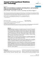
Figure 1 induces expression of CD69 without promoting cell cycle progression in resting CD4+ T cells
Prostratin
Prostratin induces expression of CD69 without promoting cell cycle progression in resting CD4+ T cells. Resting
CD4+ T cells were analyzed immediately after isolation (Untreated), or were cultured for 48 hours in the presence of DMSO
as a control or prostratin (250 ng/ml) before analysis. (A) Cells were assayed for expression of CD25 and CD69 by flow
cytometry. (B) Cells were stained with propidium iodide to evaluate DNA content by flow cytometry.
does not induce cellular proliferation nor enhance apoptosis, in agreement with previous studies [18,19].
We and others have reported that activation of PBLs and
resting CD4+ T cells with PHA, a lectin mitogen, induces
cell proliferation and the expression of Cyclin T1 and
CDK9, components of the Cyclin T1/P-TEFb complex
[8,24-27]. To examine whether prostratin induces Cyclin
T1 and CDK9, immunoblots were performed with extracts
prepared from cells treated for 48 hours with DMSO or
prostratin. We examined resting CD4+ T cells isolated
from a number of donors, and results from six representative donors are shown in Fig. 2A. The levels of β-actin, a
loading control, were equivalent in each extract. After
prostratin treatment, Cyclin T1 level increased in Donors
38, 40, and 45 from almost undetectable levels in control
cells to levels of induction ranging from approximately
four- to 14-fold after normalization to β-actin levels. For
Donors 39, 66, and 67, Cyclin T1 was expressed at a basal
level in the resting cells and was induced from two- to fivefold by prostratin. In contrast to Cyclin T1, the major
CDK9 42 kDa protein was present at readily detectable
levels in control cells and was not further up-regulated in
Donors 38 and 45, while in Donors 39, 40, 66, and 67,
CDK9 levels increased from 1.5- to 2.5-fold following
prostratin treatment. We observed that the 55 kDa minor
form of CDK9 was generally presented at low levels in
resting CD4+ T cells and in a few donors it was induced
approximately two-fold (data not shown).
We also examined the levels of Cyclins T2a and T2b, two
additional cyclin partners of CDK9. Cyclin T2a expression
Page 3 of 14
(page number not for citation purposes)
Retrovirology 2006, 3:66
/>
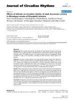
Figure 2
Effects of prostratin on the expression levels of Cyclin T1, Cyclin T2a, and CDK9
Effects of prostratin on the expression levels of Cyclin T1, Cyclin T2a, and CDK9. Cell extracts were prepared from
resting CD4+ T cells from different donors cultured in DMSO or prostratin for 48 hours, and immunoblots were performed to
examine the levels of Cyclin T1, CDK9, β-actin (A) and Cyclin T2a (B). The immunoblots were quantified as described in Material and Methods using β-actin for normalization; the value for fold-induction is given below the panels.
was not significantly affected by prostratin (Fig. 2B), while
the less abundant Cyclin T2b was below the level of detection in our system (data not shown). We conclude from
these experiments that prostratin up-regulates Cyclin T1
expression and has a relatively modest stimulatory effect
on CDK9 expression levels in resting CD4+ T cells. Because
expression of Cyclin T2a was not affected by prostratin
activation, this P-TEFb regulatory subunit may be generally involved in constitutive gene expression in resting
and activated CD4+ T cells, whereas Cyclin T1 may play a
larger role in the expression of genes induced by T cell activation.
Effects of prostratin on the levels of 7SK snRNA and
HEXIM1 associated with CDK9
We next wished to examine if 7SK snRNA and HEXIM1,
two molecules which are known to associate with P-TEFb
and repress catalytic activity in vitro, are affected by prostratin treatment. For detection of total 7SK levels, we carried out Northern blots of total RNA isolated from control
and prostratin-treated cells (Fig. 3A). Total 7SK snRNA
levels were increased in prostratin-treated cells compared
to control cells, while U1 snRNA remained constant.
When normalized to U1 RNA, total 7SK snRNA increased
3.4- and 6-fold in the two donors examined. To evaluate
the amount of 7SK associated with P-TEFb, we immuno-
precipitated CDK9 and measured 7SK levels in precipitates by Northern blots (Fig. 3B). Although there was a
considerable variation between the two donors examined,
the association between 7SK snRNA and CDK9 increased
significantly after prostratin treatment when normalized
to the amount of CDK9 protein in immunoprecipitates,
consistent with our previous findings in activated PBLs
[14].
We also examined HEXIM1 protein expression levels and
its association with CDK9. HEXIM1 was readily detectable
in control cells and prostratin treatment induced its
expression approximately two-fold in both donors (Fig.
3C). To examined the amount of HEXIM1 associated with
P-TEFb, co-immunoprecipitations were performed with
antibodies against CDK9 (Fig. 3D). Similar to 7SK snRNA,
the levels of HEXIM1 associated with CDK9 increased in
prostratin-treated cell. After normalization to the amount
of CDK9 present in immunoprecipitates, this increase was
an approximate 9-fold induction in Donor 66 and a 4fold induction in Donor 68. These data are consistent
with previous studies which demonstrated that 7SK
snRNA enhances binding of HEXIM1 to P-TEFb [9,28].
Furthermore, these data indicate that prostratin treatment
leads to a large increase in the proportion of CDK9 molecules that are associated with 7SK and HEXIM1. However,
Page 4 of 14
(page number not for citation purposes)
Retrovirology 2006, 3:66
/>
Figure 3
Levels of 7SK snRNA and HEXIM1 and association with P-TEFb
Levels of 7SK snRNA and HEXIM1 and association with P-TEFb. (A) Total RNA was isolated from DMSO control and
prostratin-treated resting CD4+ T cells. Northern blots were performed to measure 7SK snRNA and U1 snRNA levels;
amounts of 7SK and U1 snRNA were quantified using a Phosphoimager and are shown below each panel. Levels of 7SK snRNA
were normalized to U1 snRNA. (B) Immunoprecipitations were performed using α-CDK9 antibodies with extracts from control and prostratin-treated cells. CDK9 levels present in a portion of immunoprecipitates were examined by immunoblots (IPImmunoblot). RNA was extracted from the remaining protein of immunoprecipitates and the levels of 7SK snRNA were determined by Northern blots (IP-Northern blot). Amounts of 7SK snRNA were quantified by a PhosphoImager and normalized the
amounts of CDK9 present in immunoprecipitates. (C) Cell extracts were prepared from resting CD4+ T cells cultured in
DMSO or prostratin for 48 hours to examine the levels of HEXIM1 and β-actin. Protein levels were quantified by the Densitometer and are shown below each panel. Levels of HEXIM1 were normalized to β-actin levels. (D) Immunoprecipitations were
performed using antiserum against CDK9 with extracts adjusted to precipitate equivalent amount of CDK9. Immunoprecipitation products were subjected to immunoblot analysis to evaluate the levels of HEXIM1 associated with CDK9. Levels of
HEXIM1 were normalized to CDK9 levels in immunoprecipitates.
this increase does not appear to result in a general repression of gene expression, as the cells are responding to a
program of T cell activation (see Fig. 1).
Prostratin increases CDK9 kinase activity in resting CD4+ T
cells
Cell activation by a combination of cytokines or PHA has
been shown to increase P-TEFb catalytic activity in PBLs
and purified resting CD4+ T cells [8,24,26]. Induction of
kinase activity by those treatments correlates with
increased protein levels for both Cyclin T1 and CDK9. We
performed kinase assay with a recombinant CTD substrate
to examine if prostratin increases P-TEFb activity in resting
CD4+ T cells. Antibodies against CDK9 were chosen for
immunoprecipitation (IP) due to the low level of Cyclin
T1 in resting cells from some donors. Because the levels of
CDK9 can be higher in prostratin-treated cells compared
to control cells (see Fig. 2A), amounts of cell extracts used
in immunoprecipitation were adjusted so that equivalent
amounts of CDK9 would be precipitated and kinase activities would therefore be normalized to CDK9 levels. As
shown in Figure 4, equivalent amounts of CDK9 were
immunoprecipitated from control and prostratin-treated
cell extracts under these conditions. Additionally, we
examined the levels of Cyclin T1 that were associated with
CDK9. The levels of Cyclin T1 that were co-immunoprecipitated with CDK9 were significantly higher in prostratin-treated cells, likely the result of the low levels of
Page 5 of 14
(page number not for citation purposes)
Retrovirology 2006, 3:66
/>
Figure 4
Effects of prostratin on CDK9 kinase activity
Effects of prostratin on CDK9 kinase activity. The amount of cell extracts from DMSO and prostratin-treated cells used
in immunoprecipitations were adjusted to precipitate equivalent amount of CDK9 using antiserum against CDK9. Immunoprecipitates were subjected to CTD kinase assays to examine relative kinase activities. Products of kinase assay were examined on
SDS-polyacrylamide gels, and the CTD substrate hyperphosphorylated form (CTDo) and hypophosphorylated form (CTDa)
are shown at the top. PHA-treated cells were used as a positive control. A portion of immunoprecipitates were analyzed in
immunoblots shown at the bottom to confirm that equivalent amounts of CDK9 were precipitated; Cyclin T1 levels present in
immunoblots were also evaluated by immunoblots.
Cyclin T1 in control extracts. Phosphorylation of the
CTDo (hyperphosphorylated form) substrate in kinase
reactions was significantly higher in immunoprecipitates
from prostratin-treated cells than from control cells. PHAtreated cells were used as a positive control and to show
the relative positions of the CTDo and CTDa (hypophosphorylated form) substrates. Since equal amounts of
CDK9 were precipitated, the data in Fig. 4 indicate that
prostratin not only up-regulates protein levels of Cyclin
T1 and to some extent CDK9, but it also up-regulates
CDK9 kinase activity, which is likely to contribute to the
level of transcriptional elongation in prostratin-treated
cells. The amounts of resting and prostratin-treated CD4+
T cells that can be obtained for use in biochemical experiments are limited. We were therefore unable to carry out
more detailed studies on the effects of prostratin on PTEFb function as regulated by 7SK snRNA, HEXIM1, and
Brd4, a recently identified positive mediator of P-TEFb
function [29,30].
HIV-1 Tat function is likely to be important in prostratin
stimulation of viral gene expression
The data presented in Figure 4 showed that prostratin
enhances CDK9 kinase activity, and this may be utilized
by the HIV-1 Tat protein to activate expression of the integrated provirus. To examine this possibility, an HIV-1
luciferase reporter virus (NL4-3-Luc-Tat+) was used to
infect resting CD4+ T cells. After overnight incubation
with virus, cells were washed and cultured in the presence
of DMSO or prostratin. Additionally, flavopiridol, a selective inhibitor of P-TEFb and therefore Tat function [31],
was added to some of the cultures at the time of prostratin
treatment. A concentration at 10 nM of flavopiridol was
chosen based on previous studies showing sufficient
inhibitory effects of P-TEFb activities without significant
cytotoxicity at this concentration [31,32]. Cells lysates
were prepared 48 hours after prostratin/flavopiridol treatment and luciferase activity was measured to determine
reporter virus gene expression. At the time of preparation
of extracts, no significant difference in cell viability was
observed in cultures treated with flavopiridol. As shown
in Figure 5A, prostratin treatment enhanced HIV-1 gene
expression, from a 2-fold induction in Donor 61 to large
increases in Donor 63 and 64, where fold-activation could
not be quantified due to the low background levels of
luciferase expression in control infected cells. Addition of
flavopiridol at a concentration of 10 nM antagonized the
effects of prostratin, resulting in 70% or greater reduction
Page 6 of 14
(page number not for citation purposes)
Retrovirology 2006, 3:66
/>
FigureT1/P-TEFb is likely to be important for prostratin stimulation of HIV reporter virus expression
Cyclin 5
Cyclin T1/P-TEFb is likely to be important for prostratin stimulation of HIV reporter virus expression. Resting
CD4+ T cells were infected with wild type HIV NL4-3-Luc-Tat+ luciferase reporter virus (A) or NL4-3-Luc-Tat- (B), a mutant
reporter virus with a non-functional Tat. After overnight incubation, cells were washed and cultured with DMSO or prostratin.
Flavopiridol, a selective P-TEFb inhibitor, was added as indicated simultaneously with prostratin. Cells were harvest 48 hours
after prostratin/flavopiridol treatment and reporter plasmid expression was examined by luciferase assays. Dashed lines indicate the background signal in the luciferase assay (~0.025) as determined from the signal in uninfected cell extract.
in all three donors. Increasing the flavopiridol concentration to 50 nM resulted in an even greater reduction in
HIV-1 gene expression.
To determine if the prostratin enhancement of HIV-1 gene
expression is likely to be dependent upon the viral Tat
protein, a mutant reporter virus encoding a non-functional Tat protein (NL4-3-Luc-Tat-) was used to infect resting CD4+ T cell. In contrast to the virus expressing a
functional Tat protein, the Tat- virus infections did not
show a significant stimulatory effect when treated with
prostratin. In Donors 61 and 62, prostratin treatment
actually decreased luciferase activities (Fig. 5B). Luciferase
expressions in control cells from Donors 63 and 64 were
near background levels (indicated by dashed lines) of the
assay and prostratin had a small stimulatory effect whose
significance is uncertain. Adding flavopiridol at 10 nM
also had variable effects on luciferase expression in the
Tat- virus. These data indicate that in the absence of Tat,
prostratin has small and variable effects on reporter virus
expression, consistent with the proposal that the prostratin induction of Cyclin T1/P-TEFb plays a role in the
stimulation of the NL4-3-Luc-Tat+ virus. We note that it is
possible that prostratin may also affect steps in the virus
life cycle prior to transcription of the integrated provirus,
such as reverse transcription or integration. Additionally,
the data with the Tat- virus suggest that prostratin might
induce cellular factors that negatively affect the HIV-1
gene expression, potentially acting at a stage from uncoating of the virion through translation of luciferase mRNA.
Prostratin microarray analysis
Prostratin appears to induce both negative and positive
functions for HIV-1 gene expression as inferred from
infections with the Tat+ and Tat- reporter viruses. We therefore wished to investigate the global effects of prostratin
on cellular gene expression. To identify genes affected by
prostratin, RNA was isolated from resting CD4+ T cells cultured in the presence of DMSO or prostratin for 48 hours,
a time of treatment which had no observable effect on cellular proliferation or apoptosis (Fig. 1B). Gene expression
profiles were examined using the Affymetrix GeneChip
Human Genome U133 PLUS 2.0 array, which contains
about 54,000 probe sets representing approximately
21,000 human genes. Three biological replicates from 3
donors (Donor 32, 33, and 44) were prepared from both
Page 7 of 14
(page number not for citation purposes)
Retrovirology 2006, 3:66
DMSO- and prostratin-treated cells, and the data were
analyzed by the GeneSifter microarray data analysis system. The analyzed microarray data can be downloaded
from Herrmann-Rice laboratory website [33].
We identified a total of 3094 probe sets that are significantly affected by prostratin treatment using filtering criteria of ≥ 1.5 fold-change in expression, the method of
Benjamini and Hochberg for multiple testing correction
[34], and an adjusted p-value < 0.05. We found that 983
non-redundant transcripts were up-regulated ≥ 1.5-fold
and 1531 non-redundant transcripts were down-regulated
≥ 1.5-fold by prostratin. A detailed analysis of the microarray data is presented as Supplemental Data (see Additional files 1, 2, 3, 4).
Interestingly, our statistical analysis of the microarray data
indicated that the mRNAs for Cyclin T1 and CDK9 were
not significantly affected by prostratin treatment. To verify
this microarray data, we performed reverse transcription
followed by quantitative real-time PCR; in the case of Cyclin T1, we used two sets of primers for the real-time PCR
(Fig. 6). The data for Cyclin T1 with primer set A (Cyclin
T1-A) showed no induction by prostratin; primer set B
(Cyclin T1-B) showed a < 1.5-fold induction in Donors 32
and 33 and a 1.5-fold induction in Donor 44. These data
are consistent with the microarray data and suggest that
Figure
mRNAs were
PCR assays reverse T1, CDK9, donors for microarray
analysis 6 for Cyclin transcribed for control α-tubulin
An aliquot of RNA from the threeand quantitative real-time
An aliquot of RNA from the three donors for microarray
analysis were reverse transcribed for quantitative real-time
PCR assays for Cyclin T1, CDK9, and control α-tubulin
mRNAs. Two sets of primers were designed to amplify different regions of Cyclin T1 mRNA (see Methods). Foldchange was calculated as the change in transcript levels in
prostratin-treated cells relative to DMSO-treated cells after
normalization to α-Tubulin levels.
/>
there is less than a 1.5-fold stimulation of Cyclin T1
mRNA by prostratin. The real-time PCR data for CDK9 is
somewhat variable between the three donors, but they are
consistent with the statistical analysis of the microarray
data which indicated that there is not a statistically significant induction of CDK9 mRNA that is ≥ 1.5-fold. Quantitative RT-PCR for other selected mRNAs indicated that
the microarray data are in general reliable (see Additional
file 2).
Genes with a ≥ 5-fold change by prostratin were identified, and those with relevance to T cell or HIV biology are
listed in Table 1. In agreement with GO and KEGG pathway analyses (see Additional files 3 and 4), most of the
genes related to T cell biology are involved in cellular activation or apoptosis. The transcripts of CD25 and CD69
were up-regulated > 5-fold by prostratin, indicating the
increased protein levels detected by flow cytometry
involves transcriptional inductions (Fig. 1A). Expression
of LKLF transcript was down-regulated, consistent with its
role in regulating T cell quiescence [35,36]. Of relevance
to HIV biology, IL7R (interleukin-7 receptor) mRNA level
was up-regulated by prostratin, which may affect HIV
pathogenesis. Signaling via the IL7R is essential for T cell
homeostasis and maintenance of T cell memory, and
down-regulation of IL7R correlates with depletion of
CD4+ T cells and AIDS (acquired immune deficiency syndrome) progression [37,38]. Interestingly, genes identified in our analysis have conflicting effects on HIV
replication. Two up-regulated genes, APOBEC3B and
TNFSF4, have been shown to have negative and positive
effects on HIV replication, respectively. APOBEC3B is able
to suppress the infectivity of HIV-1 [39], while stimulation of TNFSF4 (tumor necrosis factor receptor superfamily, member 4) by its ligand enhances HIV-1 infection
[40]. DEFA1, the most highly down-regulated gene in our
analysis (24-fold), has been reported to have anti-HIV
activity involving steps following reverse transcription
and integration [41]. The S100 calcium-binding protein
transcripts (S100A8, 9, 12), which were down-regulated
by prostratin, induce HIV-1 transcriptional activity and
viral replication in infected CD4+ T lymphocytes [42].
These observations that prostratin affects cellular mRNAs
with both positive and negative effects on HIV-1 replication suggest that the net effect of prostratin on HIV-1
infection of CD4+ T cells may reflect a balance of different
gene functions, including stimulation of Tat function by
the induction of Cyclin T1/P-TEFb activity.
Discussion
In this study, we found that prostratin up-regulated Cyclin
T1 protein expression and had a modest induction on
CDK9 protein expression. The induction of Cyclin T1 may
involve post-transcriptional mechanisms, as Cyclin T1
mRNA levels were not significantly induced by prostratin.
Page 8 of 14
(page number not for citation purposes)
Retrovirology 2006, 3:66
/>
Table 1: Genes relevant to HIV or T cell biology
Direction
APOBEC3B
CCL3
EGR1,2
HIVEP3
IL7R
TNFSF4
DEFA1
S100A8, 9, 12
Inhibits HIV-1 replication [41]
Induce HIV-1 transcription and replication [42]
Up
CD25, 69, 96
DUSP4,5,6,10
PMAIP1
TNFRSF9
Cell activation markers
Involved in MAPK pathway [62–65]
Involved in p53-mediated apoptosis [66]
Inhibits proliferation of activated T lymphocytes, induces programmed cell death [67]
Down
T cell biology
Up
Down
HIV biology
Gene ID
Relevance
LKLF
LIME1
GILZ
T cell quiescence [35]
Involved in T cell activation [68]
Involved in T cell activation, anti-inflammatory and immunosuppressive effects [69, 70]
Anti-HIV-1 activity [39]
An HIV-suppressive factor produced by activated CD8+ T cells [59]
HIV-1 Tat binds EGRs and induces FasL up-regulation [60]
A zinc finger protein regulating transcription via the kappa-B enhancer motif [61]
Correlates with CD4+ T cell depletion in HIV-infected patients [37]
Enhances HIV-1 replication [40]
The increased Cyclin T1 protein expression by prostratin
is likely to be a main cause of the increased association of
Cyclin T1 with CDK9 as measured by co-immunoprecipitation (Fig. 4). The expression of Cyclin T2a, another Cyclin partner of CDK9, remained largely unchanged by
prostratin (Fig. 2B), suggesting the presence of Cyclin T2a
does not prevent the induced Cyclin T1 from binding to
CDK9. The increased association of Cyclin T1 with CDK9
leads to elevated Cyclin T1/P-TEFb kinase activity, and
this appears to be utilized by the HIV-1 Tat protein to
stimulate viral LTR-directed transcription as suggested by
our results with Tat+ reporter virus infection (Fig. 5A).
Thus, the prostratin reactivation of latent HIV-1 provirus
is likely to induce Tat function through an up-regulation
of Cyclin T1/P-TEFb.
In the absence of a functional Tat protein, prostratin
exhibited variable and somewhat negative effects on virus
gene expression (Fig. 4B). It has been shown previously
that prostratin inhibits reverse transcription but facilitates
proviral integration [19], which may contribute to the variable effects of prostratin in the absence of Tat when viral
gene expression is low. The overall outcome of viral gene
expression observed in this study may represents the net
result of different effects of prostratin, among which the
Tat-mediated transactivation through Cyclin T1/P-TEFb
plays a major role. Additionally, P-TEFb has been shown
to bind to NF-κB and contribute to stimulation of elongation by this transcription factor [43]. Thus, prostratin
stimulation of NF-κB through the up-regulation of Cyclin
T1/P-TEFb is also likely to contribute to stimulation of
viral gene expression [44].
The expression levels of 7SK snRNA and HEXIM1 protein,
two negative regulators of P-TEFb, were also induced by
prostratin treatment, and this lead to a large increase in
the proportion of CDK9 molecules that were associated
with 7SK and HEXIM1 (Fig. 3). These observations indicate that a large increase in the association of 7SK/
HEXIM1 with P-TEFb does not generally repress gene
expression in CD4+ T cells, consistent with our previous
studies in PBLs [14]. These data are not inherently in disagreement with 7SK/HEXIM1 acting as a negative regulator of P-TEFb. In resting CD4+ T cells, only low levels of
transcriptional elongation are needed and the levels of PTEFb are low. Upon T cell activation, there is an increase
in the overall level of P-TEFb to fulfill the requirements for
the transcriptional program of T cell activation. With
higher overall levels of P-TEFb, it may be necessary to
increase the levels of 7SK and HEXIM1 to maintain a precise balance between active and inactive P-TEFb. In this
scenario, the total level of P-TEFb, both active and inactive
in the 7SK/HEXIM1 complex, is always in excess over the
transcriptional requirements of the cell. If there is a need
for increased transcription, active P-TEFb can be rapidly
recruited from the pool of inactive molecules in the 7SK/
HEXIM1 complex.
The phosphorylation of CDK9 at threonine-186 in the Tloop is crucial for the association between 7SK snRNA and
Cyclin T1/P-TEFb [45]. This phosphorylation is likely to
be induced by prostratin and play a key role in the
increased association of 7SK snRNA with P-TEFb. Therefore, identifying the kinase responsible for this phosphorylation of CDK9 is important for further insight into this
Page 9 of 14
(page number not for citation purposes)
Retrovirology 2006, 3:66
issue. The binding of HEXIM1 to CyclinT1/P-TEFb is
known to be dependent on 7SK snRNA, and the carboxylterminus of HEXIM1 itself is important for interaction
between HEXIM1 and Cyclin T1 [13,28,46]. Although the
assembly sequence and signals required for Cyclin T1 PTEFb/7SK/HEXIM1 associations are complex and interdependent, the increased Cyclin T1 levels very likely contribute to the increased association.
Supplemental data
We performed a comprehensive transcriptional profile
analysis with an Affymetrix GeneChip Human Genome
U133 PLUS 2.0 array. Several transcripts were selected for
validation of the Affymetrix data by reverse transcription
followed by quantitative real-time PCR. CD69, dual-specificity phosphatase 4 (DUSP4), and early growth
response 1 (EGR1) genes were selected to represent upregulated transcripts identified in the microarray analysis.
Lung Kruppel-like transcription factor (LKLF), defensin
α1 (DEFA1), and S100 calcium-binding protein A8
(S100A8) genes were selected to represent down-regulated transcripts. The α-Tubulin gene was selected to represent a transcript that was present but not affected by
prostratin treatment for normalization. The results of
reverse transcription/real-time PCR assays agreed well
with the microarray data in all cases, indicating that the
microarray data are in general reliable (see Additional file
2).
To identify the biological processes to which the prostratin-regulated genes belong, predominant functional
themes were mapped by GeneSifter on the Gene Ontology
(GO) hierarchy in combination with Cytoscape using
BiNGO (see Methods). To generate a hierarchic illustration of the GO categories in biological process, nonredundant gene lists generated by GeneSifter and modified by removal of redundant genes were analyzed by
BiNGO. Additional file 3 illustrates the GO categories that
were over-represented among genes up-regulated by prostratin treatment in resting CD4+ T cells. The size of individual nodes is indicative of the numbers of genes
involved in the category and the color represents the level
of significance, with orange indicating the highest significance. Immune responses and apoptosis-related genes
were highly over-represented, consistent with the roles of
prostratin in CD4+ T cell activation [47,48]. We noted that
"regulation of IκB kinase/NF-κB cascade" was over-represented among up-regulated genes, agreeing well with a
previous study which showed that prostratin activates NFκB [17]. For the biological ontology processes of downregulated genes shown in Additional file 3, processes
related to metabolism, growth, and apoptosis were overrepresented, especially processes related to protein modification. Although both pro- and anti-apoptotic genes
were over-represented in prostratin-regulated transcripts,
/>
it is notable that apoptosis is not enhanced in prostratintreated cells.
KEGG (Kyoto Encyclopedia of Genes and Genomes) pathway is a collection of pathway maps representing molecular interaction and reaction networks for cellular processes
or pathways. Using the KEGG pathway analysis provided
by GeneSifter, we identified several pathways that are significantly affected by prostratin with a z-score >2 (see
Additional file 4). A z-score >2 is considered to represent
a pathway that is over-represented among a given gene list
[49]. In agreement with the gene ontology analysis, the
apoptotic pathway was over-represented in both up- and
down-regulated genes. Pathways related to protein degradation, proteasome and ubiquitin mediated proteolysis
were also identified, in agreement with the enriched
ontology categories related to protein metabolism shown
in Figure 6B. In addition, cytokine-cytokine receptor interaction, MAPK signaling pathway, and phosphatidylinositol signaling system were also identified in the analysis,
suggesting that prostratin affects signal transduction pathways.
In our transcriptional profiling and GO analysis, the
majority of the over-represented gene categories match
expected characteristics of prostratin in resting CD4+ T cell
activation, such as death, immune response, and metabolism. Importantly, we identified pathways and genes
worth further investigation. KEGG pathway analysis indicated certain prostratin-regulated pathways that have not
been examined. The MAPK signaling pathway and the
phosphatidylinositol signaling system have been associated with T cell activation, proliferation, and death
[50,51], and our observation that prostratin may activate
these two pathways provides insight into the effects of
prostratin on resting CD4+ T cells.
Conclusion
We found that prostratin induced expression of Cyclin T1
and P-TEFb function which appears to be utilized by the
HIV-1 Tat protein to enhance viral gene expression. Cyclin
T2a, an alternative regulatory subunit of P-TEFb, was not
induced by prostratin. Prostratin increased expression of
7SK snRNA and HEXIM1 protein and their association
with CDK9. Because 7SK and HEXIM1 are negative regulators of P-TEFb, these results suggest that as the overall
level of P-TEFb increases in CD4+ T cells, there is a requirement to maintain a precise balance between active P-TEFb
and inactive P-TEFb. Using microarray to analyze the global pattern of gene expression, we identified a number of
genes of significance to HIV-1 replication, both positive
and negative regulators that are affected by prostratin.
Page 10 of 14
(page number not for citation purposes)
Retrovirology 2006, 3:66
Methods
Isolation of resting CD4+ T cells
Peripheral blood mononuclear cells (PBMC) were isolated from healthy donors (Gulf Coast Regional Blood
Center, Houston, TX) by Isolymph density gradient centrifugation (Gallard-Schlesinger). CD4+ T cells were purified from PBMCs by negative selection with a CD4+ T cell
isolation kit II (Miltenyi Biotec) based upon a cocktail of
biotin-conjugated antibodies against CD8, CD14, CD16,
CD19, CD36, CD56, CD123, TCR γ/δ, and glycosphorin
A and α-biotin magnetic microBeads. Purity of CD4+ T cell
preparations was between 92 to 98% pure as evaluated by
flow cytometry using a Beckman-Coulter XL-MCL cytometer with fluorescein isothiocyanate (FITC)-conjugated αCD4 antibodies and phycoerythrin (PE)-conjugated αCD3 antibodies (BD PharMingen). To obtain resting
CD4+ T cells, activated cells were further depleted with αCD30 magnetic microbeads (Miltenyi Biotec).
Prostratin treatment and propidium iodide (PI) staining
Purified resting CD4+ T cells were cultured in RPMI with
10% fetal bovine serum (FBS) and 1% penicillin/streptomycin and were treated with prostratin (12-deoxyphorbol
13-acetate, kindly provided by Dr. Stephen Brown, AIDS
ReSearch Alliance, West Hollywood, CA) at a concentration of 250 ng/ml or DMSO as the solvent control. Cells
were harvested at 48 hours after treatment and were
washed with phosphate-buffered saline (PBS) containing
2% FBS. Expression of cell activation markers were examined by flow cytometry with FITC-conjugated α-CD25
antibodies and PE-conjugated α-CD69 antibodies (BD
PharMingen). Propidium iodide staining was performed
using a Cellular DNA Flow Cytometric Analysis Kit
(Roche Molecular Biochemical) and cell cycle analyses
were carried out by flow cytomertry.
Cell extracts, immunoblotting, and immunoprecipitation
Cells were collected 48 hours after DMSO, PHA (10 ng/
ml) or prostratin treatment, washed with PBS, and lysed
with EBCD buffer (50 mM Tris-HCl [pH 8.0], 120 mM
NaCl, 0.5% NP-40, 5 mM dithiothreitol) containing a
protease inhibitor cocktail (Sigma) and a ribonuclease
inhibitor (20 U/ml, Invitrogen). Total protein concentrations were determined by the Bio-Rad protein assay, and
25 μg of total protein was analyzed by sodium dodecyl
sulfate (SDS)-polyacrylamide gel electrophoresis. Immunoblotting was performed as described previously using
enhanced chemiluminescence (ECL) Western Blotting
Substrate (Pierce) for detection [52]. Antibodies against
Cyclin T1 and CDK9 were purchased from Santa Cruz Biotechnology, antibodies against HEXIM1 were kindly provided by Dr. Jiemin Wong (Baylor College of Medicine),
and antibodies for β-actin were purchased from Sigma.
Immunoprecipitations were carried out as previously
described [11] using α-CDK9 antibodies. In some experi-
/>
ments, a portion of immunoprecipitates were examined
in immunoblots and the remaining products were used
for CTD kinase assays or Northern blots. The band intensity on immunoblots was quantified using the Personal
Densitometer SI (Molecular Dynamics).
CTD kinase assay
CTD kinase assay was performed as described [53].
Briefly, immunoprecipitates obtained as described above
were incubated with a kinase reaction mixture (50 mM
Tris-HCl [pH 7.4], 5 mM MgCl2, 2.5 mM MnCl2, 5 mM
dithiothreitol, 5 μM ATP, 5 μCi of [γ-32P]-ATP [3000 Ci/
mmol], and 200 ng GST-CTD substrate) at room temperature for one hour. Reaction products were analyzed on a
9% SDS-polyacrylamide gel, and the amounts of 32P
incorportaed into the hyperphosphorylated form of CTD
(CTDo) were quantified using the Storm 860 PhosphorImager system (Molecular Dynamics).
Northern blots
Total RNA from cell extracts with the same amount of
total protein or from immunoprecipitates was isolated
using TRIzol reagent (Life Technologies) and Northern
blot analyses were performed as described [14]. Briefly,
RNA samples were resolved on a 10% urea-polyacrylamide gel, transferred and cross-linked to a nylon membrane (Perkin Elmer Life Sciences). Hybridizations were
performed with the ULTRAHyb Northern blot kit
(Ambion). Oligonucleotide probes for 7SK snRNA and
U1 snRNA were 5' end-labeled with a T4 polynucleotide
kinase (Invitrogen) and [γ-32P]-ATP, and purified by passing through Sephadex G-50 (Amersham Biosciences) spin
columns. Hybridization signals were quantified with a
Storm 860 PhosphorImager system.
Virus infection and luciferase assay
5 × 10⁶ resting CD4+ T cells were infected by a wild type
HIV luciferase reporter virus NL4-3-Luc or a mutant virus
NL4-3-Luc-Tat- with a non-functional Tat protein. These
viruses contain a deletion in the env gene, a replacement
of the nef gene with firefly luciferase gene, and are pseudotyped with VSV-G for entry. The NL4-3-Luc-Tat- virus additionally contains an EcoRI site inserted after proline 18 of
the Tat open reading frame, which abolishes Tat functions
[54]. Viruses were produced as previously described [55]
and 1 ml of culture medium containing the viruses was
used for each infection. After overnight incubation, cells
were washed twice with PBS and cultured in complete
RPMI with DMSO or prostratin in the presence or absence
of flavopiridol (10 or 50 nM). Cells were harvested 48
hours after treatment, washed with PBS, and luciferase
assay was performed using the Luciferase Assay System
(Promega) according to manufacturer's instructions, and
the luciferase activity was measured with a luminometer.
Page 11 of 14
(page number not for citation purposes)
Retrovirology 2006, 3:66
Microarray analysis and data validation
RNA samples for microarray analysis was isolated using
the Qiagen RNeasy Mini Kit from resting CD4+ T cell from
three independent donors cultured in the presence of
prostratin or DMSO for 48 hours. Microarray analysis was
carried out by the Baylor Microarray Core Facility (Baylor
College of Medicine, Houston, TX) according to the protocol described at the webpage of Baylor microarray core
facility [56]. Briefly, RNA quality and concentration were
analyzed by an Agilent 2100 Bioanalyzer and the NanoDrop ND-1000 Spectrophotometer. RNA samples were
reverse transcribed to cDNA and transcribed using T7 RNA
polymerase and biotinylated ribonucleotides to generate
labeled cRNA. Fragmented cRNA was hybridized to
Human Genome U133 Plus 2.0 Array (Affymetrix) containing ~54,000 probe sets representing over ~47,000
transcripts. Following washing and staining, arrays were
scanned by an Affymetrix Gene Chip Scanner 3000, normalized to the medium intensity, and analyzed by GeneSifter (VizX Labs), a web-based microarray analysis
system. Comparison tests were performed by t-test to
assign a confidence level as to whether the gene is differentially expressed. Raw P-values were adjusted by Benjamini and Hochberg (False Discovery Rate) method for
multiple testing corrections. Ontology analyses were done
by the GeneSifter in combination with BiNGO, a Cytoscape 2.1 plugin to determine which Gene Ontology
(GO) categories are statistically over-represented in a set
of genes [57,58]. BiNGO maps the predominant functional themes of a given gene set on the GO hierarchy, and
outputs this mapping as a Cytoscape graph. KEGG (Kyoto
Encyclopedia of Genes and Genomes) pathway reports
were used to categorize genes according to their involvement in biological pathways.
Microarray data were validated by reverse transcription
and quantitative real-time PCR using the Bio-Rad MyIQ
single color detection system as previously described [36].
Briefly, cDNA was synthesized from RNA samples using
the iScript™ cDNA synthesis kit (Bio-Rad) and quantitative real-time PCR was performed using the iQ™ SYBR
Green Supermix (Bio-Rad). Primers for PCR designed by
the Beacon Desinger 2.0 (Premier Biosoft) were: LKLF-F
(F: forward primer) 5'-GCACCGCCACTCACACCTG-3',
LKLF-R (R: reverse primer) 5'-CCGCAGCCGTCCCAGTTG-3', S100A8-F 5'-GCTAGAGACCGAGTGTCCTCAG-3', S100A8-R 5'-CTGCCACGCCCATCTTTATCAC-3',
EGR1-F 5'-TGGTGCCTTTTGTGTGATGCG-3', EGR1-R 5'GCTCAGCTCAGCCCTCTTCC-3',
DEFA1-F
5'TGCCCTCTCTGGTCACCCTG-3', DEFA1-R 5'-AGGAGAATGGCAGCAAGGATGG-3', CD69-F 5'-ACACAGAGGTCAGCAGCATGG-3',
CD69-R
5'ACCACAGAGCAGCATCCACTG-3', α-Tubulin-F 5'-CCTGACCACCCACACCACAC-3', α-Tubulin-R 5'-TCTGACTGATGAGGCGGTTGAG-3',
DUSP4-F
5'-
/>
GTGCTGCGGAGGCTGCTAG-3', DUSP4-R 5'-TGAAGACGAACTGCGAGGTGG-3', Cyclin T1-A-F 5'-CACAACACGACCCAGACAATAGAC-3',
Cyclin
T1-A-R
5'CCACCAGACCGAGGATTCAGATAG-3', Cyclin T1-B-F 5'GGCGTGGACCCAGATAAAG-3, Cyclin T1-B-R 5'-CTGTGTGAAGGACTGAATCATG-3',
CDK9-F
5'-AGCACCAACTCGCCCTCATC-3',
CDK9-R
5'TTCAGCCTGTCCTTCACCTTCC-3'. Analyses were done
by the MyIQ software program (Bio-Rad) and the foldchanges were calculated as previously described [36].
Competing interests
The author(s) declare that they have no competing interests.
Authors' contributions
T-LS performed the experiments, performed the analysis
of microarray data, and wrote the manuscript. AR conceived the study, participated in its design, and wrote the
manuscript. All authors read and approved the final manuscript.
Additional material
Additional File 1
Additional File Figure Legends. Figure legends for Additional Files 2–4.
Click here for file
[ />
Additional File 2
Validation of microarray data by quantitative real-time PCR. The data
show the quantitative real-time PCR validation of prostratin microarray
analysis.
Click here for file
[ />
Additional File 3
GO categories in biological process of transcripts regulated by prostratin.
The data show the over-represented ontology pathways in biological process in prostratin microarray analysis.
Click here for file
[ />
Additional File 4
KEGG pathways that are significantly affected by prostratin treatment.
The table shows the over-represented KEGG pathways in prostratin microarray analysis.
Click here for file
[ />
Page 12 of 14
(page number not for citation purposes)
Retrovirology 2006, 3:66
/>
21.
Acknowledgements
We thank Lisa White and members of the Baylor College of Medicine
Microarray Core Laboratory for microarray experiments. We thank Yan
Wang for providing the reporter virus and Wendong Yu and Rich Haaland
for useful discussions on microarray analysis and experimental setup. This
work was supported by NIH grant AI35381.
22.
23.
References
1.
2.
3.
4.
5.
6.
7.
8.
9.
10.
11.
12.
13.
14.
15.
16.
17.
18.
19.
20.
Blankson JN, Persaud D, Siliciano RF: The challenge of viral reservoirs in HIV-1 infection. Annu Rev Med 2002, 53:557-593.
Lassen K, Han Y, Zhou Y, Siliciano J, Siliciano RF: The multifactorial
nature of HIV-1 latency. Trends Mol Med 2004, 10(11):525-531.
Marcello A: Latency: the hidden HIV-1 challenge. Retrovirology
2006, 3(1):7.
Jordan A, Defechereux P, Verdin E: The site of HIV-1 integration
in the human genome determines basal transcriptional
activity and response to Tat transactivation. Embo J 2001,
20(7):1726-1738.
Karn J: Tackling Tat. J Mol Biol 1999, 293(2):235-254.
Peterlin BM, Trono D: Hide, shield and strike back: how HIVinfected cells avoid immune eradication. Nat Rev Immunol 2003,
3(2):97-107.
Rice AP, Herrmann CH: Regulation of TAK/P-TEFb in CD4+ T
lymphocytes and macrophages.
Curr HIV Res 2003,
1(4):395-404.
Ghose R, Liou LY, Herrmann CH, Rice AP: Induction of TAK (cyclin T1/P-TEFb) in purified resting CD4(+) T lymphocytes by
combination of cytokines. J Virol 2001, 75(23):11336-11343.
Michels AA, Fraldi A, Li Q, Adamson TE, Bonnet F, Nguyen VT,
Sedore SC, Price JP, Price DH, Lania L, Bensaude O: Binding of the
7SK snRNA turns the HEXIM1 protein into a P-TEFb
(CDK9/cyclin T) inhibitor. Embo J 2004, 23(13):2608-2619.
Yang Z, Zhu Q, Luo K, Zhou Q: The 7SK small nuclear RNA
inhibits the CDK9/cyclin T1 kinase to control transcription.
Nature 2001, 414(6861):317-322.
Haaland RE, Herrmann CH, Rice AP: siRNA depletion of 7SK
snRNA induces apoptosis but does not affect expression of
the HIV-1 LTR or P-TEFb-dependent cellular genes. J Cell
Physiol 2005, 205(3):463-470.
Fraldi A, Varrone F, Napolitano G, Michels AA, Majello B, Bensaude
O, Lania L: Inhibition of Tat activity by the HEXIM1 protein.
Retrovirology 2005, 2(1):42.
Schulte A, Czudnochowski N, Barboric M, Schonichen A, Blazek D,
Peterlin BM, Geyer M: Identification of a cyclin T-binding
domain in Hexim1 and biochemical analysis of its binding
competition with HIV-1 Tat.
J Biol Chem 2005,
280(26):24968-24977.
Haaland RE, Herrmann CH, Rice AP: Increased association of 7SK
snRNA with Tat cofactor P-TEFb following activation of
peripheral blood lymphocytes. Aids 2003, 17(17):2429-2436.
Kulkosky J, Culnan DM, Roman J, Dornadula G, Schnell M, Boyd MR,
Pomerantz RJ: Prostratin: activation of latent HIV-1 expression suggests a potential inductive adjuvant therapy for
HAART. Blood 2001, 98(10):3006-3015.
Brooks DG, Hamer DH, Arlen PA, Gao L, Bristol G, Kitchen CM,
Berger EA, Zack JA: Molecular characterization, reactivation,
and depletion of latent HIV. Immunity 2003, 19(3):413-423.
Williams SA, Chen LF, Kwon H, Fenard D, Bisgrove D, Verdin E,
Greene WC: Prostratin antagonizes HIV latency by activating
NF-kappaB. J Biol Chem 2004, 279(40):42008-42017.
Korin YD, Brooks DG, Brown S, Korotzer A, Zack JA: Effects of
prostratin on T-cell activation and human immunodeficiency
virus latency. J Virol 2002, 76(16):8118-8123.
Biancotto A, Grivel JC, Gondois-Rey F, Bettendroffer L, Vigne R,
Brown S, Margolis LB, Hirsch I: Dual role of prostratin in inhibition of infection and reactivation of human immunodeficiency virus from latency in primary blood lymphocytes and
lymphoid tissue. J Virol 2004, 78(19):10507-10515.
Bocklandt S, Blumberg PM, Hamer DH: Activation of latent HIV1 expression by the potent anti-tumor promoter 12-deoxyphorbol 13-phenylacetate. Antiviral Res 2003, 59(2):89-98.
24.
25.
26.
27.
28.
29.
30.
31.
32.
33.
34.
35.
36.
37.
38.
39.
40.
Kulkosky J, Sullivan J, Xu Y, Souder E, Hamer DH, Pomerantz RJ:
Expression of latent HAART-persistent HIV type 1 induced
by novel cellular activating agents. AIDS Res Hum Retroviruses
2004, 20(5):497-505.
Witvrouw M, Pannecouque C, Fikkert V, Hantson A, Van Remoortel
B, Hezareh M, De Clercq E, Brown SJ: Potent and selective inhibition of HIV and SIV by prostratin interacting with viral
entry. Antivir Chem Chemother 2003, 14(6):321-328.
Hezareh M, Moukil MA, Szanto I, Pondarzewski M, Mouche S, Cherix
N, Brown SJ, Carpentier JL, Foti M: Mechanisms of HIV receptor
and co-receptor down-regulation by prostratin: role of conventional and novel PKC isoforms. Antivir Chem Chemother 2004,
15(4):207-222.
Herrmann CH, Carroll RG, Wei P, Jones KA, Rice AP: Tat-associated kinase, TAK, activity is regulated by distinct mechanisms in peripheral blood lymphocytes and promonocytic
cell lines. J Virol 1998, 72(12):9881-9888.
Yang X, Gold MO, Tang DN, Lewis DE, Aguilar-Cordova E, Rice AP,
Herrmann CH: TAK, an HIV Tat-associated kinase, is a member of the cyclin-dependent family of protein kinases and is
induced by activation of peripheral blood lymphocytes and
differentiation of promonocytic cell lines. Proc Natl Acad Sci U
S A 1997, 94(23):12331-12336.
Marshall RM, Salerno D, Garriga J, Grana X: Cyclin T1 expression
is regulated by multiple signaling pathways and mechanisms
during activation of human peripheral blood lymphocytes. J
Immunol 2005, 175(10):6402-6411.
Garriga J, Peng J, Parreno M, Price DH, Henderson EE, Grana X:
Upregulation of cyclin T1/CDK9 complexes during T cell
activation. Oncogene 1998, 17(24):3093-3102.
Egloff S, Van Herreweghe E, Kiss T: Regulation of polymerase II
transcription by 7SK snRNA: two distinct RNA elements
direct P-TEFb and HEXIM1 binding. Mol Cell Biol 2006,
26(2):630-642.
Jang MK, Mochizuki K, Zhou M, Jeong HS, Brady JN, Ozato K: The
bromodomain protein Brd4 is a positive regulatory component of P-TEFb and stimulates RNA polymerase II-dependent transcription. Mol Cell 2005, 19(4):523-534.
Yang Z, Yik JH, Chen R, He N, Jang MK, Ozato K, Zhou Q: Recruitment of P-TEFb for stimulation of transcriptional elongation
by the bromodomain protein Brd4.
Mol Cell 2005,
19(4):535-545.
Chao SH, Fujinaga K, Marion JE, Taube R, Sausville EA, Senderowicz
AM, Peterlin BM, Price DH: Flavopiridol inhibits P-TEFb and
blocks
HIV-1
replication.
J
Biol
Chem
2000,
275(37):28345-28348.
Parker BW, Kaur G, Nieves-Neira W, Taimi M, Kohlhagen G, Shimizu
T, Losiewicz MD, Pommier Y, Sausville EA, Senderowicz AM: Early
induction of apoptosis in hematopoietic cell lines after exposure to flavopiridol. Blood 1998, 91(2):458-465.
Herrmann-Rice Laboratory website
[ />Reiner A, Yekutieli D, Benjamini Y: Identifying differentially
expressed genes using false discovery rate controlling procedures. Bioinformatics 2003, 19(3):368-375.
Buckley AF, Kuo CT, Leiden JM: Transcription factor LKLF is sufficient to program T cell quiescence via a c-Myc--dependent
pathway. Nat Immunol 2001, 2(8):698-704.
Haaland RE, Yu W, Rice AP: Identification of LKLF-regulated
genes in quiescent CD4+ T lymphocytes. Mol Immunol 2005,
42(5):627-641.
Rethi B, Fluur C, Atlas A, Krzyzowska M, Mowafi F, Grutzmeier S, De
Milito A, Bellocco R, Falk KI, Rajnavolgyi E, Chiodi F: Loss of IL7Ralpha is associated with CD4 T-cell depletion, high interleukin-7 levels and CD28 down-regulation in HIV infected
patients. Aids 2005, 19(18):2077-2086.
Koesters SA, Alimonti JB, Wachihi C, Matu L, Anzala O, Kimani J,
Embree JE, Plummer FA, Fowke KR: IL-7Ralpha expression on
CD4+ T lymphocytes decreases with HIV disease progression and inversely correlates with immune activation. Eur J
Immunol 2006, 36(2):336-344.
Doehle BP, Schafer A, Cullen BR: Human APOBEC3B is a potent
inhibitor of HIV-1 infectivity and is resistant to HIV-1 Vif.
Virology 2005, 339(2):281-288.
Takahashi Y, Tanaka Y, Yamashita A, Koyanagi Y, Nakamura M,
Yamamoto N: OX40 stimulation by gp34/OX40 ligand
Page 13 of 14
(page number not for citation purposes)
Retrovirology 2006, 3:66
41.
42.
43.
44.
45.
46.
47.
48.
49.
50.
51.
52.
53.
54.
55.
56.
57.
58.
59.
60.
61.
enhances productive human immunodeficiency virus type 1
infection. J Virol 2001, 75(15):6748-6757.
Chang TL, Vargas JJ, DelPortillo A, Klotman ME: Dual role of alphadefensin-1 in anti-HIV-1 innate immunity. J Clin Invest 2005,
115(3):765-773.
Ryckman C, Robichaud GA, Roy J, Cantin R, Tremblay MJ, Tessier PA:
HIV-1 transcription and virus production are both accentuated by the proinflammatory myeloid-related proteins in
human CD4+ T lymphocytes.
J Immunol 2002,
169(6):3307-3313.
Barboric M, Nissen RM, Kanazawa S, Jabrane-Ferrat N, Peterlin BM:
NF-kappaB binds P-TEFb to stimulate transcriptional elongation by RNA polymerase II. Mol Cell 2001, 8(2):327-337.
Williams SA, Chen LF, Kwon H, Ruiz-Jarabo CM, Verdin E, Greene
WC: NF-kappaB p50 promotes HIV latency through HDAC
recruitment and repression of transcriptional initiation.
Embo J 2006, 25(1):139-149.
Chen R, Yang Z, Zhou Q: Phosphorylated positive transcription
elongation factor b (P-TEFb) is tagged for inhibition through
association with 7SK snRNA.
J Biol Chem 2004,
279(6):4153-4160.
Barboric M, Kohoutek J, Price JP, Blazek D, Price DH, Peterlin BM:
Interplay between 7SK snRNA and oppositely charged
regions in HEXIM1 direct the inhibition of P-TEFb. Embo J
2005, 24(24):4291-4303.
Alimonti JB, Ball TB, Fowke KR: Mechanisms of CD4+ T lymphocyte cell death in human immunodeficiency virus infection and AIDS. J Gen Virol 2003, 84(Pt 7):1649-1661.
Lenardo M, Chan KM, Hornung F, McFarland H, Siegel R, Wang J,
Zheng L: Mature T lymphocyte apoptosis--immune regulation
in a dynamic and unpredictable antigenic environment. Annu
Rev Immunol 1999, 17:221-253.
Doniger SW, Salomonis N, Dahlquist KD, Vranizan K, Lawlor SC,
Conklin BR: MAPPFinder: using Gene Ontology and GenMAPP to create a global gene-expression profile from
microarray data. Genome Biol 2003, 4(1):R7.
Huang Y, Wange RL: T cell receptor signaling: beyond complex
complexes. J Biol Chem 2004, 279(28):28827-28830.
Rincon M, Pedraza-Alva G: JNK and p38 MAP kinases in CD4+
and CD8+ T cells. Immunol Rev 2003, 192:131-142.
Gold MO, Yang X, Herrmann CH, Rice AP: PITALRE, the catalytic subunit of TAK, is required for human immunodeficiency virus Tat transactivation in vivo.
J Virol 1998,
72(5):4448-4453.
Herrmann CH, Rice AP: Lentivirus Tat proteins specifically
associate with a cellular protein kinase, TAK, that hyperphosphorylates the carboxyl-terminal domain of the large
subunit of RNA polymerase II: candidate for a Tat cofactor.
J Virol 1995, 69(3):1612-1620.
Rice AP, Carlotti F: Mutational analysis of the conserved
cysteine-rich region of the human immunodeficiency virus
type 1 Tat protein. J Virol 1990, 64(4):1864-1868.
Wang Y, Rice AP: Interleukin-10 inhibits HIV-1 LTR-directed
gene expression in human macrophages through the induction of cyclin T1 proteolysis. Virology 2006.
Baylor Microarray Core Facility [ />mcfweb]. .
Maere S, Heymans K, Kuiper M: BiNGO: a Cytoscape plugin to
assess overrepresentation of gene ontology categories in
biological networks. Bioinformatics 2005, 21(16):3448-3449.
Shannon P, Markiel A, Ozier O, Baliga NS, Wang JT, Ramage D, Amin
N, Schwikowski B, Ideker T: Cytoscape: a software environment
for integrated models of biomolecular interaction networks.
Genome Res 2003, 13(11):2498-2504.
Cocchi F, DeVico AL, Garzino-Demo A, Arya SK, Gallo RC, Lusso P:
Identification of RANTES, MIP-1 alpha, and MIP-1 beta as
the major HIV-suppressive factors produced by CD8+ T
cells. Science 1995, 270(5243):1811-1815.
Yang Y, Dong B, Mittelstadt PR, Xiao H, Ashwell JD: HIV Tat binds
Egr proteins and enhances Egr-dependent transactivation of
the Fas ligand promoter.
J Biol Chem 2002,
277(22):19482-19487.
Hicar MD, Liu Y, Allen CE, Wu LC: Structure of the human zinc
finger protein HIVEP3: molecular cloning, expression, exonintron structure, and comparison with paralogous genes
HIVEP1 and HIVEP2. Genomics 2001, 71(1):89-100.
/>
62.
63.
64.
65.
66.
67.
68.
69.
70.
Guan KL, Butch E: Isolation and characterization of a novel
dual specific phosphatase, HVH2, which selectively dephosphorylates the mitogen-activated protein kinase. J Biol Chem
1995, 270(13):7197-7203.
Ishibashi T, Bottaro DP, Michieli P, Kelley CA, Aaronson SA: A novel
dual specificity phosphatase induced by serum stimulation
and heat shock. J Biol Chem 1994, 269(47):29897-29902.
Groom LA, Sneddon AA, Alessi DR, Dowd S, Keyse SM: Differential
regulation of the MAP, SAP and RK/p38 kinases by Pyst1, a
novel cytosolic dual-specificity phosphatase. Embo J 1996,
15(14):3621-3632.
Tanoue T, Moriguchi T, Nishida E: Molecular cloning and characterization of a novel dual specificity phosphatase, MKP-5. J
Biol Chem 1999, 274(28):19949-19956.
Oda E, Ohki R, Murasawa H, Nemoto J, Shibue T, Yamashita T,
Tokino T, Taniguchi T, Tanaka N: Noxa, a BH3-only member of
the Bcl-2 family and candidate mediator of p53-induced
apoptosis. Science 2000, 288(5468):1053-1058.
Schwarz H, Blanco FJ, von Kempis J, Valbracht J, Lotz M: ILA, a
member of the human nerve growth factor/tumor necrosis
factor receptor family, regulates T-lymphocyte proliferation
and survival. Blood 1996, 87(7):2839-2845.
Brdickova N, Brdicka T, Angelisova P, Horvath O, Spicka J, Hilgert I,
Paces J, Simeoni L, Kliche S, Merten C, Schraven B, Horejsi V: LIME:
a new membrane Raft-associated adaptor protein involved
in CD4 and CD8 coreceptor signaling. J Exp Med 2003,
198(10):1453-1462.
Ayroldi E, Migliorati G, Bruscoli S, Marchetti C, Zollo O, Cannarile L,
D'Adamio F, Riccardi C: Modulation of T-cell activation by the
glucocorticoid-induced leucine zipper factor via inhibition of
nuclear factor kappaB. Blood 2001, 98(3):743-753.
Berrebi D, Bruscoli S, Cohen N, Foussat A, Migliorati G, BouchetDelbos L, Maillot MC, Portier A, Couderc J, Galanaud P, Peuchmaur
M, Riccardi C, Emilie D: Synthesis of glucocorticoid-induced leucine zipper (GILZ) by macrophages: an anti-inflammatory
and immunosuppressive mechanism shared by glucocorticoids and IL-10. Blood 2003, 101(2):729-738.
Publish with Bio Med Central and every
scientist can read your work free of charge
"BioMed Central will be the most significant development for
disseminating the results of biomedical researc h in our lifetime."
Sir Paul Nurse, Cancer Research UK
Your research papers will be:
available free of charge to the entire biomedical community
peer reviewed and published immediately upon acceptance
cited in PubMed and archived on PubMed Central
yours — you keep the copyright
BioMedcentral
Submit your manuscript here:
/>
Page 14 of 14
(page number not for citation purposes)