Báo cáo y học: "Blockade of chemokine-induced signalling inhibits CCR5-dependent HIV infection in vitro without blocking gp120/CCR5 interaction" pps
Bạn đang xem bản rút gọn của tài liệu. Xem và tải ngay bản đầy đủ của tài liệu tại đây (420 KB, 9 trang )
BioMed Central
Page 1 of 9
(page number not for citation purposes)
Retrovirology
Open Access
Research
Blockade of chemokine-induced signalling inhibits CCR5-dependent
HIV infection in vitro without blocking gp120/CCR5 interaction
David J Grainger* and Andrew ML Lever
Address: Department of Medicine, University of Cambridge, Box 157, Addenbrooke's Hospital, Hills Road, Cambridge, CB2 2QQ, UK
Email: David J Grainger* - ; Andrew ML Lever -
* Corresponding author
ChemokinesCoreceptorsseven transmemberane receptors
Abstract
Background: Cellular infection with human immunodeficiency virus (HIV) both in vitro and in vivo
requires a member of the chemokine receptor family to act as a co-receptor for viral entry.
However, it is presently unclear to what extent the interaction of HIV proteins with chemokine
receptors generates intracellular signals that are important for productive infection.
Results: In this study we have used a recently described family of chemokine inhibitors, termed
BSCIs, which specifically block chemokine-induced chemotaxis without affecting chemokine ligands
binding to their receptors. The BSCI termed Peptide 3 strongly inhibited CCR5 mediated HIV
infection of THP-1 cells (83 ± 7% inhibition assayed by immunofluoresence staining), but had no
effect on gp120 binding to CCR5. Peptide 3 did not affect CXCR4-dependent infection of Jurkat T
cells.
Conclusion: These observations suggest that, in some cases, intracellular signals generated by the
chemokine coreceptor may be required for a productive HIV infection.
Background
Human immunodeficiency virus (HIV) enters target cells
by forming a ternary complex between the viral envelope
protein gp120 and two cellular receptor proteins: CD4
and a chemokine receptor [[1-6], reviewed in [7]]. HIV
viral strains have been described which use a wide range
of different chemokine receptors, although the majority
use either CCR5 (R5 strains), CXCR4 (X4 strains) or both
of these receptors. Consistent with a requirement for
chemokine receptors as cofactors for viral entry, the chem-
okine ligands have been reported to reduce HIV infectivity
in vitro [8-10]. Furthermore, mutations in the gene encod-
ing CCR5, such as the CCR5-∆ 32 allele, provide some
protection against HIV infection in vivo [11-13]. Conse-
quently, agents which block HIV interaction with chem-
okine receptors are candidate antiviral therapies which
can be used in conjunction with protease inhibitors and
reverse transcriptase inhibitors to attenuate a third phase
of the virus life-cycle: cell entry [7,10,14,15], in the same
way as the novel fusion inhibitor enfuvrtide [16]
Interestingly, the HIV gp120 protein which interacts with
the chemokine co-receptor primarily through its V3 loop
can induce leukocyte chemotaxis, demonstrating that
some intracellular signals are generated through the the
virus:receptor interaction [17,18]. This signalling occurs
Published: 04 April 2005
Retrovirology 2005, 2:23 doi:10.1186/1742-4690-2-23
Received: 02 March 2005
Accepted: 04 April 2005
This article is available from: />© 2005 Grainger and Lever; licensee BioMed Central Ltd.
This is an Open Access article distributed under the terms of the Creative Commons Attribution License ( />),
which permits unrestricted use, distribution, and reproduction in any medium, provided the original work is properly cited.
Retrovirology 2005, 2:23 />Page 2 of 9
(page number not for citation purposes)
even though the site of the gp120 interaction with the
chemokine receptors appears to be only partially overlap-
ping with the natural ligand binding site [14,19-22].
It has been proposed that this chemotactic signalling
might play a role during HIV infection in vivo, possibly by
recruiting susceptible T-cells to sites of viral replication
[18]. In other retroviruses envelope/receptor interactions
are known to be mitogenic [23] and this may facilitate
nuclear translocation and integration of the provirus. In
HIV, however, it is not known whether the ability to pro-
ductively engage the chemokine receptors in this way
plays any direct role in acute viral entry and subsequent
productive infection of the target cell. Guntermann and
colleagues showed that pertussis toxin (which blocks G
i
-
mediated signalling through chemokine receptors) block
cellular infection with HIV in vitro [24]. Montes et al.
obtained similar results, and also showed that the MEK
inhibitor U0126 could block both chemokine-receptor-
induced ERK activity and HIV infection in vitro [25]. How-
ever, neither pertussis toxin nor MEK inhibition are spe-
cific for chemokine signalling pathways: G
i
and ERKs
participate in other intracellular signalling pathways, so it
is possible that HIV infection was inhibited because of
blockade of downstream pathways not initiated through
productive occupancy of the chemokine receptors.
Recently, we have described a new class of chemokine
inhibitors, termed Broad Spectrum Chemokine Inhibitors
(BSCIs) which block chemokine-induced chemotaxis in a
range of leukocytes, irrespective of the chemokine used
[26,27]. These BSCIs are highly selective for chemokines,
however, and have no effect on chemotaxis induced by a
range of other chemoattractants such as TGF-β, fMLP or
C5a. Importantly, the molecular target of the BSCIs is not
the chemokine receptors themselves: BSCIs do not bind to
chemokine receptors, do not affect chemokine receptor
levels on the cell surface, and do not interefere with the
binding of chemokine ligands to the receptors [27].
Instead, they are thought to specifically inhibit intracellu-
lar signals required for chemokine-induced migration but
not for migration induced by non-chemokine pathways
[27], although their molecular target has not yet been
published. As a result, members of the BSCI family have
been shown to be potentially useful new anti-inflamma-
tory agents in a wide range of diseases [27].
BSCIs provide an ideal tool to probe the importance of
chemokine-induced intracellular signalling in HIV infec-
tion. Since the effects of these compounds are apparently
selective for chemokine receptor-induced signals, if BSCIs
interefere with cellular infection by HIV in vitro this will
indicate that productive signalling by the chemokine co-
receptor is likely to be important for successful infection.
In the present study, we have investigated whether the first
BSCI to be described, termed Peptide 3 [26], affects gp120
binding to chemokine receptors or cellular infection by
HIV in vitro.
Results
Effect of Peptide 3 on gp120 binding
The binding of gp120 to chemokine receptors is likely to
involve sequences in the V3 loop of gp120 [28,20]. We
therefore synthesised peptide sequences from the V3 loop
of the M-tropic BaL strain and the T-tropic IIIb strain and
analysed the binding of these biotinylated peptides to the
THP-1 and Jurkat cells. Specific (competable) binding of
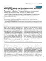
gp120:V3(BaL) to THP-1 cells was detected at 100 µM (Fig
1a). In contrast, specific binding of gp120:V3(BaL) to Jur-
kat cells was not detected even at concentrations up to 500
µM (Fig 1a). These observations are consistent with the
hypothesis that gp120:V3(BaL) binds specifically to
CCR5, which is expressed on the surface of THP-1 mono-
cytic cells but not on Jurkat T-cells.
Specific binding of gp120:V3(IIIb) at 100 µM to both Jur-
kat T-cells and THP-1 cells was detected. There was
approximately 5-fold greater specific binding to the Jurkat
cells than the THP-1 cells (Fig 1b). These observations are
consistent with the hypothesis that gp120:V3(IIIb) binds
specifically to CXCR4, which is expressed on both THP-1
and Jurkat cells, but at higher levels on the T-cell line.
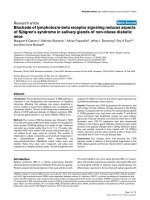
We next incubated THP-1 and Jurkat cells with 100 µM of
each biotinylated gp120:V3 peptide in the presence of var-
ious concentrations of Peptide 3. Peptide 3 had no effect
on the binding of gp120:V3(IIIb) to Jurkat cells (Fig 2a),
even though it powerfully inhibited SDF-1α induced
chemotaxis over the same concentration range (Fig 2b).
Under the same conditions, the CXCR4 receptor antago-
nist AMD3100 [31] blocked both gp120:V3(IIIb) binding
and SDF-1α-induced migration with similar IC50s (Fig
2c, d). Similarly, Peptide 3 had no effect on the binding of
gp120:V3(BaL) to THP-1 cells (Fig 2e).under conditions
where RANTES-induced chemotaxis was powerfully
inhibited (Fig 2f). Taken together, these data confirm that
Peptide 3 blocks chemokine signalling without blocking
gp120 interaction with the chemokine receptors, consist-
ent with previous observations that BSCIs such as Peptide
3 do not block chemokine ligand interactions with their
receptors [27].
Effect of peptides on HIV infection in vitro
HIV infection of Jurkat T-cells using the laboratory-
adapted T-tropic isolate IIIb was monitored using two dif-
ferent assays. Firstly, Jurkat T-cells in 96-well plates were
pre-treated with either Peptide 3, vehicle (as a negative
control) or SDF1α (as a positive control) for 4 hours, then
exposed to HIV virus (10
6
TCID
50
) and pulsed at 2–3 day
intervals with Peptide 3, SDF1α or medium alone as
Retrovirology 2005, 2:23 />Page 3 of 9
(page number not for citation purposes)
appropriate. After two weeks in culture, the extent of viral
infection was assayed by measuring the reverse tran-
scriptase activity in the supernatant, as a measure of viral
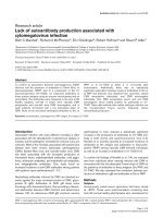
replication in the culture [32] In six separate experiments,
Peptide 3 (100 µM) had no effect on virus replication fol-
lowing HIV exposure (Fig 3a), while SDF1α inhibited
reverse transcriptase activity by an average of 75%. No
effect was seen on cell viability under any of the treatment
conditions.
HIV infection of Jurkat T-cells was also monitored by high
sensitivity quantitative immunofluoresence detection of
viral p24
gag
expression. Jurkat cells were infected with HIV
in the presence or absence of Peptide 3 (100 µM) or
SDF1α (100 ng/ml) as described above. Approximately 48
h after infection, the cells were attached to glass slides
using a cytospin and then fixed by immersion in ice-cold
70% ethanol for 90 seconds. Expression of p24
gag
was
determined using quantitative immunofluoresence as
previously described [33], except that the primary anti-
body was post-fixed to the section using paraformalde-
hyde to increase the sensitivity of the technique (see
Methods). Viral infectivity was expressed as the number of
cells stained for p24
gag
expressed as a proportion of the
total number of cells (detected using Hoechst 33342
nuclear dye). Consistent with the reverse transcriptase
assay results, SDF1α inhibited viral infectivity by more
than 80% (Fig 3b), while peptide 3 had no effect.
Infection of THP-1 cells with M-tropic isolates does not
generate high levels of virus particles and hence the
reverse transcriptase assay is not sufficiently sensitive to
monitor the progress of the infection. However, it was
possible to assess HIV infectivity of THP-1 cells using high
sensitivity immunofluorescent detection of p24
gag
. THP-1
cells were differentiated with hydrocortisone and PMA,
then treated with TNFα, resulting in adherent monolayers
on glass chamber slides. The THP-1 cells were then treated
with either Peptide 3 (100 µM), MIP1α (100 ng/ml) or
SDF1α (100 ng/ml) as for the Jurkat cells. THP-1 cells
were infected with HIV strain MN at a concentration pre-
viously validated to produce easily detectable infection
and grown for 72 h prior to fixation and staining for
p24
gag
. In contrast to the findings with HIV strain IIIb
infection of Jurkat T cells, Peptide 3 inhibited infection of
THP-1 cells by more than 80% (Fig 3c), very similar to the
effect of MIP1α. SDF-1a had no statistically significant
effect on HIV strain MN infection of THP-1 cells, confirm-
ing that the infection was entirely CCR5-dependent, even
though THP-1 cells express CXCR4.
Discussion
Taken together our results suggest that, at least under
some conditions, the generation of intracellular signals by
the chemokine co-receptor during HIV infection might be
necessary for productive infection. Since Peptide 3 power-
fully inhibited CCR5-dependent HIV infection of THP-1
cells under conditions where gp120 binding to CCR5 was
unaffected but chemotaixs in response to RANTES was
profoundly blocked, it is likely that at least some of the
signals elicted by CCR5 occupation that result in
Specific binding of gp120:V3 loop peptides to THP-1 and Jur-kat cellsFigure 1
Specific binding of gp120:V3 loop peptides to THP-1
and Jurkat cells. (a) Binding of gp120:V3(BaL)-biotin 10
6
Jurkat cells or THP-1 cells per reaction were incubated with
various concentrations of N-terminal biotinylated peptide in
the presence and absence of 10 mM unlabelled peptide. Spe-
cific binding is expressed as the absorbance in the absence of
unlabelled peptide minus the absorbance in the presence of
competitor (b) Binding of gp120:V3(IIIb)-biotin under the
same conditions as in (a). All reactions were performed in
100 µl of binding medium at 4°C (see Materials and Meth-
ods). Values are mean ± SEM from triplicate determinations.
* p < 0.05 Student's t-test for specific binding.
10M 100M 500M 10M 100M 500M
0.0
0.5
1.0
1.5
Binding to
Jurkat T cells
Binding to
THP-1 cells
*
*
A
10M 100M 500M 10M 100M 500M
0.0
0.5
1.0
1.5
Binding to
THP-1 cells
Binding to
Jurkat T cells
*
*
*
*
*
B
Retrovirology 2005, 2:23 />Page 4 of 9
(page number not for citation purposes)
Effect of Peptide 3 on gp120:V3 loop peptide binding to cellsFigure 2
Effect of Peptide 3 on gp120:V3 loop peptide binding to cells. (a) The binding of 100 µM N-terminal biotinylated
gp120:V3(IIIb) to THP-1 cells was measured in the presence of various concentrations of peptide 3. In each case, the non-spe-
cific binding (in the presence of 10 mM unlabelled gp120:V3 loop peptide) has been subtracted. (b) Chemotaxis in response to
100 ng/ml SDF1α was measured in the presence of various concentrations of peptide 3. (c) and (d) As for (a) and (b) except
that the CXCR4 receptor antagonist AMD3100 was used in place of Peptide 3. (e) As for (a) except that the effect of Peptide
3 on the binding of gp120:V3(BaL) to THP-1 cells was determined. (f) As for (b) except that the effect of Peptide 3 on chemo-
taxis induced by 25 ng/ml MIP1α was determined. All binding reactions were performed with 10
6
cells in 100 µl of binding
medium at 4°C. Chemotaxis assays were performed with 5 × 10
4
cells per well. Values are mean ± SEM of triplicate
determinations.
1 10 100 1000
0
25
50
75
100
*
*
*
*
B
[Peptide 3] (M)
1 10 100 1000
0.00
0.25
0.50
0.75
1.00
*
*
*
*
C
[AMD 3100] (nM)
1 10 100 1000
0
25
50
75
100
*
*
*
*
*
D
[AMD 3100] (nM)
1 10 100 1000
0.00
0.25
0.50
0.75
1.00
E
[Peptide 3] (M)
1 10 100 1000
0
25
50
75
100
*
*
*
*
F
[Peptide 3] (M)
1 10 100 1000
0.00
0.25
0.50
0.75
1.00
A
[Peptide 3] (M)
Retrovirology 2005, 2:23 />Page 5 of 9
(page number not for citation purposes)
Peptide 3 inhibition of HIV infectivity in vitroFigure 3
Peptide 3 inhibition of HIV infectivity in vitro. (a) HIV (IIIb) replication in cultures of Jurkat T-cells was estimated by
measuring the supernatant reverse transcriptase activity two weeks after infection. Peptide 3 was at 100 µM final concentra-
tion and SDF-1α was added at 100 ng/ml final concentration 1 hour prior to exposure to virus. Values are mean ± SEM from
12 wells, expressed as the percentage of the reverse transcriptase activity in the supernatant from the control wells. The
experiment shown is typical of six separate experiments. (b) HIV (IIIb) infectivity of Jurkat T-cells was estimated by staining
cells treated identically to those in (a) for p24
gag
expression. Values are mean ± S.D. percentage of cells stained for p24
gag
aver-
aged from 12 fields of view from each of two separate wells. (c) HIV (MN) infectivity of THP-1 cells measured as in (b). MIP1α
and SDF1α were used at 100 ng/ml final concentration.
Vehicle Peptide 3 SDF1
0
25
50
75
100
125
A
*
Vehicle Peptide 3 SDF1
0
5
10
15
*
B
Vehicle Peptide 3 SDF1 MIP1
0
2
4
6
8
*
*
C
Retrovirology 2005, 2:23 />Page 6 of 9
(page number not for citation purposes)
chemotaxis are required for successful infection of the cell
by HIV. Since BSCIs, such as Peptide 3, do not block
chemokine receptor internalisation induced by ligand
binding [27], it seems likely that the HIV successfully
entered the cell in the presence of Peptide 3, but that some
later stage in the viral life cycle was dependent on one or
more intracellular signal generated by chemokine recep-
tor occupancy. These results are consistent with, but
extend, the findings of Guntermann [24] and Montes [25]
who saw similar effects with pertussis toxin and a MEK
inhibitor.
It is unclear why Peptide 3 blocked CCR5-dependent HIV
infection of THP-1 cells but had no effect on CXCR4-
dependent infection of Jurkat T-cells under similar cond-
tions (even though Peptide 3 efficiently blocks SDF1α
dependent chemotaxis). It is possible that infection of cer-
tain cell types (such as monocyte/macrophage cells) is
more dependent on a chemokine receptor-induced intra-
cellular signal than infection of other cell types (such as T-
lymphocytes). This may reflect the fact the the Jurkat cells
were proliferating at the time of infection, whereas the
THP-1 derived macrophages were quiescent. However, it
is also possible that this difference is due to the
particularly high levels of CXCR4 which are expressed on
Jurkat T cells. The high levels of receptor on this cell line
might render infection relatively insensitive to intracellu-
lar signalling requirements compared with native T-cells
or other cell types expressing physiological levels of chem-
okine co-receptors.
Irrespective of the reasons for this difference, our prelimi-
nary studies illustrate the need to further investigate the
role of intracellular signals induced by co-receptor occu-
pancy as participants in the viral life cycle. Furthermore,
the recent discovery of much more potent non-peptide
BSCIs, such as the acylaminocaprolactams [34,35], opens
up the possibility that interfering with chemokine recep-
tor-induced signalling might offer an alternative thera-
peutic strategy to blocking chemokine receptor binding.
The best acylaminocaprolactam BSCIs are cheap, orally
bioavailable and apparently free of acute toxicity [35],
and, on the basis of studies such as ours, warrant further
investigation as part of an antiviral combination therapy
for HIV.
Conclusion
We conclude that selective inhibition of the intracellular
signal(s) generated through interaction of the HIV gp120
envelope protein with its chemokine co-receptor can
block productive HIV infection in THP-1 derived macro-
phages in vitro. However, blockade of CXCR4-mediated
signalling in Jurkat T-cells has no effect on HIV infection
in vitro. Under certain condtions, therefore, HIV infection
may require activation of the chemokine co-receptor sig-
nalling pathway.
Methods
Peptides
Peptides were prepared by Affiniti (Exeter, U.K.) using
standard solid phase chemistry, followed by reverse-phase
HPLC purification to greater than 95% purity. Peptide 3
(derived from amino acids 51–62 of mature human MCP-
1) had the sequence EICADPKQKWVQ [26]. Labelled
peptides were also synthesised corresponding to the
sequence of the full length V3 loop (including terminal
cysteine residues) of gp120 from HIV-1 IIIb and HIV-1
BaL with an N-terminal co-synthetic biotin label. All pep-
tides were prepared as TFA salts and dissolved in sterile
MilliQ and stored at -20°C until used.
Chemotaxis experiments
Chemotaxis experiments were performed essentially as
previously described [26,36], using the ChemoTx dispos-
able 96-well transwell migration plates, with PVP-free
membrane (6 µm pore size). Chemoattractant (lower
compartment) and cells upper compartment) were sus-
pended in Gey's Balanced salt solution + 1 mg/ml BSA.
Consistent with our previous recommendations [36],
putative inhibitors were added at equal concentration to
both the upper and lower compartments. Migration was
allowed to proceed for 2 hours at 37°C. The number of
cells which had migrated to the lower compartment was
determined using the vital dye MTT and interpolation of a
standard curve. Each condition was determined in tripli-
cate, and the number of cells migrating in the absence of
chemoattractant was subtracted to determine the chemok-
ine-dependent chemotaxis which was reported.
Binding assays
Cells (either Jurkat T-cells or THP-1 cells) were grown in
RPMI 1640 medium supplemented with 10% fetal calf
serum, 2 mM glutamine, 20 µM β-mercaptoethanol, 100
U/ml penicillin and 100 µg/ml streptomycin and main-
tained between 2 × 10
5
and 1 × 10
6
cells/ml. Prior to per-
forming a binding assay, cells were spun out (100 × g; 4
mins) and washed 3 times in ice-cold PBS. A volume of
cell suspension in PBS containing 10
6
cells was pipetted
into each well of a V-bottom 96-well plate (Gibco BRL)
and spun out (100 × g; 4 mins). Cells from triplicate wells
were then resuspended in 100 µl binding medium (PBS
pH 7.2 containing 0.1% fatty-acid free bovine serum albu-
min (BSA)) containing labelled peptide in the presence or
absence of 10 mM unlabelled peptide. The plate was then
incubated on ice for 90 minutes. Cells were washed 3
times with 380 µl of ice-cold PBS, spinning out the cells
each time (100 × g; 4 mins), and resuspended in 100 µl
binding medium containing streptavidin-peroxidase
(Amersham International) at 1:1000 dilution. Cells were
Retrovirology 2005, 2:23 />Page 7 of 9
(page number not for citation purposes)
incubated for a further 15 minutes on ice to allow label-
ling of any bound biotinylated peptide, then washed 4
times as above. Cells were finally incubated with 200 µl
TMB substrate (K-Blue, Bionostics) for 20 minutes at
room temperature, and the reaction stopped by addition
of 50 µl 2 M HCl. The plate was spun (3,000 × g ; 3 mins)
and 200 µl of the coloured product was transferred to an
empty 96-well ELISA plate and the absorbance at 450 nm
determined.
HIV-1 infection and reverse transcriptase assays
HIV-1 stocks were prepared in the following manner. HIV-
1 IIIb (provided by the MRC AIDS Reagent Programme)
was used to infect Jurkat T cells and the progression of
infection was monitored using the reverse transcriptase
assay for 10–14 days. Virus was then harvested and
assayed for infectivity using a TCID50 assay with Jurkat T
cells as targets as previously described [37]. Virus contain-
ing supernatants were centrifuged to remove cellular
debris and then stored in aliqouts in liquid nitrogen until
used. Stocks of HIV-1 MN strain were prepared in a similar
manner, except that H9 T cells were used and the progres-
sion of infection was monitored by immunofluoresence
(because of the low RT activity) in this strain.
Experimental infection of Jurkat T cells was performed by
incubating cells win the presence of test peptide or
chemokine with aliquots of stored virus at the titres
described in the text. Thereafter, the cells were fed fresh
medium containing test peptide or chemokine where
appropriate, every 48 h. Viral replication two weeks after
infection was estimated by measuring reverse tran-
scriptase activity in the supernatant using the Potts Mini
RT assay as previously described [32].
THP-1 cells were differentiated prior to infection with
hydrocortisone and PMA in 8-well chamber slides. Sixteen
hours prior to infection, TNFα was added (100 ng/ml).
Twelve hours later, the medium was aspirated and
replaced with fresh medium containing the test peptides
or chemokines as appropriate. After a further four hours,
virus from the frozen stocks was adeed to the cells, which
were the processed for immunofluoresence between 28 h
and 72 h after infection.
Immunofluorescence detection of p24
gag
Jurkat cells following HIV infection were attached to 8-
well chamber slides (Becton-Dickinson) by spinning the
slides using a plate rotor in a Labofuge centrifuge (Her-
aeus) at 3,000 × g for 5 minutes. Attached Jurkat cells or
THP-1 cells were then fixed by dipping the slides into ice-
cold 70% ethanol for 90 seconds. Non-specific binding
was blocked by incubation with 3% fatty acid-free BSA in
TBS for 1 hour at room temprature. Cells were incubated
with the mouse monoclonal anti-HIV-1 p24
gag
antibody
EH12E1 (ref 38;AIDS Reagent Program, NIBSC) at 10 µg/
ml in 3% BSA in TBS at room temperature overnight.
Unbound antibody was removed with 3 × 3 min washes
in PBS, and bound antibody was then fixed to the slide by
incubation with 3.8% phosphate buffered formalin
pH7.2 for 10 mins at room temperature, followed by 3
further 3 min washes in PBS. Bound antibody was visual-
ised using donkey anti-mouse IgG FITC conjugate (715-
095-150; Jackson Immunoresearch) at 30 µg/ml in 3%
BSA/TBS + 1 ng/ml Hoescht 33342 for 6 hours at room
temperature. Twelve fields of view (100× magnification)
were captured from each well of the chamber slide using
an Olympus Provis AX electronic microscope connected
to a Power Macintosh 8500, running OpenLab image
analysis software (Improvision), under both FITC illumi-
nation conditions (NIBA filter block; λex = 470–490 nm,
dichroic mirror = 505 nm, λem = 515–550 nm) and UV
illumination conditions (Chroma 31000; λex = 340–380
nm, dichroic mirror = 400 nm, λem = 435–485 nm).
Images were acquired with a Hamamatsu C4742-05 mon-
ochrome digital camera with 10-bit depth in a 1280-1024
pixel field connected to a DIG Snapper frame grabber. The
exposure time, amplifier gain and offset values were con-
trolled by the OpenLab software and were held constant
throughout the experiment. A background (an image
captured without a slide under the objective) was digitally
subtracted from every image. A threshold was then
applied to each image which was the lowest threhold that
detected <1% of the pixels of an image of uninfected cells
stained under identical conditions. The number of objects
exceeding this threshold in each field of view were
counted. A similar procedure was used to determine the
total number of nuclei in the same field of view, using the
image captured under UV illumination conditions. The
ratio of positively stained objects to nuclei in each field of
view was reported as the percentage of cells stained for
p24
gag
.
Competing interests
DJG is an inventor on a range of patents filed by the Uni-
versity of Cambridge containing composition of matter
and pharmaceutical use claims for a wide range of BSCIs,
including Peptide 3 used in this manuscript. The patent
specifically claims the use of BSCIs for the prevention
and/or treatment of HIV infection. An exclusive license to
these patents have been granted by the University of Cam-
bridge to Ipsen (Paris, France) and DJG may gain finan-
cially from the successful exploitation of this intellectual
property.
Authors' contributions
DJG and AML jointly conceived of these studies; AML per-
formed the HIV infection experiments and RT assays; DJG
performed the immunofluoresence detection analyses
and the in vitro binding assays and functional migration
Retrovirology 2005, 2:23 />Page 8 of 9
(page number not for citation purposes)
assays. DJG drafted this manuscript, which was critically
reviewed by AML and both authors approve the final ver-
sion for submission and publication.
Acknowledgements
This work was supported by grants from NeoRx Corporation (Seattle,
Wa., USA) and the Wellcome Trust to D.J.G., who was a Royal Society Uni-
versity Research Fellow. We are grateful to Paul Sheppard and his col-
leagues at Affiniti (now Biomol International) for help and advice on
designing peptides, labelled peptides and their derivatives. The monoclonal
antibody to p24
gag
was obtained from the AIDS Reagent Program at NIBSC.
References
1. Alkhatib G, Combadiere C, Broder CC, Feng Y, Kennedy PE, Murphy
PM, Berger EA: CC-CKR5: a RANTES, MIP-1α, MIP-1β recep-
tor as a fusion cofactor for macrophage-tropic HIV-1. Science
1996, 272:1955-1958.
2. Feng Y, Broder CC, Kennedy PE, Berger EA: HIV-1 entry cofactor:
functional cDNA cloning of a seven-transmembrane, G pro-
tein-coupled receptor. Science 1996, 272:872-877.
3. Deng H, Liu R, Ellmeier W, Choe S, Unutmaz D, Burkhart M, Di Mar-
zio P, Marmon S, Sutton RE, Hill CM, Davis CB, Peiper SC, Schall TJ,
Littman DR, Landau NR: Identification of a major co-receptor
for primary isolates of HIV-1. Nature 1996, 381:661-666.
4. Dragic T, Litwin V, Allaway GP, Martin SR, Huang Y, Nagashima KA,
Cayanan C, Maddon PJ, Koup RA, Moore JP, Paxton WA: HIV-1
entry into CD4
+
cells is mediated by the chemokine receptor
CC-CKR-5. Nature 1996, 381:667-673.
5. Doranz BJ, Rucker J, Yi Y, Smyth RJ, Samson M, Peiper SC, Parmentier
M, Collman RG, Doms RW: A dual-tropic primary HIV-1 isolate
that uses fusin and the beta-chemokine receptors CKR-5,
CKR-3, and CKR-2b as fusion cofactors. Cell 1996,
85:1149-1158.
6. Choe H, Farzan M, Sun Y, Sullivan N, Rollins B, Ponath PD, Wu L,
Mackay CR, LaRosa G, Newman W, Gerard N, Gerard C, Sodroski J:
The beta-chemokine receptors CCR3 and CCR5 facilitate
infection by primary HIV-1 isolates. Cell 1996, 85:1135-1148.
7. Cairns JS, D'Souza MP: Chemokines and HIV-1 second recep-
tors: the therapeutic connection. Nat Med 1998, 4:563-568.
8. Kelly MD, Naif HM, Adams SL, Cunningham AL, Lloyd AR: Dichoto-
mous effects of beta-chemokines on HIV replication in
monocytes and monocyte-derived macrophages. J Immunol
1998, 160:3091-3095.
9. Cocchi F, DeVico AL, Garzino-Demo A, Arya SK, Gallo RC, Lusso P:
Identification of RANTES, MIP-1α, and MIP-1β as the major
HIV-suppressive factors produced by CD8
+
T cells. Science
1995, 270:1811-1815.
10. Zagury D, Lachgar A, Chams V, Fall LS, Bernard J, Zagury JF, Bizzini B,
Gringeri A, Santagostino E, Rappaport J, Feldman M, O'Brien SJ, Burny
A, Gallo RC: C-C chemokines, pivotal in protection against
HIV type 1 infection. Proc Natl Acad Sci U S A 1998, 95:3857-3861.
11. Alvarez V, Lopez-Larrea C, Coto E: Mutational analysis of the
CCR5 and CXCR4 genes (HIV-1 co-receptors) in resistance
to HIV-1 infection and AIDS development among intrave-
nous drug users. Hum Genet 1998, 102:483-486.
12. Samson M, Libert F, Doranz BJ, Rucker J, Liesnard C, Farber CM, Sara-
gosti S, Lapouméroulie C, Cognaux J, Forceille C, Muyldermans G,
Verhofstede C, Burtonboy G, Georges M, Imai T, Rana S, Yi YJ, Smyth
RJ, Collman RG, Doms RW, Vassart G, Parmentier M: Resistance to
HIV-1 infection in caucasian individuals bearing mutant alle-
les of the CCR-5 chemokine receptor gene. Nature 1996,
382:722-725.
13. Zimmerman PA, BucklerWhite A, Alkhatib G, Spalding T, Kubofcik J,
Combadiere C, Weissman D, Cohen O, Rubbert A, Lam G, Vacca-
rezza M, Kennedy PE, Kumaraswami V, Giorgi JV, Detels R, Hunter J,
Chopek M, Berger EA, Fauci AS, Nutman TB, Murphy PM: Inherited
resistance to HIV-1 conferred by an inactivating mutation in
CC chemokine receptor 5: Studies in populations with con-
trasting clinical phenotypes, defined racial background, and
quantified risk. Mol Med 1997, 3:23-36.
14. Wells TNC, Proudfoot AEI, Power CA, Lusti-Narasimhan M, Alouani
S, Hoogewerf AJ, Peitsch MC: The molecular basis of the chem-
okine/chemokine receptor interaction- scope for design of
chemokine antagonists. Methods 1996, 10:126-134.
15. Bushman F, Landau NR, Emini EA: New developments in the biol-
ogy and treatment of HIV. Proc Natl Acad Sci U S A 1998,
95:11041-11042.
16. Altmeyer R: Virus attachment and entry offer numerous tar-
gets for antiviral therapy. Curr Pharm Des 2004, 10:3701-3712.
17. Iyengar S, Schwartz DH, Hildreth JE: T cell-tropic HIV gp120
mediates CD4 and CD8 cell chemotaxis through CXCR4
independent of CD4: implications for HIV pathogenesis. J
Immunol 1999, 162:6263-6267.
18. Balabanian K, Harriague J, Decrion C, Lagane B, Shorte S, Baleux F,
Virelizier JL, Arenzana-Seisdedos F, Chakrabarti LA: CXCR4-tropic
HIV-1 envelope glycoprotein functions as a viral chemokine
in unstimulated primary CD4+ T lymphocytes. J Immunol 2004,
173:7150-7160.
19. Ross TM, Bieniasz PD, Cullen BR: Multiple residues contribute to
the inability of murine CCR-5 to function as a coreceptor for
macrophage-tropic human immunodeficiency virus type 1
isolates. J Virol 1998, 72:1918-1924.
20. Alkhatib G, Ahuja SS, Light D, Mummidi S, Berger EA, Ahuja SK: CC
chemokine receptor 5-mediated signaling and HIV-1 co-
receptor activity share common structural determinants.
Critical residues in the third extracellular loop support HIV-
1 fusion. J Biol Chem 1997, 272:19771-19776.
21. Dragic T, Trkola A, Lin SW, Nagashima KA, Kajumo F, Zhao L, Olson
WC, Wu L, Mackay CR, Allaway GP, Sakmar TP, Moore JP, Maddon
PJ: Amino-terminal substitutions in the CCR5 coreceptor
impair gp120 binding and human immunodeficiency virus
type 1 entry. J Virol 1998, 72:279-285.
22. Farzan M, Choe H, Martin KA, Sun Y, Sidelko M, Mackay CR, Gerard
NP, Sodroski J, Gerard C: HIV-1 entry and macrophage inflam-
matory protein-1 beta-mediated signaling are independent
functions of the chemokine receptor CCR5. J Biol Chem 1997,
272:6854-6857.
23. Kimata JT, Palker TJ, Ratner L: The mitogenic activity of human
T-cell leukemia virus type I is T-cell associated and requires
the CD2/LFA-3 activation pathway. J Virol 1993, 67:3134-3141.
24. Guntermann C, Murphy BJ, Zheng R, Qureshi A, Eagles PA, Nye KE:
Human immunodeficiency virus-1 infection requires pertus-
sis toxin sensitive G-protein-coupled signalling and mediates
cAMP downregulation. Biochem Biophys Res Commun 1999,
256:429-435.
25. Montes M, Tagieva NE, Heveker N, Nahmias C, Baleux F, Trautmann
A: SDF-1-induced activation of ERK enhances HIV-1
expression. Eur Cytokine Netw 2000, 11:470-477.
26. Reckless J, Grainger DJ: Identification of oligopeptide sequences
which inhibit migration induced by a wide range of
chemokines. Biochem J 1999, 240:803-811.
27. Grainger DJ, Reckless J: Broad-spectrum chemokine inhibitors
(BSCIs) and their anti-inflammatory effects in vivo. Biochem
Pharmacol 2003, 65:1027-34.
28. Ross TM, Cullen BR: The ability of HIV type 1 to use CCR-3 as
a coreceptor is controlled by envelope V1/V2 sequences act-
ing in conjunction with a CCR-5 tropic V3 loop. Proc Natl Acad
Sci U S A 1998, 95:7682-7686.
29. Cocchi F, DeVico AL, Garzino-Demo A, Cara A, Gallo RC, Lusso P:
The V3 domain of the HIV-1 gp120 envelope glycoprotein is
critical for chemokine-mediated blockade of infection. Nat
Med 1996, 2:1244-1247.
30. Jiang S: HIV-1: co-receptors binding. Nat Med 1997, 3:367-368.
31. Schols D, Este JA, Henson G, De Clercq E: Bicyclams, a class of
potent anti-HIV agents, are targeted at the HIV coreceptor
fusin/CXCR-4. Antiviral Res 1997, 35:147-156.
32. Potts BJ: 'Mini' reverse transcriptase (RT) assay. In Techniques in
HIV research Edited by: Aldovini A, Walker BD. New York: Stockton
Press; 1990:102-106.
33. Mosedale DE, Metcalfe JC, Grainger DJ: Optimization of immun-
ofluorescence methods by quantitative image analysis. J His-
tochem Cytochem 1996, 44:1043-1050.
34. Fox DJ, Reckless J, Warren SG, Grainger DJ: Design, synthesis, and
preliminary pharmacological evaluation of N-acyl-3-
aminoglutarimides as broad-spectrum chemokine inhibitors
in vitro and anti-inflammatory agents in vivo. J Med Chem 2002,
45:360-370.
Publish with BioMed Central and every
scientist can read your work free of charge
"BioMed Central will be the most significant development for
disseminating the results of biomedical research in our lifetime."
Sir Paul Nurse, Cancer Research UK
Your research papers will be:
available free of charge to the entire biomedical community
peer reviewed and published immediately upon acceptance
cited in PubMed and archived on PubMed Central
yours — you keep the copyright
Submit your manuscript here:
/>BioMedcentral
Retrovirology 2005, 2:23 />Page 9 of 9
(page number not for citation purposes)
35. Fox DJ, Reckless J, Wilbert SM, Greig I, Warren S, Grainger DJ: Iden-
tification of 3-(Acylamino)azepan-2-ones as Stable Broad-
Spectrum Chemokine Inhibitors Resistant to Metabolism in
Vivo. J Med Chem 2005, 48:867-874.
36. Frow EK, Reckless J, Grainger DJ: Tools for anti-inflammatory
drug design: in vitro models of leukocyte migration. Med Res
Rev 2004, 24:276-298.
37. Peden KWC, Martin MA: Virological and molecular genetic
techniques. In HIV: A practical approach Volume 1. Edited by: Karn J.
Oxford: Oxford University Press; 1995:21-45.
38. Ferns RB, Tedder RS, Weiss RA: Characterization of monoclonal
antibodies against the human immunodeficiency virus (HIV)
gag products and their use in monitoring HIV isolate
variation. J Gen Virol 1987, 68:1543-51.