Báo cáo y học: "Elevated expression of CD30 in adult T-cell leukemia cell lines: possible role in constitutive NF-κB activation" pps
Bạn đang xem bản rút gọn của tài liệu. Xem và tải ngay bản đầy đủ của tài liệu tại đây (450.62 KB, 12 trang )
BioMed Central
Page 1 of 12
(page number not for citation purposes)
Retrovirology
Open Access
Research
Elevated expression of CD30 in adult T-cell leukemia cell lines:
possible role in constitutive NF-κB activation
Masaya Higuchi
1
, Takehiro Matsuda
2
, Naoki Mori
2
, Yasuaki Yamada
3
,
Ryouichi Horie
4
, Toshiki Watanabe
5
, Masahiko Takahashi
1
, Masayasu Oie
1
and Masahiro Fujii*
1
Address:
1
Division of Virology, Niigata University Graduate School of Medical and Dental Sciences, Niigata 951-8510, Japan,
2
Division of
Molecular Virology and Oncology, Faculty of Medicine, University of the Ryukyus, Nishihara, Okinawa 903-0215, Japan,
3
Department of
Laboratory Medicine, Nagasaki University Graduate School of Biomedical Sciences, Nagasaki 825-8501, Japan,
4
Fourth Department of Internal
Medicine, Faculty of Medicine, Kitasato University, Sagamihara, Kanagawa 228-8555, Japan and
5
Laboratory of Tumor Cell Biology, Department
of Medical Genome Sciences, Graduate School of Frontier Sciences, The University of Tokyo, Minato-ku, Tokyo 108-109, Japan
Email: Masaya Higuchi - ; Takehiro Matsuda - ; Naoki Mori -
ryukyu.ac.jp; Yasuaki Yamada - ; Ryouichi Horie - ;
Toshiki Watanabe - ; Masahiko Takahashi - ; Masayasu Oie - ;
Masahiro Fujii* -
* Corresponding author
Abstract
Background: Human T-cell leukemia virus type 1 (HTLV-1) is associated with the development
of adult T-cell leukemia (ATL). HTLV-1 encoded Tax1 oncoprotein activates the transcription of
genes involved in cell growth and anti-apoptosis through the NF-κB pathway, and is thought to play
a critical role in the pathogenesis of ATL. While Tax1 expression is usually lost or minimal in ATL
cells, these cells still show high constitutive NF-κB activity, indicating that genetic or epigenetic
changes in ATL cells induce activation independent of Tax1. The aim of this study was to identify
the molecules responsible for the constitutive activation of NF-κB in ATL cells using a retroviral
functional cloning strategy.
Results: Using enhanced green fluorescent protein (EGFP) expression and blasticidin-resistance as
selection markers, several retroviral cDNA clones exhibiting constitutive NF-κB activity in Rat-1
cells, including full-length CD30, were obtained from an ATL cell line. Exogenous stable expression
of CD30 in Rat-1 cells constitutively activated NF-κB. Elevated expression of CD30 was identified
in all ATL lines examined, and primary ATL cells from a small number of patients (8 out of 66 cases).
Conclusion: Elevated CD30 expression is considered one of the causes of constitutive NF-κB
activation in ATL cells, and may be involved in ATL development.
Background
Adult T-cell leukemia (ATL) is an extremely aggressive
human CD4+ T-cell leukemia (reviewed in [1]). ATL is
resistant to chemotherapy and most patients die within
one year of diagnosis. Human T-cell leukemia virus type 1
(HTLV-1) infection of CD4+ T-cells is the first step in ATL
development. However, this alone is not sufficient for the
development of leukemia because a minority of HTLV-1
Published: 06 May 2005
Retrovirology 2005, 2:29 doi:10.1186/1742-4690-2-29
Received: 07 February 2005
Accepted: 06 May 2005
This article is available from: />© 2005 Higuchi et al; licensee BioMed Central Ltd.
This is an Open Access article distributed under the terms of the Creative Commons Attribution License ( />),
which permits unrestricted use, distribution, and reproduction in any medium, provided the original work is properly cited.
Retrovirology 2005, 2:29 />Page 2 of 12
(page number not for citation purposes)
infected subjects (approximately 5%) develop ATL on
average 60–70 years after the infection (reviewed in [2,3]).
In vitro, HTLV-1 transforms primary human CD4+ T-cells
in an interleukin (IL)-2-dependent or an IL-2-independ-
ent manner. HTLV-1 encoded Tax1 protein is thought to
play a critical role in T-cell transformation and leukemo-
genesis, as Tax1 itself immortalizes primary human CD4+
T-cells in vitro [4,5] and inhibits apoptosis induced by var-
ious stimuli in T-cell lines [6-9].
Tax1 is a multifunctional protein (reviewed in [2,3]). It
activates the transcription of many cellular genes associ-
ated with cell growth, such as genes encoding cytokines
[10-13], cytokine receptors [14-17], anti-apoptotic pro-
tein [8,18], cell cycle regulators [19-22], and proto-onco-
genes [23]. Those proteins are thought to contribute to the
deregulated proliferation of HTLV-1-infected cells. Accu-
mulating evidence suggests that activation of cellular
genes by Tax1, particularly through the nuclear factor-kap-
paB (NF-κB) pathway, is a critical process in transforma-
tion as well as the inhibition of apoptosis. For example,
the transforming activity of Tax1 is abrogated by muta-
tions that impair the ability of Tax1 to activate NF-κB [24-
26]. Tax1 inhibits apoptosis of mouse T-cell lines by
induction of the anti-apoptotic gene Bcl-xL through NF-
κB activation [8,18].
In resting T-cells, NF-κB factors are sequestered in the
cytoplasm, tightly associated with inhibitory proteins
IκBs. Activation of NF-κB generally involves phosphoryla-
tion and degradation of IκBs, followed by nuclear translo-
cation of NF-κB dimers and subsequent activation of the
genes containing NF-κB binding sites (reviewed in [27]).
Alternatively, NF-κB activation occurs by inducible
processing of NFKB2/p100 with IκB-like inhibitory activ-
ity, into p52 with DNA binding activity, followed by
nuclear translocation of p52 containing NF-κB dimers
(reviewed in [28]). These two processes are largely
dependent on an IκB kinase (IKK) complex comprised of
two catalytic subunits, IKKα and IKKβ and a regulatory
subunit IKKγ/NEMO. Tax1 interacts with the IKK complex
through these three subunits and stimulates the catalytic
activity [29-32].
In primary ATL cells as well as cell lines established from
ATL patients, NF-κB is constitutively active as seen in
HTLV-1 transformed cells [33]. It appears that this consti-
tutive NF-κB activation contributes to the survival and
chemotherapy resistance of ATL cells, since treatment of
ATL cells with a NF-κB inhibitor, Bay 11-7082, induces
apoptosis of these cells [34]. However, how NF-κB is con-
stitutively activated in ATL cells is still largely unknown
since the tax gene is mutated in some ATL cases [35,36] or
the level of expression of Tax1 in these cells is extremely
low, thereby being clearly insufficient to activate NF-κB
[37,38]. There may be genetic or epigenetic changes that
lead to tax-independent NF-κB activation, such as a gain
of function of the NF-κB activating molecule(s) or a loss
of function of the NF-κB regulator(s). The elucidation of
the molecular mechanism of NF-κB activation in ATL cells
is quite important in the light of prevention, diagnosis
and treatment of ATL.
In order to identify the molecule(s) responsible for the
constitutive NF-κB activation in ATL, we took a functional
screening approach using a retroviral cDNA library from
an ATL cell line and a reporter cell line that is easily distin-
guishable as a positive clone once NF-κB is activated. We
obtained several cDNA clones that constitutively activate
NF-κB. One of these, the full-length CD30, is a member of
the TNF receptor superfamily and a marker of malignant
Hodgkin and Reed-Sternberg (H-RS) cells in Hodgkin's
lymphoma (HL) (reviewed in [39,40]). It is suggested that
overexpression of CD30 in H-RS cells and HL cell lines
contributes to CD30 ligand-independent constitutive NF-
κB activation in these cells [41]. The results showed that
CD30 is strongly expressed in all ATL cell lines examined,
and that CD30 is expressed in primary ATL cells in a small
number of ATL patients.
Results and Discussion
Screening of NF-
κ
B activating molecules
In order to identify the molecule(s) responsible for the
constitutive NF-κB activation in ATL cells, we employed a
functional screening strategy using a retroviral cDNA
library from an ATL cell line. In theory, if ATL cells express
NF-κB activating molecules leading to the constitutive
activation, it would be possible to obtain such clones
using NF-κB activation as a positive selection marker (Fig-
ure 1A). We generated a retroviral cDNA library from ATL
cell line TL-OmI, which had already been shown to have
constitutive NF-κB activity in the absence of Tax1 [33]. As
a reporter cell line, we generated a Rat-1 fibroblast cell line
with a stably integrated blasticidin deaminase gene (bsr)
fused to enhanced green fluorescent protein (EGFP)
under five repeats of the NF-κB binding sequences from
the IL-2 receptor α chain and the minimal HTLV-1 pro-
moter [42]. The bsr and EGFP enabled us to easily identify
NF-κB activated cells as surviving cells with green fluores-
cence in the presence of blasticidin. A pilot experiment,
however, showed that the green fluorescent signal from
this fusion protein in the cells after NF-κB activation stim-
uli (such as TNF-α treatment) was extremely low, proba-
bly due to the short half life of the fusion protein or a
conformational change that interferes with EGFP activity
(data not shown). Thus, we further stably transfected the
EGFP gene regulated under the same NF-κB responsive
promoter into the reporter cell line. This new reporter cell
line, named Rat-1 κB-bsrEGFPx2, showed bright EGFP sig-
nals and blasticidin resistance after TNF-α treatment
Retrovirology 2005, 2:29 />Page 3 of 12
(page number not for citation purposes)
(Figure 1B and data not shown). This doubly transfected
cell line has a critical advantage in this screening system.
It is possible that retroviral cDNA is inserted near the
bsrEGFP gene and the retroviral long terminal repeat
(LTR) constitutively activates the expression of the
bsrEGFP gene, resulting in a false positive clone. However,
if it occurs in the new reporter cell line, these cells should
have minimal EGFP signals because of the extremely low
fluorescence intensity of the fusion protein and such cells
could be easily eliminated during the screening process.
After converting the plasmid library to the retroviral
library by introduction into packaging cells, the resultant
viruses were transduced into the Rat-1 κB-bsrEGFPx2
reporter cells. After selection in the presence of blasticidin,
under an inverted fluorescence microscope, EGFP-posi-
tive cells were picked up and expanded, followed by
genomic DNA extraction. PCR products amplified by the
primers specific for the retroviral vector were cloned and
the sequences were determined. Following three inde-
pendent screenings, we obtained a total of 64 clones
(Table 1).
NF-κB inducing kinase (NIK) is a mitogen-activated pro-
tein kinase kinase kinase (MAP3K), which is involved in
NFKB2/p100 processing and nuclear translocation of
p52/RelB dimers, the so-called noncanonical pathway
[43]. This pathway is activated by lymphotoxin-β (LT-β),
CD40 ligand, and B cell activating factor (BAFF) and
depends on IKKβ (reviewed in [28]). All the NIK clones
we obtained possessed the intact kinase domain and the
N-terminal amino acid deletion, starting at codon 417. It
has been reported that the N-terminus of NIK contains a
negative-regulatory domain and an N-terminal truncation
mutant has higher NF-κB inducing activity than the wild
type [44]. It is likely that this deletion was introduced by
incomplete reverse transcription with oligo dT primer
during the cDNA library construction process. It is inter-
esting to note that none of the other MAP3Ks that can acti-
vate NF-κB, such as MEKK1 [45], were cloned. This
selective isolation of NIK as well as its high frequency
among the NF-κB-inducing clones indicates that NIK and/
or the noncanonical pathway may play a central role in
the constitutive NF-κB activation seen in various tumors.
The sequences of the two LT-β receptor (LT-βR) clones
were identical and encoded a part of the cytoplasmic
Strategy for cloning NF-κB activating moleculesFigure 1
Strategy for cloning NF-κB activating molecules. A) A
retroviral cDNA library from an ATL cell line is transduced
to a reporter cell line expressing EGFP and bsr in response to
NF-κB activation. Blasticidin-resistant and EGFP expressing
cell clones are expanded and cDNA clones are obtained by
PCR using the retrovirus vector specific primers. B) Visuali-
zation of NF-κB activation in reporter cells. Reporter cells
were stimulated with TNF-α for 48 hours and tested for the
expression of EGFP by FACS analysis.
A
B
bsr
NF- κ
κκ
κB
NF- κ
κκ
κB
EGFP
Retroviral
cDNA library
Reporter cell
Screening by Blasticidin resistance
and EGFP expression
TNF-α (-)
TNF-α (+)
Table 1: NF-κB activators isolated from the TL-OmI cDNA
library.
cDNA No. of isolates Characteristics
NIK 58 N terminal deletion
CD30 3 Full length
LT-βR 2 Cytoplasmic region
RIP2 1 Full length
Retrovirology 2005, 2:29 />Page 4 of 12
(page number not for citation purposes)
domain of the receptor (from codon 268 to 395). The ret-
rovirus vector used in our experiments transcribes two
mRNAs, one spliced and one unspliced. The unspliced
mRNA may translate fusion genes of gag with inserted
cDNA when they are in frame. The isolated LT-βR clone is
in frame with gag and could be expressed as a fusion pro-
tein, which might induce constitutive NF-κB activation.
This cloned LT-βR mutant is likely to be an artificial one
generated during the library construction process as dis-
cussed above.
The receptor-interacting protein 2 (Rip2) is a serine/thre-
onine kinase that contains a caspase-recruitment domain
(CARD) at its carboxyl terminus and has been shown to
induce NF-κB activation in an over-expression system
[46]. Rip2 has been implicated in regulating both the
innate and adaptive immune responses [47,48]. Recently,
it has been reported that Rip2 participates in Bcl10-medi-
ated NF-κB activation [49]. The Rip2 clone isolated in our
study is full length and not in frame with gag. It is possible
that Rip2 is over-expressed in ATL cells and this contrib-
utes to constitutive NF-κB activation. This hypothesis is
currently under investigation.
Exogenous stable expression of CD30 induces constitutive
NF-
κ
B activation
CD30 is a member of the TNF receptor super family and
is known as a marker of malignant Hodgkin and Reed-
Sternberg (H-RS) cells in Hodgkin's lymphoma (HL). It
has been suggested that overexpression of CD30 in H-RS
cells and HL cell lines contributes to CD30 ligand
(CD30L)-independent constitutive NF-κB activation in
these cells [41]. The same possibility in ATL cells was fur-
ther examined. One of the three CD30 clones (named
kBL1) contains full-length CD30 in frame with gag (the
other two clones were not completely sequenced). As
described above, the retrovirus vector used in our experi-
ments transcribes two mRNAs, one is a spliced one and
the other is an unspliced one. The unspliced mRNA can
translate fusion genes of gag with inserted cDNA when
they are in frame. To determine that the fusion between
CD30 and gag is responsible for its constitutive NF-κB
inducing activity, we generated a retroviral vector that
expresses only full-length CD30 by introducing a frame
shift mutation upstream of the CD30 open reading frame
of the cloned gene. We also constructed a retroviral vector
for full-length CD30 cDNA (pMX CD30WT) out of frame
with gag. Retroviral vectors for CD30 either in or out of
frame with gag (pMX kBL1 or pMX kBL1∆BglII respec-
tively) and pMX CD30WT were introduced into packaging
cells and the Rat-1-bsrEGFPx2 cells were infected with the
resultant viruses. After 48 hours, EGFP signals were exam-
ined by fluorescence activated cell sorter (FACS) analysis.
In all three cases, CD30 induced constitutive NF-κB acti-
vation, although CD30 in frame with gag had stronger NF-
κB inducing activity, which means the fusion with gag
indeed augments the activity (Figure 2). This result dem-
onstrates that stably overexpressed CD30 can induce con-
stitutive NF-κB activation in a ligand independent
manner in Rat-1 cells, as described previously in human
embryonic kidney cell line 293 [41].
Exogenous stable expression of CD30 induces constitutive NF-κB activation in Rat-1 cellsFigure 2
Exogenous stable expression of CD30 induces constitutive NF-κB activation in Rat-1 cells Rat-1 κB-bsrEGFPx2
cells were infected with the pMX kBL1, pMX kBL1∆BglII, or pMX CD30WT virus and tested for the expression of EGFP by
FACS analysis. The cells infected with pMX virus were used as a negative control. CD30 expression was seen in cells infected
with all three viruses containing CD30 gene (pMX kBL1, pMX kBL1∆BglII, pMX CD30WT) but not pMX virus (data not
shown).
kBL1
kBL1 ∆
∆∆
∆Bgl II
WT CD30
Retrovirology 2005, 2:29 />Page 5 of 12
(page number not for citation purposes)
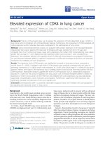
Overexpression of CD30 in ATL cell lines
We next examined CD30 expression in ATL-derived T-cell
lines, HTLV-1 transformed cell lines and HTLV-1 negative
T-cell lines using FACS analysis (Figure 3). All ATL cell
lines (TL-OmI, KOB, KK1 and ST1) showed strong CD30
expression whereas a B lymphoma cell line (BJAB)
showed no staining (Figure 3A). HTLV-1 transformed cell
lines (HUT-102, C5/MJ, MT-4 and SLB-1) also showed
CD30 expression but the amount of the expression was
various and lower than TL-OmI (Figure 3B). In HTLV-1
negative T-cell lines (Jurkat and MOLT-4), the expression
of CD30 was significantly lower than TL-OmI (Figure 3C).
Interestingly, NF-κB activity was much lower in Jurkat and
MOLT-4 than ATL cell lines. Thus CD30 expression level
is well correlated with the NF-κB activity, which suggests
that overexpression of CD30 might be at least one of the
factors that contributes to constitutive NF-κB activation in
ATL cell lines. In HTLV-1 transformed cells, NF-κB activa-
tion is thought to be largely dependent on Tax1, however
it is possible that relatively strong CD30 expression in
HUT-102 and SLB-1 also contributes to constitutive NF-
κB activation in these cells. In addition, CD30L expressed
in ATL cell lines may possibly contribute to CD30 activa-
tion by a cell-cell contact mechanism. RT-PCR analysis for
CD30 ligand showed that CD30L expression in TL-OmI
cells was extremely weak compared with a Burkitt lym-
phoma cell line (EB-1), in which CD30L is weakly
expressed (data not shown) [50]. This finding suggests
that CD30L is not involved in the constitutive NF-κB acti-
vation in TL-OmI cells.
Expression of CD30 in primary ATL cells
Next, we examined CD30 expression in primary ATL cells
by FACS analysis. Peripheral blood lymphocytes (PBLs),
lymph node cells, or ascitic fluid cells from ATL patients
were stained with anti-CD30 antibody (Figure 4 and Table
2). ATL cases in which more than 30% of the cells
expressed CD30 were classified as CD30-positive ones.
CD30 expression was seen in 8 of 66 ATL cases (12.1%)
and the CD30 expression was predominantly seen in the
acute type (5 of 25 cases), representing the advanced stage
of ATL (Figure 4B). Data of the FACS analysis (CD3, CD4,
CD8, CD25, and CD30 expression) of the CD30-positive
ATL cases are summarized in Table 2.
It has been reported that proteolytic cleavage of mem-
brane-anchored CD30 releases a soluble fragment corre-
sponding to the extracellular domain [51]. To examine
the possibility that the low frequency of CD30 expression
in primary ATL cells in the FACS analysis is due to this
proteolytic processing, CD30 mRNA expression was
examined in 8 ATL cases different from those used in the
FACS analysis. Strong CD30 mRNA expression was seen
in HUT-102 and PBLs activated by phytohemagglutinin
(PHA), whereas the CD30 expression was seen in only
one case (ATL8) diagnosed as the lymphoma type (Figure
5). The amount of CD30 mRNA expression in this case
was lower than HUT-102 and it might not be sufficient to
induce NF-κB activation by itself. However it is possible
that weak CD30 expression still contributes to the consti-
tutive NF-κB activation in cooperation with other signal-
ing molecules in vivo. In summery, these FACS and RT-
PCR data suggest that the expression of CD30 in ATL is
not a common event and is limited to a small number of
ATL cases. This is consistent with a previous report that
CD30 expression was seen in 7 out of 36 cases (19.4%)
when their lymph node biopsies were immunohisto-
chemically stained with anti-CD30 antibody [52].
The reason for the discrepancy between ATL cell lines and
primary ATL cells in terms of CD30 expression is
unknown at present. One possibility is that only CD30-
positive primary ATL cells could be established as a cell
line in vitro because of their stronger NF-κB activity or acti-
vation of other signaling pathways originating from
CD30. In fact, CD30 activates not only NF-κB but also the
mitogen activated protein kinase (MAPK) pathways, such
as extracellular regulated kinase (ERK), Jun N-terminal
kinase (JNK), and p38 MAPK pathways [53,54].
Recently, it has been reported that the noncanonical path-
way is involved in constitutive NF-κB activation in ATL
cells [55]. Although activation of the noncanonical path-
way by CD30 has not yet been reported, it is likely that
CD30 activates this pathway through association with
TNF receptor associated factors (TRAFs) like LT-βR and
CD40. In H-RS cells, which strongly express CD30, TRAF2
and TRAF5 make aggregates in the cytoplasm and co-
localize with downstream signaling molecules, such as
IKKα and IKKβ [56]. It would be interesting to see
whether TRAF2 and TRAF5 also form aggregates in ATL
cell lines and primary ATL cells expressing CD30.
In order to confirm that CD30 is involved in constitutive
NF-κB activation and cell survival in ATL cell lines, we
tried to knockdown CD30 expression in these cells by
using short-hairpin RNAs. We generated 11 different
short-hairpin RNAs for CD30 in total, but none of them
showed any RNA interference effect. We also tried to
introduce a decoy CD30 that lacks most of the
cytoplasmic region and has been shown to induce apop-
tosis in H-RS cells [41], by using an adenovirus vector.
However we were unable to obtain a sufficiently high titer
adenovirus as a decoy CD30 mutant to carry out the
experiment. Thus, whether elevated expression of CD30
actually contributes to constitutive NF-κB activation in
ATL cell lines still remains unknown.
In this regard, the mechanism by which NF-κB is constitu-
tively activated in ATL cells still remains a mystery. How-
Retrovirology 2005, 2:29 />Page 6 of 12
(page number not for citation purposes)
ever, our data suggest that the elevated expression of
CD30 plays a critical role in NF-κB activation in ATL cell
lines and a small number of primary ATL cells. Other
molecules belonging to the TNF receptor family, such as
LT-βR, OX40, or downstream signaling molecules, could
be involved in constitutive NF-κB activation in CD30-neg-
ative ATL cells, and the identification of such molecules
would contribute to the prevention, diagnosis and treat-
ment of ATL.
Conclusion
ATL cells have constitutive NF-κB activity which is impor-
tant for the cells' survival. This NF-κB activation is inde-
pendent of Tax protein expression. By screening a
retroviral cDNA library from an ATL cell line to identify
Elevated expression of CD30 in ATL cell linesFigure 3
Elevated expression of CD30 in ATL cell lines. CD30 expression was examined in A) ATL, B) HTLV-1-transformed, and
C) HTLV-1-negative cell lines by FACS analysis. A Burkitt lymphoma cell line (BJAB) was used as a negative control in A). TL-
OmI was used as a standard for the CD30 expression level in B) and C).
BJAB
TL-OmI KOB KK1 ST1
A
B
TL-OmI C5/MJ HUT-102 MT-4 SLB-1
C
Jurkat MOLT-4
TL-OmI
Retrovirology 2005, 2:29 />Page 7 of 12
(page number not for citation purposes)
NF-κB activating molecules, we obtained several cDNA
clones including full-length CD30. CD30 is strongly
expressed in ATL cell lines and primary ATL cells from a
small number of patients. Our results suggest that ele-
vated expression of CD30 is one of the factors responsible
for constitutive NF-κB activation in ATL cells.
Methods
Cell culture
Rat-1, a rat fibroblast cell line, was cultured in Dulbecco's
modified Eagle's medium (DMEM) supplemented with
10% fetal bovine serum (FBS). Human T-cell lines used in
the present experiments have been characterized previ-
ously [33,57]. Jurkat and MOLT-4 are HTLV-1 negative
human T-cell lines. HUT-102, C5/MJ, MT-4 and SLB-1 are
HTLV-1-positive human T-cell lines. TL-OmI, KK1 [58],
KOB [59], and ST1 [60] are HTLV-1-positive, ATL-derived
cell lines. These cells were cultured in RPMI 10% FBS.
Recombinant human IL-2 (Takeda Chemical Industries,
Osaka, Japan) was added at 0.5 nM to the culture of KK1,
KOB and ST1. A retrovirus packaging cell line Plat-E [61]
was cultured in DMEM 10% FBS containing 1 µg/ml puro-
mycin (Calbiochem, La Jolla, CA) and 10 µg/ml blastici-
din (Invitrogen, San Diego, CA).
cDNA library construction
Poly (A)
+
RNA was purified from TL-OmI using FastTrack
2.0 (Invitrogen). cDNA was synthesized by oligo(dT)
primers using SuperScript Choice System (Invitrogen)
according to the instructions provided by the manufac-
turer. The resulting cDNAs were size-fractionated through
agarose gel electrophoresis, and cDNA fragments longer
than 2.5 kb were extracted from the gel by using Qiaex II
(Qiagen, Hilden, Germany). The cDNA fragments were
then inserted into BstXI sites of the retroviral vector pMX
[62] using BstXI adapters (Invitrogen). The ligated DNA
was ethanol-precipitated and then electroporated into
DH10B competent cells (Electromax DH10B; Invitrogen).
About 1 × 10
6
independent clones were amplified on 150
mm LB/amp plates and plasmid DNA was purified by
using Qiagen Plasmid Giga kit (Qiagen).
Generation of a reporter cell line
The NF-κB reporter plasmid κB-EGFP was constructed by
replacing the luciferase gene (a BglII – BamHI fragment) of
the κB-Luc plasmid [42] with EGFP (a HindIII – AflII frag-
ment) from pEGFP-N3 (Clontech Laboratories, Palo Alto,
CA) by blunt-end ligation. To construct the plasmid κB-
bsrEGFP, which expresses bsrEGFP fusion protein, a PCR
amplified bsr gene fragment was inserted in the ApaI and
BamHI sites upstream of EGFP of the κB-EGFP plasmid.
To prepare a NF-κB reporter cell line, Rat-1 cells (5 × 10
6
)
were transfected with 20 µg of κB-bsrEGFP and 1 µg of
pcDNA3 (Invitrogen) by electroporation at 250 V and 975
µF. The transfected cells were cultured in 500 µg/ml G418
(Invitrogen), and resistant clones were screened for EGFP
signals after being infected with retroviruses that express
Epstein-Barr virus transforming protein LMP1. The
selected cell clone (Rat-1 κB-bsrEGFP) was further
transfected with κB-EGFP and pMik-HygB and cultured in
250 µg/ml hygromycin B (Wako Pure Chemical Indus-
tries, Osaka, Japan). Resistant clones were screened for
EGFP signals after stimulation with 20 ng/ml TNF-α
(Peprotech, London, UK).
Preparation of retroviruses and infection of reporter cells
Plat-E cells (2 × 10
6
cells) were seeded onto 60 mm dishes
one day before transfection. The cDNA library (3 µg) was
transfected using Fugene 6 (Roche Molecular Systems,
Inc., NJ) according to the protocol provided by the manu-
facturer. Cells were cultured for 48 hours and the retrovi-
ral supernatant was harvested. For infection of reporter
Table 2: Cell surface markers in CD30-positive ATL cases
% of Positive Cells
Case Sex Type Material CD3 CD4 CD8 CD25 CD30
1 M Acute PB 90.1 86.5 4.4 89.8 56.5
2 F Acute PB 18.9 78.3 3.0 81.0 48.7
3 F Acute PB 94.8 14.2 64.2 81.9 84.8
4 F Acute PB 89.3 96.3 2.6 93.3 35.5
5 M Acute LN 10.1 96.6 5.2 90.1 93.0
6 M Lymphoma LN 8.7 85.1 5.3 58.1 76.5
7 F Unknown LN 67.5 77.8 23.0 81.7 60.4
8 F Unknown Ascites 89.5 99.7 0.1 99.5 96.2
The percentage of positive cells was determined by immunofluorescence staining with respective antibodies and flow cytometric analysis.
Abbreviations: PB, peripheral blood; LN, lymph node.
Retrovirology 2005, 2:29 />Page 8 of 12
(page number not for citation purposes)
cells, 2.5 × 10
5
cells were seeded onto 100 mm dishes one
day before infection and incubated with 10 ml DMEM
10% FBS containing 0.6 ml of the virus stock for 24 hours
in the presence of polybrene (20 µg/ml). The medium was
changed to fresh DMEM 10% FBS after 24 hours. After
CD30 expression in primary ATL cellsFigure 4
CD30 expression in primary ATL cells. A) Primary ATL cells from a patient (case 8) were tested for the expression of
CD3, CD4, CD8, CD25 and CD30 by FACS analysis. B) Summary of the number of CD30-positive ATL cases.
CD30
CD4
CD25
CD8CD3
8 / 66 (12.1%)total
2/23Unknown
0/15Chronic
1/3Lymphoma
5/25Acute
A
B
Retrovirology 2005, 2:29 />Page 9 of 12
(page number not for citation purposes)
another 24 hours, the cells were incubated with medium
containing 50 µg/ml blasticidin (Invitrogen).
Isolation of cDNA fragments from blasticidin-resistant
clones
Genomic DNA was extracted from the blasticidin-resistant
clones by DNeasy kit (Qiagen) and subjected to PCR to
recover integrated cDNAs using pMX vector primers (5'-
GGTGGACCATCCTCTAGACT-3' and 5'-CCCCTTTTTCT-
GGAGACTAAAT-3'). The PCR products were cloned into
pGEM-T Easy vector (Promega, Madison, WI) and
sequenced using BigDye Terminator v1.1 cycle sequenc-
ing kit (Applied Biosystems, Foster City, CA).
Expression plasmids
The retroviral vector pMX LMP1 was prepared by inserting
an EcoRI – BamHI fragment of pSG5 F-LMP1 [63] in the
EcoRI site of pMX by blunt-end ligation. pMX CD30WT
was generated by inserting a MluI – NotI fragment of
pCD30WT [64] in the EcoRI and NotI sites of pMX by
blunt-end ligation. pMX kBL1 was generated by inserting
a BamHI – SfiI fragment of pGEMT kBL1 in the BamHI –
NotI sites of pMX. The BglII site of pMX kBL1 was
destroyed by cutting by BglII, filling in by T4 polymerase,
and self-ligation to make pMX kBL1∆BglII.
Flow cytometric analysis
Heparinized peripheral blood, a piece of a lymph node, or
ascites (in case no. 8) was collected from patients with
ATL after obtaining informed consent in accordance with
the Helsinki Declaration. Mononuclear cells were sepa-
rated by Lymphoprep™ density gradient centrifugation
(Axis-Shield PoC AS, Oslo, Norway). Morphological and
surface marker analyses indicated that ATL cells in these
samples always accounted for more than 80% of the total
cell population in most cases. The study protocol was
approved by the Human Ethics Review Committee of
Nagasaki University Graduate School of Biomedical
Sciences.
Primary ATL cells or T-cell lines were incubated for 30 min
at 4°C with each PE-labeled or FITC-labeled monoclonal
antibody (mAb). Cells were also incubated with isotype
matched control antibodies. The following antibodies
were used: PE-labeled mouse anti-human CD4 and CD25,
FITC-labeled anti CD3 and CD8 (BD Biosciences
Pharmingen, San Diego, CA); and PE-labeled mouse anti-
human CD30 (Dako Corporation, Carpinteria, CA or
Immunotech, Marseille, France). After washing with PBS,
the cells were analyzed on FACScan flow cytometer using
Cellquest software (Becton Dickinson, San Jose, CA).
CD30 mRNA expression in primary ATL cellsFigure 5
CD30 mRNA expression in primary ATL cells Primary ATL cells from ATL patients (lanes 5–12) and normal PBLs from
healthy adult donors (lanes 1–3) were tested for CD30 (upper panel) and β-actin (lower panel) mRNA expression by RT-PCR
analysis. The CD30 expression was seen in ATL8 (lane 12). PHA-stimulated PBLs (lane 4) and HUT-102 (lane 13) were used as
a positive control.
Retrovirology 2005, 2:29 />Page 10 of 12
(page number not for citation purposes)
Reverse transcription-polymerase chain reaction
Total cellular RNA was extracted with Trizol (Invitrogen)
according to the protocol provided by the manufacturer.
First-strand cDNA was synthesized from 1 µg total cellular
RNA in a 20-µl reaction volume using an RNA PCR kit
(Takara Shuzo, Kyoto, Japan) with random primers.
Thereafter, cDNA was amplified for 35 cycles for CD30
and 28 cycles for β-actin. The oligonucleotide primers
used were as follows: for CD30, sense, 5'-CTGTGTC-
CCCTACCCAATCT-3' and antisense, 5'-CTTCTTTCCCT-
TCCTCTTCCA-3'; [65] and for β-actin, sense, 5'-
GTGGGGCGCCCCAGGCACCA-3' and antisense, 5'-CTC-
CTTAATGTCACGCACGATTTC-3'. Product sizes were
860-bp for CD30 and 548-bp for β-actin. Cycling condi-
tions were as follows: denaturing at 94°C for 45 sec (for
CD30) or for 30 sec (for β-actin), annealing at 62°C for 45
sec (for CD30) or 60°C for 30 sec (for β-actin) and exten-
sion at 72°C for 60 sec (for CD30) or for 90 sec (for β-
actin). The PCR products were fractionated on 2% agarose
gels and visualized by ethidium bromide staining.
Competing interests
The author(s) declare that they have no competing
interests.
Authors' contributions
MH carried out the cDNA cloning and the functional anal-
ysis of CD30. TM and NM carried out the RT-PCR analysis.
YY carried out the FACS analysis. MH, RH, TW, MT, MO
and MF participated in the experimental design, data
interpretation, and writing of the manuscript.
Acknowledgements
We are deeply indebted to the many patients with ATL and the control
subjects who donated blood for these studies. We thank T. Kitamura for
providing the retroviral vector pMX and the packaging cell line Plat-E. We
also thank R. Fujita, S. Takizawa, and C. Yamamoto for the excellent tech-
nical assistance. This work was supported in part by a Grant-in-Aid for Sci-
entific Research of Japan, Grant for Promotion of Niigata University
Research Projects, and Tsukada Grant for Niigata University Medical
Research.
References
1. Sugamura K, Hinuma Y: Human retroviruses: HTLV-I and
HTLV-II. In The Retrovirudae Volume 2. Edited by: Levy JA. New York:
Plenum Press; 1993:399-435.
2. Yoshida M: Multiple viral strategies of HTLV-1 for dysregula-
tion of cell growth control. Annu Rev Immunol 2001, 19:475-496.
3. Matsuoka M: Human T-cell leukemia virus type I and adult T-
cell leukemia. Oncogene 2003, 22:5131-5140.
4. Grassmann R, Berchtold S, Radant I, Alt M, Fleckenstein B, Sodroski
JG, Haseltine WA, Ramstedt U: Role of human T-cell leukemia
virus type 1 X region proteins in immortalization of primary
human lymphocytes in culture. J Virol 1992, 66:4570-4575.
5. Akagi T, Shimotohno K: Proliferative response of Tax1-trans-
duced primary human T cells to anti-CD3 antibody stimula-
tion by an interleukin-2-independent pathway. J Virol 1993,
67:1211-1217.
6. Brauweiler A, Garrus JE, Reed JC, Nyborg JK: Repression of bax
gene expression by the HTLV-1 Tax protein: implications for
suppression of apoptosis in virally infected cells. Virology 1997,
231:135-140.
7. Mulloy JC, Kislyakova T, Cereseto A, Casareto L, LoMonico A, Fullen
J, Lorenzi MV, Cara A, Nicot C, Giam C, Franchini G: Human T-cell
lymphotropic/leukemia virus type 1 Tax abrogates p53-
induced cell cycle arrest and apoptosis through its CREB/
ATF functional domain. J Virol 1998, 72:8852-8860.
8. Tsukahara T, Kannagi M, Ohashi T, Kato H, Arai M, Nunez G, Iwanaga
Y, Yamamoto N, Ohtani K, Nakamura M, Fujii M: Induction of Bcl-
x(L) expression by human T-cell leukemia virus type 1 Tax
through NF-kappaB in apoptosis-resistant T-cell transfect-
ants with Tax. J Virol 1999, 73:7981-7987.
9. Kawakami A, Nakashima T, Sakai H, Urayama S, Yamasaki S, Hida A,
Tsuboi M, Nakamura H, Ida H, Migita K, Kawabe Y, Eguchi K: Inhibi-
tion of caspase cascade by HTLV-I tax through induction of
NF-kappaB nuclear translocation. Blood 1999, 94:3847-3854.
10. Siekevitz M, Feinberg MB, Holbrook N, Wong-Staal F, Greene WC:
Activation of interleukin 2 and interleukin 2 receptor (Tac)
promoter expression by the trans-activator (tat) gene prod-
uct of human T-cell leukemia virus, type I. Proc Natl Acad Sci U
S A 1987, 84:5389-5393.
11. Himes SR, Coles LS, Katsikeros R, Lang RK, Shannon MF: HTLV-1
tax activation of the GM-CSF and G-CSF promoters
requires the interaction of NF-kB with other transcription
factor families. Oncogene 1993, 8:3189-3197.
12. Azimi N, Brown K, Bamford RN, Tagaya Y, Siebenlist U, Waldmann
TA: Human T cell lymphotropic virus type I Tax protein
trans-activates interleukin 15 gene transcription through an
NF-kappaB site. Proc Natl Acad Sci U S A 1998, 95:2452-2457.
13. Waldele K, Schneider G, Ruckes T, Grassmann R: Interleukin-13
overexpression by tax transactivation: a potential autocrine
stimulus in human T-cell leukemia virus-infected
lymphocytes. J Virol 2004, 78:6081-6090.
14. Inoue J, Seiki M, Taniguchi T, Tsuru S, Yoshida M: Induction of
interleukin 2 receptor gene expression by p40x encoded by
human T-cell leukemia virus type 1. EMBO J 1986, 5:2883-2888.
15. Maruyama M, Shibuya H, Harada H, Hatakeyama M, Seiki M, Fujita T,
Inoue J, Yoshida M, Taniguchi T: Evidence for aberrant activation
of the interleukin-2 autocrine loop by HTLV-1-encoded p40x
and T3/Ti complex triggering. Cell 1987, 48:343-350.
16. Cross SL, Feinberg MB, Wolf JB, Holbrook NJ, Wong-Staal F, Leonard
WJ: Regulation of the human interleukin-2 receptor alpha
chain promoter: activation of a nonfunctional promoter by
the transactivator gene of HTLV-I. Cell 1987, 49:47-56.
17. Mariner JM, Lantz V, Waldmann TA, Azimi N: Human T cell lym-
photropic virus type I Tax activates IL-15R alpha gene
expression through an NF-kappa B site. J Immunol 2001,
166:2602-2609.
18. Mori N, Fujii M, Cheng G, Ikeda S, Yamasaki Y, Yamada Y, Tomonaga
M, Yamamoto N: Human T-cell leukemia virus type I tax pro-
tein induces the expression of anti-apoptotic gene Bcl-xL in
human T-cells through nuclear factor-kappaB and c-AMP
responsive element binding protein pathways. Virus Genes
2001, 22:279-287.
19. Akagi T, Ono H, Shimotohno K: Expression of cell-cycle regula-
tory genes in HTLV-I infected T-cell lines: possible involve-
ment of Tax1 in the altered expression of cyclin D2, p18Ink4
and p21Waf1/Cip1/Sdi1. Oncogene 1996, 12:1645-1652.
20. Santiago F, Clark E, Chong S, Molina C, Mozafari F, Mahieux R, Fujii
M, Azimi N, Kashanchi F: Transcriptional up-regulation of the
cyclin D2 gene and acquisition of new cyclin-dependent
kinase partners in human T-cell leukemia virus type 1-
infected cells. J Virol 1999, 73:9917-9927.
21. Iwanaga R, Ohtani K, Hayashi T, Nakamura M: Molecular mecha-
nism of cell cycle progression induced by the oncogene prod-
uct Tax of human T-cell leukemia virus type I. Oncogene 2001,
20:2055-2067.
22. Mori N, Fujii M, Hinz M, Nakayama K, Yamada Y, Ikeda S, Yamasaki
Y, Kashanchi F, Tanaka Y, Tomonaga M, Yamamoto N: Activation
of cyclin D1 and D2 promoters by human T-cell leukemia
virus type I tax protein is associated with IL-2-independent
growth of T cells. Int J Cancer 2002, 99:378-385.
23. Fujii M, Niki T, Mori T, Matsuda T, Matsui M, Nomura N, Seiki M:
HTLV-1 Tax induces expression of various immediate early
serum responsive genes. Oncogene 1991, 6:1023-1029.
Retrovirology 2005, 2:29 />Page 11 of 12
(page number not for citation purposes)
24. Yamaoka S, Inoue H, Sakurai M, Sugiyama T, Hazama M, Yamada T,
Hatanaka M: Constitutive activation of NF-kappa B is essential
for transformation of rat fibroblasts by the human T-cell
leukemia virus type I Tax protein. EMBO J 1996, 15:873-887.
25. Akagi T, Ono H, Nyunoya H, Shimotohno K: Characterization of
peripheral blood T-lymphocytes transduced with HTLV-I
Tax mutants with different trans-activating phenotypes.
Oncogene 1997, 14:2071-2078.
26. Robek MD, Ratner L: Immortalization of CD4(+) and CD8(+) T
lymphocytes by human T-cell leukemia virus type 1 Tax
mutants expressed in a functional molecular clone. J Virol
1999, 73:4856-4865.
27. Hayden MS, Ghosh S: Signaling to NF-kappaB. Genes Dev 2004,
18:2195-2224.
28. Bonizzi G, Karin M: The two NF-kappaB activation pathways
and their role in innate and adaptive immunity. Trends Immunol
2004, 25:280-288.
29. Chu ZL, DiDonato JA, Hawiger J, Ballard DW: The tax oncopro-
tein of human T-cell leukemia virus type 1 associates with
and persistently activates IkappaB kinases containing IKKa-
lpha and IKKbeta. J Biol Chem 1998, 273:15891-15894.
30. Harhaj EW, Good L, Xiao G, Uhlik M, Cvijic ME, Rivera-Walsh I, Sun
SC: Somatic mutagenesis studies of NF-kappa B signaling in
human T cells: evidence for an essential role of IKK gamma
in NF-kappa B activation by T-cell costimulatory signals and
HTLV-I Tax protein. Oncogene 2000, 19:1448-1456.
31. Sun SC, Harhaj EW, Xiao G, Good L: Activation of I-kappaB
kinase by the HTLV type 1 Tax protein: mechanistic insights
into the adaptor function of IKKgamma. AIDS Res Hum
Retroviruses 2000, 16:1591-1596.
32. Xiao G, Cvijic ME, Fong A, Harhaj EW, Uhlik MT, Waterfield M, Sun
SC: Retroviral oncoprotein Tax induces processing of NF-
kappaB2/p100 in T cells: evidence for the involvement of
IKKalpha. EMBO J 2001, 20:6805-6815.
33. Mori N, Fujii M, Ikeda S, Yamada Y, Tomonaga M, Ballard DW,
Yamamoto N: Constitutive activation of NF-kappaB in pri-
mary adult T-cell leukemia cells. Blood 1999, 93:2360-2368.
34. Mori N, Yamada Y, Ikeda S, Yamasaki Y, Tsukasaki K, Tanaka Y,
Tomonaga M, Yamamoto N, Fujii M: Bay 11-7082 inhibits tran-
scription factor NF-kappaB and induces apoptosis of HTLV-
I-infected T-cell lines and primary adult T-cell leukemia cells.
Blood 2002, 100:1828-1834.
35. Furukawa Y, Kubota R, Tara M, Izumo S, Osame M: Existence of
escape mutant in HTLV-I tax during the development of
adult T-cell leukaemia. Blood 2001, 97:987-993.
36. Okazaki S, Moriuchi R, Yosizuka N, Sugahara K, Maeda T, Jinnai I,
Tomonaga M, Kamihira S, Katamine S: HTLV-1 proviruses
encoding non-functional TAX in adult T-cell leukemia. Virus
Genes 2001, 23:123-135.
37. Kinoshita T, Shimoyama M, Tobinai K, Ito M, Ito S, Ikeda S, Tajima K,
Shimotohno K, Sugimura T: Detection of mRNA for the tax1/
rex1 gene of human T-cell leukemia virus type I in fresh
peripheral blood mononuclear cells of adult T-cell leukemia
patients and viral carriers by using the polymerase chain
reaction. Proc Natl Acad Sci U S A 1989, 86:5620-5624.
38. Furukawa Y, Osame M, Kubota R, Tara M, Yoshida M: Human T-
cell leukemia virus type-1 (HTLV-1) Tax is expressed at the
same level in infected cells of HTLV-1-associated myelopa-
thy or tropical spastic paraparesis patients as in asympto-
matic carriers but at a lower level in adult T-cell leukemia
cells. Blood 1995, 85:1865-1870.
39. Schneider C, Hubinger G: Pleiotropic signal transduction medi-
ated by human CD30: a member of the tumor necrosis fac-
tor receptor (TNFR) family. Leuk Lymphoma 2002, 43:1355-1366.
40. Al-Shamkhani A: The role of CD30 in the pathogenesis of hae-
matopoietic malignancies. Curr Opin Pharmacol 2004, 4:355-359.
41. Horie R, Watanabe T, Morishita Y, Ito K, Ishida T, Kanegae Y, Saito I,
Higashihara M, Mori S, Kadin ME: Ligand-independent signaling
by overexpressed CD30 drives NF-kappaB activation in
Hodgkin-Reed-Sternberg cells. Oncogene 2002, 21:2493-2503.
42. Suzuki T, Hirai H, Murakami T, Yoshida M: Tax protein of HTLV-
1 destabilizes the complexes of NF-kappa B and I kappa B-
alpha and induces nuclear translocation of NF-kappa B for
transcriptional activation. Oncogene 1995, 10:1199-1207.
43. Xiao G, Harhaj EW, Sun SC: NF-kappaB-inducing kinase regu-
lates the processing of NF-kappaB2 p100. Mol Cell 2001,
7:401-409.
44. Xiao G, Sun SC: Negative regulation of the nuclear factor
kappa B-inducing kinase by a cis-acting domain. J Biol Chem
2000, 275:21081-21085.
45. Lee FS, Peters RT, Dang LC, Maniatis T: MEKK1 activates both
IkappaB kinase alpha and IkappaB kinase beta. Proc Natl Acad
Sci U S A 1998, 95:9319-9324.
46. McCarthy JV, Ni J, Dixit VM: RIP2 is a novel NF-kappaB-activat-
ing and cell death-inducing kinase. J Biol Chem 1998,
273:16968-16975.
47. Chin AI, Dempsey PW, Bruhn K, Miller JF, Xu Y, Cheng G: Involve-
ment of receptor-interacting protein 2 in innate and adap-
tive immune responses. Nature 2002, 416:190-194.
48. Kobayashi K, Inohara N, Hernandez LD, Galan JE, Nunez G, Janeway
CA, Medzhitov R, Flavell RA: RICK/Rip2/CARDIAK mediates
signalling for receptors of the innate and adaptive immune
systems. Nature 2002, 416:194-199.
49. Ruefli-Brasse AA, Lee WP, Hurst S, Dixit VM: Rip2 participates in
Bcl10 signaling and T-cell receptor-mediated NF-kappaB
activation. J Biol Chem 2004, 279:1570-1574.
50. Gruss HJ, DaSilva N, Hu ZB, Uphoff CC, Goodwin RG, Drexler HG:
Expression and regulation of CD30 ligand and CD30 in
human leukemia-lymphoma cell lines. Leukemia 1994,
8:2083-2094.
51. Pfreundschuh M, Pohl C, Berenbeck C, Schroeder J, Jung W, Schmits
R, Tschiersch A, Diehl V, Gause A: Detection of a soluble form of
the CD30 antigen in sera of patients with lymphoma, adult
T-cell leukemia and infectious mononucleosis. Int J Cancer
1990, 45:869-874.
52. Ohtsuka E, Kikuchi H, Nasu M, Takita-Sonoda Y, Fujii H, Yokoyama
S: Clinicopathological features of adult T-cell leukemia with
CD30 antigen expression. Leuk Lymphoma 1994, 15:303-310.
53. Harlin H, Podack E, Boothby M, Alegre ML: TCR-independent
CD30 signaling selectively induces IL-13 production via a
TNF receptor-associated factor/p38 mitogen-activated pro-
tein kinase-dependent mechanism. J Immunol 2002,
169:2451-2459.
54. Zheng B, Fiumara P, Li YV, Georgakis G, Snell V, Younes M, Vauthey
JN, Carbone A, Younes A: MEK/ERK pathway is aberrantly
active in Hodgkin disease: a signaling pathway shared by
CD30, CD40, and RANK that regulates cell proliferation and
survival. Blood 2003, 102:1019-1027.
55. Hironaka N, Mochida K, Mori N, Maeda M, Yamamoto N, Yamaoka
S: Tax-independent constitutive IkappaB kinase activation in
adult T-cell leukemia cells. Neoplasia 2004, 6:266-278.
56. Horie R, Watanabe T, Ito K, Morisita Y, Watanabe M, Ishida T,
Higashihara M, Kadin M: Cytoplasmic aggregation of TRAF2
and TRAF5 proteins in the Hodgkin-Reed-Sternberg cells.
Am J Pathol 2002, 160:1647-1654.
57. Sugamura K, Fujii M, Kannagi M, Sakitani M, Takeuchi M, Hinuma Y:
Cell surface phenotypes and expression of viral antigens of
various human cell lines carrying human T-cell leukemia
virus. Int J Cancer 1984, 34:221-228.
58. Yamada Y, Nagata Y, Kamihira S, Tagawa M, Ichimaru M, Tomonaga
M, Shiku H: IL-2-dependent ATL cell lines with phenotypes dif-
fering from the original leukemia cells. Leuk Res 1991,
15:619-625.
59. Maeda T, Yamada Y, Moriuchi R, Sugahara K, Tsuruda K, Joh T, Ato-
gami S, Tsukasaki K, Tomonaga M, Kamihira S: Fas gene mutation
in the progression of adult T cell leukemia. J Exp Med 1999,
189:1063-1071.
60. Yamada Y, Ohmoto Y, Hata T, Yamamura M, Murata K, Tsukasaki K,
Kohno T, Chen Y, Kamihira S, Tomonaga M: Features of the
cytokines secreted by adult T cell leukemia (ATL) cells. Leuk
Lymphoma 1996, 21:443-447.
61. Morita S, Kojima T, Kitamura T: Plat-E: an efficient and stable
system for transient packaging of retroviruses. Gene Ther 2000,
7:1063-1066.
62. Kitamura T, Onishi M, Kinoshita S, Shibuya A, Miyajima A, Nolan GP:
Efficient screening of retroviral cDNA expression libraries.
Proc Natl Acad Sci U S A 1995, 92:9146-9150.
63. Izumi KM, Kaye KM, Kieff ED: The Epstein-Barr virus LMP1
amino acid sequence that engages tumor necrosis factor
receptor associated factors is critical for primary B lym-
Publish with BioMed Central and every
scientist can read your work free of charge
"BioMed Central will be the most significant development for
disseminating the results of biomedical researc h in our lifetime."
Sir Paul Nurse, Cancer Research UK
Your research papers will be:
available free of charge to the entire biomedical community
peer reviewed and published immediately upon acceptance
cited in PubMed and archived on PubMed Central
yours — you keep the copyright
Submit your manuscript here:
/>BioMedcentral
Retrovirology 2005, 2:29 />Page 12 of 12
(page number not for citation purposes)
phocyte growth transformation. Proc Natl Acad Sci U S A 1997,
94:1447-1452.
64. Horie R, Ito K, Tatewaki M, Nagai M, Aizawa S, Higashihara M, Ishida
T, Inoue J, Takizawa H, Watanabe T: A variant CD30 protein lack-
ing extracellular and transmembrane domains is induced in
HL-60 by tetradecanoylphorbol acetate and is expressed in
alveolar macrophages. Blood 1996, 88:2422-2432.
65. Gattei V, Degan M, Gloghini A, De Iuliis A, Improta S, Rossi FM, Aldi-
nucci D, Perin V, Serraino D, Babare R, Zagonel V, Gruss HJ, Carbone
A, Pinto A: CD30 ligand is frequently expressed in human
hematopoietic malignancies of myeloid and lymphoid origin.
Blood 1997, 89:2048-2059.