Báo cáo y học: " A novel function for spumaretrovirus integrase: an early requirement for integrase-mediated cleavage of 2 LTR circles" docx
Bạn đang xem bản rút gọn của tài liệu. Xem và tải ngay bản đầy đủ của tài liệu tại đây (938.55 KB, 18 trang )
BioMed Central
Page 1 of 18
(page number not for citation purposes)
Retrovirology
Open Access
Research
A novel function for spumaretrovirus integrase: an early
requirement for integrase-mediated cleavage of 2 LTR circles
Olivier Delelis
†1
, Caroline Petit*
†1
, Herve Leh
2
, Gladys Mbemba
3
, Jean-
François Mouscadet
3
and Pierre Sonigo*
1
Address:
1
Génétique des virus, Département des Maladies Infectieuses, Institut Cochin, INSERM U567, CNRS UMR8104, Université René
Descartes, 22 rue Méchain, 75014 Paris, France,
2
Bioalliancepharma, 59 boulevard Martial Valin, 75015 Paris, France and
3
LBPA, CNRS UMR8113,
Ecole Normale Supérieure de Cachan, 61 avenue du Président Wilson, 94235, Cachan, France
Email: Olivier Delelis - ; Caroline Petit* - ; Herve Leh - ;
Gladys Mbemba - ; Jean-François Mouscadet - ;
Pierre Sonigo* -
* Corresponding authors †Equal contributors
spumaretrovirusintegrase substratepalindrome at LTR-LTR junctions2-LTR circles DNA
Abstract
Retroviral integration is central to viral persistence and pathogenesis, cancer as well as host
genome evolution. However, it is unclear why integration appears essential for retrovirus
production, especially given the abundance and transcriptional potential of non-integrated viral
genomes. The involvement of retroviral endonuclease, also called integrase (IN), in replication
steps apart from integration has been proposed, but is usually considered to be accessory. We
observe here that integration of a retrovirus from the spumavirus family depends mainly on the
quantity of viral DNA produced. Moreover, we found that IN directly participates to linear DNA
production from 2-LTR circles by specifically cleaving the conserved palindromic sequence found
at LTR-LTR junctions. These results challenge the prevailing view that integrase essential function
is to catalyze retroviral DNA integration. Integrase activity upstream of this step, by controlling
linear DNA production, is sufficient to explain the absolute requirement for this enzyme.
The novel role of IN over 2-LTR circle junctions accounts for the pleiotropic effects observed in
cells infected with IN mutants. It may explain why 1) 2-LTR circles accumulate in vivo in mutants
carrying a defective IN while their linear and integrated DNA pools decrease; 2) why both LTRs
are processed in a concerted manner. It also resolves the original puzzle concerning the integration
of spumaretroviruses. More generally, it suggests to reassess 2-LTR circles as functional
intermediates in the retrovirus cycle and to reconsider the idea that formation of the integrated
provirus is an essential step of retrovirus production.
Background
Integration of viral genomes into host cell DNA is a key
element of the life cycle and pathogenesis of many viruses.
DNA viruses integrate by relying solely on cell machinery.
In contrast, retroviruses possess a specialized endonucle-
ase, also designated integrase (IN), which is essential for
Published: 18 May 2005
Retrovirology 2005, 2:31 doi:10.1186/1742-4690-2-31
Received: 20 April 2005
Accepted: 18 May 2005
This article is available from: />© 2005 Delelis et al; licensee BioMed Central Ltd.
This is an Open Access article distributed under the terms of the Creative Commons Attribution License ( />),
which permits unrestricted use, distribution, and reproduction in any medium, provided the original work is properly cited.
Retrovirology 2005, 2:31 />Page 2 of 18
(page number not for citation purposes)
their replication (for a review, see [1]). After entering a tar-
get cell, reverse transcriptase (RT) converts genomic RNA
into linear double-stranded cDNA with a copy of the viral
long terminal repeat (LTR) at each end. Such linear
genomic cDNA included in a preintegration complex
(PIC) [2-9] can be used as a template for integration in
vivo. Consequently, circular viral genomes that are
detected in infected cells were considered until now as
«dead-end» molecules, without essential function in the
integration process and the viral cycle in general [8].
Integration mediated by the retrovirus IN occurs in two
catalytic steps, referred to as 3'-processing and strand
transfer (or joining), respectively. Interestingly, the two
steps appeared on distinct reactions catalyzed by virus IN
in two different compartments in the infected cells. The
strand transfer reaction joins viral DNA to cellular DNA in
the cell nucleus. The viral cDNA ends are used to cut the
target DNA in a staggered manner, which covalently links
the viral 3' ends to the 5' phosphates of the cut (for
reviews see [10,11]. The 3' hydroxyl groups at the LTR ter-
mini are the nucleophiles that promote DNA strand trans-
fer [12]. Efficient strand transfer requires previous
endonucleolysis of DNA that produces recessed
3'hydroxyl ends [3,5]. This occurs in the cytoplasm very
soon after reverse transcription is completed [13-16], as
viral genomes with blunt ends are extremely rare in the
infected cytoplasm. Following these reactions, host cell
enzymes likely repair the gap remaining between host and
provirus DNA [17,18].
IN recognizes and acts on short sequences (12 to 20 bp)
called attachment (att) sites that are located at the LTRs
[19]. Att site includes the invariant CA dinucleotides,
which are conserved in all retroviruses whereas the other
nucleotides of the att site, while not conserved in
sequence, form an (imperfect) inverted repeat (IR) in all
retroviruses, that has to be maintained intact for viral rep-
lication. Att mutagenesis experiments showed that muta-
tion in one LTR precludes the processing of the other,
demonstrating that activity of IN is concerted onto the
two viral LTRs that are simultaneously cleaved in vivo [20].
The structural basis of such concerted processing of both
extremities is unknown. More surprisingly, in the case of
spumaretroviruses, a subfamily of retroviruses that share
some features of DNA viruses [21-23], the IN may process
only one of the two LTRs, although the att sites are present
at the two LTRs. Based on the sequences of both 2-LTR
DNA and integrated proviruses, an asymmetric processing
of att sites has been proposed, in which IN may cleave the
right, U5 end and may leave the left, U3 end intact
[24,25]. As the human spumaretrovirus (PFV) IN presents
the usual features of other IN and carries out in vitro an
endonucleolytic activity, as well as strand transfer and dis-
integrase activities [26,27], the reason for this unusual
mechanics is not understood at present.
The att recognition site of IN is present at least one time
on all forms of viral DNA. In addition to linear and inte-
grated forms, viral DNA is found in the infected cells as
covalently closed DNA circles containing either one or
two copies of the LTR, referred to as 1-LTR and 2-LTR cir-
cles, respectively [2]. Interestingly in the 2-LTR circles, the
att sites are in a closed configuration due to the juxtaposi-
tion of the two LTRs and are included within a palindro-
mic motif formed by the inverted repeat sequences in all
retroviruses [28-31]. These 2-LTR circles are believed to
result from a direct covalent joining of LTR ends at the so-
called circle junction [32,33]. Circularization is thought to
occur by blunt-end ligation of the ends of linear proviral
DNA, even no direct evidence has been provided until
now to support this hypothesis. 2-LTR could be formed in
part by the non-homologous end-joining (NHEJ) path-
way of DNA recombination [34]. The two-LTR circle
forms could, theoretically, serve as a potential precursor
for the integrated provirus [4]. In spleen necrosis virus
(SNV), Rous sarcoma virus (RSV), avian sarcoma virus
(ASV) and avian leukosis virus (ALV), closed circular
forms were initially proposed to act as substrates tem-
plates for integration [31,32,35], although these reports
have not been substantiated. Although they are currently
described in a productive infection as "dead end" mole-
cules, precisely because of their incapacity to be directly
integrated [8], intriguing observations invite some to
reconsider their place. First, 2-LTR molecules were shown
to be used as functional templates for the transcription
machinery in HIV infected cells [36-39]. Second, 2-LTR
viral DNA were detected in the cytoplasm of MLV and PFV
infected cells at a very early time post infection, suggesting
that they are not formed in the nucleus by an alternative
fate to the integration way [40,41]. In this context, we
asked whether 2-LTR circles, rather than being substrate
for integration nor "dead end" molecules, would be used
as substrates for a preintegrative endonucleolytic activity
of PFV IN.
Such interrogation comes within the scope of the more
global questioning concerning the pleiotropic actions of
IN. Indeed, the mechanisms underlying the essential
requirement for integration are still unclear in the retrovi-
rus cycle. Why is integration critical for viral production
when unintegrated DNA is abundant and competent for
transcription [36-39,42-45]? Is it possible that preintegra-
tive function of IN explain its essential requirement rather
than integration per se? Indeed, in addition to its roles in
the establishment of the proviral integrated state, IN par-
ticipates to other critical steps, such as reverse transcrip-
tion [23,46-52], nuclear import of HIV-1 preintegration
complex (PICs) (for a review, see [53]), and the
Retrovirology 2005, 2:31 />Page 3 of 18
(page number not for citation purposes)
postintegration step of viral particle assembly (reviewed
in [54]). Among the PIC constituents, IN is a logical and
probable candidate for facilitating the efficient nuclear
import of cDNA, since it has karyophilic properties [55-
61]. Reflecting the pleiotropic activities of IN, non-replica-
tive IN mutants of HIV were divided in two phenotypic
classes depending on their defects [54]. The properties of
IN mutants of PFV are less extensively described, and we
suspected that PFV IN could play a key role in early pre-
integrative steps.
In an attempt to better characterize the properties and
substrates of the original IN of PFV, we analyzed both its
in vivo properties and in vitro activity. We observed that the
2-LTR circles could serve as templates for the 3' processing
reaction of the IN. This allows spumaretrovirus to follow
a symmetrical mechanism of integration and leads to
reexamine the role of 2-LTR molecules and the impor-
tance of preintegrative function of IN.
Results and discussion
The mutations inPFV IN do not alter its karyophilic
property
Retroviral INs from oncoviruses [62,63], lentiviruses
[55,59,64,65] and spumavirus [66] are karyophilic pro-
teins, since they localize to cell nuclei in the absence of
any other viral protein. Nuclear accumulation of INs may
be a general feature of retroviruses. The intrinsic kary-
ophilic property of retrovirus INs could be of high impor-
tance for the import of preintegration complex containing
viral genomes in the nucleus (for a review, see [53]),
where the transcription step occurs.
The 39-kDa PFV virus IN [67] shares significant homolo-
gies with other retroviral INs including an amino-terminal
HHCC zinc finger, a D, D
35
, E typical active site, and a
DNA binding domain (Figure 1A) [68-70]. Three PFV-1
constructs with point mutations at conserved residues of
IN were generated: (1) a His
42
Leu mutation within the
HH-CC zinc finger domain that has been suggested to be
involved in DNA binding (mutant M5, Figure 1A). (2) an
Ile
106
Thr mutation which had been described to abolish
the in vitro integration activity of the protein due essen-
tially to a strong defect in strand transfer, the 3'processing
reaction being carried out with an efficacy of 35% com-
pared to the WT IN (mutant M9) [24] and; (3) an
Asp
160
Gly mutation (mutant M8) in the invariant cata-
lytic triad which has been shown to impair PFV replica-
tion [24], likely due to a defective catalytic activity of the
protein, as reported for HIV [69]. As expected, by using a
vector encoding PFV-1 IN fused to the Flag epitope, we
confirmed that PFV-1 WT IN shares the karyophilic prop-
erties as other retroviral IN. PFV-1 IN expressed in Hela-
transfected cells was indeed confined to the cell nucleus as
detected by immununofluorescence staining (figure 1B).
We then evaluated the effects of the IN mutations onto the
ability of IN to spontaneously localize into cell nucleus.
None of the mutations we introduced did affect the
nuclear accumulation of the protein (figure 1B) indicating
that these mutations do not affect the ability of IN to be
retained in the nucleus by tethering the chromosomes
and/or the karyophilic character of IN. We conclude that
the IN mutant phenotypes did not result from altered IN
cellular localization.
PFV harboring mutant IN genes are impaired in their
replication at an early step
In order to study the impact of IN mutations in the viral
context, the three mutations were introduced in the viral
molecular clone PFV-1. We first analyzed overall infectiv-
ities in situations allowing the dissociation between early
and late stages of viral replication. After transfection in
FAB cells, transient viral production was found to be sim-
ilar for both wild type parental and mutant viruses, as
measured by reverse transcriptase activity in culture super-
natants (Figure 2A). In these cells, only the late phase of
virus replication is required to produce virions as transfec-
tion allows processes related to the synthesis of viral DNA
to be bypassed. Certain point mutations in MLV or HIV IN
were indeed described to impair the late replication steps
such as virion assembly, production or maturation
(viruses classified as class II IN mutant) [38,52,71-74].
This suggested that none of the mutations affected any of
the late viral replicative steps, from viral transcription to
the release of viral particles (Figure 2A). The impact of IN
mutations on viral infectivity was further evaluated in a
one-round infection assay based on indicator FAB cells
[75]. This assay requires de novo synthesis of the viral Tas
protein that trans-activates an integrated β-galactosidase
reporter gene under the control of PFV LTR in the indica-
tor cells. All mutations were found to affect viral replica-
tion in this assay, as well as in multiple-cycle assays in
human glioblastoma U373-MG or Baby Hamster Kidney
(BHK-21) cells (not shown). Since the DNA transfection
experiments demonstrated that viral transcription itself
was not affected by the IN mutations, the inability of these
mutants to induce expression of the virus trans-activation
dependent reporter gene (Figure 2B) indicates that their
replication is impaired at an early step, between virus
entry and transcription. Of importance, the M9 virus
retained nearly 50% of the replication ability of its wild-
type counterpart, which was striking in view of the
reported inability of IN mutated at this site to integrate
DNA mimicking PFV-1 LTR ends in vitro [27]. These data
confirm that IN integrity is required for PFV replication.
As for other retroviruses, it participates at an early pre-
transcriptional stage of the replication cycle. Interestingly,
it appeared that PFV can still replicate with an IN that has
lost its in vitro strand transfer activity. Similar paradoxical
Retrovirology 2005, 2:31 />Page 4 of 18
(page number not for citation purposes)
The mutations in PFV-1 IN do not alter its karyophilic propertyFigure 1
The mutations in PFV-1 IN do not alter its karyophilic property. (A) Schematic representation of foamy virus IN showing
conserved motifs and residues between retroviral INs (IN-WT). Critical amino acid residues were mutated as indicated: M5
was mutated within the HH-CC zinc finger domain. In the M8 virus, Asp
160
in the invariant conserved catalytic triad, was
changed to a glycine residue. Such a mutation has been shown to impair PFV-1 replication [24], likely due to a defective cata-
lytic activity of the protein, as reported in HIV [50]. Another mutation was introduced at Ile
106
in the M9 mutant, since this
mutation had been described to abolish the in vitro integration activity of the protein [24, 27]. (B) Confocal microscopy analysis
of WT PFV-1 IN and of mutants M5, M8, M9 IN. HeLa cells were transfected with plasmids expressing the WT or mutant IN,
fused to the Flag epitope. After 36 hours, cells were fixed, permeabilized, and stained with anti-Flag-antibodies. Series of optical
sections at 0.7-µm intervals were recorded. One representative medial section of the immunofluorescence staining is shown.
A
I
N-WT
M
5
M
8
M
9
Z
n binding
c
atalytic core
1
3
09
42
-
- - - - - - -L- - - - - - - - - - - - - - - - - - - - - - - - - - - - - - - - - - - - - - - - - - - - - - - - - - - - - - - - - - - - -
-
-
- - - - - - - - - - - - - - - - - - - - - - - - - - - - G- - - - - - - - - - - - - - - - - - - - - - - - - - - - -
-
- - - - - - - - - - - - - - - - - - - - - - - T - - - - - - - - - - - - - - - - - - - - - - - - - - - - - - - - - - - - - - -
-
106
HH C C D I E
B
I
N-WT
M
5
M
8
M9
160
D
Retrovirology 2005, 2:31 />Page 5 of 18
(page number not for citation purposes)
observations have already been reported for HIV
[39,51,76].
PFV-1 replication defective IN mutants display an
abnormal pattern of viral DNA synthesis with an
accumulation of 2-LTR circles
To further document the early steps at which the replica-
tion of defective mutant IN viruses is impaired, detailed
kinetic analyzes of the different viral DNA forms were
conducted in infected cells. The importance of IN in the
virus replication might be very early since it participates to
reverse transcription [23,46-52], and may be even in close
contact with the viral DNA all along its synthesis since it
was shown to directly interact with the RT [46,47].
U373-MG cells were exposed to equal amounts of viral
particles. At various time-points after infection, DNA was
extracted from infected cells and analysed for total viral
DNA content by real-time PCR amplifying a gag region.
This PCR reaction amplifies all complete reverse transcrip-
tion products. As shown in Figure 3A, all IN-defective
viruses produced viral DNAs containing gag sequences
indicating that their reverse transcription proceeded
through both strand transfers. This DNA represented
newly synthesized molecules since the RT-inhibitor AZT
abolished DNA production (Figure 3A). However, the
amount of viral DNA accumulating in cells infected with
M5 and M8 mutant viruses was reduced, as compared to
the DNA contents in wild-type virus-infected cells. After
24 hours of infection, viral DNA production increases in
cells infected with wild-type or M9 virus (data not
shown), likely reflecting new viral cycles which only take
place under conditions of productive infection. These data
indicate that M5 and M8 IN mutations affect reverse tran-
scription, an IN mutant phenotype also observed in other
retroviruses [38,50,51,61].
Various DNA extracts were then analyzed for their content
in molecules carrying 2-LTR junctions. As previously
shown [40], viral DNA containing a LTR-LTR junction
could be detected as early as 3 hours post-infection, and it
continuously increased during viral replication (Figure
3B). The kinetics of production of 2-LTR species for IN
mutant viruses paralleled that of the wild-type virus, indi-
cating that their reverse transcription products were quite
compatible with the formation of viral DNA containing
LTR-LTR junctions. Using these quantitative data, we cal-
culated the ratio of 2-LTR versus gag containing DNA in the
same extracts. As for other retroviruses [77,78], viral DNA
species with an LTR-LTR junction represented a minority
of the total viral DNA, from 0.6% early in the replicative
cycle to a maximum of 9% 24-hour post-infection, in the
case of wild-type virus (Figure 3C).
Interestingly, for all IN-mutant viruses, we noticed a
marked increase in the proportion of 2-LTR species as
compared to the wild-type virus. The over-representation
of 2-LTR molecules increased all along infection, reaching
a remarkable 35% of total viral DNA in the case of the M8
mutant (Figure 3C). 2-LTR PCR does not allow to distin-
guish between 2-LTR circles and other molecules contain-
ing a LTR-LTR junction such as concatemeric linear or
circular genomes. As the later molecules were not
Impact of the IN mutations on viral replicationFigure 2
Impact of the IN mutations on viral replication. (A) The
late replicative steps – from viral transcription until the
release of new virions in the cell supernatant- were studied
by determining the reverse transcriptase (RT) activity in the
culture supernatant of FAB cells transfected with equal quan-
tities of the various proviral molecular clones. (B) To study
the early replicative steps, viral infectivity was determined in
a single-cycle replication assay using FAB-indicator cells [75].
Cells were exposed to equal amounts of wild-type or IN-
mutated viruses for 24 hours, as determined by RT-activity
measurements in viral supernatants. Infections were assessed
by measuring β-galactosidase activity in cell extracts. Data
represent the mean of triplicate infections (+/- SD).
B
A
0
0
,
5
1
1
,
5
2
2
,
5
M
ock
WT
M5
M8
M9
ß-gal activity
(O.D.)
0
5
0
1
00
1
50
RT activity
(cpm/10 µl)
Early replicative steps
Late replicative steps
M
ock
WT
M5
M8
M9
Retrovirology 2005, 2:31 />Page 6 of 18
(page number not for citation purposes)
Decreased viral DNA production by IN-defective viruses is concomitant with an abnormal accumulation of LTR-LTR junctionsFigure 3
Decreased viral DNA production by IN-defective viruses is concomitant with an abnormal accumulation of LTR-LTR
junctions. Quantification of viral DNA synthesis was carried out by real-time PCR amplification of total DNA extracts from
U373-MG infected cells (equal virion levels as measured by reverse transcriptase activity), collected 3, 6, 10, and 24 hours
post-infection. An m.o.i. of 1 for the WT infection as determined by the FAB assay was used. Data are presented for 10
6
cells
as measured by quantification of the nuclear β-globin gene and standard deviations representing variations between two quan-
tifications of the same sample are given. To ensure that only freshly synthesized DNA, and not contaminating DNA contained
in the viral particles input, was analyzed, all infections were performed in parallel control experiments under AZT treatment
that inhibits viral neosynthesis. Representative kinetics from 4 independent experiments is presented. (A) Total viral DNA was
detected using primers allowing amplification of the region of the PFV cDNA at the 5' end of the gag gene [40]. (B) Viral DNA
with 2-LTR junctions was measured using primers that cross the junction between the two LTRs as previously described [40].
(C) The abundance of 2-LTR molecules is expressed as the percentage of 2-LTR copies relative to the total viral DNA (gag) at
each infection time-point.
B
A
C
gag copies per 10
6
cells
2-LTR copies per 10
6
cells
Total viral DNA
M9
M8
M5
WT
2-LTR DNA content relative
to total viral DNA (%)
Viral DNA with LTR-LTR junctions
Relative abundance of LTR-LTR molecules
g
a
g
L
T
R
L
T
R
p
o
l
e
n
v
p
rimers: gag-ga
g
R
eal-time PCR
o
f all viral DNA species
(
late RT products included)
1
43 bp amplicon
4
16 bp amplicon
L
T
R
L
T
R
p
rimers: U5-U
3
R
eal-time PCR
o
f DNA carrying
L
TR-LTR junctions
hours post-infection
hours post-infection
hours post-infection
0
1
0
2
0
3
0
4
0
0
3
6
1
0
2
4
5
0000
1
00000
1
50000
0
0
5
1
0
1
5
2
0
2
5
0
5
000
1
0000
1
5000
2
0000
0
5
1
0
1
5
2
0
2
5
WT
M5
M8
M9
W
T + AZ
T
M
9 + AZ
T
Retrovirology 2005, 2:31 />Page 7 of 18
(page number not for citation purposes)
described, we assume that the 2-LTR junctions we quanti-
fied are indeed carried by circular genomes as in other ret-
roviruses. However, such circles were difficult to detect
during spumavirus infection by Southern blot [79], and
further studies will be required to precisely answer this
question.
Our kinetic analyses revealed that the impaired global
production of viral DNA due to inactivation of IN was
associated with an abnormal accumulation of 2-LTR DNA
species. Importantly, this overaccumulation of 2-LTR spe-
cies has also been associated with IN-defective HIV viruses
[50,80-82]. To explain this observation, it is currently
assumed that linear HIV DNA, representing the precursor
of integration [3,5], accumulates because it cannot be
integrated and is rerouted into the circularization pathway
producing 2-LTR molecules in the nucleus [29,83-85].
However, 2-LTR circles are also detected in WT infected
cells. In this case, 2-LTR formation was suggested to result
from aberrant att sequences preventing their recognition
by IN [83]. Moreover, since 2-LTR molecules have been
detected both in the cytoplasm and the nucleus of PFV WT
infected cells [40], as well as at very early time-points in
cytoplasm of MLV infected cells [41], overproduction of
2-LTR DNA cannot simply be explained by such a rerout-
ing of non-integrated viral DNA. Alternatively, PFV-1 IN
might be directly involved in the processing and/or turn-
over of viral DNA containing LTR-LTR junctions explain-
ing their accumulation when IN is defective. To address
this hypothesis, we tested whether PFV-1 IN might use
LTR-LTR circle as a substrate in vitro.
PFV IN can specifically cleave the conserved palindromic
sequence found at LTR-LTR junctions to generate 3'-end
processed LTRs
Sequences located at each end of linear proviral DNA, that
are essential for recognition by IN, define the viral attach-
ment (att) site. We analyzed sequences connecting the
LTRs in the 2-LTR viral DNAs produced in infected cells.
We found that these sequences bear a long palindrome
composed of a central 8-base motif, flanked on each side
by another 12-base palindrome separated from the central
one by a 2-nucleotide insertion (Figure 4A). This 20 nucle-
otide-long bipartite palindrome was highly conserved in
36/40 of the sequenced clones as well as in U373-MG-
infected cells, and corresponded to the juxtaposition of
blunted 5'-LTR and 3'-LTR ends [24]. Palindromic
sequences at the LTR-LTR junctions of the 2-LTR circles
were also described in ASV and HIV-1 infected cells, each
of them having its unique and specific palindrome (Figure
4D) [29,31].
Since inactivation of PFV IN led to the accumulation of 2-
LTR viral DNA containing a palindrome reminiscent of
enzymatic restriction sites, we tested whether this palin-
drome was a possible substrate for the endonuclease activ-
ity of IN, as proposed for avian retroviruses [86].
Recombinant PFV IN was produced in E. coli and purified
on nickel column. The purified IN, able to catalyze inte-
gration in vitro, was incubated with a double stranded
32
P-
labeled oligonucleotide containing the palindrome. Reac-
tion products were analyzed by electrophoresis in a poly-
acrylamide sequencing gel. A cleavage product appeared
in the presence of IN confirming that IN harbors endonu-
clease activity. Moreover, the digestion fragment was
found to be unique (Figure 4B and 4C, lanes 2 and 6) and
corresponded to a cut between the two consecutive
adenines in the middle of the palindrome, as determined
by comigration of the sequencing reaction (Figure 4B,
lane (G+A)). This digestion was dependent on IN activity
as only the initial oligonucleotide was detected when IN
was inactivated by EDTA treatment (Figure 4B and 4C,
lanes 1 and 5). Moreover, this activity of PFV-1 IN was
highly dependent on the target sequence since oligonucle-
otides carrying mutations that disrupt the palindromic
character of the LTR-LTR junction (Figure 4C lane 10 and
Figure 4D), and an irrelevant scrambled oligonucleotide
(Figure 4D) did not undergo specific cleavage. Finally,
PFV-1 IN did not cleave palindromes that are found at
HIV-1 and MLV retroviral LTR-LTR junctions (Figure 4D).
These data demonstrated that IN double-stranded DNA
cleavage activity is restricted to the palindrome at the LTR-
LTR junction found in corresponding infected cells and
thus carries the same sequence specificity as already docu-
mented for the 3'processing of LTR extremities [26].
Detailed analysis indicated that the digestion had oper-
ated on the two strands (U5- and U3-end labeling) of the
oligonucleotide substrate generating cohesive ends with a
5'-protuding AT (compare lanes 2 and 3, or 6 and 7, Figure
4C).
Altogether, these data reveal a new substrate for IN endo-
nuclease activity. This endonucleolytic activity is able to
cleave specifically the palindromic sequence generated at
the LTR-LTR junctions of viral DNA. The cleavage of 2-LTR
circles into linear genomes justifies revisiting them as
functional intermediates in the retroviral cycle. This is
reinforced by recent observations showing their stability
and contribution to the viral transcription [36,37,77,78].
Interestingly, many DNA viruses replicate by using circu-
lar intermediates resembling the retroviral 2-LTR circles,
and require the activity of a virally encoded endonuclease
reminiscent of the IN. Identification of new IN activity
should improve our understanding of the early steps of
the retroviral replication cycle, allow screening of anti-ret-
roviral drugs as well as design of new non-integrating ret-
roviral vectors.
Retrovirology 2005, 2:31 />Page 8 of 18
(page number not for citation purposes)
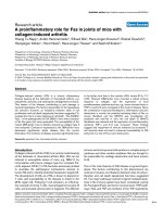
PFV-1 IN specifically cleaves the conserved palindromic sequence found at LTR-LTR junctionsFigure 4
PFV-1 IN specifically cleaves the conserved palindromic sequence found at LTR-LTR junctions. (A) The LTR-LTR junc-
tion in infected cells forms a 20 nucleotide-long bipartite palindrome. The LTR-LTR viral DNAs were PCR-amplified, cloned
and sequenced following 5-days infection of BHK-21 cells with wild type virus. The vast majority of sequences (90%) were sim-
ilar whereas approximately 10% had some divergence of the U3 junction. (B) The LTR-LTR junction is cleaved by recombinant
PFV IN. This purified IN was shown to be functional by its 3' processing activity on the blunt-ends of PFV LTR (see lanes 3 and
7, panel C) and its strand transfer activity (not shown). The U5 strand of an oligonucleotide spanning over the WT LTR-LTR
palindromic junction was labelled at its 5' extremity, annealed to its U3 complementary strand and incubated in the presence of
PFV-1 IN. Products were resolved on a 15% denaturing polyacrylamide gel. A G+A chemical sequencing reaction was run
alongside to identify the cleavage site. A specific cleavage immediately downstream of the conserved 5'CA was obtained. The
complementary strand was used for the U3 LTR-LTR junction. (C) The cleavage of the LTR-LTR junction by IN is operating on
the two strands of the palindrome leading to cohesive digestion fragments (lanes 2 and 6) indistinguishable from the products
generated by the classical 3' processing in vitro reaction on the blunt-ended LTRs (lanes 3 and 7). Cleavage products were
obtained as for panel B. 3' processing of either U5 or U3 blunt double-stranded LTRs was carried out under similar conditions
and products were run alongside to confirm the structure of the palindrome cleavage products. Lanes 2, 3, 6, 7 and 10: 150 nM
PFV-1 IN; Lanes 1, 4, 5, 8 and 9: 150 nM IN + 20 mM EDTA. EDTA was used to impair the cation-dependant activity of IN. This
digestion is highly specific of the viral palindromic sequence since a mutated palindrome (which sequence is indicated panel D)
was not cleaved by IN (lane 10). (D) A palindrome motif is required for cleavage by PFV-1 IN. Cleavage of oligonucleotides
with mutations that disrupt the palindrome motif (mutated nucleotides different from the PFV wild-type sequence are marked
with an asterisk), and with a scrambled sequence was assessed. Oligonucleotides carrying different palindromes chosen
because they correspond to LTR-LTR junctions of other retroviruses such as HIV-1 and MLV were also tested as putative sub-
strates of the PFV-1 IN. Assays were performed under the same conditions as in Fig. 3C. The ability of the IN to cleave the oli-
gonucleotides onto their two strands is indicated in the right column. The vertical arrow indicates the cleavage site of the wild-
type PFV LTR-LTR junction. These experiments were found reproducible in four independent assays.
B
C
A
a
ctive integrase:
s
ubstrate:
D
+
-
c
leavage site in the
p
alindromic LTR-LTR
j
unction
a
ctive integrase
:
A
C
T
G
A
T
T
G
A
G
T
G
G
A
A
T
G
T
A
C
C
T
A
T
(G + A)
LTR-LTR junction
in PFV-1 infected cells
U
5
U
3
AA T
A A
GAAT
AGGA-A GTGTGGTGG-ATGC
1
0 %
CAAAATTCCATGACAATTGTGGTGGAATGCCACTAGAAA
A
9
0 %
3
’ processed LT
R
(
U3 end)
3
’ processed LTR
(
U5 end)
1
2
3
4
LTR-LTR
+
-
+
-
LTR
7
8
10
+
-
LTR-LTR
+
-
LTR-LTR mutant 1
+
-
LTR
9
U
5 end
U
3 end
6
5
substrate
CAAAATTCCATGACAATTGTGGTGGAATGCCACTAGAAA
CAAAAAACGATGAGTATGTAGGTCCATTGCCACTAGAAA
CAAAATTCCATGATTATTATGGTTTAATGCCACTAGAAA
CAGAGATAGGTTTGAATGTTGTTACAGTTTGGAACAAGA
GAAAATCTCTAGCAGTACTGGAAGGGCTAATTCACTCCC
CAGCGGGGGTCTTTCATTAATGAAAGACCCCACCTGTAG
*
*
*
*
*
*
*
*
*
*
*
*
*
*
*
*
-
-
-
-
-
-
-
-
-
-
-
origin cleavage ( PFV 1 IN
)
y
e
s
n
o
n
o
n
o
n
o
n
o
P
FV-1 LTR-LTR W
T
LTR-LTR mutant
1
LTR-LTR mutant
2
S
cramble sequenc
e
H
IV-1 LTR-LTR WT
M
LV LTR-LTR W
T
Retrovirology 2005, 2:31 />Page 9 of 18
(page number not for citation purposes)
That IN operates on 2-LTR molecules to produce linear
DNA with each LTR end 3'-processed avoids the need for
asymmetrical integration in spumavirus
PFV IN was suggested to be unrelated to other retrovirus
INs because of its apparent inactivity on the U3 LTR end
of linear molecules, and the integration process of spuma-
virus was proposed to be asymmetrical [24,25]. The asym-
metric integration has been deduced from the sequences
of both integrated and 2-LTR viral molecules (Figure 5A).
The usual replication model supposes that the reverse
transcription stage leads to linear DNA with blunt-ends.
However, these ends are difficult to detect and sequence.
Their structure had been previously deduced from the
sequence at the LTR-LTR junctions. Indeed, the latter are
themselves supposed to be formed by the intramolecular
ligation between the two blunt-ends of linear DNA by an
unidentified mechanism. As only two nucleotides are lost
during integration, the PFV integration process was pro-
posed to be unusual (figure 5A).
Asymmetric integration is not required to understand the sequences of integrated and 2-LTR molecules observed in PFV-1 infected cellsFigure 5
Asymmetric integration is not required to understand the sequences of integrated and 2-LTR molecules observed in
PFV-1 infected cells. (A) The asymmetric integration in PFV-1 virus was proposed to account for the sequences of both inte-
grated and 2-LTR viral molecules as observed in the infected cells [24, 25]. This unusual proposed integration was able to solve
the problematic lost of only 2 nucleotides between U5 extremity of the integrated molecules and the putative U5 free end,
whereas the U3 end remains unchanged. This assertion was based on the following model: the linear substrate for integration
is produced by two 3'-processing reactions at each end of a blunt molecule. Of note, such blunt linear molecules have never
been detected in infected cells and their structure was deduced from the observed 2-LTR circles sequences. Such deduction is
based on the idea that 2-LTR circles result from the ligation of blunt linear DNA. However the actors of this reaction are still
unknown. (B) We propose a revised version where the PFV-1 integration remains classical. A single reaction of PFV-1 IN onto
the palindrome at the LTR-LTR circle junction can generate a linear DNA with its two 3' ends processed. The subsequent inte-
gration then eliminates the two nucleotides that are lost between the observed sequences of the LTR-LTR junction and the
integrated provirus.
A
DNA with
LTR-LTR junction
i
ntegrated DNA
v
iral integrated DNA
a
nd 2-LTR circles
(
observed
s
tructures)
U
3
U
5
U
5
U
3
a
symmetric viral
D
NA (proposed
s
tructure)
2
-nt lost
TGT ACA
ACA TGTTA
U
3
U
5
ACAAT TGT
TGTTA ACA
TGT ACA
ACA TGT
l
inear DNA
b
lunting and
l
igation
integration
a
symmetric 3’-processing (IN
)
b
lunt viral DNA, sequence deduced
f
rom observed integrated and 2-LTR
j
unctions
B
s
ymmetric 3’ processing
i
ntegrated DNA
U
3
U
5
TGT ACA
ACA TGT
c
lassical integration
v
iral DNA 3’-processed at eac
h
L
TR: sequence deduced from th
e
I
N-cleavage of 2-LTR molecules
2
-nt lost
TGT ACAAT
ACA TGTTA
I
N
ACAATTGT
TGTTAACA
DNA with
LTR-LTR junction
U
5
U
3
I
N
I
N
ATTGT ACA
ACA TGTTA
r
esulting from LTR-LTR
c
ircle junction cleavage (I
N)
Retrovirology 2005, 2:31 />Page 10 of 18
(page number not for citation purposes)
In light of our observation that 2-LTR molecules are pos-
sible substrates for PFV-1 IN (Figure 4), the 3'-processing
of both ends of the linear DNA might be generated in a
single reaction that produces the two 3'-processed ends
simultaneously (Figure 5B). Such concerted processing
might explain the influence of one LTR on the processing
of the other, as observed for HIV-1 [20]. The subsequent
integration of such processed extremities would eliminate
the two nucleotides that are lost between the LTR-LTR
junction and the integrated provirus. No asymmetric inte-
gration is required to account for the previous observa-
tions [24,25]. This mechanic, when generalized to other
retroviruses carrying a different palindrome at the LTR-
LTR junction, would result during integration in the loss
of the number of nucleotides comprised between the con-
served CA.
In support of our symmetrical integration model, Pahl
and Flügel [26] previously reported an efficient 3'-process-
ing activity of PFV IN on LTR containing the two addi-
tional nucleotides AT. The substrate of concerted
processing corresponds to the extended substrate they
tested. We confirmed the 3'-processing cleavage of the
extended U3 LTR carrying an additional AT (Figure 4C), as
well as the fact that the 3'-processing does not occur onto
the shorter U3 LTR lacking these nucleotides (not shown).
Integration depends on preintegrative IN activity
Integration was reported to be a very rare event in spuma-
viruses [87,88], except in chronically infected cell situa-
tions [89]. To document this point in our conditions, we
quantified the integration events for PFV-1 WT and IN
mutants. To this end, we designed a highly sensitive quan-
titative real-time RACE-PCR reaction, amplifying Alu-LTR
junctions between the cell genome and integrated provi-
ruses (detecting 25 integrated proviruses per 50 000 cells,
Figure 6A). U373-MG cells were infected with equivalent
amounts of viral particles as measured by RT activity and
the quantity of integrated viral molecules was analyzed 24
hours later, a time-point at which the first round of infec-
tion is achieved. As shown in Figure 6A, and as expected
[87,88], only a small fraction of total wild-type PFV DNA
was integrated (range of 0.9–2.1%). The M8 and M9
mutant INs used in our study failed to integrate oligonu-
cleotides mimicking the PFV LTR DNA ends into a target
plasmid in vitro [26]. We therefore assessed the ability of
viruses carrying the same IN mutations to integrate in vivo.
We could detect integrated DNA after infection with
viruses carrying inactive INs (Figure 6B upper panel).
However, with the exception of the semi-replicative M9
virus, IN mutants yielded significantly fewer integrated
proviruses than the wild-type (Figure 6B). Similar obser-
vations have been reported in cells infected with IN-defec-
tive HIV and the presence of integrated proviruses was
attributed to integrase-independent integration events
depending on cell enzymes [81]. Another explanation
could rely on the fact that IN mutants produced less linear
DNA as a substrate for integration. The altered viral DNA
production is likely reflected by the reduced amounts of
total viral DNA quantified in the same extracts (Figure 6B
lower panel). We compared integration ratios with and
without functional IN by normalizing integrated provi-
ruses values with the total number of viral DNA copies
present in infected cells. Strikingly, the percentage of inte-
grated DNA was not modified by the presence of a defec-
tive IN (Figure 6C). Thus, the level of integrated provirus
depends on the global viral DNA pool available in the
infected cells. And such global viral DNA content itself
depends on the early activity of the viral IN as shown
above.
Role of IN in PFV retrovirus replication cycle
We conclude from these experiments that PFV IN displays
a specific activity on the 2-LTR circles, which may consti-
tute a substrate for the 3'processing reaction in vivo. This
action of IN generates linear DNA that might be then inte-
grated in the cell genome following a classical symmetri-
cal integration process. The fact that early actions of IN
may influence later steps of replication, including integra-
tion, certainly participates in the pleiotropic effects of IN
mutations. Finally, IN seems to be essential not because of
its participation to the integration per se but for its
upstream activities able to influence integration efficacy.
Our findings that a loss of endonuclease IN activity results
in both LTR-LTR accumulation and an associated reduc-
tion in viral DNA production leads us to propose a direct
role for retroviral integrase in the production of viral
DNA. Thus, a modified replication model is presented in
Fig. 7B. It is accepted that the encounter between viral
DNA and IN occurs very shortly after viral DNA synthesis,
since cytoplasmic viral DNA is mostly found as linear
molecules with 3' processed ends resulting from IN endo-
nucleolytic action in the cytoplasm [13-15]. In our model,
DNA molecules containing LTR-LTR junction would be
generated during the reverse transcription process and
cleaved rapidly by the IN, leading to the production of lin-
ear DNA harboring 3'-processed ends. This would account
for the rarity of linear DNA with blunt ends in the cyto-
plasm of infected cell, as well as for the presence of 2-LTR
circles in the cytoplasm of retrovirus infected cells at early
times post infection [40,41]. Additionally, it would
explain the data from att site mutagenesis experiments
showing that mutation of one LTR precludes the process-
ing of the other LTR [20]. These results were initially inter-
preted to represent a concerted activity of IN on the two
viral LTRs ends that must be simultaneously cleaved in
infected cells. In view of our results, these data might be
understood as resulting from the endonucleolytic activity
of IN on palindromic LTR-LTR junctions. Such processed
Retrovirology 2005, 2:31 />Page 11 of 18
(page number not for citation purposes)
DNA could then undergo integration. In this interpreta-
tion, a unique endonucleolytic action of IN at an early
step would explain many of the phenotypes associated
with IN mutations, including the increasing abundance of
2-LTR molecules at the expense of linear and integrated
DNA in IN-defective viruses. It underlines that in vivo inte-
gration is performed in two steps that are uncoupled both
in time and in space, ie 3' processing in the cytoplasm and
Integration of IN-defective virusesFigure 6
Integration of IN-defective viruses. (A) A quantitative assay based on a real-time RACE-PCR reaction was designed, amplify-
ing Alu-LTR junctions between the cell genome and integrated proviruses twenty-four hours post-infection. PCR amplifications
of existing Alu-PFV-1 LTR junctions were subjected to a second quantitative round of real time PCR with PFV-1 LTR-specific
primers. Fluorogenic hybridization probes were used to quantify the amplification products. Infected cells with known copy
numbers of integrated proviruses were used as quantification standards. The assay is highly sensitive since it allows detecting
25 proviruses copies in 50,000 human cells. Control reactions are detailed in the Material and methods section. (B) Detection
of integrated viral DNA following infection of IN-mutated viruses. Quantitation of viral DNA accumulated in PFV-1 infected
cells was carried out by real-time PCR of total DNA extracts from U373-MG infected cells (m.o.i. of 1) collected at the com-
pletion of the first viral replication cycle, 24 hours post-infection. Total viral DNA (gag quantifications) and integrated provi-
ruses were quantified in duplicate using real-time PCRs. Data obtained in one representative infection from four independent
experiments are expressed as integrated DNA copies per million cells (logarithmic scale) as determined by a human β-globin
quantification in cell extracts ("Integrated provirus" panel). Total DNA copies per million cells (logarithmic scale) present in the
same extracts are presented in the lower panel. Standard deviations representing variations between two quantifications of the
same sample are given. (C) Integration efficiency in PFV-1 infected cells. Integration efficiency was determined by normalizing
the number of integrated proviruses (mean of duplicates) with the total number of viral DNA molecules (mean of duplicates)
present in the same extract. Raw LightCycler data from four independent experiments are presented in the upper table. Mean
of integration efficiencies from these four experiments are figured in the lower histogram.
Exp#1
integrated
copies
total viral
copies
integration
efficiency
100
90
10 869
9 977
0.91 %
Exp#2
250
186
19 700
16 938
1.19 %
Exp#3
195
178
17 964
18 766
1.02 %
Exp#4
120
156
6 652
6 423
2.10 %
1.32 % – 0.39
Mean
WT
integrated
copies
total viral
copies
integration
efficiency
55
42
2 350
1 820
2.33 %
23
20
2 026
2 298
0.99 %
31
27
3 409
3 421
0.85 %
31
37
3 024
3 189
1.09 %
M5
integrated
copies
total viral
copies
integration
efficiency
34
46
1 388
1 080
3.23 %
12
15
1 431
1 324
0.98 %
28
23
3 299
2 719
0.85 %
48
45
3 724
3 581
1.27 %
M8
integrated
copies
total viral
copies
integration
efficiency
86
75
3 161
2 744
2.72 %
53
42
4 950
4 697
0.99 %
102
89
6 993
7 655
1.30 %
105
117
5 460
5569
2.01 %
M9
virus:
1.32 % – 0.51 1.58 % – 0.82 1.75 % – 0.61
C
In vivo integration efficiency
W
T
M
5
M
8
M
9
integrated DNA content
relative to total viral DNA (%)
0
0,5
1
1,5
2
2,5
S
econd round PCR of integrated provirus
a
nd quantification
P
reamplification of Alu-spumavirus junction
s
λ
λ
p
rime
r
U
3 prime
r
λ
A
lu
A
lu
h
ybridization probe
s
Quantification of integrated proviruses
by real-time PCR
B
log copies per 10
6
cells
Integrated provirus
1
00
1
000
1
0 000
W
T
M
5
M
8
M
9
Total virus
logcopies per 10
6
cells
1
0 000
1
00 000
1
000 000
W
T
M
5
M
8
M
9
A
Retrovirology 2005, 2:31 />Page 12 of 18
(page number not for citation purposes)
integration per se in the nucleus. It also illustrates why and
how certain in vitro integration-defective viruses such as
our M9 mutant or HIV mutants [39,51,76] are still repli-
cative. The IN activity demonstrated in this report allows
processing the circles – currently considered as dead-end
molecules- into the replication pathway. Additional sup-
port to this conclusion is present in the HIV literature
where episomal circular DNA were shown to turn over by
degradation rather than through death or tissue redistri-
bution of the infected cell itself in HIV-1 infected individ-
uals [42]. Finally, our data imply that circular retroviral
genomes are fully functional replication intermediates,
first as substrates for transcription and second as precur-
sors of linear unintegrated DNA.
Although the consensus sequences in the C ter region of
IN may differ between the lentiviruses and the nonlentivi-
ruses, the carboxyterminal region of IN is well conserved
in all retroviruses [80], and further studies are now
required to evaluate whether the revised replication
model we propose here, applies to all retroviruses. The
fact that the typical phenotype associated with a defective
IN, either due to mutations or inhibitors, resulting in
reduced DNA synthesis but a persistence of integration
and an accumulation of 2-LTR molecules, is commonly
observed among retroviruses [73,82,90], argues in favour
of a conserved IN function. Such an early participation of
IN sheds new light on reports showing both that viral
transcription occurs from nonintegrated HIV DNA
[38,44,45,91], and that the most prevalent form of HIV
DNA during the asymptomatic phase of infection is full-
length unintegrated DNA [42,92]. Whereas IN activity is
clearly required, formation of integrated provirus as an
obligate step of retroviral replication now needs to be
reconsidered. On the other hand, early preintegrative
activities of IN are of capital importance. This provides
new answers to the puzzling question of why is
integration essential to retrovirus replication, when many
authors have shown that unintegrated genomes are abun-
dant and expressed [36-39,42-45,93]. Our proposal is
simply: integrase is essential, integration is not; and IN is
required given its critical preintegrative influence on
genomic DNA production in vivo, as we precisely meas-
ured here.
Given the above, retroviruses better fit the classical
schemes of distinct lytic and lysogenic phases exemplified
by the lambda phage: integration (lysogeny) contributes
to viral persistence and pathogenesis, but it is not essential
for acute viral production (lytic cycle). Finally, a fascinat-
ing evolutionary conservation appears between retrovi-
ruses and DNA viruses (such as poxviruses). All use
circular DNA intermediates and a specialized endonucle-
ase activity for genome production.
Methods
Cells, virus infections and reagents
BHK-21, FAB, HeLa and U373-MG cells were cultivated in
DMEM with 10% foetal calf serum, 1 µg per ml of strepto-
mycine-streptavidine. For FAB indicator cells, 1 µg per ml
of G418 (Sigma) was added.
PFV-1 virus stocks were prepared by transfecting BHK-21
cells with the PFV-1 molecular WT and mutant clones
using the calcium phosphate method. Cells were infected
by WT and mutant viruses with same amounts of viral par-
ticles, as evaluated by a reverse transcription assay. The
culture medium was changed two hours post-infection
with fresh medium.
Cell free virus stocks were titrated on FAB cells [75]. In
some experiments, infected cells were treated with 3'-
azido-3'-deoxythymidine (AZT, Sigma) at 100 µM.
DNA quantifications by real time PCR
Total DNAs were extracted from 10
6
cells using the DNA
Blood Mini kit (Qiagen) in a final volume of 200 µl and
analysed by real time PCR as described previously [40].
Integrated viral DNA was also quantified by two rounds of
PCR [94]. The first one amplifies integrated DNA using
primers ALU1 (5'-CCT CAG CCT CCC GAG TAG CTG
GGA-3'), ALU2 (5'-CTG TAA TCC CAG CAC TTT GGG
AGG C-3'), and λ TSPA (5'-ATG CCA CGT AAG CGA AAC
TTA GTA TAA TCA TTT CCG CTT TCG-3'). Sequence in
bold represents a sequence in the lambda phage, which is
unknown in all mammals' databanks. The other part of
the sequence of λ TSPA primer can hybridize in PFV LTR.
Amplification was performed in a 20 µl reaction volume
containing 1X Light Cycler Fast Start DNA Hybridation
probes, 3.5 mM MgCL
2
, 300 nM of primer ALU1, ALU2
and 10 nM of primer λ TSPA. The same mix, containing
only primer λ TSPA, was prepared. DNA from U373-MG
chronically infected cells was used as a standard for inte-
grated copies. All reactions were further diluted in a final
volume of 200 µl of water. 2 µl over 200 µl was used for
the second PCR. This amplification was performed with
300 nM of each primers Nested R (5'-GAA ACT AGG GAA
AAC TAG G-3'), lambdaT (5'-ATG CCA CGT AAG CGA
AAC T-3') and 100 nM of each hybridation probes SpuFL
(5'-CAC TCT CGA CGC AGC GAG TAG TGA A X-3') and
SpuLC (5'-GCC TCC CGT ACA ATC TAG AAA CTA TCC T
p-3'). This assay is quite specific of integrated provirus
only, as attested by performing the following control reac-
tions: – a carry-over control in which all primers were
omitted in the first PCR, data obtained indicated always
that the second-round amplification of nonpreamplified
viral DNA is efficiently prevented; -a parallel reaction with
the Alu primers in the first-round PCR, in order to calcu-
late the linear amplifications resulting from all the viral
DNA species. The copy number due to the linear
Retrovirology 2005, 2:31 />Page 13 of 18
(page number not for citation purposes)
Role of IN in retrovirus replication cycleFigure 7
Role of IN in retrovirus replication cycle. (A) Classical model of early steps in retrovirus replication. IN plays a role in the 3'
processing as well as in the integration itself, these two steps being separated both in time and in space. Following synthesis of
linear blunt-ended DNA in the cytoplasm (step 1 in Fig. 7A), IN cleaves their 3' termini, thus eliminating the terminal two bases
from each 3'end (step 2). The resulting recessed 3'OH groups provide the attachment sites of the provirus to host DNA, an
attachment which is performed only after import of 3'processed DNA into the nucleus where the final step of the integration
process occurs (step 3). Circular DNA carrying LTR-LTR junctions are reportedly formed from linear DNA via the action of
cellular ligases (step 4). The circularization is considered to be an alternate fate of linear DNA that has not integrated, and may
indirectly explain why DNA bearing LTR-LTR junctions accumulates to high levels in cells harboring integration-defective
viruses. This classical model considers that functions of IN in processes other than integration are secondary. (B) Alternate
retrovirus replication model. IN cleaves the LTR-LTR junction generated at the reverse transcription step (step 1) to produce
3'end-processed linear DNA (step 2). This specific activity of the IN explains the pleiotropic effects of this protein and the phe-
notypes associated with its mutagenesis. First, since linear DNA is the direct product of a reaction that is catalyzed by IN, its
levels would decrease under IN-defective conditions. Moreover if LTR-LTR junction molecules indeed constitute the substrate
for IN, their amount would increase as a direct consequence of defective IN. Second, decreased levels of integrated proviruses
would be an indirect result of the decreased pool of 3'processed IN-catalyzed linear DNA molecules that are available for inte-
gration (step 3). In this model, 2-LTR molecules are a replication-intermediate. Low levels of these molecules would be due to
their rapid processing by IN in the wild-type infections. Rapid processing might also explain the presence of linear molecules
with 3' processed ends in the cell cytoplasm during diverse retroviral infections, even though no blunt-ended linear molecules
can be recovered from infected cells. Thus, apart from participating in retroviral DNA integration per se, IN would act
upstream by controlling linear DNA production. This function of IN, as included in the modified replication model presented
here, provides a parsimonious interpretation of the pleiotropic effects observed in cells infected with IN mutants.
A
B
I
N
reverse
transcription
3’ processing
l
inear DNA
3
’ processed
i
ntegrated DNA
strand transfer
l
inear DNA
c
ell enzyme
DNA with
LTR-LTR junction
reverse
transcription
DNA with
LTR-LTR junction
IN
palindrome
cleavage
l
inear DNA
3
’ processed
i
ntegrated DNA
I
N
(
1
)
(
2
)
(
3
)
(
4
)
(
1
)
(
2
)
(
3
)
c
ell enzyme
(
4
)
R
NA
R
NA
IN
Retrovirology 2005, 2:31 />Page 14 of 18
(page number not for citation purposes)
amplification was systematically subtracted from the sig-
nal obtained in the presence of Alu primer. We evaluated
that this interfering amplification never exceeded 6.7 % of
the global amplification.
Quantifications were performed with the LightCycler soft-
ware Version 3.5 according to manufacturer's instructions.
Virion-associated RT assays
48 hours post transfection viral supernatants were col-
lected. 10 µl of viral supernatant was incubated with 20 µl
of reaction buffer (Tris pH 8 50 mM – KCl 75 mM – Dithi-
otreitol 2 mM – rA/dT 25 µg/ml – NP40 0,05% – MnCl
2
5
mM – dTTP α-
32
P 20 µCi/ml). The reaction mixtures were
incubated at 37°C for 90 min. 10 µl of the reaction was
spotted onto DE81 filter and allowed to dry. The filters
were washed four times with 2xSSC (1xSSC is 0.15 M
NaCl plus 0.015 M sodium citrate) for 5 min each, fol-
lowed by two washes with 95% ethanol. The filters were
then dried and counting by scintillation fluid.
Construction of Flag-PFV IN mutants and their cell
localisation by immunofluorescence staining
To express the INs in the absence of other viral products,
we used the pFlag expression vector [95]; in which we
inserted the PFV-1 IN sequence under the control of the
simian virus 40 promoter. The IN fragment was amplified
by PCR with the following primers, which created a
BamH1 and an XhoI restriction site at the 5' and 3' ends,
respectively, of the IN sequence: 5'-GGA TCC TAC ATA
TTT TTT AGA AGA TGG C-3'; and 5'-CTC GAG TTA TTC
ATT TTT TTC CAA TGA TCC-3'. The resulting PCR frag-
ment was digested with BamHI and XhoI and ligated into
the corresponding cloning sites of pSG-Flag [95], in the
plasmid called pSG-FlagIN PFV. The pSG-FlagIN PFV
expression vector was used for the mutagenesis, with the
Quick Change mutagenesis kit (Stratagene), and the
primers: 5'-CAA TTT GGC TCT CAC AGG ACG TGA AGC
C-3' and 5'-GGC TTC ACG TCC TGT GAG AGC CAA ATT
G-3' for the M5 mutant; 5'-ATT CAC TCT GGT CAA GGT
GCA GC-3' and 5'-GCT GCA CCT TGA CCA GAG TGA AT-
3' for the M8 mutant; and 5'-GGC AAA GGG CCA GTA
TAG TCA AT-3' and 5'-ATT GAC TAT ACT GGC CCT TTG
CC-3' for the M9 mutant.
HeLa cells (2 × 10
5
) were spread on glass coverslips in 24-
well plates, transfected with 1 µg of the corresponding
plasmids, and stained for immunofluorescence 36 hours
later. Cells were fixed in 3.7% formaldehyde-PBS for 20
min, washed three times in PBS, and incubated for 10 min
in 50 mM NH
4
Cl to quench free aldehydes. Cells were
washed three times in PBS and incubated in a
permeabilization buffer (0.05% saponin, 0.01% Triton X-
100, 2% bovine serum albumin, PBS) for 15 min and
incubated 1 h with the first MAb (M2 anti-Flag MAb at 7.5
µg/ml) in permeabilization buffer. Cells were washed
three times in permeabilization buffer and incubated with
Cy3-conjugated anti-mouse MAbs (Amersham) at a final
dilution of 1:200. Cells were washed three times in per-
meabilization buffer and once in PBS and mounted in
133 mg of Mowiol (Hoechst) per ml-33% glycerol-133
mM Tris HCl (pH 8.5). Confocal microscopy was per-
formed and optical sections were recorded. One repre-
sentative medial section was mounted by using Adobe
Photoshop software.
Construction of PFV proviruses
We inserted a DNA fragment containing the PFV-1 IN
sequence into a Litmus 38 plasmid, in which a PacI site
had been added. The viral fragment was amplified by PCR
with the following primers: 5'-GGA TCC TAC ATA TTT
TTT AGA AGA TGG C-3' and 5'-CTC GAG TTA TTC ATT
TTT TTC CAA TGA TCC-3', and cloned after a BspEI-PacI
digestion into the modified Litmus. This plasmid contain-
ing the WT IN was used for the mutagenesis, with the
Quick Change mutagenesis kit and the primers used
above for the expression IN vector mutagenesis. After the
mutagenesis, the PacI-BspEI digestion fragments from the
mutated Litmus vectors were substituted for the corre-
sponding sequence of the PFV-1 full-length clone. All con-
structions were confirmed by DNA sequencing of the
entire PCR-amplified fragment.
2 LTR junction sequence analysis
Total DNA from acutely BHK-21 infected cells of two
independent infections were extracted and analyzed by a
PCR amplification specific for the LTR-LTR junction from
the 2-LTR circles, using the following primers: R, 5'-TAC
GAG ACT CTC CAG GTT TG-3'; and U3, 5'-CGA CGC
AGC GAG TAG TGA AG-3' and the Pfu polymerase (Strat-
agene) [40]. PCR products were cloned in a pSK+ plasmid
(PCR-Script cloning kit, Stratagene). 50 independent
cloned were sequenced.
Construction and purification of PFV recombinant IN
Histidine-tagged PFV-1 IN, corresponding to aminoacids
752-1143 of the Pol polyprotein, was expressed and
purified by nickel affinity. The preparation and purifica-
tion of recombinant PFV-1 IN protein were performed as
described for HIV IN [96]. To obtain wild type IN protein,
plasmid pET15b (Novagen) was digested with NdeI and
BamHI. The DNA fragment containing the PFV IN was
obtained from pHSRV clone C55 by PCR using the Pfu
DNA polymerase (Stratagene). The sequence of the prim-
ers used to amplify the fragment were 5'-ACA TAT GTG
TAA TAC CAA AAA ACC AAA CCT GG-3' and 5'-AGG ATC
CTT ACT CGA GTT CAT TTT TTT C-3'. PCR amplifications
were done at 92°C for 1 min, 55°C for 45 s, and at 72°C
for 90 s; the cycle was repeated 28 times. The resulting
PCR fragment were digested with NdeI and BamHI and
Retrovirology 2005, 2:31 />Page 15 of 18
(page number not for citation purposes)
ligated into the corresponding cloning sites of pET15b.
Plasmid pET15bIN was used to express the His-tagged IN
in E. coli BL21 (DE3) cells. 500 ml of BL21 (DE3)
pET15bIN cells was grown at 37°C in LB medium (sup-
plemented with 50 mg/ml ampicilin) to an A
600
of 0.6–
0.8. To induce IN protein expression, isopropyl-1-thio-β-
D-galactopyranoside was added to a final concentration
of 1 mM; bacteria were grown for another 4 hours and
harvested by low speed centrifugation. The pellet was
resuspended in 24 ml of 50 mM Tris-HCl, pH8, 1 M NaCl,
4 mM β-mercaptoethanol (buffer A). Cells were lysed with
French Press and centrifugated at 14,000 rpm and 4°C for
30 min to remove cells debris
The supernatant was filtered (0.45 µm) and incubated
over night with Ni-NTA agarose beads (Qiagen). The
beads were washed with 10 volumes of buffer A. Then, IN
was purified under native conditions according to manu-
facturer's instructions using batch procedure. His-tagged
IN was eluted with buffer A supplemented with 50 µM
ZnSO
4
and 1 M imidazole. The IN concentration was
adjusted to 0.1 mg/ml in buffer A and dialysed over night
against 20 mM Tris-HCl, pH 8, 1 M NaCl, and 4 mM β-
mercaptoethanol. Fractions were aliquoted and rapidly
frozen at -80°C.
Nucleic acid substrates
All oligonuleotides U5B (5'-CCT TAG GAT AAT CAA TAT
ACA AAA TTC CAT GAC AAT-3'), (U5A 5'-ATT GTC ATG
GAA TTT TGT ATA TTG ATT ATC CTA AGG-3'), U3 B (5'-
ATT GTG GTG GAA TGC CAC TAG AAA T-3'), U3A (5'-
ATT TCT AGT GGC ATT CCA CCA CAA T-3'), LTR-LTRB
(5'-CCT TAG GAT AAT CAA TAT ACA AAA TTC CAT GAC
AAT TGT GGT GGA ATG CCA CTA GAA AT-3') and LTR-
LTRA (5'-ATT TCT AGT GGC ATT CCA CCA CAA TTG TCA
TGG AAT TTT GTA TAT TGA TTA TCC TAA GG-3') were
purchased from Eurogentec and further purified on an
15% denaturing acrylamide/urea gel. 100 pmol of U5 B,
U3 B and LTR-LTR B were radiolabeled using T4 polynu-
cleotide kinase and 50 µCi of [γ-
32
P]ATP (3000 Ci/mmol)
during 2 hours at 37°C. The T4 kinase was heat inacti-
vated, and unincorporated nucleotides were removed
using a Sephadex G-10 column (Pharmacia). NaCl was
added to a final concentration of 100 mM and comple-
mentary unlabeled strand was added to either U5 B, U3 B
or LTR-LTR B. The mixture was heated to 90°C for 3 min,
and the DNA was annealed by slow cooling.
LTR processing, LTR-LTR junction cleavage
Processing and LTR-LTR cleavage were performed in
buffer containing 50 mM Hepes, 5 mM DTT and 10 mM
MgCl
2
. 150 nM of PFV-1 IN was used for reaction. The
reaction was initiated by addition of substrate DNA, and
the mixture was incubated 2 hours at 37°C and stopped
by phenol/chloroform extraction. DNA products were
precipitated with ethanol, dissolved in TE containing 7 M
urea and electrophoresed on a 15% denaturing acryla-
mide/urea gel. Gels were analysed using a STORM Molec-
ular Dynamics phosphorimager.
List of abbreviations
Att, attachment site
HIV, human immunodeficiency virus
IN, integrase
LTR, long terminal repeat
PFV, primate foamy virus
PIC, preintegration complex
RT, reverse transcriptase
WT, wild-type
Authors' contributions
OD carried out all the experiments concerning the pheno-
type analysis of the viruses in the cell context including
constructions, viral kinetics and real-time PCR, and partic-
ipated to the analysis of the data. CP contributed to the
design and coordination of the study, supervised the
experimental work, participated in the analysis and inter-
pretation of the data, and drafted figures and the manu-
script. HL participated in the acquisition of the
biochemical datas and in their interpretation. GM con-
tributed to the acquisition of biochemical datas. JFM con-
tributed and supervised biochemical analysis of integrase
in vitro. PS conceived the original ideas, designed and
coordinated the study, and took part in writing the man-
uscript. All authors read and approved the final
manuscript.
Acknowledgements
We warmly acknowledge Olivier Neyrolles, Sebastien Petit and the OCU
for stimulating remarks and daily help. We are grateful to William Jacques
Speare and Alexandre Matet for their corrections and for continued enthu-
siastic discussion regarding this research. We also thank Marc Alizon,
Olivier Danos and Olivier Schwartz for stimulating and thoughtful com-
ments, and constructive criticisms on the manuscript. We finally thank
Naomi Taylor and Marc Sitbon for insightful discussions concerning the ret-
rovirus replication models, as well as for their meticulous reading of our
original manuscript.
References
1. Rice P, Craigie R, Davies DR: Retroviral integrases and their
cousins. Cur Opin in Struct Biol 1996, 6(1):76-83.
2. Brown PO, Bowerman B, Varmus HE, Bishop JM: Correct integra-
tion of retroviral DNA in vitro. Cell 1987, 49:347-356.
3. Brown PO, Bowerman B, Varmus HE, Bishop JM: Retroviral inte-
gration: structure of the initial covalent product and its pre-
Retrovirology 2005, 2:31 />Page 16 of 18
(page number not for citation purposes)
cursor, and a role for the viral IN protein. Proc Natl Acad Sci USA
1989, 86(8):2525-2529.
4. Brown DP, Idler KB, Katz L: Characterization of the genetic ele-
ments required for site-specific integration of plasmid
pSE211 in Saccharopolyspora erythraea. J Bacteriol 1990,
172(4):1877-1888.
5. Fujiwara T, Mizuuchi K: Retroviral DNA integration : structure
of an integration intermediate. Cell 1988, 54:497-504.
6. Lee YMH, Coffin JM: Relationship of avian retrovirus DNA syn-
thesis to integration in vitro. Mol Cell Biol 1991, 11:1419-1430.
7. Bowerman B, Brown PO, Bishop JM, Varmus HE: A nucleoprotein
complex mediates the integration of retroviral DNA. Genes
& Development 1989, 3(4):469-478.
8. Lobel LI, Murphy JE, Goff SP: The palindromic LTR-LTR junction
of Moloney murine leukemia virus is not an efficient sub-
strate for proviral integration. J Virol 1989, 63(6):2629-2637.
9. Ellis J, Bernstein A: Retrovirus vectors containing an internal
attachment site: evidence that circles are not intermediates
to murine retrovirus integration. J Virol 1989, 63(6):2844-2846.
10. Hindmarsh P, Leis J: Retroviral DNA integration. Microbiol Mol
Biol Rev 1999, 63(4):836-43, table of contents
11. Turlure F, Devroe E, Silver PA, Engelman A: Human cell proteins
and human immunodeficiency virus DNA integration. Front
Biosci 2004, 9:3187-3208.
12. Engelman A, Mizuuchi K, Craigie R: HIV-1 DNA integration:
mechanism of viral DNA cleavage and DNA strand transfer.
Cell 1991, 67(6):1211-1221.
13. Chen H, Engelman A: Characterization of a replication-defec-
tive human immunodeficiency virus type 1 att site mutant
that is blocked after the 3' processing step of retroviral
integration. J Virol 2000, 74(17):8188-8193.
14. Chen H, Engelman A: Asymmetric processing of human immu-
nodeficiency virus type 1 cDNA in vivo: implications for func-
tional end coupling during the chemical steps of DNA
transposition. Mol Cell Biol 2001, 21(20):6758-6767.
15. Miller MD, Farnet CM, Bushman FD: Human immunodeficiency
virus type 1 preintegration complexes: studies of organiza-
tion and composition. J Virol 1997, 71(7):5382-5390.
16. Roth M, Schwartzberg P, Goff SP: Structure of the termini of
DNA intermediates in the integration of retroviral DNA. Cell
1989, 58:47-54.
17. Daniel R, Katz RA, Skalka AM: A role for DNA-PK in retroviral
DNA integration. Science 1999, 284(5414):644-647.
18. Yoder KE, Bushman FD: Repair of gaps in retroviral DNA inte-
gration intermediates. J Virol 2000, 74(23):11191-11200.
19. Katzman M, Katz RA: Substrate recognition by retroviral
integrases. Adv Virus Res 1999, 52:371-395.
20. Murphy JE, Goff SP: A mutation at one end of Moloney murine
leukemia virus DNA blocks cleavage of both ends by the viral
integrase in vivo. J Virol 1992, 66(8):5092-5095.
21. Yu SF, Baldwin DN, Gwynn SR, Yendapalli S, Linial ML: Human
foamy virus replication: a pathway distinct from that of ret-
roviruses and hepadnaviruses. Science 1996,
271(5255):1579-1582.
22. Yu SF, Sullivan MD, Linial ML: Evidence that the human foamy
virus genome is DNA. J Virol 1999, 73(2):1565-1572.
23. Moebes A, Enssle J, Bieniasz PD, Heinkelein M, Lindemann D, Bock M,
McClure MO, Rethwilm A: Human foamy virus reverse tran-
scription that occurs late in the viral replication cycle. J Virol
1997, 71(10):7305-7311.
24. Enssle J, Moebes A, Heinkelein M, Panhuysen M, Mauer B, Schweizer
M, Neumann-Haefelin D, Rethwilm A: An active foamy virus inte-
grase is required for virus replication. J Gen Virol 1999, 80(Pt
6):1445-1452.
25. Juretzek T, Holm T, Gartner K, Kanzler S, Lindemann D, Herchen-
roder O, Picard-Maureau M, Rammling M, Heinkelein M, Rethwilm A:
Foamy virus integration. J Virol 2004, 78(5):2472-2477.
26. Pahl A, Flugel RM: Endonucleolytic cleavages and DNA-joining
activities of the integration protein of human foamy virus. J
Virol 1993, 67(9):5426-5434.
27. Pahl A, Flugel RM: Characterization of the human spuma ret-
rovirus integrase by site- directed mutagenesis, by comple-
mentation analysis, and by swapping the zinc finger domain
of HIV-1. J Biol Chem 1995, 270(7):2957-2966.
28. Boyer PL, Ferris AL, Hughes SH: Cassette mutagenesis of the
reverse transcriptase of human immunodeficiency virus type
1. J Virol 1992, 66(2):1031-1039.
29. Hong T, Drlica K, Pinter A, Murphy E: Circular DNA of human
immunodeficiency virus : analysis of circle junction nucle-
otide sequences. J Virol 1991, 65:551-555.
30. Russell RA, Critchley R, Vassaux G, McClure MO: Human foamy
virus integrase fails to catalyse the integration of a circular
DNA molecule containing an LTR junction sequence. Gene
Ther 2002, 9(19):1326-1332.
31. Duyk G, Longiaru M, Cobrinik D, Kowal R, deHaseth P, Skalka AM,
Leis J: Circles with two tandem long terminal repeats are spe-
cifically cleaved by pol gene-associated endonuclease from
avian sarcoma and leukosis viruses: nucleotide sequences
required for site-specific cleavage. J Virol 1985, 56(2):589-599.
32. Panganiban AT, Temin HM: Circles with two tandem LTRs are
precursors to integrated retrovirus DNA. Cell 1984,
36(3):673-679.
33. Shoemaker C, Goff S, Gilboa E, Paskind M, Mitra SW, Baltimore D:
Structure of a cloned circular Moloney murine leukemia
virus DNA molecule containing an inverted segment: impli-
cations for retrovirus integration. Proc Natl Acad Sci U S A 1980,
77(7):3932-3936.
34. Li L, Olvera JM, Yoder KE, Mitchell RS, Butler SL, Lieber M, Martin SL,
Bushman FD: Role of the non-homologous DNA end joining
pathway in the early steps of retroviral infection. Embo J 2001,
20(12):3272-3281.
35. Luciw PA, Oppermann H, Bishop JM, Varmus HE: Integration and
expression of several molecular forms of Rous sarcoma virus
DNA used for transfection of mouse cells. Mol Cell Biol 1984,
4(7):1260-1269.
36. Brussel A, Sonigo P: Evidence for gene expression by uninte-
grated human immunodeficiency virus type 1 DNA species.
J Virol 2004, 78(20):11263-11271.
37. Cara A, Cereseto A, Lori F, Reitz MSJ: HIV-1 protein expression
from synthetic circles of DNA mimicking the extrachromo-
somal forms of viral DNA. J Biol Chem 1996, 271(10):5393-5397.
38. Engelman A, Englund G, Orenstein JM, Martin MA, Craigie R: Multi-
ple effects of mutations in Human Immunodeficiency Virus
type 1 integrase on viral replication. J Virol 1995,
69(5):2729-2736.
39. Wiskerchen M, Muesing MA: Human Immunodeficiency Virus
type 1 integrase: effects of mutations on viral ability to inte-
grate, direct viral gene expression from unintegrated viral
DNA templates, and sustain viral propagation in primary
cells. J Virol 1995, 69(1):376-386.
40. Delelis O, Saib A, Sonigo P: Biphasic DNA synthesis in
spumaviruses. J Virol 2003, 77(14):8141-8146.
41. Serhan F, Penaud M, Petit C, Leste-Lasserre T, Trajcevski S, Klatz-
mann D, Duisit G, Sonigo P, Moullier P: Early detection of a two-
long-terminal-repeat junction molecule in the cytoplasm of
recombinant murine leukemia virus-infected cells. J Virol
2004, 78(12):6190-6199.
42. Sharkey M, Triques K, Kuritzkes DR, Stevenson M: In vivo evidence
for instability of episomal human immunodeficiency virus
type 1 cDNA. J Virol 2005, 79(8):5203-5210.
43. Vargas JJ, Gusella GL, Najfeld V, Klotman ME, Cara A: Novel inte-
grase-defective lentiviral episomal vectors for gene transfer.
Hum Gene Ther 2004, 15(4):361-372.
44. Wu Y, Marsh JW: Selective transcription and modulation of
resting T cell activity by preintegrated HIV DNA. Science
2001, 293(5534):1503-1506.
45. Wu Y, Marsh JW: Early transcription from nonintegrated DNA
in human immunodeficiency virus infection. J Virol 2003,
77(19):10376-10382.
46. Hehl EA, Joshi P, Kalpana GV, Prasad VR: Interaction between
human immunodeficiency virus type 1 reverse transcriptase
and integrase proteins. J Virol 2004, 78(10):5056-5067.
47. Wu X, Liu H, Xiao H, Conway JA, Hehl E, Kalpana GV, Prasad V, Kap-
pes JC: Human immunodeficiency virus type 1 integrase pro-
tein promotes reverse transcription through specific
interactions with the nucleoprotein reverse transcription
complex. J Virol 1999, 73(3):2126-2135.
48. Zhu K, Dobard C, Chow SA: Requirement for integrase during
reverse transcription of human immunodeficiency virus type
1 and the effect of cysteine mutations of integrase on its
Retrovirology 2005, 2:31 />Page 17 of 18
(page number not for citation purposes)
interactions with reverse transcriptase. J Virol 2004,
78(10):5045-5055.
49. Lai L, Liu H, Wu X, Kappes JC: Moloney murine leukemia virus
integrase protein augments viral DNA synthesis in infected
cells. J Virol 2001, 75(23):11365-11372.
50. Leavitt AD, Robles G, Alesandro N, Varmus HE: Human Immuno-
deficiency Virus type 1 integrase mutants retain in vitro inte-
grase activity yet fail to integrate viral DNA efficiently
during infection. J Virol 1996, 70(2):721-728.
51. Masuda T, Planelles V, Krogstad P, Chen ISY: Genetic analysis of
Human Immunodeficiency Virus type 1 integrase and the U3
att site : unusual phenotype of mutants in the zinc finger-like
domain. J Virol 1995, 69:6687-6696.
52. Shin CG, Taddeo B, Haseltine WA, Farnet CM: Genetic analysis of
the Human Immunodeficiency Virus type 1 integrase
protein. J Virol 1994, 68(3):1633-1642.
53. Piller SC, Caly L, Jans DA: Nuclear import of the pre-integration
complex (PIC): the Achilles heel of HIV? Curr Drug Targets
2003, 4(5):409-429.
54. Engelman A: In vivo analysis of retroviral integrase structure
and function. Adv Virus Res 1999, 52:411-426.
55. Bouyac-Bertoia M, Dvorin JD, Fouchier RA, Jenkins Y, Meyer BE, Wu
LI, Emerman M, Malim MH: HIV-1 infection requires a functional
integrase NLS. Mol Cell 2001, 7(5):1025-1035.
56. Depienne C, Mousnier A, Leh H, Le Rouzic E, Dormont D, Benichou
S, Dargemont C: Characterization of the nuclear import path-
way for HIV-1 integrase. J Biol Chem 2001, 276(21):18102-18107.
57. Gallay P, Swingler S, Song J, Bushman F, Trono D: HIV nuclear
import is governed by the phosphotyrosine-mediated bind-
ing of matrix to the core domain of integrase. Cell 1995,
83(4):569-576.
58. Limon A, Devroe E, Lu R, Ghory HZ, Silver PA, Engelman A: Nuclear
localization of human immunodeficiency virus type 1 pre-
integration complexes (PICs): V165A and R166A are pleio-
tropic integrase mutants primarily defective for integration,
not PIC nuclear import. J Virol 2002, 76(21):10598-10607.
59. Petit C, Schwartz O, Mammano F: The karyophilic properties of
human immunodeficiency virus type 1 integrase are not
required for nuclear import of proviral DNA. J Virol 2000,
74(15):7119-7126.
60. Pluymers W, Cherepanov P, Schols D, De Clercq E, Debyser Z:
Nuclear localization of human immunodeficiency virus type
1 integrase expressed as a fusion protein with green fluores-
cent protein. Virology 1999, 258(2):327-332.
61. Tsurutani N, Kubo M, Maeda Y, Ohashi T, Yamamoto N, Kannagi M,
Masuda T: Identification of critical amino acid residues in
human immunodeficiency virus type 1 IN required for effi-
cient proviral DNA formation at steps prior to integration in
dividing and nondividing cells. J Virol 2000, 74(10):4795-4806.
62. Kukolj G, Jones KS, Skalka AM: Subcellular localization of avian
sarcoma virus and human immunodeficiency virus type 1
integrases. J Virol 1997, 71(1):843-847.
63. Kukolj G, Katz RA, Skalka AM: Characterization of the nuclear
localization signal in the avian sarcoma virus integrase. Gene
1998, 223(1-2):157-163.
64. Gallay P, Hope T, Chin D, Trono D: HIV-1 infection of nondivid-
ing cells through the recognition of integrase by the impor-
tin/karyopherin pathway. Proc Natl Acad Sci USA 1997,
94(18):9825-9830.
65. Woodward CL, Wang Y, Dixon WJ, Htun H, Chow SA: Subcellular
localization of feline immunodeficiency virus integrase and
mapping of its karyophilic determinant. J Virol 2003,
77(8):4516-4527.
66. Imrich H, Heinkelein M, Herchenroder O, Rethwilm A: Primate
foamy virus Pol proteins are imported into the nucleus. J Gen
Virol 2000, 81(Pt 12):2941-2947.
67. Netzer KO, Schliephake A, Maurer B, Watanabe R, Aguzzi A, Reth-
wilm A: Identification of pol-related gene products of human
foamy virus. Virology 1993, 192(1):336-338.
68. Dias HW, Aboud M, Flugel RM: Analysis of the phylogenetic
placement of different spumaretroviral genes reveals com-
plex pattern of foamy virus evolution. Virus Genes 1995, 11(2-
3):183-190.
69. Engelman A, Craigie R: Identification of conserved amino acid
residues critical for human immunodeficiency virus type 1
integrase function in vitro. J Virol 1992, 66(11):6361-6369.
70. Schweizer M, Neumann-Haefelin D: Phylogenetic analysis of pri-
mate foamy viruses by comparison of pol sequences. Virology
1995, 207(2):577-582.
71. Ansari-Lari MA, Gibbs RA: Expression of human immunodefi-
ciency virus type 1 reverse transcriptase in trans during vir-
ion release and after infection. J Virol 1996, 70(6):3870-3875.
72. Bukovsky A, Gottlinger H: Lack of integrase can markedly affect
Human Immunodeficiency Virus type 1 particle production
in the presence of an active viral protease. J Virol 1996,
70(10):6820-6825.
73. Roth PJ, Schwartzberg P, Tanese N, Goff SP: Analysis of mutations
in the integration function of Moloney murine leukemia virus
: effects on DNA binding and cutting. J Virol 1990,
64:4709-4717.
74. Schwartzberg P, Colicelli J, Goff SP: Construction and analysis of
deletion mutations in the pol gene of Moloney murine leuke-
mia virus: a new viral function required for productive
infection. Cell 1984, 37(3):1043-1052.
75. Yu SF, Linial ML: Analysis of the role of the bel and bet open
reading frames of human foamy virus by using a new quanti-
tative assay. J Virol 1993, 67(11):6618-6624.
76. Engelman A, Liu Y, Chen H, Farzan M, Dyda F: Structure-based
mutagenesis of the catalytic domain of human immunodefi-
ciency virus type 1 integrase. J Virol 1997, 71(5):3507-3514.
77. Butler SL, Johnson EP, Bushman FD: Human immunodeficiency
virus cDNA metabolism: notable stability of two-long termi-
nal repeat circles. J Virol 2002, 76(8):3739-3747.
78. Pierson TC, Kieffer TL, Ruff CT, Buck C, Gange SJ, Siliciano RF:
Intrinsic stability of episomal circles formed during human
immunodeficiency virus type 1 replication. J Virol 2002,
76(8):4138-4144.
79. Saib A, Puvion-Dutilleul F, Schmid M, Peries J, de The H: Nuclear
targeting of incoming human foamy virus Gag proteins
involves a centriolar step. J Virol 1997, 71(2):1155-1161.
80. Cannon PM, Byles ED, Kingsman SM, Kingsman AJ: Conserved
sequences in the carboxyl terminus of integrase that are
essential for human immunodeficiency virus type 1
replication. J Virol 1996, 70(1):651-657.
81. Gaur M, Leavitt AD: Mutations in the human immunodefi-
ciency virus type 1 integrase D,D(35)E motif do not elimi-
nate provirus formation. J Virol 1998, 72(6):4678-4685.
82. Hazuda DJ, Felock P, Witmer M, Wolfe A, Stillmock K, Grobler JA,
Espeseth A, Gabryelski L, Schleif W, Blau C, Miller MD: Inhibitors of
strand transfer that prevent integration and inhibit HIV-1
replication in cells. Science 2000, 287(5453):646-650.
83. Randolph CA, Champoux JJ: The majority of simian immunode-
ficiency virus/mne circle junctions result from ligation of
unintegrated viral DNA ends that are aberrant for
integration. Virology 1993, 194(2):851-854.
84. Whitcomb JM, Kumar R, Hughes SH: Sequence of the circle junc-
tion of human immunodeficiency virus type 1 : implications
for reverse transcription and integration. J Virol 1990,
64:4903-4906.
85. Whitcomb JM, Hughes SH: The sequence of human immunode-
ficiency virus type 2 circle junction suggests that integration
protein cleaves the ends of linear DNA asymmetrically. Virol
1991, 65:3906-3910.
86. Cobrinik D, Katz R, Terry R, Skalka AM, Leis J: Avian sarcoma and
leukosis virus pol-endonuclease recognition of the tandem
long terminal repeat junction: minimum site required for
cleavage is also required for viral growth. J Virol 1987,
61(6):1999-2008.
87. Hirata RK, Miller AD, Andrews RG, Russell DW: Transduction of
hematopoietic cells by foamy virus vectors. Blood 1996,
88(9):3654-3661.
88. Schweizer M, Fleps U, Jackle A, Renne R, Turek R, Neumann-Haefelin
D: Simian foamy virus type 3 (SFV-3) in latently infected
Vero cells: reactivation by demethylation of proviral DNA.
Virology 1993, 192(2):663-666.
89. Meiering CD, Comstock KE, Linial ML: Multiple integrations of
human foamy virus in persistently infected human erythro-
leukemia cells. J Virol 2000, 74(4):1718-1726.
90. Donehower LA, Varmus HE: A mutant murine leukemia virus
with a single missense codon in pol is defective in a function
affecting integration. Proc Natl Acad Sci 1984, 81:6461-6465.
Publish with Bio Med Central and every
scientist can read your work free of charge
"BioMed Central will be the most significant development for
disseminating the results of biomedical research in our lifetime."
Sir Paul Nurse, Cancer Research UK
Your research papers will be:
available free of charge to the entire biomedical community
peer reviewed and published immediately upon acceptance
cited in PubMed and archived on PubMed Central
yours — you keep the copyright
Submit your manuscript here:
/>BioMedcentral
Retrovirology 2005, 2:31 />Page 18 of 18
(page number not for citation purposes)
91. Stevenson M, Haggerty S, Lamonica CA, Meier CM, Welch SK,
Wasiak AJ: Integration is not necessary for expression of
Human Immunodeficiency Virus type 1 protein products. JVi-
rol 1990, 64(5):2421-2425.
92. Chun TW, Carruth L, Finzi D, Shen X, DiGiuseppe JA, Taylor H, Her-
mankova M, Chadwick K, Margolick J, Quinn TC, Kuo YH, Brook-
meyer R, Zeiger MA, Barditch-Crovo P, Siliciano RF: Quantification
of latent tissue reservoirs and total body viral load in HIV-1
infection. Nature 1997, 387(6629):183-188.
93. Saenz DT, Loewen N, Peretz M, Whitwam T, Barraza R, Howell KG,
Holmes JM, Good M, Poeschla EM: Unintegrated lentivirus DNA
persistence and accessibility to expression in nondividing
cells: analysis with class I integrase mutants. J Virol 2004,
78(6):2906-2920.
94. Delelis O, Brussel A, Sonigo P: Quantification of HFV-integrated
DNA in Human Cells by Alu-LTR Real-Time PCR. Human Ret-
rovirus Protocols: Virology and Molecular Biology 2004, Part I. Methods
for Virus Isolation and Detection:chapter 12.
95. Rousset R, Desbois C, Bantignies F, Jalinot P: Effects on NF-kappa
B1/p105 processing of the interaction between the HTLV-1
transactivator Tax and the proteasome. Nature 1996,
381(6580):328-331.
96. Leh H, Brodin P, Bischerour J, Deprez E, Tauc P, Brochon JC, LeCam
E, Coulaud D, Auclair C, Mouscadet JF: Determinants of Mg2+-
dependent activities of recombinant human immunodefi-
ciency virus type 1 integrase. Biochemistry 2000,
39(31):9285-9294.