Báo cáo y học: " APOBEC3G targets human T-cell leukemia virus type 1" docx
Bạn đang xem bản rút gọn của tài liệu. Xem và tải ngay bản đầy đủ của tài liệu tại đây (602.65 KB, 10 trang )
BioMed Central
Page 1 of 10
(page number not for citation purposes)
Retrovirology
Open Access
Research
APOBEC3G targets human T-cell leukemia virus type 1
Amane Sasada
1
, Akifumi Takaori-Kondo*
1
, Kotaro Shirakawa
1
,
Masayuki Kobayashi
1
, Aierkin Abudu
1
, Masakatsu Hishizawa
1
,
Kazunori Imada
1
, Yuetsu Tanaka
2
and Takashi Uchiyama
1
Address:
1
Department of Hematology and Oncology, Graduate School of Medicine, Kyoto University, 54 Shogoin-Kawaracho, Sakyo-ku, Kyoto
606-8507, Japan and
2
Department of Immunology, Graduate School and Faculty of Medicine, University of the Ryukyus, Uehara 207, Nishihara-
cho, Nakagami-gun, Okinawa 903-0215, Japan
Email: Amane Sasada - ; Akifumi Takaori-Kondo* - ;
Kotaro Shirakawa - ; Masayuki Kobayashi - ;
Aierkin Abudu - ; Masakatsu Hishizawa - ; Kazunori Imada -
u.ac.jp; Yuetsu Tanaka - ; Takashi Uchiyama -
* Corresponding author
Abstract
Background: Apolipoprotein B mRNA-editing enzyme-catalytic polypeptide-like 3G
(APOBEC3G) is a host cellular protein with a broad antiviral activity. It inhibits infectivitiy of a wide
variety of retroviruses by deaminating deoxycytidine (dC) into deoxyuridine (dU) in newly
synthesized minus strand DNA, resulting in G-to-A hypermutation of the viral plus strand DNA.
To clarify the mechanism of its function, we have examined the antiviral activity of APOBEC3G on
human T-cell leukemia virus type 1 (HTLV-1), the first identified human retrovirus.
Results: In this study, we have demonstrated that overexpressed as well as endogenous
APOBEC3G were incorporated into HTLV-1 virions and that APOBEC3G inhibited the infection
of HTLV-1. Interestingly, several inactive mutants of APOBEC3G also inhibited HTLV-1 and no G-
to-A hypermutation was induced by APOBEC3G in HTLV-1 genome. Furthermore, we introduced
the human immunodeficiency virus type 1 (HIV-1) vif gene into HTLV-1 producing cell line, MT-2,
to antagonize APOBEC3G by reducing its intracellular expression and virion incorporation, which
resulted in upregulation of the infectivity of produced viruses.
Conclusion: APOBEC3G is incorporated into HTLV-1 virions and inhibits the infection of HTLV-
1 without exerting its cytidine deaminase activity. These results suggest that APOBEC3G might act
on HTLV-1 through different mechanisms from that on HIV-1 and contribute to the unique features
of HTLV-1 infection and transmission.
Background
APOBEC3G, also known as CEM15 [1], is a host cellular
protein which has a broad antiviral activity on a wide vari-
ety of retroviruses including HIV-1, other lentiviruses, and
murine leukemia virus (MLV) [2-4]. The protein belongs
to the Apobec superfamily of cytidine deaminases [5] and
inhibits the infectivity of these viruses by being packaged
into virions. During reverse transcription, it deaminates
deoxycytidine (dC) into deoxyuridine (dU) in newly syn-
thesized minus strand DNA, resulting in either G-to-A
Published: 19 May 2005
Retrovirology 2005, 2:32 doi:10.1186/1742-4690-2-32
Received: 21 April 2005
Accepted: 19 May 2005
This article is available from: />© 2005 Sasada et al; licensee BioMed Central Ltd.
This is an Open Access article distributed under the terms of the Creative Commons Attribution License ( />),
which permits unrestricted use, distribution, and reproduction in any medium, provided the original work is properly cited.
Retrovirology 2005, 2:32 />Page 2 of 10
(page number not for citation purposes)
hypermutation of the viral plus strand DNA or degrada-
tion of dU-rich reverse transcripts [3,6-8], though several
resent studies suggest cytidine deaminase adtivity is essen-
tial but not a sole determinant for antiviral activity of
APOBEC3G. [7]. Most lentiviruses express an accessory
protein called virion infectivity factor (Vif) which blocks
the antiviral function of APOBEC3G by preventing its
packaging into virions. Vif binds to APOBEC3G and
induces its ubiquitination and subsequent degradation by
the proteasome [9-13]. It has also been reported that
APOBEC3G inhibits the replication of hepatitis B virus
(HBV) without inducing G-to-A hypermutation [14]. This
suggests that APOBEC3G has a broad antiviral activity not
only on retroviruses but also on other viruses through dif-
ferent mechanisms from that on retroviruses.
HTLV-1 is a member of retroviruses which is the etiologic
agent of adult T-cell leukemia(ATL) [15] and HTLV-1
associated myelopathy/tropical spastic paraparesis
(HAM/TSP) [16]. HTLV-1 has a unique feature of its infec-
tivity and transmission, that is, cell-to-cell contacts are
necessary for HTLV-1 transmission, because HTLV-1-
infected lymphocytes produce very few cell-free virions, of
which, only 1 in 10
5
to 10
6
is infectious [17]. The fact that
infusion of fresh frozen plasma from the seropositive
individuals did not cause the transmission also supports
the notion that living infected cells are essential for the
transmission in vivo [18,19]. Furthermore, the genetic
diversity of HTLV-1 is much lower than that of other ret-
roviruses such as HIV-1, although the most frequent
mutations in HTLV-1 are also G-to-A transitions [20]. In
addition to gag, pol, and env genes, HTLV-1 genome has
four open reading frame (ORF) regions at its 3' end, which
encode regulatory proteins including Rex and Tax.
Although the functions of other encoded proteins such as
p12, p13, and p30 have been under investigation [21,22],
any counterparts of HIV-1 Vif have not been identified in
HTLV-1. These findings suggest the involvement of
APOBEC3G in the characteristic infectious and genetic
features of HTLV-1 and lead us to investigate this
possibility.
In this report, we have investigated the antiviral activity of
APOBEC3G on HTLV-1. We examined the packaging of
APOBEC3G into HTLV-1 virions, induction of mutations
in the viral genome, and regulation of the viral infectivity.
Our finding would be a clue to understand the unique
infectious mechanism of HTLV-1.
Results
APOBEC3G was incorporated into HTLV-1 virions
We first examined the incorporation of APOBEC3G into
HTLV-1 virions. We transfected HEK293T cells with an
infectious molecular clone of HTLV-1 (K30) and infec-
tious molecular clones of HIV-1 with or without vif
(pNL43-Luc or pNL43/∆vif-Luc, respectively) with or
without an expression vector for HA-APOBEC3G and per-
formed Western blotting to detect APOBEC3G in pro-
ducer cells and produced virions. Incorporation of
APOBEC3G was clearly detected in HTLV-1 virions pro-
duced from cells cotransfected with HTLV-1 K30 and
APOBEC3G expression vector (Fig. 1A, lane 2). Expres-
sion of APOBEC3G and its incorporation into HIV-1 were
reduced by expression of Vif as reported previously (Fig.
1A, lane 4) [3,4,7,8]. Packaging of APOBEC3G into viri-
ons was also confirmed by Western blotting of HTLV-1
K30 virions purified by sucrose density equilibrium gradi-
ents method (Fig. 1B). APOBEC3G were detected and
colocalized with HTLV-1 Gag (p19) proteins (lanes 4, 5),
indicating the incorporation of APOBEC3G into HTLV-1
virion. APOBEC3G mutants and murine APOBEC3G
(muAPOBEC3G) were also detected in HTLV-1 virions
(Fig. 1C). Since we detected the incorporation of overex-
pressed APOBEC3G into HTLV-1 virions, we next exam-
ined the incorporation of endogenous APOBEC3G into
HTLV-1 virions using an HTLV-1 producing cell line, MT-
2, which expressed endogenous APOBEC3G (Fig. 1D,
lane 1, upper panel). We also detected the incorporation
of endogenous APOBEC3G in HTLV-1 virions produced
from MT-2 cells (Fig. 1D, lane 1, lower panel). An abun-
dant cytoplasmic protein, β-tubulin, was not detected in
MT-2 virion, which excluded the possibility of contamina-
tion of the MT-2 virion preparations by cytoplasmic pro-
teins (Fig. 1D lane 2). These indicate that APOBEC3G
cannot be excluded from HTLV-1 virions.
HTLV-1 infectivity was inhibited by APOBEC3G
We next examined whether APOBEC3G packaged into
HTLV-1 virions deteriorated the infectivity of the virus.
For this purpose, we employed the PCR-based infectivity
assay as previously described [23] with modification
because of very low infectivity of HTLV-1 virions. In brief,
we prepared viruses from HEK293T cells transfected with
K30 and expression vectors for APOBEC3G or its mutants
and challenged these viruses to target SupT1 cells. Infectiv-
ity was determined by measuring HTLV-1 proviral DNA
load in target cells with real-time quantitative polymerase
chain reaction (RQ-PCR) [24]. To exclude the possibility
that the residual viral DNA in the supernatant was
detected by PCR method, we treated viruses with DNase
before assay and prepared heat-inactivated virus as a neg-
ative control. Infectivity of K30 was suppressed almost to
the level of that of heat-inactivated virus when expressed
with APOBEC3G, its mutants, and muAPOBEC3G (Fig. 2
and data not shown). Interestingly, all the APOBEC3G
inactive mutants also lowered the infectivity, suggesting
that the enzymatic activity of APOBEC3G was dispensable
for the antiviral activity on HTLV-1 and that APOBEC3G
might act on HTLV-1 through different mechanisms.
Retrovirology 2005, 2:32 />Page 3 of 10
(page number not for citation purposes)
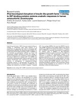
Incorporation of APOBEC3G into HTLV-1 virionsFigure 1
Incorporation of APOBEC3G into HTLV-1 virions. (A) Overexpressed APOBEC3G was incorporated into
HTLV-1 virions. HEK293T cells were cotransfected with K30, pNL43-Luc (WT), or pNL43/∆vif-Luc (∆Vif) with or without
an expression vector for HA-APOBEC3G. Western blotting was performed to detect HA-APOBEC3G in HEK293T cells and
produced virions with anti-HA mAb. APOBEC3G was expressed in producer cells and efficiently incorporated into produced
virions (lane 2). Expression of APOBEC3G and its incorporation into HIV-1 virions were reduced by expression of Vif as
described previously (lane 4). Western blotting with anti-p19 and anti-p24 mAbs showed that similar amounts of virions were
produced from each transfection (bottom panel). (B) Incorporation of APOBEC3G was confirmed in HTLV-1 virions
purified by sucrose density equilibrium gradient analysis. HTLV-1 K30 virions were purified by sucrose density equilib-
rium gradient analysis. Gradient fractions were collected and used for analyzing incorporation of APOBEC3G into virions.
APOBEC3G were detected and colocalized with HTLV-1 Gag (p19) proteins (lanes 4, 5). (C) APOBEC3G, its mutants,
and muAPOBEC3G were incorporated into HTLV-1 virions. Expression vectors for HA-APOBEC3G, its mutants, or
HA-muAPOBEC3G were cotransfected with K30 into HEK293T cells and APOBEC3G was detected with anti-HA mAb. HA-
APOBEC3G, its mutants, and HA-muAPOBEC3G were all incorporated into virions. A3G and muA3G indicate human and
murine APOBEC3G, respectively. E67Q, E259Q, and E67Q/E259Q were inactive mutants of human APOBEC3G that have a
point mutation in N-terminal active site, C-terminal active site, and both, respectively, as described previously [7]. (D) Endog-
enous APOBEC3G was also incorporated into HTLV-1 virions. Western blotting with anti-APOBEC3G Ab revealed
expression of endogenous APOBEC3G in MT-2 cells (lane 1, upper panel) and its incorporation into produced virions (lane 1,
lower panel). No cytoplasmic proteins were detected with anti-β-tubulin mAb in MT-2 virions (lane 2, lower panel).
Retrovirology 2005, 2:32 />Page 4 of 10
(page number not for citation purposes)
APOBEC3G did not induce G-to-A hypermutation in
HTLV-1 genome
To confirm the above hypothesis, we examined whether
APOBEC3G induces G-to-A hypermutation in HTLV-1
DNA. p12 region was amplified from target cell DNA and
sequenced. We detected a few G-to-A mutations in HTLV-
1 K30 genome integrated into target cell DNA in the pres-
ence of APOBEC3G (Fig. 3C), but not in the absence of
APOBEC3G (Fig. 3D). These G-to-A mutations were only
seen with expression of APOBEC3G and mostly occurred
in the context of G
pG sequence which is the preferred sub-
strate for APOBEC3G, suggesting that these mutations
were induced by APOBEC3G, although the frequency is
very low as seen with HBV [14]. In contrast, G-to-A hyper-
mutation was induced in HIV-1∆Vif DNA by APOBEC3G
(Fig. 3A) as previously reported [3,6-8]. Accordingly, this
again suggests the former notion that hypermutation may
not be necessary for the antiviral activity of APOBEC3G
on HTLV-1.
HIV-1 Vif reverses the infectivity of HTLV-1 suppressed by
endogenous APOBEC3G
Finally, we examined the antiviral activity of endogenous
APOBEC3G. First, we confirmed the function of endog-
enous APOBEC3G in MT-2 cells by infection with HIV-1
wild type (WT) and ∆Vif virions. WT virus could replicate
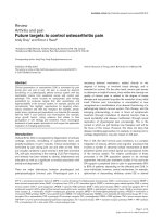
Inhibition of HTLV-1 infection by APOBEC3GFigure 2
Inhibition of HTLV-1 infection by APOBEC3G. APOBEC3G as well as its mutants inhibited the infectivity of HTLV-1.
Infectivity of HTLV-1 was measured as described in Materials and Methods. HTLV-1 proviral DNA load in target SupT1 cells
was suppressed by APOBEC3G and its mutants to the level of that of heat-inactivted virus. Six independent experiments gave
similar results and the data was presented as the mean of these values. Values are presented as infectivity ratio relative to K30
virus without expression of APOBEC3G.
Retrovirology 2005, 2:32 />Page 5 of 10
(page number not for citation purposes)
in MT-2 cells, but ∆Vif virus not (data not shown), indi-
cating that endogenous APOBEC3G in MT-2 cells may be
able to function as an anti-HIV-1 factor or that there may
exist other APOBEC3 protein members sensitive to Vif.
Based on this result, we performed an infectivity assay
using HTLV-1 virions produced from MT-2 cells. Since we
found that endogenous APOBEC3G was incorporated
into HTLV-1 virions produced from MT-2 cells (Fig. 1D),
we introduced HIV-1 Vif into MT-2 cells to see whether Vif
can upregulate the infectivity of HTLV-1 virions produced
from MT-2 cells by blocking the virion incorporation of
APOBEC3G. MT-2/Mock and MT-2/Vif cell lines were
established for this purpose using retrovirus vectors. We
confirmed that Vif reduced expression of APOBEC3G in
MT-2/Vif cells as well as its incorporation into produced
virions (Fig. 4A). Unfortunately, expression of Vif was not
enough to totally suppress the expression of APOBEC3G
in MT-2/Vif cells and there were some levels of virion
incorporation of APOBEC3G left. In order to affirm the
inhibitory activity of HIV-1 Vif against APOBEC3G, we
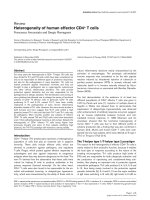
No G-to-A hypermutation in HTLV-1 genome was induced by APOBEC3GFigure 3
No G-to-A hypermutation in HTLV-1 genome was induced by APOBEC3G. Mutations in HTLV-1 and HIV-1 ∆Vif
viruses were detected by sequencing p12 and Env regions, respectively. G-to-A hypermutation was induced by APOBEC3G in
HIV-1 ∆Vif DNA, but not in HTLV-1 DNA. We detected very few G-to-A mutations in HTLV-1 K30 genome with expression
of APOBEC3G (C), but not without expression of APOBEC3G (D), whereas G-to-A hypermutation was induced in HIV-1∆Vif
DNA by APOBEC3G (A). We also detected a very few G-to-A mutations in MT-2/Mock virus DNA (E) as well as MT-2/Vif
virus DNA (F). G-to-A mutations are shown in red, while other mutations are denoted in black. The numbers before the
sequence indicate the number of each clone, while those in parentheses indicate the total number of clones sequenced. WT
indicates no mutations in this region.
Retrovirology 2005, 2:32 />Page 6 of 10
(page number not for citation purposes)
HIV-1 Vif reduced the incorporation of APOBEC3G into HTLV-1 virions, resulting in the upregulation of the infectivityFigure 4
HIV-1 Vif reduced the incorporation of APOBEC3G into HTLV-1 virions, resulting in the upregulation of the
infectivity. (A) Expression of APOBEC3G in MT-2 cells and its incorporation into produced virions were
reduced by HIV-1 Vif. Expression level of APOBEC3G was reduced in MT-2/Vif cells (lane 2, middle panel) as compared to
MT-2/Mock cells (lane 1, middle panel). Incorporation of APOBEC3G into produced virions was also reduced in virions pro-
duced from MT-2/Vif cells (lane 2, bottom panel). Expression of Vif protein in MT-2/Vif cells was detected with anti-Vif mAb
(top panel). (B) HIV Vif upregulated the infectivity of HTLV-1 produced from MT-2 cells. Infectivity of HTLV-1 virus
produced from MT-2 cells was determined as described in Materials and Methods. Infectivity of viruses produced from MT-2/
Vif cells was more than 4 times higher than that from MT-2/Mock cells. Four independent experiments gave similar results and
the data was presented as the mean of these values. Values are presented as infectivity ratio relative to viruses from MT-2/
Mock cells.
Retrovirology 2005, 2:32 />Page 7 of 10
(page number not for citation purposes)
performed an infectivity assay using virions produced
from these cell lines. The infectivity of viruses produced
from MT-2/Vif cells was more than 4 times higher than
that from MT-2/Mock cells (Fig. 4B). The infectivity assay
on target cells after 10 days of culture also showed similar
results (data not shown), suggesting that the possible
detection of residual viral DNA in the culture was
unlikely. These results indicate that endogenous
APOBEC3G incorporated into HTLV-1 virions is func-
tional and suppresses the infectivity of HTLV-1, which can
be overcome by HIV-1 Vif. We also examined whether
these proviruses have G-to-A hypermutation when inte-
grated into the infected target cell DNA and again found
very few G-to-A mutations in both viruses (Fig. 3E and
3F), suggesting that G-to-A hypermutation was not neces-
sary for the inhibition of virus infectivity.
Discussion
In this study, we have demonstrated that APOBEC3G has
an antiviral activity on HTLV-1. APOBEC3G was
efficiently incorporated into HTLV-1 virions and inhibited
the infectivity of HTLV-1 without inducing G-to-A hyper-
mutation. First, we showed that APOBEC3G, overex-
pressed or endogenous, was efficiently incorporated into
HTLV-1 virions. Our finding suggests that HTLV-1 cannot
exclude this protein from visions unlike HIV-1 [2-4,6-8].
Previous reports have shown that some accessory proteins
encoded in open reading frames of HTLV-1 genome could
enhance the infectivity of the virus. For example, deletion
or mutants of p12 led to impaired infectivity of HTLV-1
both in vivo and in vitro [21,25]. We could not fully
exclude the possibility that both K30 and the provirus in
MT-2 cells possess mutations in some of these accessory
genes so that these viruses could not exclude APOBEC3G
from virions, although the possibility is quite low.
Whether p12 potentially overcomes APOBEC3G has not
been clarified and further investigations are necessary.
Second, we also showed that APOBEC3G inhibited the
infection of HTLV-1. Because of low infectivity of cell-free
HTLV-1 virions, we could not detect p19 production in
the supernatant of infection culture (data not shown).
Instead, we performed an infectivity assay as described
previously with modification [23], in which RQ-PCR
methods enabled us to quantify HTLV-1 genome inte-
grated into target cells and measure the infectivity of cell
free virions of HTLV-1, which was very low [24]. Using
this method, we demonstrated that APOBEC3G sup-
pressed the infectivity of HTLV-1. Interestingly, not only
APOBEC3G but also its inactive mutants inhibited the
infectivity of HTLV-1. Taken together with the data that
APOBEC3G doesn't induce G-to-A hypermutation in
HTLV-1 genome, these results indicate that the enzymatic
activity is dispensable for the anti-HTLV-1 activity of
APOBEC3G and that it may inhibit HTLV-1 through dif-
ferent mechanisms. In contrast, we previously reported
that point mutants of C-terminal active site of APOBEC3G
(E259Q, E67Q/E259Q) abrogated its antiviral activity on
HIV-1, indicating that the enzymatic activity is essential
for anti-HIV-1 activity of APOBEC3G [7]. Furthermore,
some groups recently reported that APOBEC3G acts as an
antiviral factor on HBV through several mechanisms
[14,26]. One is induction of G-to-A mutations in cell type
dependent manner, and the other is interference with
pregenomic HBV RNA packaging without inducing G-to-
A hypermutation. The reason why APOBEC3G inhibits
HTLV-1 without inducing G-to-A hypermutation as seen
with other retroviruses, even though it is a member of ret-
roviruses, remains unclear. In order to elucidate the pre-
cise mechanisms of the antiviral activity of APOBEC3G on
HTLV-1, further studies, such as its effects on translation
of viral proteins, packaging of viral genome, and budding
of virions, other than its cytidine deaminase activity,
should be performed in the future.
To confirm the notion above, we prepared MT-2/Vif cells
to block incorporation of endogenous APOBEC3G into
HTLV-1 virions. Expression of Vif in MT-2 cells reduced
the expression of APOBEC3G and its incorporation into
virions. In the presence of Vif, APOBEC3G in MT-2 cells
seemed to be ubiquitinated and degraded by the proteas-
ome, because we detected two bands of APOBEC3G in
MT-2/Vif cells by immunoblotting, of which the upper
band might indicate mono-ubiquitinated APOBEC3G,
while the faded lower band indicate the intact
APOBEC3G remained (Fig. 4A, lanes 1 and 2, middle
panel). Interestingly, we demonstrated that viruses
released from MT-2/Vif cells recovered their infectivity
which had been suppressed in MT-2/Mock cells. Then, we
sequenced integrated HTLV-1 genome in target cells
infected with viruses produced from MT-2/Vif and MT-2/
Mock cells, and detected no G-to-A hypermutation (Fig.
3E and 3F). We hereby propose that the presence of
functional endogenous APOBEC3G in virions from MT-2
cells inhibited the infectivity of the virus and that it might
be linked to very low infectious titers of cell free HTLV-1
viruses. Taken together, our findings suggest that
APOBEC3G might contribute to the unique features of
HTLV-1 transmission, such as low infectivity of the virions
[17] with very low genetic diversity [20].
During the preparation of this manuscript, Navarro et al.
reported that HTLV-1 is relatively resistant to the antiviral
effect of encapsidated APOBEC3G [27]. In that paper,
they have shown that AOBEC3G is incorporated into
HTLV-1 virion and suppresses the infectivity of HTLV-1,
although the antiviral activity on HTLV-1 is very weak. We
speculate that this discrepancy between their study and
ours may originate from different assay systems to meas-
ure the infectivity of HTLV-1. They used a luciferase
Retrovirology 2005, 2:32 />Page 8 of 10
(page number not for citation purposes)
reporter HTLV-1 molecular clone in their study. However,
luciferase activity was very low (below 10,000 cps) as
compared to that of HIV-1 (more than 20 million cps).
Taken together with our data that we could not detect the
elevation of p19 levels in the supernatant of infection cul-
ture, we suspect that after integration the transcription
level of viral gene is very low, resulting in low levels of
luciferase activity and p19 production. In such a situation,
luciferase reporter system might be inappropriate for eval-
uation of the infectivity of HTLV-1. Furthermore, in our
study, we have shown that APOBEC3G inhibits HTLV-1
infection without exerting its cytidine deaminase activity,
suggesting that APOBEC3G might act on HTLV-1 through
different mechanisms from that on HIV-1. We believe that
this is the first detailed report on the anti-HTLV-1 function
of APOBEC3G and first description of possible involve-
ment of other mechanisms than inducing G-to-A hyper-
mutation in anti-HTLV-1 activity.
Finally, our findings have also broadened the spectrum of
antiviral activity of APOBEC3G and further studies on the
mechanisms of the antiviral activity of APOBEC3G on
HTLV-1 will provide us with new insights into the func-
tion of this molecule as an antiviral innate immunity.
Conclusion
APOBEC3G is incorporated into HTLV-1 virions and
inhibits the infection of HTLV-1 without exerting its cyti-
dine deaminase activity. This suggests that APOBEC3G
might act on HTLV-1 through different mechanisms from
that on HIV-1 and contribute to the unique features of
HTLV-1 infection and transmission.
Materials and methods
Expression vectors and molecular clones
Expression vectors for hemagglutinin (HA)-tagged human
APOBEC3G (APOBEC3G), its point mutants (E67Q,
E259Q, and E67Q/E259Q), and murine APOBEC3G
(muAPOBEC3G) were described previously [4,7]. pNL43-
Luc and pNL43/∆vif-Luc were also constructed as previ-
ously described [7]. HTLV-1 K30 was a kind gift from Dr.
Thomas Kindt through the AIDS Research and Reference
Reagent Program [28]. The vif gene was amplified by PCR
method from pNL43 and cloned into pDON-AI (Takara
Bio Inc., Otsu, Japan) to construct a retrovirus vector,
pDON/Vif.
Cell lines
HEK293T cells were maintained in Dulbecco's modified
Eagle's medium (Invitrogen, Carlsbad, California) con-
taining 10% fetal calf serum, penicillin, streptomycin, and
glutamine (Invitrogen). SupT1 cells and MT-2 cells were
maintained in RPMI 1640 (Sigma, St. Louis, Missouri)
containing 10% fetal calf serum, penicillin, streptomycin,
and glutamine. MT-2/Mock and MT-2/Vif cells were estab-
lished by transduction of retrovirus vectors (pDON-AI
and pDON/Vif, respectively) and selection with Neomy-
cin (Nacalai tesque, Kyoto, Japan).
Expression of APOBEC3G in producer cells and its
incorporation into visions
Western blotting was performed to detect expression of
APOBEC3G, its mutants, and muAPOBEC3G in producer
cells, and their incorporation into virions as described
previously [4]. In brief, expression vectors for HA-
APOBEC3G, its mutants, or HA-muAPOBEC3G were
cotransfected with K30, pNL43-Luc, or pNL43/∆vif-Luc
into HEK293T cells. Two days after transfection, viruses in
the supernatant were collected and ultracentrifuged with
Beckman TL-100s ultracentrifuge at 60,000 × g for 10min
and subjected to sodium dodecyl sulfate-polyacrylamide
gel electrophoresis (SDS-PAGE) together with whole cell
lysates of producer HEK293T cells. To detect HA-tagged
proteins, they were immunoblotted with anti-HA mono-
clonal antibody (mAb) (12CA5) (F. Hoffmann-La Roche
Ltd., Basel, Switzerland). Virus production was confirmed
by immunoblotting with the following antibodies; GIN-
7(anti-p19 mAb)[29] for HTLV-1 and anti-p24 mAb (Zep-
toMetrix Corporation, Buffalo, New York) for HIV-1. To
detect endogenous APOBEC3G in MT-2 cells and its
incorporation into virions, whole cell lysates of MT-2 cells
and precipitated virions were subjected to immunoblot-
ting with anti-APOBEC3G antibody (a kind gift from Dr.
Warner C. Greene, Gladstone Institute of Virology and
Immunology, University of California, San Francisco). Vif
expression in MT-2/Vif cells was detected with anti-Vif
mAb (#319) (a kind gift from Dr. Michael H. Malim
through the AIDS Research and Reference Reagent
Program) (18). Cytoplasmic proteins were detected with
anti-β-tubulin mAb (D-10)(Santa Cruz Biotechnology,
Santa Cruz, California). Samples applied to Western blot-
ting were equalized according to p19 antigen levels for
HTLV-1 and p24 antigen levels for HIV-1.
Purification of HTLV-1 virions by sucrose density
equilibrium gradients and analysis of APOBEC3G
packaging
To confirm the incorporation of APOBEC3G into virion,
HTLV-1 K30 virions were purified by sucrose density equi-
librium gradients as previously reported with slight mod-
ifications [30]. Briefly, HTLV-1 K30 virions were prepared
as described above and pelleted by ultracentrifugation,
then resuspended in 150µl of PBS. They were laid on top
of the sucrose gradient, prepared in PBS ranging from 10
to 60%, and centrifuged for 13 h at 20,000 rpm in an SW-
41Ti rotor (Beckman, Palo Alto, California). Gradient
fractions were collected from the top of the gradient.
These samples were used for analyzing protein profiles of
the virion by Western blotting. They were subjected to
Retrovirology 2005, 2:32 />Page 9 of 10
(page number not for citation purposes)
immunoblotting with anti-HA mAb (12CA5) and GIN-7
for detection of HA-APOBEC3G and p19, respectively.
Assessment of HTLV-1 infectivity
Infectivity of HTLV-1 was detected as previously reported
with slight modifications [23]. In brief, expression vectors
for HA-APOBEC3G, its mutants, or HA-muAPOBEC3G
were cotransfected with K30 into HEK293T cells. Viruses
in the supernatants were collected 2 days after transfec-
tion, then treated with DNase (80 U/ml) (Roche Diagnos-
tics GmbH, Germany) at 37°C for 1 h and filtrated
through a 0.45-µm-pore-size filter. Viruses from MT-2
cells were also collected and treated in the same way. We
also used noninfectious HTLV-1 as a negative control that
had been heat inactivated at 56°C for 1 h. Virus titers were
measured with an enzyme-linked immunosorbent assay
kit for the p19 antigen (RETRO-TEK, ZeptoMetrix Corpo-
ration). SupT1 cells were challenged with viruses whose
amounts were equalized according to p19 antigen levels,
and washed five times after incubation at 37°C for 8 h.
These target cells were cultivated for 2 to 10 days and total
cellular DNA was extracted with DNA Mini kit (Quiagen,
Valencia, California). HTLV-1 proviral DNA loads were
measured by RQ-PCR as described previously [24].
Detection of mutations in the viral DNA
Mutations in HTLV-1 DNA were detected by sequencing
p12 region of HTLV-1 integrated into target cells [4]. Prep-
aration of total cellular DNA of target cells infected with
HTLV-1 is described above [23]. The p12 region of HTLV-
1 was amplified with the following primer pairs:op-
32.1(ATAGTCGACCTGTTTCGCCTTCTCAGCCC) and
op-32.3(TATCTCGAGGAAGCTGTGCTTGACGG). The
PCR products were cloned into pT7-Blue (Novagen,
Darmstadt, Germany) and the inserts of individual clones
were sequenced. Mutations in HIV-1 NL43 Env region
were also detected as previously described [7].
Competing interests
The author(s) declare that they have no competing
interests.
Authors' contributions
AS designed research, performed research, contributed
vital new reagents, analyzed data, and wrote the paper.
AT-K designed research, performed research, contributed
vital new reagents, analyzed data, wrote the paper, and
organized research. KS performed a part of research. MK
performed a part of research. AA performed a part of
research. MH performed a part of research and contrib-
uted vital new analytical tools. KI contributed vital new
analytical tools and analyzed data. YT contributed vital
new reagents. TU analyzed data, drafted the paper, and
organized research.
Acknowledgements
The following reagents were obtained through the AIDS Research and Ref-
erence Reagent Program, Divirion of AIDS, NIDS, NIH: HTLV-1 K30 DNA
from Dr. Thomas Kindt, anti-HIV-1 Vif mAb (#319) from Dr. Michael H.
Malim. We also thank Dr. Warner C. Greene for providing us with the anti-
APOBEC3G Ab.
References
1. Sheehy AM, Gaddis NC, Choi JD, Malim MH: Isolation of a human
gene that inhibits HIV-1 infection and is suppressed by the
viral Vif protein. Nature 2002, 418:646-650.
2. Harris RS, Bishop KN, Sheehy AM, Craig HM, Petersen-Mahrt SK,
Watt IN, Neuberger MS, Malim MH: DNA deamination mediates
innate immunity to retroviral infection. Cell 2003, 113:803-809.
3. Mangeat B, Turelli P, Caron G, Friedli M, Perrin L, Trono D: Broad
antiretroviral defence by human APOBEC3G through lethal
editing of nascent reverse transcripts. Nature 2003, 424:99-103.
4. Kobayashi M, Takaori-Kondo A, Shindo K, Abudu A, Fukunaga K,
Uchiyama T: APOBEC3G Targets Specific Virus Species. J Virol
2004, 78:8238-8244.
5. Jarmuz A, Chester A, Bayliss J, Gisbourne J, Dunham I, Scott J, Navar-
atnam N: An anthropoid-specific locus of orphan C to U RNA-
editing enzymes on chromosome 22. Genomics 2002,
79:285-296.
6. Lecossier D, Bouchonnet F, Clavel F, Hance AJ: Hypermutation of
HIV-1 DNA in the absence of the Vif protein. Science 2003,
300:1112.
7. Shindo K, Takaori-Kondo A, Kobayashi M, Abudu A, Fukunaga K,
Uchiyama T: The enzymatic activity of CEM15/Apobec-3G is
essential for the regulation of the infectivity of HIV-1 virion
but not a sole determinant of its antiviral activity. J Biol Chem
2003, 278:44412-44416.
8. Zhang H, Yang B, Pomerantz RJ, Zhang C, Arunachalam SC, Gao L:
The cytidine deaminase CEM15 induces hypermutation in
newly synthesized HIV-1 DNA. Nature 2003, 424:94-98.
9. Yu X, Yu Y, Liu B, Luo K, Kong W, Mao P, Yu XF: Induction of
APOBEC3G ubiquitination and degradation by an HIV-1 Vif-
Cul5-SCF complex. Science 2003, 302:1056-1060.
10. Sheehy AM, Gaddis NC, Malim MH: The antiretroviral enzyme
APOBEC3G is degraded by the proteasome in response to
HIV-1 Vif. Nat Med 2003, 9:1404-1407.
11. Marin M, Rose KM, Kozak SL, Kabat D: HIV-1 Vif protein binds
the editing enzyme APOBEC3G and induces its degradation.
Nat Med 2003, 9:1398-1403.
12. Stopak K, de Noronha C, Yonemoto W, Greene WC: HIV-1 Vif
blocks the antiviral activity of APOBEC3G by impairing both
its translation and intracellular stability. Mol Cell 2003,
12:591-601.
13. Kobayashi M, Takaori-Kondo A, Miyauchi Y, Iwai K, Uchiyama T:
Ubiquitination of APOBEC3G by an HIV-1 Vif-Cullin5-
Elongin B-Elongin C Complex Is Essential for Vif Function. J
Biol Chem 2005, 280:18573-18578.
14. Turelli P, Mangeat B, Jost S, Vianin S, Trono D: Inhibition of Hepa-
titis B Virus Replication by APOBEC3G. Science 2004,
303:1829.
15. Uchiyama T, Yodoi J, Sagawa K, Takatsuki K, Uchino H: Adult T-cell
leukemia: clinical and hematologic features of 16 cases. Blood
1977, 50:481-492.
16. Uchiyama T: Human T cell leukemia virus type I (HTLV-I) and
human diseases. Annu Rev Immunol 1997, 15:15-37.
17. Igakura T, Stinchcombe JC, Goon PK, Taylor GP, Weber JN, Griffiths
GM, Tanaka Y, Osame M, Bangham CR: Spread of HTLV-I
between lymphocytes by virus-induced polarization of the
cytoskeleton. Science 2003, 299:1713-1716.
18. Derse D, Hill SA, Lloyd PA, Chung H, Morse BA: Examining human
T-lymphotropic virus type 1 infection and replication by cell-
free infection with recombinant virus vectors. J Virol 2001,
75:8461-8468.
19. Matsuoka M: Human T-cell leukemia virus type I and adult T-
cell leukemia. Oncogene 2003, 22:5131-5140.
20. Mansky LM: In vivo analysis of human T-cell leukemia virus
type 1 reverse transcription accuracy. J Virol 2000,
74:9525-9531.
Publish with BioMed Central and every
scientist can read your work free of charge
"BioMed Central will be the most significant development for
disseminating the results of biomedical research in our lifetime."
Sir Paul Nurse, Cancer Research UK
Your research papers will be:
available free of charge to the entire biomedical community
peer reviewed and published immediately upon acceptance
cited in PubMed and archived on PubMed Central
yours — you keep the copyright
Submit your manuscript here:
/>BioMedcentral
Retrovirology 2005, 2:32 />Page 10 of 10
(page number not for citation purposes)
21. Albrecht B, Collins ND, Burniston MT, Nisbet JW, Ratner L, Green
PL, Lairmore MD: Human T-lymphotropic virus type 1 open
reading frame I p12(I) is required for efficient viral infectivity
in primary lymphocytes. J Virol 2000, 74:9828-9835.
22. Franchini G, Fukumoto R, Fullen JR: T-cell control by human T-
cell leukemia/lymphoma virus type 1. Int J Hematol 2003,
78:280-296.
23. Fan N, Gavalchin J, Paul B, Wells KH, Lane MJ, Poiesz BJ: Infection
of peripheral blood mononuclear cells and cell lines by cell-
free human T-cell lymphoma/leukemia virus type I. J Clin
Microbiol 1992, 30:905-910.
24. Hishizawa M, Imada K, Ishikawa T, Uchiyama T: Kinetics of proviral
DNA load, soluble interleukin-2 receptor level and tax
expression in patients with adult T-cell leukemia receiving
allogeneic stem cell transplantation. Leukemia 2004,
18:167-169.
25. Collins ND, Newbound GC, Albrecht B, Beard JL, Ratner L, Lairmore
MD: Selective ablation of human T-cell lymphotropic virus
type 1 p12I reduces viral infectivity in vivo. Blood 1998,
91:4701-4707.
26. Rosler C, Kock J, Malim MH, Blum HE, von Weizsacker F: Comment
on "Inhibition of Hepatitis B Virus Replication by
APOBEC3G". Science 2004, 305:1403a.
27. Navarro F, Bollman B, Chen H, Konig R, Yu Q, Chiles K, Landau NR:
Complementary function of the two catalytic domains of
APOBEC3G. Virology 2005, 333:374-386.
28. Zhao TM, Robinson MA, Bowers FS, Kindt TJ: Characterization of
an infectious molecular clone of human T-cell leukemia virus
type I. J Virol 1995, 69:2024-2030.
29. Tanaka Y, Lee B, Inoi T, Tozawa H, Yamamoto N, Hinuma Y: Anti-
gens related to three core proteins of HTLV-I (p24, p19 and
p15) and their intracellular localizations, as defined by mon-
oclonal antibodies. Int J Cancer 1986, 37:35-42.
30. Yonezawa A, Hori T, Takaori-Kondo A, Morita R, Uchiyama T:
Replacement of the V3 region of gp120 with SDF-1 preserves
the infectivity of T-cell line-tropic human immunodeficiency
virus type 1. J Virol 2001, 75:4258-4267.