Báo cáo y học: " Conservation of functional domains and limited heterogeneity of HIV-1 reverse transcriptase gene following vertical transmission" pdf
Bạn đang xem bản rút gọn của tài liệu. Xem và tải ngay bản đầy đủ của tài liệu tại đây (440.78 KB, 17 trang )
BioMed Central
Page 1 of 17
(page number not for citation purposes)
Retrovirology
Open Access
Research
Conservation of functional domains and limited heterogeneity of
HIV-1 reverse transcriptase gene following vertical transmission
Vasudha Sundaravaradan, Tobias Hahn and Nafees Ahmad*
Address: Department of Microbiology and Immunology, College of Medicine, The University of Arizona Health Sciences Center, Tucson, Arizona
85724, USA
Email: Vasudha Sundaravaradan - ; Tobias Hahn - ;
Nafees Ahmad* -
* Corresponding author
Abstract
Background: The reverse transcriptase (RT) enzyme of human immunodeficiency virus type 1
(HIV-1) plays a crucial role in the life cycle of the virus by converting the single stranded RNA
genome into double stranded DNA that integrates into the host chromosome. In addition, RT is
also responsible for the generation of mutations throughout the viral genome, including in its own
sequences and is thus responsible for the generation of quasi-species in HIV-1-infected individuals.
We therefore characterized the molecular properties of RT, including the conservation of
functional motifs, degree of genetic diversity, and evolutionary dynamics from five mother-infant
pairs following vertical transmission.
Results: The RT open reading frame was maintained with a frequency of 87.2% in five mother-
infant pairs' sequences following vertical transmission. There was a low degree of viral
heterogeneity and estimates of genetic diversity in mother-infant pairs' sequences. Both mothers
and infants RT sequences were under positive selection pressure, as determined by the ratios of
non-synonymous to synonymous substitutions. Phylogenetic analysis of 132 mother-infant RT
sequences revealed distinct clusters for each mother-infant pair, suggesting that the
epidemiologically linked mother-infant pairs were evolutionarily closer to each other as compared
with epidemiologically unlinked mother-infant pairs. The functional domains of RT which are
responsible for reverse transcription, DNA polymerization and RNase H activity were mostly
conserved in the RT sequences analyzed in this study. Specifically, the active sites and domains
required for primer binding, template binding, primer and template positioning and nucleotide
recruitment were conserved in all mother-infant pairs' sequences.
Conclusion: The maintenance of an intact RT open reading frame, conservation of functional
domains for RT activity, preservation of several amino acid motifs in epidemiologically linked
mother-infant pairs, and a low degree of genetic variability following vertical transmission is
consistent with an indispensable role of RT in HIV-1 replication in infected mother-infant pairs.
Background
The vertical transmission of human immunodeficiency
virus type 1 (HIV-1) accounts for more than 90% of all
HIV-1 infections in children. HIV-1 infected pregnant
Published: 26 May 2005
Retrovirology 2005, 2:36 doi:10.1186/1742-4690-2-36
Received: 18 February 2005
Accepted: 26 May 2005
This article is available from: />© 2005 Sundaravaradan et al; licensee BioMed Central Ltd.
This is an Open Access article distributed under the terms of the Creative Commons Attribution License ( />),
which permits unrestricted use, distribution, and reproduction in any medium, provided the original work is properly cited.
Retrovirology 2005, 2:36 />Page 2 of 17
(page number not for citation purposes)
women can transmit the virus to their infants during all
stages of their pregnancy, including prepartum (trans-pla-
cental passage), intrapartum (exposure of infants' skin
and mucous membranes to contaminated maternal blood
and vaginal secretions) and post-partum (via breast milk)
at an estimated rate of 30% [1-4]. However, the rate of ver-
tical transmission can be reduced by antiretroviral therapy
during pregnancy. The risk of vertical transmission
increases with several parameters, including advanced
maternal disease status, low maternal CD4 cell count,
high maternal viral load, recent infection of the mother,
prolonged exposure of infant to ruptured membranes dur-
ing parturition, and higher viral heterogeneity in the
mother [5-8].
Viral heterogeneity is one of the classical means by which
HIV-1 evades the host immune system. The heterogeneity
of HIV-1 is attributed to the error-prone reverse tran-
scriptase (RT) enzyme, which is responsible for converting
the single stranded viral genomic RNA to double-stranded
DNA that integrates into the host chromosome. As reverse
transcription is the first step of the viral replication cycle
[9], errors made at this stage ensures propagation of the
erroneously copied genome to form the quasi-species of
HIV-1 found in the infected individuals. These quasi-spe-
cies infect other uninfected target cells and the cycle of
error-prone reverse transcription continues. We have pre-
viously demonstrated that HIV-1 sequences from trans-
mitting mothers (mothers who transmitted HIV-1 to their
infants) were more heterogeneous compared with HIV-1
sequences from non-transmitting mothers (mothers who
failed to transmit HIV-1 to their infants) [10]. This finding
further suggests that the reverse transcription step that is
responsible for generation of viral heterogeneity, may also
play an important role in vertical transmission. The RT
gene is unique in that it is also exposed to the same mutat-
ing effects of the RT enzyme as other part of the HIV-1
genome. Therefore, we sought to examine HIV-1 RT
sequences from five infected mother-infant pairs follow-
ing perinatal transmission.
The HIV-1 RT shows significant sequence and structural
similarity to other viral reverse transcriptases as well as
viral and bacterial RNA polymerases [11-13]. HIV-1 RT is
a heterodimeric protein comprising of two subunits, 66
kDa and 51 kDa. It is encoded as a Gag-Pol precursor,
Pr160
gag-pol
, which is cleaved by viral protease to yield the
Gag protein and the viral polymerase which codes for RT
[9,14]. The larger subunit (p66) of the heterodimer acts as
an RNA-dependant DNA polymerase, a DNA-dependant
DNA polymerase and has RNase H activity associated
with the C-terminus [15,16], whereas the p51 subunit
lacks the C-terminus RNase H activity, is folded differently
from the p66 subunit and is thus inactive [17-20]. The
p66 is folded to form a structure similar to a right hand
with palm, finger and thumb subdomains [21-23] that are
connected to the RNase H by the "connexion" subdomain
[22,24,25]. Each domain has several secondary structural
elements which are critical for primer binding, template
binding [14,22,23,26,27] and nucleotide recruitment
[28]. More specifically, the aspartate residues at position
110, 185 and 186 are believed to be the active sites of the
polymerase and are located in the palm subdomain at the
bottom of the DNA binding cleft [14,16,20,28,29]. Muta-
tions in this subdomain and the active site abolish the
enzymatic activity of HIV-1 RT [2,19,22,30-32] and alter
viral replication, which may also affect HIV-1 mother-to-
infant transmission.
In this study, we characterized the HIV-1 RT quasi-species
from five mother-infant pairs following vertical transmis-
sion, including a mother with infected twin infants. We
show that the open reading frame of the RT gene was
highly conserved in the sequences from five mother-
infant pairs. In addition, there was a low degree of heter-
ogeneity and high conservation of functional domains
essential for RT activity. These findings may be helpful in
the understanding of the molecular mechanisms of HIV-1
vertical transmission.
Results
Patient population and sample collection
Blood samples were collected from five HIV-1-infected
mother-infant pairs following perinatal transmission,
including samples from a set of twins (IH1 and IH2) in
the case of mother H. The demographic, clinical and lab-
oratory findings on these mother-infant pairs are summa-
rized in Table 1. The Human Subjects Committee of the
University of Arizona, and the Institutional Review Board
of the Children's Hospital Medical Centre, Cincinnati
Ohio, approved this study. Written informed consent was
obtained for participation in the study from mothers of
infected mother-infant pairs.
Phylogenetic analysis of RT sequences of mother-infant
isolates
We first performed multiple independent polymerase
chain reaction (PCR) amplifications from peripheral
mononuclear cells (PBMC) DNA of five mother-infant
pairs and obtained 10 to 14 clones from each patient fol-
lowed by nucleotide sequencing of these clones. We then
performed the phylogenetic analysis by constructing a
neighbor-joining tree of the 132 RT sequences from these
mother-infant pairs, including the set of twins from
mother H and the reference strain NL4-3, as shown in Fig-
ure 1. A model of evolution was optimized for the entire
nucleotide sequence data set using the approach outlined
by Huelsenbeck and Crandall [33]. The model of choice
was incorporated into PAUP [34] to estimate a neighbor-
joining tree and the tree was bootstrapped 1000 times to
Retrovirology 2005, 2:36 />Page 3 of 17
(page number not for citation purposes)
ensure fidelity. The phylogenetic tree demonstrated that
the RT sequences from five mother-infant pairs were well
discriminated in separate clusters and that the mother and
infant sequences were generally separated in distinct sub-
clusters. However, there was some intermingling between
mother and infant sequences in pair C. Furthermore, the
formation of separate subclusters of RT sequences from
twins of mother H suggests that the there was probably
compartmentalization of HIV-1 in the two fetuses causing
independent evolution. We also compared our mother-
infant pairs' RT sequences with the RT sequences of several
clades present in the HIV databases and found that our RT
sequences grouped with clade or subtype B sequences
(not shown). The data on phylogenetic analysis indicate
that the epidemiologically linked mother-infant
sequences are closer to each other than epidemiologically
unlinked sequences and that there was no PCR cross con-
tamination. It is important to note that the mother-infant
pairs grouped in the same subtree, even when some of the
infants' ages were more than 2 to 3 years, suggesting that
the epidemiological relationships are maintained in
mother-infant pairs no matter how long the infection in
the infants has progressed.
Coding potential of RT gene sequences
The multiple sequence alignments of the deduced amino
acid sequences of HIV-1 RT genes from five mother-infant
pairs, B, C, D, F, mother H and her twin infants IH1 and
IH2 are shown in Figures 2, 3, 4, 5, 6, and 7, respectively.
These sequences were aligned with consensus subtype B
RT sequence (CON B). We found that 115 of the 132
sequences analyzed contained a complete RT open read-
ing frame (ORF), with an 87.2% frequency of intact RT
open reading frames thus indicating that the coding
potential of the RT ORF was maintained in most of the
sequences in 1680 bp sequenced. Moreover, the infected
mothers' sequences showed a frequency of 85.5% of
intact RT ORF while infants demonstrated a frequency of
88.5%. Several clones in mother-infant pair B and mother
H were found to be defective due to a single nucleotide
substitution, insertion or deletion resulting either in
frame-shift or stop codons. The RT sequences also dis-
played patient and pair specific amino acid sequence pat-
terns. Several amino acid motifs changes were observed in
majority of the mother-infant pairs' sequences, including
a glutamic acid (E) or proline (P) at position 122, an
arginine (R) at 277, and a threonine (T) or serine (S) at
376 and 400.
Variability of RT gene sequences in mother-infant isolates
The degree of genetic variability of RT sequences, meas-
ured as nucleotide and amino acid distances based on
pairwise comparison (as described in Methods), was
determined for the five mother-infant pairs' sequences,
and is shown in Table 2. The nucleotide sequences of RT
within mothers (mothers B, C, D, F and H) differed by
0.80, 1.76, 1.37, 1.21 and 2.90% (median values), respec-
tively, ranging from 0 to 3.46%. The variability in the
infant sets (infants B, C, D, F, H1 and H2) was similar to
the mother sequences and differed by 0.80, 1.49, 1.37,
1.31, 0.64 and 1.24% (median values), respectively, rang-
ing from 0 to 2.21%. Interestingly, the variability between
epidemiologically linked mother and infant sets (pairs B,
C, D, F and H) was also on the same order of 1.05, 1.7.
1.74, 1.22 and 1.45 (median values) respectively, ranging
from 0 to 4.48%. Moreover, the amino acid sequence var-
iability of RT within mothers (mothers B, C, D, F and H)
differed by 1.26, 2.81, 1.98, 1.26 and 2.27% (median val-
ues), respectively, ranging from 0 to 5.51%. The variabil-
ity within infants (infants B, C, D, F, H1 and H2) differed
Table 1: Demographic, Clinical, and Laboratory Parameters of HIV-1 Infected Mother-Infant Pairs
Patient Age Sex CD4+ cells/mm3 Length of infection
a
Antiviral drug Clinical Evaluation
b
MB 28 yr 509 11 mo None Asymptomatic
IB 4.75 mo M 1942 4.75 mo None Asymptomatic, P1A
MC 23 yr 818 1 yr6 mo None Asymptomatic
IC 14 mo F 772 14 mo ZDV Symptomatic AIDS;P2A,D1,3,F
MD 31 yr 480 2 yr6 mo None Asymptomatic
ID 28 mo M 46 28 mo ddC
c
Symptomatic AIDS, P2AB,F; failed ZDV therapy
MF 23 yr 692 2 yr10 mo None Asymptomatic
IF 1 wk M 2953 1 wk ZDV Asymptomatic,P1A
MH 33 yr 538 5 mo None Asymptomatic
IHT1 7 mo F 3157 7 mo ACTG152 Hepatosplenomeglay lymphadenopathy
IHT2 7 mo F 2176 7 mo ACTG152 Hepatosplenomegaly lymphadenopathy
M: mother; I: infant.
a
Length of infection: The closest time of infection that we could document was the first positive HIV-1 serology date or the first
visit of the patient to the AIDS treatment Center, where all the HIV-1 positive patients were referred to as soon as an HIV-1 test was positive.
Therefore, these dates may not reflect the exact dates of infection.
b
Evaluation for infants is based on CDC criteria,
c
ddC, Zalcitibine
Retrovirology 2005, 2:36 />Page 4 of 17
(page number not for citation purposes)
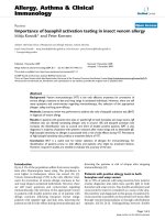
Phylogenetic analysis of HIV-1 RT of 132 RT sequences from five mother-infant pairs, including B, C, D, F and HFigure 1
Phylogenetic analysis of HIV-1 RT of 132 RT sequences from five mother-infant pairs, including B, C, D, F and H. The neighbor-
joining tree is based on the distance calculated between the nucleotide sequences from the five mother-infant pairs. Each ter-
minal node represents one RT gene sequence. The numbers on the branch points indicate the percent occurrence of branches
over 1,000 bootstrap resamplings of the data set. The sequences from each mother formed distinct clusters and are well dis-
criminated and in confined subtrees, indicating that the variants from the same mother-infant pair are closer to each other than
to other sequences and that there was no PCR cross-contamination. These data were strongly supported by the high boot-
strap values indicated on the branch points.
hivnl43
mb.1
mb.12
mb.4
mb.5
mb.8
mb.11
mb.2
mb.6
mb.3
mb.7
mb.9
ib.1
ib.7
ib.2
ib.3
ib.4
ib.5
ib.6
ib.8
ib.9
ib.10
ib.11
ib.12
mb.10
mc.1
mc.2
mc.3
ic.7
ic.8
ic.10
ic.11
ic.12
ic.13
ic.9
mc.4
mc.5
mc.6
mc.7
mc.8
ic.4
ic.1
ic.2
ic.3
mc.9
mc.10
mc.11
mc.12
ic.5
ic.6
mf.1
mf.2
mf.5
mf.9
mf.13
mf.11
mf.3
mf.4
mf.6
mf.7
mf.8
mf.10
mf.14
if.1
if.2
if.3
if.4
if.5
if.6
if.7
if.8
if.9
if.10
if.11
if.12
mh.1
mh.2
mh.8
mh.9
mh.14
mh.13
mh.5
mh.11
mh.12
mh.10
mh.3
mh.4
mh.6
mh.7
ih1.1
ih1.2
ih1.3
ih1.11
ih1.4
ih1.5
ih1.9
ih1.6
ih1.7
ih1.8
ih1.10
ih2.1
ih2.2
ih2.9
ih2.3
ih2.6
ih2.4
ih2.5
ih2.7
ih2.8
ih2.10
ih2.11
md.1
md.2
md.3
md.4
md.5
md.6
md.7
md.11
md.8
md.9
md.10
id.1
id.2
id.3
id.4
id.5
id.6
id.10
id.7
id.8
id.9
0.005 substitutions/site
Pair H
Pair F
Pair C
Pair B
61
100
100
Pair H
Pair F
Pair D
Pair C
Pair B
100
100
100
Retrovirology 2005, 2:36 />Page 5 of 17
(page number not for citation purposes)
by 1.44, 2.35, 1.80, 1.62, 1.44 and 1.62% (median val-
ues), ranging from 0 to 4.57%, and between mother-
infant pairs (pairs B, C, D, F and H) by 1.44, 2.90, 2.53,
1.44 and 2.17% (median values), ranging from 0 to
6.47%, respectively. We also determined sequence varia-
bility between epidemiologically unlinked individuals
and found that the nucleotide distances ranged from 0 to
9.1% (median 5.4%) and amino acid from 0 to 12.4%
(median 6.34%). The variability in general was lower
between epidemiologically linked mother-infant pairs'
sequences than epidemiologically unlinked individuals,
suggesting that epidemiologically linked mother-infant
pair sequences are closer to each other.
We also investigated if the low variability of RT sequences
seen in our mother-infant pair isolates is due to errors
made by LA Taq polymerase used in our study. We did not
find any errors made by the LA Taq polymerase when we
used a known sequence of HIV-1 NL 4–3 for PCR ampli-
fication and DNA sequencing of the RT gene.
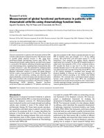
Multiple sequence alignment of deduced amino acids of HIV-1 reverse transcriptase (RT) gene from mother-infant pair B involved in vertical transmissionFigure 2
Multiple sequence alignment of deduced amino acids of HIV-1 reverse transcriptase (RT) gene from mother-infant pair B
involved in vertical transmission. In the alignment, the top sequence is the consensus RT sequence of subtype or clade B (CON
B) to which mother-infant pair-B RT sequences are aligned. In mother-infant pair B sequences, each line refers to a clone iden-
tified by a clone number with M referring to mothers and I referring to infants. The structural elements of RT are indicated
above the alignment. Dots represent amino acid agreement with CON-B and substitutions are shown by single letter codes for
the changed amino acid. Stop codons are shown as x and dashes represent gaps or truncated protein. Relevant amino acid
motifs and domains essential for RT activity are shown by spanning arrowheads indicated above the alignment.
Finger Template grip (73-90)
D110 Palm CTL epitope Active site
1 50 110 150 187
CON B PISPIETVPV KLKPGMDGPK VKQWPLTEEK IKALVEICTE MEKEGKISKI GPENPYNTPV FAIKKKDSTK WRKLVDFREL NKRTQDFWEV QLGIPHPAGL KKKKSVTVLD VGDAYFSVPL DKDFRKYTAF TIPSINNETP GIRYQYNVLP QGWKGSPAIF QSSMTKILEP FRKQNPDIVI YQYMDDL
MB.1 .EN .G
MB.2 A .EN
MB.3 E .EN R
MB.4 A .EN
MB.5 A D. .EN S
MB.6 D .EN
MB.7 A A.HMAIDRR. A A .EN
MB.8 D .EN
MB.9 D A .EN
MB.10 D .EN
MB.11 .EN
MB.12 G .EN
IB.1 D .EN D L
IB.2 D I .EN
IB.3 D S .EN
IB.4 AP V R .EN L
IB.5 DP .EN
IB.6 D .EN L
IB.7 D .EN
IB.8 A .EN A
IB.9 A TG .EN
IB.10 DP .EN
IB.11 D .EN
IB.12 A .EN
Thumb Connection
Template and primer binding helices
Primer grip(227-235)
α
αα
αH α
αα
αI
188 250 300 374
CON B YVGSDLEIGQ HRTKIEELRQ HLLRWGFTTP DKKHQKEPPF LWMGYELHPD KWTVQPIVLP EKDSWTVNDI QKLVGKLNWA SQIYAGIKVK QLCKLLRGTK ALTEVIPLTE EAELELAENR EILKEPVHGV YYDPSKDLIA EIQKQGQGQW TYQIYQEPFK NLKTGKYARM RGAHTNDVKQ LTEAVQK
MB.1 K L A D V.P R Y R.
MB.2 K L P D. R Y
MB.3 K L P R Y D
MB.4 G K L E. P R Y
MB.5 K L P R R Y
MB.6 K L P R Y
MB.7 K L SP G G R Y
MB.8 K L .E P R Y V
MB.9 K L P R Y
MB.10 K L R P R Y
MB.11 K L P R Y
MB.12 K L A P R Y
IB.1 TK L G SP R Y I
IB.2 K L FP PNR RSRARAGRKQ RDS.RTSTWS VLX.I.R.NS RNTEA.VRPM DISNLSRAIX KSENR.ICKN E.C
IB.3 K L P R Y
IB.4 V K L P R Y
IB.5 K L P.T R Y
IB.6 G K L P R Y
IB.7 K V L GH SP R A Y
IB.8 K L P R Y
IB.9 K L T P R Y
IB.10 K L N D P R Y
IB.11 K L P GP R Y
IB.12 GK X.L P R Y
Connection RNase H
RNase H Active sites
D443 E478 D498 D549
375
↓
455
↓
↓
505
↓
560
CON B IATESIVIWG KTPKFKLPIQ KETWEAWWTE YWQATWIPEW EFVNTPPLVK LWYQLEKEPI VGAETFYVDG AANRETKLGK AGYVTDRGRQ KVVPLTDTTN QKTELQAIHL ALQDSGLEVN IVTDSQYALG IIQAQPDKSE SELVSQIIEQ LIKKEKVYLA WVPAHKGIGG NEQVDKLVSA GIRKVL
MB.1 .SM T ID A F I.N .V
MB.2 .SM T ID A A.F G I.N .V
MB.3
MB.4 .SM T ID A F I.N .V
MB.5 .SM S T ID A F X I.N .V
MB.6 .SM T ID V A F R G I.N .V
MB.7 .SM T ID A F I.N .V
MB.8 .SM T ID A F R I.N .V
MB.9 .SM T ID A F G P G I.HP P MV T N E
MB.10 .SM T ID A F I.N .V
MB.11 .SM T ID A F I.N .V
MB.12 .SM T ID A F S .GI.N .V
IB.1 .SM T ID A F I.N .V A
IB.2 .SM T ID A F Y I.N .V
IB.3 .SM T ID A F I.N .V
IB.4 .SM T ID .S A F R I.N .V
IB.5
IB.6 .SM T ID A F I.NR .V
IB.7 .SM T ID A F I.N .V P.
IB.8 .SM T ID A F I.N .V R
IB.9 .SM T ID A F D I.N .V
IB.10 .SM T ID .X A F T G I.N .V D
IB.11 .SM .A T ID A F I.N .V
IB.12 .SM T ID A F I.N .V
Retrovirology 2005, 2:36 />Page 6 of 17
(page number not for citation purposes)
Dynamics of HIV-1 RT gene evolution in mother-infant
isolates
The maximum likelihood estimates and chi square tests
performed by Modeltest 3.06 [35] suggested different
models of evolution for each patient sample. The esti-
mates of genetic diversity of RT sequences from the five
mother-infant pairs were determined by using the Watter-
son model, assuming segregating sites and the Coalesce
method assuming a constant population size. The esti-
mates of genetic diversity shown as theta values
(estimated as nucleotide substitutions per site per genera-
tion) are shown in Table 3. The levels of genetic diversity
among infected mothers and infants, as estimated by Wat-
terson method, ranged from 0.012 to 0.025 and 0.009 to
0.021, respectively. Similar results were obtained when
the mother-infant pair populations were analyzed by the
Coelesce method, with the values ranging from 0.020 to
0.058 in mothers and from 0.016 to 0.060 in infants.
Multiple sequence alignment of deduced amino acids of HIV-1 reverse transcriptase (RT) gene from mother-infant pair C in reference to consensus subtype B (CON B) RT sequenceFigure 3
Multiple sequence alignment of deduced amino acids of HIV-1 reverse transcriptase (RT) gene from mother-infant pair C in
reference to consensus subtype B (CON B) RT sequence. In the alignment, the top sequence is CON B RT sequence and the
bottom sequences are mother-infant pair C sequences (M refers to mother sequences and I to sequences). The number of
clones sequenced is represented with clone numbers. The structural elements of RT are indicated above the alignment. Dots
represent amino acid agreement with CON-B and substitutions are shown by single letter codes for the changed amino acid.
Stop codons are shown as x and dashes represent gaps or truncated protein. Spanning arrowheads indicated above the align-
ment shows relevant amino acid motifs and domains essential for RT function.
Finger
Template grip (73-90)
D110
Palm
CTL epitope Active site
1 50 110 150 187
CON B PISPIETVPV KLKPGMDGPK VKQWPLTEEK IKALVEICTE MEKEGKISKI GPENPYNTPV FAIKKKDSTK WRKLVDFREL NKRTQDFWEV QLGIPHPAGL KKKKSVTVLD VGDAYFSVPL DKDFRKYTAF TIPSINNETP GIRYQYNVLP QGWKGSPAIF QSSMTKILEP FRKQNPDIVI YQYMDDL
MC.1 HE T. E E.
MC.2 L N R Q HE E E
MC.3 L N R Q HE E
MC.4 H HE I E
MC.5 HE I E
MC.6 HE I E
MC.7 .R HE I E
MC.8 R E HE I EV
MC.9 R K HE E
MC.10 L R K HE E
MC.11 K HE N E E
MC.12 K V HEG L E
IC.1 D S HE S .S I E
IC.2 R HE I E
IC.3 V HE I E
IC.4 R HE I E
IC.5 K R HE .C I E
IC.6 HE .C E
IC.7 L N R Q HE .C Y E
IC.8 L R N R Q
IC.9 L N R A Q HE .C E
IC.10 N R Q A HE .C E
IC.11 K N R Q H HE .C E G
IC.12 K N R Q HE H .C E
IC.13 N R Q HE .C A E G
Thumb Connection
Template and primer binding helices
Primer grip(227-235)
α
αα
αH α
αα
αI
188 250 300 374
CON B YVGSDLEIGQ HRTKIEELRQ HLLRWGFTTP DKKHQKEPPF LWMGYELHPD KWTVQPIVLP EKDSWTVNDI QKLVGKLNWA SQIYAGIKVK QLCKLLRGTK ALTEVIPLTE EAELELAENR EILKEPVHGV YYDPSKDLIA EIQKQGQGQW TYQIYQEPFK NLKTGKYARM RGAHTNDVKQ LTEAVQK
MC.1 N P R I
MC.2 .Q S F P N P R V T
MC.3 .Q D N H P R P V G
MC.4 G N P R
MC.5 N P P R G
MC.6 N P R
MC.7 G N P R A
MC.8 H P .E R A N H D P R K A
MC.9 A N P R G D
MC.10 N N H P P R
MC.11 N V P R
MC.12 I R E N P R
IC.1 N P R V
IC.2 NQ P R
IC.3 E N P R
IC.4 A N A P R
IC.5 N P R .S V P
IC.6 R. N P R V G
IC.7 .Q N P R V
IC.8
IC.9 S N P R Y
IC.10 .Q N P R V D V
IC.11 .Q N P R V G
IC.12 .Q N R P R V S
IC.13 G .R C N X P R V R
RNase H
Connection
RNase H active sites
D443 E478 D498 D549
375
↓
455
↓
↓
505
↓
560
CON B IATESIVIWG KTPKFKLPIQ KETWEAWWTE YWQATWIPEW EFVNTPPLVK LWYQLEKEPI VGAETFYVDG AANRETKLGK AGYVTDRGRQ KVVPLTDTTN QKTELQAIHL ALQDSGLEVN IVTDSQYALG IIQAQPDKSE SELVSQIIEQ LIKKEKVYLA WVPAHKGIGG NEQVDKLVSA GIRKVL
MC.1 .S I R N S D L I T T S
MC.2 .S R R N S D L I T T S
MC.3 .S R N S D L I N R T T P.N S
MC.4 .S R N S D T L C I .I T S
MC.5 .S R N S D L C I .I T S
MC.6 .S R R N E S AD L C I .I T S
MC.7 .S R N S D L I .I T P
MC.8 .S R N S D A L C I .I I T S
MC.9 .S R N S D L I G. S S
MC.10 .S R N S D L I G .Q T R… S
MC.11 .S R N S D L I .Q T T F
MC.12 .S R R N S D L I .Q R T F
IC.1 .S R N S D L I .I T S
IC.2 .S R N S D L I .I .M T S
IC.3 .S R N S D L I .I .M T S
IC.4 .S R N S D L C I .I V R T S
IC.5 .S R N S D L I T T S
IC.6 .S R N S D L I .I T T S
IC.7 .S R N S D G L I T T S
IC.8
IC.9 .S R N S D L I IT T S
IC.10 .S E R N S D L S I A A T S
IC.11 .S R.S.N S D L I T S
IC.12 .S R N S D L I A T T D S
IC.13 .S R N S D L I T T S
Retrovirology 2005, 2:36 />Page 7 of 17
(page number not for citation purposes)
These data suggest that the mother and infant populations
evolved very slowly and at similar rates. The differences
observed in the estimates of genetic diversity between and
mothers and infants sequences are not statistically
significant.
Rates of accumulation of nonsynonymous and
synonymous substitutions
Selection pressure on the RT gene was estimated as a ratio
of accumulation of non-synonymous to non-synony-
mous substitutions using the Nielsen and Yang model
[36] as implemented in codeML [37]. Although there are
several models to predict the rate of positive selection,
most of these models assume that all sites in a sequence
are under the same selection pressure with the same
underlying dN/dS ratio [38]. As substitutions of critical
regions of a protein can lead to deleterious mutations, it is
unrealistic to make assumptions about equal degree of
selection throughout the protein. In cases where positive
selection is operating on proteins, it has been shown that
only a limited number of amino acids may be responsible
for adaptive evolution. In such a case, methods that esti-
mate dN/dS ratios over an entire sequence may fail to
detect positive selection even when it exists [39]. The
codeML method uses the codon as a unit of evolution as
opposed to a nucleotide, and thus allows us to estimate
the percentage of positions that are being positively
selected instead of averaging the rates of positive selection
Multiple sequence alignment of deduced amino acids of HIV-1 reverse transcriptase (RT) gene from mother-infant pair DFigure 4
Multiple sequence alignment of deduced amino acids of HIV-1 reverse transcriptase (RT) gene from mother-infant pair D. The
patient sequences are aligned in reference to consensus RT sequence of HIV-1 subtype or clade B (CON B) at the top. In the
mother-infant pair sequences, each line refers to a clone identified by a clone number with M referring to mother and I to
infants. The structural elements of RT are indicated above the alignment. Dots represent amino acid agreement with CON-B
and substitutions are shown by single letter codes for the changed amino acid. Stop codons are shown as x and dashes repre-
sent gaps or truncated protein. Relevant amino acid motifs and domains essential for RT activity are shown by spanning arrow-
heads indicated above the alignment.
Finger Template grip (73-90)
D110 Palm CTL epitope Active site
1 50 110 150 187
CON B PISPIETVPV KLKPGMDGPK VKQWPLTEEK IKALVEICTE MEKEGKISKI GPENPYNTPV FAIKKKDSTK WRKLVDFREL NKRTQDFWEV QLGIPHPAGL KKKKSVTVLD VGDAYFSVPL DKDFRKYTAF TIPSINNETP GIRYQYNVLP QGWKGSPAIF QSSMTKILEP FRKQNPDIVI YQYMDDL
MD.1 S A .E T .C
MD.2 A .E.S T .C
MD.3 .E .T T .C T
MD.4 .E .T T .C T
MD.5 G .E T .CR
MD.6 M .E T .C
MD.7 .EG M. .C
MD.8 R .E P. T. .C
MD.9 M .EG C
MD.10 A .EG HC
MD.11 A .EG .C
ID.1 I L .E T .C
ID.2 I .E T .C
ID.3 .E T .C
ID.4 .E T .C
ID.5 R I .E S T.H R H E .C S
ID.6 T I L E .E T .C R
ID.7 I .E T F. .C I
ID.8 .EG T .C
ID.9 I .E T .C
ID.10 I N R A .E T .C
Thumb Connection
Template and primer binding helices
Primer grip(227-235)
α
αα
αH α
αα
αI
188 250 300 374
CON B YVGSDLEIGQ HRTKIEELRQ HLLRWGFTTP DKKHQKEPPF LWMGYELHPD KWTVQPIVLP EKDSWTVNDI QKLVGKLNWA SQIYAGIKVK QLCKLLRGTK ALTEVIPLTE EAELELAENR EILKEPVHGV YYDPSKDLIA EIQKQGQGQW TYQIYQEPFK NLKTGKYARM RGAHTNDVKQ LTEAVQK
MD.1 .R A. V V.
MD.2 A. V V.
MD.3 G T E A. A V R V. H
MD.4 G T E A. C V. D
MD.5 R N I.H X A. A V.
MD.6 A. Q V.
MD.7 A. .V V.
MD.8 L S R A. V.
MD.9 A. V.
MD.10 H A. V V.
MD.11 E A. .V A V. .T R
ID.1 F .Q R A. .P V.
ID.2 F .Q A. V.
ID.3 F .Q R A. V.
ID.4 K. F .Q G A. V.
ID.5 V F LP P A.P.L A. V.
ID.6 Y W F A. V.
ID.7 P A V A. V.
ID.8 A V A. X V.
ID.9 V A. V.
ID.10 F .Q A. V.
Connection RNase H
RNase H Active sites
D443 E478 D498 D549
375
↓
455
↓
↓
505
↓
560
CON B IATESIVIWG KTPKFKLPIQ KETWEAWWTE YWQATWIPEW EFVNTPPLVK LWYQLEKEPI VGAETFYVDG AANRETKLGK AGYVTDRGRQ KVVPLTDTTN QKTELQAIHL ALQDSGLEVN IVTDSQYALG IIQAQPDKSE SELVSQIIEQ LIKKEKVYLA WVPAHKGIGG NEQVDKLVSA GIRKVL
MD.1 .S R M. I P N. V .T I.
MD.2 .S R M. I P N. V .T I.
MD.3 .S R M. I P N. V .T I.
MD.4 .SP R P M. H W. I P N. V .T I.
MD.5 .S X M. I P N. V .T I.
MD.6 .S R M. I P N. V .T I.
MD.7 .S R M. IP N. V .T I.
MD.8 .S R M. .E I P N. T V .T I.
MD.9 .S R R M. R I P N. V .T I.
MD.10 .S R M. S I P N. V .T I.
MD.11 .S R M. T I P N. V .T I.
ID.1 .S R M. I P N. Q V .T T
ID.2 .S R T M. I P N. Q V .T T
ID.3 .S R T M. I P N. G Q V .T T
ID.4 .S R T M. I P N. I Q V .L .T T
ID.5 .S R T M. A I P N. Q V LL .L .T T
ID.6 .S R T M. A I.VL P N. V .T T
ID.7 .S R T M. I P N. Q V .T T
ID.8 .S R T M. H A P N. Q .GV H .T T
ID.9 .S R M. I P N. V .T T
ID.10 .S R T M. A P N. Q V .T T
Retrovirology 2005, 2:36 />Page 8 of 17
(page number not for citation purposes)
over the entire gene [39]. This method also provides the
percentage of mutations that are conserved, neutral or
positively selected based on dN/dS values of 0, 1 or > 1,
respectively. The dN/dS values as well as the proportions
of each site category estimated using the Nielsen and Yang
model are shown in Table 4. As described in the methods,
a dN/dS value of greater than 1 suggests positive selection.
The percentage of the substitutions being positively
selected is shown in column p3. Except for viral popula-
tions in infants C and F, all isolated populations were
associated with dN/dS ratio >1, indicating positive selec-
tion. In case of infants C and F, there was no positive selec-
tion on the mutations and most of the substitutions were
neutral. All mothers generally displayed a higher propor-
tion of positively selected p3 sites as compared to the
infants. Although the dN/dS values for infant H1 and H2
seem higher than mother H, closer observation shows that
the percentage of sites undergoing positive selection is
higher in the mother than in the twin infants. Table 4
shows that in mothers, over half the sites (66.6%) belong
to the conserved p1 category, whereas the frequency of
neutral and positively selected sites was equally distrib-
Multiple sequence alignment of deduced amino acids of HIV-1 reverse transcriptase gene from mother-infant pair FFigure 5
Multiple sequence alignment of deduced amino acids of HIV-1 reverse transcriptase gene from mother-infant pair F. In the
alignment, the top sequence (CON B) is the consensus subtype B RT sequence and the bottom sequences are from mother-
infant pair F sequences (M stands for mother sequences and I for infant sequences and the number of clones for mother and
infant are indicated by clone number). The structural elements of RT are indicated above the alignment. Dots represent amino
acid agreement with CON-B and substitutions are shown by single letter codes for the changed amino acid. Stop codons are
shown as x and dashes represent gaps or truncated protein. Relevant amino acid motifs and domains essential for RT functions
are shown by spanning arrowheads indicated above the alignment.
Finger
Template grip (73-90)
D110
Palm
CTL epitope Active site
1 50 110 150 187
CON B PISPIETVPV KLKPGMDGPK VKQWPLTEEK IKALVEICTE MEKEGKISKI GPENPYNTPV FAIKKKDSTK WRKLVDFREL NKRTQDFWEV QLGIPHPAGL KKKKSVTVLD VGDAYFSVPL DKDFRKYTAF TIPSINNETP GIRYQYNVLP QGWKGSPAIF QSSMTKILEP FRKQNPDIVI YQYMDDL
MF.1 K .P E
MF.2 Q K .P E
MF.3 I L K .P .S I S E
MF.4 Q K .R .P .N E
MF.5 K .P S E
MF.6 Q K .R .P .N E
MF.7 L K .P E
MF.8 L K .P E
MF.9 A K .P E
MF.10 L G. K .P E
MF.11 Q K .P E
MF.13 Q K S .P .N E
MF.14 L K .P G
IF.1 K .P E
IF.2 .R.R K .R .P S E
IF.3 K .P L E
IF.4 K .P E
IF.5 .K .P E
IF.6 .K .P E
IF.7 A .K GP E
IF.8 N .K .P E
IF.9 .K .P X K. E
IF.10 D .K A .P E
IF.11 .K N .P A. R G. E
IF.12 D I .K .P X E
Thumb Connection
Template and primer binding helices
Primer grip(227-235)
α
αα
αH α
αα
αI
188 250 300 374
CON B YVGSDLEIGQ HRTKIEELRQ HLLRWGFTTP DKKHQKEPPF LWMGYELHPD KWTVQPIVLP EKDSWTVNDI QKLVGKLNWA SQIYAGIKVK QLCKLLRGTK ALTEVIPLTE EAELELAENR EILKEPVHGV YYDPSKDLIA EIQKQGQGQW TYQIYQEPFK NLKTGKYARM RGAHTNDVKQ LTEAVQK
MF.1 GH G R E N
MF.2 .P R E
MF.3 R E .A
MF.4 R E
MF.5 R E
MF.6 R E
MF.7 R E
MF.8 R E
MF.9 R E
MF.10 R E
MF.11 L A R E
MF.13 R E G
MF.14 R E
IF.1 V Q R E
IF.2 R E T L
IF.3 R E S
IF.4 M SQ R E
IF.5 R E G
IF.6 R E
IF.7 E
IF.8 R E
IF.9 R E
IF.10 P R E A. T
IF.11 MD R E
IF.12 L R E
Connection RNase H
RNase H active sites
D443 E478 D498 D549
375
↓
455
↓
↓
505
↓
560
CON B IATESIVIWG KTPKFKLPIQ KETWEAWWTE YWQATWIPEW EFVNTPPLVK LWYQLEKEPI VGAETFYVDG AANRETKLGK AGYVTDRGRQ KVVPLTDTTN QKTELQAIHL ALQDSGLEVN IVTDSQYALG IIQAQPDKSE SELVSQIIEQ LIKKEKVYLA WVPAHKGIGG NEQVDKLVSA GIRKVL
MF.1 M R T A. K S N D T
MF.2 M R T A. K S N N
MF.3 M R T K S N N
MF.4 M R T A. S N
MF.5 M R T A. K S NP NQ T
MF.6 M R T A. S N
MF.7 V R T K S N T
MF.8 M R T K S N T
MF.9 M R T A. K S N N G T
MF.10 M R T K S N T
MF.11 M R T A. K S N N T
MF.13 M R T A. K S N N T
MF.14 M R T A. S .A N T
IF.1 M R T G K S N
IF.2 M R T A. K S N G.
IF.3 M R A T A. K S N
IF.4 M R T A. L. K .A S N T
IF.5 M R T A. K .A S I.N
IF.6 M R T A. K L S N
IF.7 M V R T K .A S N T
IF.8 M R T A. K S V N
IF.9 M R T A. K .A S V N
IF.10 M R T A. K .A .S S P N R
IF.11 M R T A. C K S G N
IF.12 M R T A. K .A A S N
Retrovirology 2005, 2:36 />Page 9 of 17
(page number not for citation purposes)
uted. This is in contrast to the viral population from the
infants where the conserved site category (p1) had a fre-
quency of only 36.5% and close to half the sites (55.7%)
belongs to the neutral p2 category. Statistical analysis
revealed that only the proportion of the neutral p2 cate-
gory was significantly different between mothers' and
infants' sequence viral populations (p < 0.05). This is sig-
nified by the case that all the sites in Infant F belonged to
the p2 category. Higher proportion of p2 sites in infants
have also been shown in the nef gene product in these
same mother infant pairs [40]. The variable (positively
selected) sites (p3) in the mothers' sequences were associ-
ated with dN/dS ratios that ranged from 2.34 to 8.9, with
viral sequence populations from three mothers (MD, MF,
MH) that displayed a dN/dS ratio of below three. This is
in contrast to the infants' viral populations that were
either associated with a dN/dS of below 1, indicating no
directional selection (IC and IF), a dN/dS ratio between 3
and 4 (IB and ID) or a very high dN/dS ratio as found in
the sequences isolated from the twins H1 and H2. This
analysis showed that the RT gene in both the mothers and
infants is under positive selection pressure.
Analysis of functional domains of RT in mother-infant
pairs
HIV-1 RT is a heterodimeric protein comprising of two
subunits, p66 and p51. The larger subunit of the het-
erodimer acts as an RNA-dependant DNA polymerase, a
DNA-dependant DNA polymerase and an RNase H that is
associated with the C-terminus [15,16]. The p66 is folded
to form a structure similar to the right hand with palm,
finger and thumb subdomains [21,23,32] that are con-
nected to the RNase H by the "connexion" subdomain
[22,24,25]. Each domain has several secondary structural
elements, which are critical for primer binding, template
binding [14,22,23,26,27,41] and nucleotide recruitment
[28]. The active sites of the polymerase comprise of aspar-
tic acid (D) residues at positions 110, 185 and 186, which
are located in the palm subdomain at the bottom of the
DNA binding cleft [22,23]. Mutations of these aspartic
Multiple sequence alignment of deduced amino acids of HIV-1 reverse transcriptase (RT) gene from mother H, who had given birth to infected twins, H1 and H2 (alignment shown in Figure 7)Figure 6
Multiple sequence alignment of deduced amino acids of HIV-1 reverse transcriptase (RT) gene from mother H, who had given
birth to infected twins, H1 and H2 (alignment shown in Figure 7). In the mother H sequences, each line refers to a clone iden-
tified by a clone number with M referring to mother. The mother sequences are aligned in reference to consensus RT
sequence of HIV-1 subtype or clade B (CON B) shown at the top. The structural elements of RT are indicated above the align-
ment. Dots represent amino acid agreement with CON-B and substitutions are shown by single letter codes for the changed
amino acid. Stop codons are shown as x and dashes represent gaps or truncated protein. Spanning arrowheads indicated above
the alignment shows relevant amino acid motifs and domains required for RT activity.
Finger
Template grip (73-90) D110
Palm
CTL epitope Active site
1 50 110 150 187
CON B PISPIETVPV KLKPGMDGPK VKQWPLTEEK IKALVEICTE MEKEGKISKI GPENPYNTPV FAIKKKDSTK WRKLVDFREL NKRTQDFWEV QLGIPHPAGL KKKKSVTVLD VGDAYFSVPL DKDFRKYTAF TIPSINNETP GIRYQYNVLP QGWKGSPAIF QSSMTKILEP FRKQNPDIVI YQYMDDL
MH.1 D R .K K R
MH.2 A R .K T R
MH.3 .K
MH.4 .K .V
MH.5 .K L K R
MH.6 I .K H
MH.7 .K S
MH.8 D T .K K R
MH.9 .K K R
MH.10 .K
MH.11 A .K K T R
MH.12 .K K R
MH.13 E. .K K T L R
MH.14 .K K R
Thumb Connection
Template and primer binding helices
Primer grip(227-235) α
αα
αH α
αα
αI
188 250 300 374
CON B YVGSDLEIGQ HRTKIEELRQ HLLRWGFTTP DKKHQKEPPF LWMGYELHPD KWTVQPIVLP EKDSWTVNDI QKLVGKLNWA SQIYAGIKVK QLCKLLRGTK ALTEVIPLTE EAELELAENR EILKEPVHGV YYDPSKDLIA EIQKQGQGQW TYQIYQEPFK NLKTGKYARM RGAHTNDVKQ LTEAVQK
MH.1 R P K R I G R R I X N I
MH.2 R K I AR R I X I
MH.3 K E P R I E I
MH.4 K R I G E I
MH.5 R K I R L XI R I G E I
MH.6 K R I E I S
MH.7 K R I E V I
MH.8 R K I R R I X I
MH.9 R K I R R I X .R Y I
MH.10 K P R I E I
MH.11 R K I AR R I E I G
MH.12 R K R I E I
MH.13 R K L I G R ATGL P R R I I
MH.14 R K I R R I I
Connection RNase H
RNase H Active sites
D443 E478 D498 D549
375
↓
455
↓
↓
505
↓
560
CON B IATESIVIWG KTPKFKLPIQ KETWEAWWTE YWQATWIPEW EFVNTPPLVK LWYQLEKEPI VGAETFYVDG AANRETKLGK AGYVTDRGRQ KVVPLTDTTN QKTELQAIHL ALQDSGLEVN IVTDSQYALG IIQAQPDKSE SELVSQIIEQ LIKKEKVYLA WVPAHKGIGG NEQVDKLVSA GIRKVL
MH.1 .T X. G X.T X A R IR. N V E R R T R
MH.2 .T X. R X.T X A R IR. N V E R R S T R
MH.3 .T R T A I N V E T
MH.4 .T R T A I N .R V E .L RT
MH.5 .T R T A I N V E R R T R
MH.6 .T R T A I N G V P E .L RT
MH.7 .T R T A I N V E RT
MH.8 .T X. R X.T X A R A.I N V E S R R T R
MH.9 .T X. R X.T X A I N V E S R R T R
MH.10 .T R T S A I N V P E G P.R T R
MH.11 .T R T A I N G R V E P.R T R
MH.12 .T R T A I N P V LL.E .X.RK P.R T R
MH.13 .T R T A. A R IR. N V E R R T R
MH.14 .T R T A R IR. N .G V E M R R T R
Retrovirology 2005, 2:36 />Page 10 of 17
(page number not for citation purposes)
acid residues abrogates the polymerase activity of RT
[22,23,29,32]. These aspartate residues of the RT active
site were conserved within the five mother-infant pairs RT
sequences. Furthermore, the D185 and D186 that form a
part of an essential highly conserved YMDD [32,42,43]
motif involved in binding to the 3'OH of the primer
strand [14,26], were highly conserved in our mother-
infant pairs' RT sequences (Figures 2 to 7). The amino
acids at positions 73–90 that constitute the template grip
required for positioning and binding the RT template near
the active site of the RT [23], were also conserved in most
of our RT sequences. The primer grip responsible for
primer binding extends from amino acids 227 to 235
[22,23] and these amino acids were also conserved in the
mother-infant RT sequences. The K263, K353 and R358
that form salt bridges with the phosphate groups
[14,21,22,30,44] of the template and primer were found
to be conserved in most of the RT sequences analyzed. The
thumb subdomain of RT is comprised of two anti-parallel
α helices, αH and αI, which bind to the opposite strand of
dsDNA. The αH also directly inserts into the minor groove
of the DNA [14,22,41]. Both these helices were generally
conserved in our mother-infant RT sequences.
The connexion subdomain that links the RT to the RNase
H and forms the floor of the template binding cleft
[22,24,25,42], showed some substitutions, including
V293I, A376S and A400T in our mother-infant RT
Multiple sequence alignment of deduced amino acids of HIV-1 reverse transcriptase gene (RT) from infected twin infants, H1 and H2 of mother H (alignment shown in Figure 6)Figure 7
Multiple sequence alignment of deduced amino acids of HIV-1 reverse transcriptase gene (RT) from infected twin infants, H1
and H2 of mother H (alignment shown in Figure 6). In the alignment, the top sequence is the consensus subtype B RT sequence
(CON B) and the bottom sequences are of infants H1 and H2 represented by I and clone numbers. Dots represent amino acid
agreement with CON-B and substitutions are shown by single letter codes for the changed amino acid. Stop codons are shown
as x and dashes represent gaps or truncated protein. Relevant amino acid motifs and domains essential for RT activity are
shown by spanning arrowheads indicated above the alignment.
Finger
Template grip (73-90)
D110
Palm
CTL epitope Active site
1 50 110 150 187
CON B PISPIETVPV KLKPGMDGPK VKQWPLTEEK IKALVEICTE MEKEGKISKI GPENPYNTPV FAIKKKDSTK WRKLVDFREL NKRTQDFWEV QLGIPHPAGL KKKKSVTVLD VGDAYFSVPL DKDFRKYTAF TIPSINNETP GIRYQYNVLP QGWKGSPAIF QSSMTKILEP FRKQNPDIVI YQYMDDL
IH1.1 A R A. .K
IH1.2 A R A. .K
IH1.3 .K E
IH1.4 D .K
IH1.5 .K
IH1.6 .K
IH1.7 .K L
IH1.8 S .K
IH1.9 .K
IH1.10 G. .K
IH1.11 .K E
IH2.1 A D .K
IH2.2 D R R .K M.
IH2.3 D A .K G E
IH2.4 A .K L
IH2.5 .K
IH2.6 .K Q
IH2.7 D .K .K
IH2.8 M .K T .K
IH2.9 R .K
IH2.10 .K G
IH2.11 GD P .K L E
Thumb Connection
Template and primer binding helices
Primer grip(227-235)
α
αα
αH α
αα
αI
188 250 300 374
CON B YVGSDLEIGQ HRTKIEELRQ HLLRWGFTTP DKKHQKEPPF LWMGYELHPD KWTVQPIVLP EKDSWTVNDI QKLVGKLNWA SQIYAGIKVK QLCKLLRGTK ALTEVIPLTE EAELELAENR EILKEPVHGV YYDPSKDLIA EIQKQGQGQW TYQIYQEPFK NLKTGKYARM RGAHTNDVKQ LTEAVQK
IH1.1 V K S R I I
IH1.2 V K S R I I
IH1.3 K H .E P R I I
IH1.4 G K S R I X I .A
IH1.5 P K M.L P D S R I I
IH1.6 M K R I R I.R
IH1.7 K R I G I
IH1.8 K G S R I C I
IH1.9 K M S R I I
IH1.10 K .E S R I I
IH1.11 K H .E P R I I
IH2.1 K R I I
IH2.2 K R R I I
IH2.3 K R I E I
IH2.4 T K L R I R I
IH2.5 K R I N I
IH2.6 K R I I
IH2.7 K V R I I
IH2.8 K R I I
IH2.9 K R I .N R I
IH2.10 K T I
IH2.11 K L R R I I
Connection RNase H
RNase H Active sites
D443 E478 D498 D549
375
↓
455
↓
↓
505
↓
560
CON B IATESIVIWG KTPKFKLPIQ KETWEAWWTE YWQATWIPEW EFVNTPPLVK LWYQLEKEPI VGAETFYVDG AANRETKLGK AGYVTDRGRQ KVVPLTDTTN QKTELQAIHL ALQDSGLEVN IVTDSQYALG IIQAQPDKSE SELVSQIIEQ LIKKEKVYLA WVPAHKGIGG NEQVDKLVSA GIRKVL
IH1.1 .T R T A I N A V E T
IH1.2 .T R T A I N A V E T
IH1.3 .T R T A I N A V E T
IH1.4 .T R T A I A V E T
IH1.5 .T R T A I N P A V E R T
IH1.6 .T R T A I N A V E R T
IH1.7 .T R T A H I N A V E R T
IH1.8 .T R T A I N A V E R T
IH1.9 .T R A I N A V E T
IH1.10 .T R T A I N G A V E T
IH1.11 .T R T A I N A V E T
IH2.1 .T R T A I N A V E T
IH2.2 .T R T A I N V E T
IH2.3 .T R T A A.I N A V M.E T
IH2.4 .T R T A I N V E T
IH2.5 .T R T A I N V E T
IH2.6 .T R T G A I S A V E T
IH2.7 .T R T A I N R V E R T
IH2.8 .T R T T I N V E T
IH2.9 .T V R T A I N V E R T
IH2.10 .T R R T A I N A V E T
IH2.11 .T R VT A I N A V E T
Retrovirology 2005, 2:36 />Page 11 of 17
(page number not for citation purposes)
sequences. Mutations at positions H361 and Y501
reduces RNase H activity [24]. Examination of the five
mother-infant pairs' sequences revealed that these two
positions were intact in all RT sequences (Figures 2 to 7).
Furthermore, the RNase H active sites contain four acidic
amino acid residues, D443, E478, D498 and D549
Table 2: Distances in the RT sequences within mother sets, within infant sets, and betweenmother-infant pairs
Nucleotide distances
Within mothers Within infants Between mother and infants
Pair Min Med Max Pair Min Med Max Pair Min Med Max
MB 0.0 0.80 2.10 IB 0.0 0.80 1.30 B 0.0 1.05 2.05
MC 0.0 1.76 3.46 IC 0.0 1.49 2.17 C 0.0 1.70 3.26
MD 0.0 1.37 2.21 ID 0.0 1.37 2.21 D 0.0 1.74 4.48
MF 0.0 1.21 1.54 IF 0.0 1.31 2.93 F 0.0 1.22 2.08
MH 0.0 2.90 2.60 IH1 0.0 0.64 1.34 H 0.0 1.45 3.30
IH2 0.0 1.24 1.75
Total 0.0 1.34 3.46 Total 0.0 1.48 2.21 Total 0.0 1.32 4.48
Amino acid distances
Within mothers Within infants Between mother and infants
Pair Min Med Max Pair Min Med Max Pair Min Med Max
MB 0.0 1.26 4.61 IB 0.0 1.44 2.72 B 0.0 1.44 4.57
MC 0.0 2.81 5.51 IC 0.0 2.35 4.01 C 0.0 2.90 5.51
MD 0.0 1.98 3.83 ID 0.0 1.80 4.57 D 0.0 2.53 6.47
MF 0.0 1.26 2.35 IF 0.0 1.62 3.09 F 0.0 1.44 3.09
MH 0.0 2.27 3.09 IH1 0.0 1.44 2.17 H 0.0 2.17 6.27
IH2 0.0 1.62 2.72
Total 0.0 1.52 5.51 Total 0.0 1.42 4.57 Total 0.0 2.90 6.47
M: mother; I: infant. Min: Minimum; Med: Median; Max: Maximum. Totals were calculated for all pairs together
Table 3: Estimates of genetic diversity of HIV-1 RT within mother sets and infant sets
MOTHERS INFANTS
N θ
w
θ
c
θ
w
θ
c
Mother B 12 0.015 0.038 Infant B 12 0.014 0.033
Mother C 12 0.025 0.058 Infant C 13 0.021 0.060
Mother D 11 0.017 0.042 Infant D 10 0.019 0.040
Mother F 14 0.012 0.029 Infant F 12 0.018 0.053
Mother H 14 0.020 0.020 Infant H1 11 0.009 0.016
Infant H2 11 0.015 0.044
Totals 63 0.018 0.037 69 0.016 0.041
N – number of RT clones sequenced. θ
w
– genetic diversity as calculated by the Watterson method; θ
c
– genetic diversity as calculated by the
Coelesce method. Totals were indicated as an average of all values.
Retrovirology 2005, 2:36 />Page 12 of 17
(page number not for citation purposes)
[22,24,25,41,42], which were highly conserved in our
mother-infant pairs sequences. In addition, several substi-
tutions were seen in regions of RT that are not known to
have critical function. The relevance of these changes is
not known.
Mutations associated with anti-retroviral drug resistance
Several naturally occurring mutations in the pol gene in
treatment-naïve patients have been reported [45,46],
although most of these mutations are not seen in our RT
gene sequences. In addition, these mutations found in
treatment-naïve patients were usually seen in non-sub-
type B infections and our patient population was from
subtype B infected individuals. These changes were
usually in amino acids where the mutations did not actu-
ally confer nucleoside reverse transcriptase inhibitor
(NRTI) drug resistance but were accessory mutations [46-
48]. Several amino acid changes in RT seen in patients
undergoing NRTI therapy are selected primarily with zido-
vudine (ZDV) treatment. These mutations referred to as
thymidine analog mutations (TAMs) include M41L,
D67N, K70R, L210N, T215Y/F and K219Q [47,49]. Since
most of our infected mothers were treatment naïve but
infants were actively on ZDV therapy or on other drugs
(Table 1), we examined the RT sequences for ZDV
resistant mutations (Figure 2). Several TAMs associated
with drug resistance were observed in our infants C and D
who were either on prolonged or failed ZDV therapy.
These mutations included M41L in three clones from
infant C and two clones in infant D, D67N and K70R in
five clones from infant C, L210W in one clone from infant
D and T215F in seven clones from infant D and K219Q in
four clones from infant C and D. In addition, one clone
from infant C had all the above mutations, indicating sig-
nificant resistance to ZDV [46,50]. Although Mother C
was not on any antiretroviral therapy two clones had
TAMs at M41L and K219Q positions, suggesting that these
mutations were naturally occurring. It is interesting to
note that the infant of this mother yielded several clones
with these two mutations. An R211K mutation known as
an accessory mutation associated with NRTI resistance
[46] was also observed in all mother-infant pair H clones.
Immunologically relevant mutations in the CTL epitopes of
RT
The cytotoxic T lymphocyte (CTL) responses have been
shown to exert significant immune pressure during HIV-1
infection. Strong CTL responses are maintained in long-
term nonprogressors and these responses correlate with
decrease in viral load [51-55]. It has been shown that
transmitting mothers have larger numbers of CTL escape
variants as compared to non-transmitting mothers [56],
emphasizing that CTL escape variants may become a part
of circulating virus that influences vertical transmission
[56,57]. Several regions in the RT gene have been shown
to elicit strong CTL responses during HIV-1 infection. The
CTL eptitope, TVLDVGDAY, between amino acid posi-
tions 107–115 />nology/ctl_search, is highly conserved among known
HIV-1 isolates [57]. This epitope contains the amino acid
D110 which is part of the RT active site. This epitope was
highly conserved in most of the mother-infant RT clones
sequenced (Fig. 2).
Another motif, TAFTIPSI, between amino acid positions
128–135 is an HLA-B51 restricted epitope http://
www.hiv.lanl.gov/content/immunology/ctl_search. This
epitope is present in the palm region consisting of posi-
tions A129 and I135 as anchor residues [57]. This motif
was mostly conserved in the RT sequences of the five
mother-infant pairs analyzed. In addition, I135T muta-
tion decreases CTL response but increasing concentration
of mutant peptide re-establishes appropriate responses
Table 4: dN/dS values in HIV-1 RT sequences within mother sets and within infant sets.
MOTHER INFANT
N P1P2P3dN/dS N P1P2P3dN/dS
Mother B125318.8278.9Infant B124142163.31
Mother C 12 55.5 43 1.3 6.09 Infant C 13 0 81.2 18.8 0.01
Mother D 11 70.6 5.7 23.6 2.52 Infant D 10 74.8 19.2 5.9 4.44
Mother F 14 81.7 7.8 10.4 2.67 Infant F 12 0 100 0 0.001
Mother H 14 72 0 27 2.34 Infant H1 11 47 50 2.8 14.04
Infant H2 11 56 42 0.6 16.58
Totals 66.5 15.1 18.4 4.50 69 36.5 55.7 7.8 6.39
N – number of RT clones sequenced.; P1 = proportion of conserved codons as a percent; P2 = proportion of neutral codons as a percent; P3 =
proportion of positively selected codons as a percent. dN/dS = ratio of synonymous to non-synonymous at P3 sites. Totals were calculates as an
average of all values.
Retrovirology 2005, 2:36 />Page 13 of 17
(page number not for citation purposes)
[57]. The I135T mutation was seen in several of our
mother-infant pair's D sequences.
The next motif AIFQSSMTK from amino acid positions
158–166, comprising of I159, F160, K166 anchor resi-
dues and recognized by several HLA types, is conserved
among known HIV-1 isolates and believed to be associ-
ated with vertical transmission [56,57]. Our mother-
infant pairs' RT sequences showed conservation in this
motif. Another CTL epitope YPGIKVRQL from positions
271–279 has been reported to be conserved in transmit-
ting mothers and infants with several natural occurring
variants [56], was also found to be conserved in our
mother-infant pairs' RT sequences. In addition, a P272H
mutation that causes significant loss of CTL response for
this epitope [56] was not seen in any of the RT clones
analyzed.
Discussion
In this study, we show for the first time that reverse tran-
scriptase open reading frames from five mother-infant
pairs following perinatal transmission were maintained
with a frequency of 87.2%. The functional domains
required for reverse transcriptase activity in HIV-1 replica-
tion were highly conserved in most of the mother-infants
sequences. We also demonstrate a low degree of sequence
variability and estimates of genetic diversity for reverse
transcriptase genes after mother-to-infant transmission.
However, epidemiologically unlinked individual's
sequences were more heterogeneous than epidemiologi-
cally linked mother-infant pair's sequences. Several motifs
in reverse transcriptase responsible for primer and tem-
plate binding and positioning and motifs involved in
nucleotide recruitment were conserved in all mother-
infant pairs' sequences. The data we show here are compa-
rable to those of our previously analyzed conserved genes,
including gagP17MA, vif, vpr, tat and nef [58-62]. Our
findings suggest that an intact and functional reverse
transcriptase open reading frame is essential for HIV-1
replication in mothers and their infants and low degree of
viral heterogeneity is maintained following vertical
transmission.
The RT open reading frame was maintained in 115 of the
132 sequences (1680 base pairs sequenced), whereas 17
sequences contained stop codons (Figure 2). The fre-
quency of conservation in five mother-infant pairs was
found to be 87.2%. The comparison of the RT sequences
with those of other conserved genes from HIV-1 infected
mother-infant pairs showed comparable frequency of
conversation, including gag p17 (86.2%), vif (89.8%), vpr
(92.1%), tat (90.9%), nef (86.2%) and vpu (90.12%).
There was no significant correlation between the conser-
vation of RT open reading frame and disease progression
in mothers and infants [63-65]. Several amino acid motifs
were found to be a signature characteristic of each mother-
infant pair, even in older infants where infection has pro-
gressed for more than 3 years. Phylogenetic analysis of the
RT sequences revealed that the five mother-infant pairs
were well discriminated, separated and confined within
subtrees (Fig. 1), indicating that the epidemiologically
linked mother-infant pairs were closer to each other and
that there was no PCR product cross-contamination
[66,67]. In addition, most of the mother and infant
sequences of the same pair formed separate subclusters,
with little intermingling between sequences of mother
and infant in some pairs. In some mother-infant pairs,
minor variants of the mothers seem to be predominating
in the infants, which was also seen in our previous V3
region analysis [68]. We also observed intermingling of
sequences in mother-H and her infected twins, indicating
that different mother's variants were transmitted to the
twins. With respect to viral heterogeneity, there was a low
degree of genetic variability in the RT sequences from
mother-infant pairs estimated by several methods. Similar
levels of genetic diversity were seen in other conserved
genes of the same mother-infant pairs, including gag, vif,
vpr and tat [59-61,69]. The low degree of genetic variabil-
ity was observed in RT sequences of mothers and main-
tained in the infants following transmission, suggesting
the essential nature of this gene in viral pathogenesis. It is
important to note that the mother-infant pairs retained
the same epidemiological relationship, even when some
of the infant's age was more than 2 to 3 years. We believe
this is an important finding that the epidemiological rela-
tionships as well as certain signature sequence motifs are
maintained in mother-infant pairs or transmitter-recipi-
ent partners no matter how long the infection has pro-
gressed. This information may be critical in terms of
vaccine development.
Examining the motifs of the deduced amino acid
sequences of the RT gene from five mother-infant pairs,
we found that the essential motifs required for RT activity
were mostly conserved in our mother-infant pairs'
sequences (Figure 2). The sites essential for primer bind-
ing, template binding, positioning of template and
primer, which are located in α-Helix H and α-Helix I
[22,23], were are all conserved in RT sequences (Figure 2).
Specifically, the amino acids involved in recruitment of
nucleotides during reverse transcription [28] were mostly
conserved. The active sites of the polymerase are located
in the palm subdomain at the bottom of the DNA binding
cleft comprising of aspartic acid (D) residues at positions
110, 185 and 186 were conserved within the five mother-
infant pairs' RT sequences. Furthermore, the D185 and
D186 also form a part of an essential YMDD motif, which
is highly conserved in known HIV-1 isolates
[14,22,23,26,32,43], was also conserved in our mother-
infant pairs' RT sequences analyzed.
Retrovirology 2005, 2:36 />Page 14 of 17
(page number not for citation purposes)
Some of the amino acids of the connexion subdomain
that are critical for RNase H activity and replication
[9,24,25] are conserved in our RT sequences with several
substitutions of compatible nature, including V293I,
K358R, A376S, and A390T. These substitutions were
located in the regions of the connexion that forms the
base of the binding cleft. It is possible that such mutations
in the binding cleft may change the size of the cleft and
affect fidelity of the reverse transcriptase without affecting
the active site. Further assessment also shows that our RT
sequences harbor mutations in the connexion and RNase
H subdomains that are not at the critical sites required for
RT activity. The implications of these mutations can be
studied by performing the biological characterization of
these RT clones in the context of HIV-1 replication. It
would be interesting to determine whether the degree of
genetic variability and conservation of RT functional
domains in non-transmitting mothers and compare their
sequences with the data presented here. Nonetheless, the
data described here suggest that functional domains of the
RT enzyme, including reverse transcriptase, DNA
polymerase and RNase H, were highly conserved in our
five mother-infant pair sequences.
In terms of CTL epitopes in the RT gene, Wilson et al.,
have shown that the transmitting mothers have larger
numbers of CTL escape variants as compared to non-
transmitting mothers but the transmitted viruses carrying
epitopes are not escape variants [56]. It is possible that the
CTL responses studied are tissue specific and a representa-
tion of peripheral blood, and the virus and the CTL vari-
ants in the placenta, birth canal, and breast milk are
different [70]. In addition, there is evidence suggesting
that Nef and Pol specific CTLs found in breast milk
showed no detectable responses in peripheral blood.
Although several previously defined CTL motifs in the RT
gene [56,57] were conserved in our RT sequences, other
mutations that either abrogated or improved the CTL
responses [56,57] were not seen in our sequences. The
possibilities exist that the mutants observed in the CTL
epitopes in our study may contribute to differential
responses in a tissue specific manner and thus influence
vertical transmission.
While antiretroviral treatment during pregnancy has
reduced the risk of vertical transmission in the United
States, HIV-1 infection in children, as a result of perinatal
transmission, is still increasing rapidly in developing
countries. There is a global need of better preventive
strategies of HIV-1 vertical transmission. If we characterize
the properties of the transmitted viruses, we can then
develop interventions against the properties of the trans-
mitted viruses. We have already shown that the minor
genotypes with R5 phenotypes are transmitted from
mothers to infants and are initially maintained in the
infants with the same properties [71]. Additional data on
the properties of HIV-1 from mothers and infants follow-
ing perinatal transmission presented in this study may aid
in a better understanding of the molecular mechanisms of
vertical transmission and development of effective
strategies for prevention and control of HIV-1 infection in
children.
Conclusion
We have demonstrated that an intact and functional RT
gene was maintained in infected mother-infant pairs fol-
lowing perinatal transmission. In addition, there was a
lower degree of viral heterogeneity and estimates of
genetic diversity in epidemiologically linked mother-
infant pairs compared with epidemiologically unlinked
individuals. Several amino acid motifs were found as a
signature sequences in each mother-infant pair. We also
found that the functional motifs of RT responsible for
reverse transcription, DNA polymerization and RNase H
were highly conserved in mother-infant RT sequences.
These findings support the notion that RT is essential for
HIV-1 replication in mothers and their infected infants.
Methods
PCR amplification, cloning and nucleotide sequencing
Peripheral blood mononuclear cells (PBMCs) were iso-
lated by a single step Ficoll-Hypaque procedure (Pharma-
cia-LKB) from whole blood samples of HIV-1-infected
mother-infant pairs. DNA was isolated as described previ-
ously [68]. The HIV-1 RT gene was amplified by a two-step
PCR method, first using outer primers RT1 (5 GTACAG-
TATTAGTAGGACCTACACCTGTC, 2470 to 2498, sense)
and RT2 (5'AAAATCACTAGCCATTGCTCTCCAATTAC,
4307 to 4279, antisense) and then with nested primers
RT3 (5'TGGAAGAAATCTGTTGACTCAGATTGG, 2507 to
2533, sense) and RT4, (5'TTCTCATGTTCTTGGGCCT-
TATCT, 4270 to 4244, antisense). Equal amounts of
PBMC DNA (approximately 25 to 50 copies from each
patient) as determined by end-point dilution was sub-
jected to multiple (5 to 8) independent PCRs to obtain
clones that were sequenced and analyzed. PCRs were per-
formed according the modified procedure of Ahmad et
al., [68] in a 25 µl reaction mixture containing 2.5 µl of
10X PCR buffer (100 mM Tris-HCL, pH 8.3, 100 mM KCl,
0.02% Tween 20), 2.5 mM MgCl
2
, 400 µM each of dATP,
dCTP, dGTP and dTTP, 0.2 to 1.0 µM of each of outer
primers, and 2.5 U of TaKaRa LA Taq polymerase (TaKaRa
Biomedicals, Shiga, Japan). The reactions were carried out
at 94°C for 30s, 45°C for 45s and 72°C for 3 min for 35
cycles, with the last cycle allowing for seven minutes of
additional polymerization. After the first round of PCR,
4µl of the first-PCR product was used for nested PCR,
using inner primers and same reagents at 94°C for 30s,
52°C for 45s and 72°C for 3 min for 35 cycles. We used
negative control with each PCR amplification and a
Retrovirology 2005, 2:36 />Page 15 of 17
(page number not for citation purposes)
known HIV-1 DNA, pNL4-3, to assess errors generated by
the LA Taq polymerase. To avoid contamination, all
samples, reagents and PCR products were stored sepa-
rately and dispensed in a separate room free of all DNA
used in the lab. The PCR products were then visualized on
a 1% agarose gel, excised ad extracted by using a QIAquick
Gel Extraction kit (Qiagen Inc.). These DNAs were cloned
into the TA cloning system (pCR 2.1-TOPO vector, Invit-
rogen Inc.) and transformed into chemically competent
TOP10 cells (Invitrogen Inc.). The white colonies were
screened for correct size inserts and 10 to 14 clones from
each patient obtained from multiple independent PCRs
were initially manually sequenced and then sequenced
using University of Arizona Biotechnology Center auto-
mated system.
Sequence analysis
The nucleotide sequences of HIV-1 RT gene (approxi-
mately 1680 bp) from five mother-infant pairs were ana-
lyzed with the Wisconsin package 10.1 version of the
Genetics Computer group (GCG) and were translated to
corresponding deduced amino acid sequences (560
amino acids). A multiple sequence alignment was
performed for the nucleotide and amino acid sequences
with a reference HIV-1 consensus clade or subtype B RT
sequences with a gap-opening penalty of 10 and a gap
extension penalty of 5 using Clustal X. The transitions
were not weighted and the amino acids were scored using
a BLOSUM matrix. A model of evolution was optimized
for the entire nucleotide sequence data set using the
approach outlined by Huelsenbeck and Crandall [33].
Likelihood scores for different models of evolution were
calculated using PAUP [34] and a chi square test was per-
formed by Modeltest 3.06 [34,35,40,72]. Using the Model
test and Akaike Information Criterion [72], all the null
hypotheses were rejected except a GTR+G model. The five
rate categories were as follows: R (A-C) = 2.962, R (A-G) =
10.5176, R (A-T) 1.3663, R (C-G) = 0.6563, R (C-T)
12.5484, R (G-T) = 1. A gamma distribution with the
shape parameter (α) of the distribution estimated from
the data matrix via maximum likelihood was used to
account for the rate of heterogeneity. This shape parame-
ter α was = 0.7775. The model of choice was incorporated
into PAUP [34] to estimate a neighbor-joining tree and
the tree was bootstrapped 1000 times to ensure fidelity.
Models to represent patterns of evolution of variants of
each patient population were identified and were used to
estimate corrected pairwise nucleotide distances using
PAUP [34]. Amino acid distances were also estimated
using the Jukes-Cantor model with the Wisconsin package
10.1 of GCG. The minimum, median and maximum
nucleotide and amino acid distances for each patient and
linked patient pairs were calculated from these data (Table
2). To analyze the evolutionary processes acting on the RT
gene, we estimated the ratio of non-synonymous (dN) to
synonymous (dS) substitutions by a maximum likelihood
model using codeML, a part of the PAML [37] package.
The Nielsen and Yang [36] model considers the codon
instead of the nucleotide as the unit of evolution and
incorporates three distinct categories of sites. Every
mutation is three times more likely to cause a nonsynon-
ymous than a synononymous substitution and codeML
accounts for this bias. The first category p1 represents the
sites that are conserved and invariable where dN/dS = 0.
The second category p2 represents neutral sites where dN/
dS = 1 and represents sites at which the dN and the dS are
fixed at the same rate. The third category p3 represents
sites that are under positive selection where the dN have a
higher rate of fixation than dS proportionally and dN/dS
>1. The dynamics of HIV-1 evolution was assessed using
techniques of population genetics. In population genetics,
genetic diversity is defined as θ = 2N
ei
µ, where N
ei
is the
inbreeding effective population size and µ is the per
nucleotide mutation rate per generation. The Watterson
model based on segregating sites and the Kuhner model
assuming constant population size were used to estimate
differences in genetic diversity, using the program Coa-
lesce, />which is part of the Lamarc software package. The tree files
and the data matrixes from PAUP were used to estimate θ
values as a measure of genetic diversity.
Nucleotide sequence accession numbers
The sequences have been submitted to GenBank with
accession numbers AY560388
to AY560528.
Competing interests
The author(s) declare that they have no competing
interests.
Authors' contributions
VS carried out the PCR, cloning, and sequencing. VS and
TH performed the sequence analysis by computer pro-
grams. VS and NA participated in the experimental design,
data interpretation and writing of the manuscript. All the
authors read and approved the final manuscript.
Acknowledgements
This work was supported by grants to NA from the National Institute of
Allergy and Infectious Disease (AI 40378, AI 40378-06) and the Arizona
Disease Control Research Commission (ADCRC-7002, 8001). We thank
Raymond C. Baker, Children's Hospital Medical Center, Cincinnati, Ohio
and Ziad M. Shehab Department of Pediatrics, University of Arizona Col-
lege of Medicine for providing HIV-1-infected mother-infant pairs blood
samples. We thank members of Ahmad Lab, including Tiffany Davis and
Kamlesh Patel for their help in cloning of the RT genes and Rajesh Ram-
akrishnan, Roshni Mehta and Brian Wellensiek for critically reading this
manuscript and providing helpful suggestions.
Retrovirology 2005, 2:36 />Page 16 of 17
(page number not for citation purposes)
References
1. Lepage P, Van de Perre P, Carael M, Nsengumuremyi F, Nkurunziza J,
Butzler JP, Sprecher S: Postnatal transmission of HIV from
mother to child. Lancet 1987, 2:400.
2. Lowe DM, Parmar V, Kemp SD, Larder BA: Mutational analysis of
two conserved sequence motifs in HIV-1 reverse
transcriptase. FEBS Lett 1991, 282:231-234.
3. Weinbreck PLV, Denis F, Vidal B, Muvnier M, DeLumley I: Postnatal
transmission of HIV infection. Lancet 1988, 1:482.
4. Ziegler JB, Cooper DA, Johnson RO, Gold J: Postnatal transmis-
sion of AIDS-associated retrovirus from mother to infant.
Lancet 1985, 1:896-898.
5. Ahmad N: Molecular mechanisms of human immunodefi-
ciency virus type 1 mother-infant transmission. Adv Pharmacol
2000, 49:387-416.
6. Blanche S, Rouzioux C, Moscato ML, Veber F, Mayaux MJ, Jacomet C,
Tricoire J, Deville A, Vial M, Firtion G: A prospective study of
infants born to women seropositive for human immunodefi-
ciency virus type 1. HIV Infection in Newborns French Col-
laborative Study Group. N Engl J Med 1989, 320:1643-1648.
7. Mok JQ, Giaquinto C, De Rossi A, Grosch-Worner I, Ades AE, Peck-
ham CS: Infants born to mothers seropositive for human
immunodeficiency virus. Preliminary findings from a multi-
centre European study. Lancet 1987, 1:1164-1168.
8. Ryder RW, Nsa W, Hassig SE, Behets F, Rayfield M, Ekungola B, Nel-
son AM, Mulenda U, Francis H, Mwandagalirwa K: Perinatal trans-
mission of the human immunodeficiency virus type 1 to
infants of seropositive women in Zaire. N Engl J Med 1989,
320:1637-1642.
9. Gotte M, Li X, Wainberg MA: HIV-1 reverse transcription: a
brief overview focused on structure-function relationships
among molecules involved in initiation of the reaction. Arch
Biochem Biophys 1999, 365:199-210.
10. Matala E, Crandall KA, Baker RC, Ahmad N: Limited heterogene-
ity of HIV type 1 in infected mothers correlates with lack of
vertical transmission. AIDS Res Hum Retroviruses 2000,
16:1481-1489.
11. Larder BA, Kemp SD, Darby G: Related functional domains in
virus DNA polymerases. Embo J 1987, 6:169-175.
12. Kamer G, Argos P: Primary structural comparison of RNA-
dependent polymerases from plant, animal and bacterial
viruses. Nucleic Acids Res 1984, 12:7269-7282.
13. Toh H, Hayashida H, Miyata T: Sequence homology between ret-
roviral reverse transcriptase and putative polymerases of
hepatitis B virus and cauliflower mosaic virus. Nature 1983,
305:827-829.
14. Ding J, Hughes SH, Arnold E: Protein-nucleic acid interactions
and DNA conformation in a complex of human immunode-
ficiency virus type 1 reverse transcriptase with a double-
stranded DNA template-primer. Biopolymers 1997, 44:125-138.
15. di Marzo Veronese F, Copeland TD, DeVico AL, Rahman R, Oroszlan
S, Gallo RC, Sarngadharan MG: Characterization of highly immu-
nogenic p66/p51 as the reverse transcriptase of HTLV-III/
LAV. Science 1986, 231:1289-1291.
16. Gotte M, Maier G, Gross HJ, Heumann H: Localization of the
active site of HIV-1 reverse transcriptase-associated RNase
H domain on a DNA template using site-specific generated
hydroxyl radicals. J Biol Chem 1998, 273:10139-10146.
17. Hizi A, McGill C, Hughes SH: Expression of soluble, enzymati-
cally active, human immunodeficiency virus reverse tran-
scriptase in Escherichia coli and analysis of mutants. Proc Natl
Acad Sci U S A 1988, 85:1218-1222.
18. Prasad VR, Goff SP: Linker insertion mutagenesis of the human
immunodeficiency virus reverse transcriptase expressed in
bacteria: definition of the minimal polymerase domain. Proc
Natl Acad Sci U S A 1989, 86:3104-3108.
19. Larder BA, Purifoy DJ, Powell KL, Darby G: Site-specific mutagen-
esis of AIDS virus reverse transcriptase. Nature 1987,
327:716-717.
20. Le Grice SF, Naas T, Wohlgensinger B, Schatz O: Subunit-selective
mutagenesis indicates minimal polymerase activity in het-
erodimer-associated p51 HIV-1 reverse transcriptase. Embo J
1991, 10:3905-3911.
21. Boyer PL, Ferris AL, Clark P, Whitmer J, Frank P, Tantillo C, Arnold
E, Hughes SH: Mutational analysis of the fingers and palm sub-
domains of human immunodeficiency virus type-1 (HIV-1)
reverse transcriptase. J Mol Biol 1994, 243:472-483.
22. Jacobo-Molina A, Ding J, Nanni RG, Clark AD Jr, Lu X, Tantillo C,
Williams RL, Kamer G, Ferris AL, Clark P: Crystal structure of
human immunodeficiency virus type 1 reverse transcriptase
complexed with double-stranded DNA at 3.0 A resolution
shows bent DNA. Proc Natl Acad Sci U S A 1993, 90:6320-6324.
23. Kohlstaedt LA, Wang J, Friedman JM, Rice PA, Steitz TA: Crystal
structure at 3.5 A resolution of HIV-1 reverse transcriptase
complexed with an inhibitor. Science 1992, 256:1783-1790.
24. Julias JG, McWilliams MJ, Sarafianos SG, Alvord WG, Arnold E,
Hughes SH: Mutation of amino acids in the connection domain
of human immunodeficiency virus type 1 reverse tran-
scriptase that contact the template-primer affects RNase H
activity. J Virol 2003, 77:8548-8554.
25. Julias JG, McWilliams MJ, Sarafianos SG, Arnold E, Hughes SH: Muta-
tions in the RNase H domain of HIV-1 reverse transcriptase
affect the initiation of DNA synthesis and the specificity of
RNase H cleavage in vivo. Proc Natl Acad Sci U S A 2002,
99:9515-9520.
26. Ding J, Jacobo-Molina A, Tantillo C, Lu X, Nanni RG, Arnold E: Bur-
ied surface analysis of HIV-1 reverse transcriptase p66/p51
heterodimer and its interaction with dsDNA template/
primer. J Mol Recognit 1994, 7:157-161.
27. Gao G, Orlova M, Georgiadis MM, Hendrickson WA, Goff SP: Con-
ferring RNA polymerase activity to a DNA polymerase: a
single residue in reverse transcriptase controls substrate
selection. Proc Natl Acad Sci U S A 1997, 94:407-411.
28. Harris D, Kaushik N, Pandey PK, Yadav PN, Pandey VN: Functional
analysis of amino acid residues constituting the dNTP bind-
ing pocket of HIV-1 reverse transcriptase. J Biol Chem 1998,
273:33624-33634.
29. Harris D, Yadav PN, Pandey VN: Loss of polymerase activity due
to Tyr to Phe substitution in the YMDD motif of human
immunodeficiency virus type-1 reverse transcriptase is com-
pensated by Met to Val substitution within the same motif.
Biochemistry 1998, 37:9630-9640.
30. Boyer PL, Ferris AL, Hughes SH: Cassette mutagenesis of the
reverse transcriptase of human immunodeficiency virus type
1. J Virol 1992, 66:1031-1039.
31. Chao SF, Chan VL, Juranka P, Kaplan AH, Swanstrom R, Hutchison
CA 3rd: Mutational sensitivity patterns define critical resi-
dues in the palm subdomain of the reverse transcriptase of
human immunodeficiency virus type 1. Nucleic Acids Res 1995,
23:803-810.
32. Mulky A, Sarafianos SG, Arnold E, Wu X, Kappes JC: Subunit-spe-
cific analysis of the human immunodeficiency virus type 1
reverse transcriptase in vivo. J Virol 2004, 78:7089-7096.
33. Huelsenbeck JP, Crandall KA: Phylogeny estimation and hypoth-
esis testing using maximum likelihood. Annu Rev Ecol Sys
1997:437-466.
34. Swofford DI: PAUP* Phylogenetic analysis using parsimony
and other methods 4.0.0b2. Sinauer associated, Sunderland, MA;
1999.
35. Posada D, Crandall KA: MODELTEST: testing the model of
DNA substitution. Bioinformatics 1998, 14:817-818.
36. Nielsen R, Yang Z: Likelihood models for detecting positively
selected amino acid sites and applications to the HIV-1 enve-
lope gene. Genetics 1998, 148:929-936.
37. Yang Z: Phylogenetic Analysis of Maximum Likelihood
(PAML). 3.0th edition. University College of London: London; 2000.
38. Nei M, Gojobori T: Simple methods for estimating the num-
bers of synonymous and nonsynonymous nucleotide
substitutions. Mol Biol Evol 1986, 3:418-426.
39. Zanotto PM, Kallas EG, de Souza RF, Holmes EC: Genealogical evi-
dence for positive selection in the nef gene of HIV-1. Genetics
1999, 153:1077-1089.
40. Hahn T, Ramakrishnan R, Ahmad N: Evaluation of genetic diver-
sity of human immunodeficiency virus type 1 NEF gene asso-
ciated with vertical transmission. J Biomed Sci 2003, 10:436-450.
41. Jacobo-Molina A, Arnold E: HIV reverse transcriptase structure-
function relationships. Biochemistry 1991, 30:6351-6356.
42. Sarafianos SG, Das K, Tantillo C, Clark AD Jr, Ding J, Whitcomb JM,
Boyer PL, Hughes SH, Arnold E: Crystal structure of HIV-1
reverse transcriptase in complex with a polypurine tract
RNA:DNA. Embo J 2001, 20:1449-1461.
Publish with BioMed Central and every
scientist can read your work free of charge
"BioMed Central will be the most significant development for
disseminating the results of biomedical research in our lifetime."
Sir Paul Nurse, Cancer Research UK
Your research papers will be:
available free of charge to the entire biomedical community
peer reviewed and published immediately upon acceptance
cited in PubMed and archived on PubMed Central
yours — you keep the copyright
Submit your manuscript here:
/>BioMedcentral
Retrovirology 2005, 2:36 />Page 17 of 17
(page number not for citation purposes)
43. Huang H, Chopra R, Verdine GL, Harrison SC: Structure of a cov-
alently trapped catalytic complex of HIV-1 reverse tran-
scriptase: implications for drug resistance. Science 1998,
282:1669-1675.
44. Boyer PL, Ding J, Arnold E, Hughes SH: Subunit specificity of
mutations that confer resistance to nonnucleoside inhibitors
in human immunodeficiency virus type 1 reverse
transcriptase. Antimicrob Agents Chemother 1994, 38:1909-1914.
45. Cornelissen M, van den Burg R, Zorgdrager F, Lukashov V, Goudsmit
J: pol gene diversity of five human immunodeficiency virus
type 1 subtypes: evidence for naturally occurring mutations
that contribute to drug resistance, limited recombination
patterns, and common ancestry for subtypes B and D. J Virol
1997, 71:6348-6358.
46. Vergne L, Peeters M, Mpoudi-Ngole E, Bourgeois A, Liegeois F,
Toure-Kane C, Mboup S, Mulanga-Kabeya C, Saman E, Jourdan J, et
al.: Genetic diversity of protease and reverse transcriptase
sequences in non-subtype-B human immunodeficiency virus
type 1 strains: evidence of many minor drug resistance
mutations in treatment-naive patients. J Clin Microbiol 2000,
38:3919-3925.
47. Tantillo C, Ding J, Jacobo-Molina A, Nanni RG, Boyer PL, Hughes SH,
Pauwels R, Andries K, Janssen PA, Arnold E: Locations of anti-
AIDS drug binding sites and resistance mutations in the
three-dimensional structure of HIV-1 reverse transcriptase.
Implications for mechanisms of drug inhibition and
resistance. J Mol Biol 1994, 243:369-387.
48. Turner D, Brenner B, Wainberg MA: Relationships among vari-
ous nucleoside resistance-conferring mutations in the
reverse transcriptase of HIV-1. J Antimicrob Chemother 2004,
53:53-57.
49. Turner D, Roldan A, Brenner B, Moisi D, Routy JP, Wainberg MA:
Variability in the PR and RT genes of HIV-1 isolated from
recently infected subjects. Antivir Chem Chemother 2004,
15:255-259.
50. Shafer RW, Hsu P, Patick AK, Craig C, Brendel V: Identification of
biased amino acid substitution patterns in human immuno-
deficiency virus type 1 isolates from patients treated with
protease inhibitors. J Virol 1999, 73:6197-6202.
51. Borrow P, Lewicki H, Wei X, Horwitz MS, Peffer N, Meyers H, Nel-
son JA, Gairin JE, Hahn BH, Oldstone MB, Shaw GM: Antiviral pres-
sure exerted by HIV-1-specific cytotoxic T lymphocytes
(CTLs) during primary infection demonstrated by rapid
selection of CTL escape virus. Nat Med 1997, 3:205-211.
52. Harrer T, Harrer E, Kalams SA, Barbosa P, Trocha A, Johnson RP,
Elbeik T, Feinberg MB, Buchbinder SP, Walker BD: Cytotoxic T
lymphocytes in asymptomatic long-term nonprogressing
HIV-1 infection. Breadth and specificity of the response and
relation to in vivo viral quasispecies in a person with pro-
longed infection and low viral load. J Immunol 1996,
156:2616-2623.
53. Harrer T, Harrer E, Kalams SA, Elbeik T, Staprans SI, Feinberg MB,
Cao Y, Ho DD, Yilma T, Caliendo AM, et al.: Strong cytotoxic T
cell and weak neutralizing antibody responses in a subset of
persons with stable nonprogressing HIV type 1 infection.
AIDS Res Hum Retroviruses 1996, 12:585-592.
54. Klein MR, van Baalen CA, Holwerda AM, Kerkhof Garde SR, Bende
RJ, Keet IP, Eeftinck-Schattenkerk JK, Osterhaus AD, Schuitemaker
H, Miedema F: Kinetics of Gag-specific cytotoxic T lymphocyte
responses during the clinical course of HIV-1 infection: a lon-
gitudinal analysis of rapid progressors and long-term
asymptomatics. J Exp Med 1995, 181:1365-1372.
55. Rinaldo CR Jr, Beltz LA, Huang XL, Gupta P, Fan Z, Torpey DJ 3rd:
Anti-HIV type 1 cytotoxic T lymphocyte effector activity and
disease progression in the first 8 years of HIV type 1 infection
of homosexual men. AIDS Res Hum Retroviruses 1995, 11:481-489.
56. Wilson CC, Brown RC, Korber BT, Wilkes BM, Ruhl DJ, Sakamoto
D, Kunstman K, Luzuriaga K, Hanson IC, Widmayer SM, Wiznia A,
Clapp S, Aman AJ, Koup RA, Wolinsky SM, Walker BD: Frequent
detection of escape from cytotoxic T-lymphocyte recogni-
tion in perinatal human immunodeficiency virus (HIV) type
1 transmission: the ariel project for the prevention of trans-
mission of HIV from mother to infant. J Virol 1999,
73:3975-3985.
57. Menendez-Arias L, Mas A, Domingo E: Cytotoxic T-lymphocyte
responses to HIV-1 reverse transcriptase (review). Viral
Immunol 1998, 11:167-181.
58. Hahn T, Ahmad N: Genetic characterization of HIV type 1 gag
p17 matrix genes in isolates from infected mothers lacking
perinatal transmission. AIDS Res Hum Retroviruses 2001,
17:1673-1680.
59. Husain M, Hahn T, Yedavalli VR, Ahmad N: Characterization of
HIV type 1 tat sequences associated with perinatal
transmission. AIDS Res Hum Retroviruses 2001, 17:765-773.
60. Yedavalli VR, Chappey C, Ahmad N: Maintenance of an intact
human immunodeficiency virus type 1 vpr gene following
mother-to-infant transmission. J Virol 1998, 72:6937-6943.
61. Yedavalli VR, Chappey C, Matala E, Ahmad N: Conservation of an
intact vif gene of human immunodeficiency virus type 1 dur-
ing maternal-fetal transmission. J Virol 1998, 72:1092-1102.
62. Yedavalli VR, Husain M, Horodner A, Ahmad N: Molecular charac-
terization of HIV type 1 vpu genes from mothers and infants
after perinatal transmission. AIDS Res Hum Retroviruses 2001,
17:1089-1098.
63. Albert J, Wahlberg J, Leitner T, Escanilla D, Uhlen M: Analysis of a
rape case by direct sequencing of the human immunodefi-
ciency virus type 1 pol and gag genes. J Virol 1994, 68:5918-5924.
64. Holmes EC, Zhang LQ, Simmonds P, Rogers AS, Brown AJ: Molecu-
lar investigation of human immunodeficiency virus (HIV)
infection in a patient of an HIV-infected surgeon. J Infect Dis
1993, 167:1411-1414.
65. Huang Y, Zhang L, Ho DD: Characterization of gag and pol
sequences from long-term survivors of human immunodefi-
ciency virus type 1 infection. Virology 1998, 240:36-49.
66. Korber BT, Learn G, Mullins JI, Hahn BH, Wolinsky S: Protecting
HIV databases. Nature 1995, 378:242-244.
67. Wolinsky SM, Korber BT, Neumann AU, Daniels M, Kunstman KJ,
Whetsell AJ, Furtado MR, Cao Y, Ho DD, Safrit JT: Adaptive evo-
lution of human immunodeficiency virus-type 1 during the
natural course of infection. Science 1996, 272:537-542.
68. Ahmad N, Baroudy BM, Baker RC, Chappey C: Genetic analysis of
human immunodeficiency virus type 1 envelope V3 region
isolates from mothers and infants after perinatal
transmission. J Virol 1995, 69:1001-1012.
69. Hahn T, Matala E, Chappey C, Ahmad N: Characterization of
mother-infant HIV type 1 gag p17 sequences associated with
perinatal transmission. AIDS Res Hum Retroviruses 1999,
15:875-888.
70. Sabbaj S, Edwards BH, Ghosh MK, Semrau K, Cheelo S, Thea DM,
Kuhn L, Ritter GD, Mulligan MJ, Goepfert PA, et al.: Human immu-
nodeficiency virus-specific CD8(+) T cells in human breast
milk. J Virol 2002, 76:7365-7373.
71. Matala E, Hahn T, Yedavalli VR, Ahmad N: Biological characteriza-
tion of HIV type 1 envelope V3 regions from mothers and
infants associated with perinatal transmission. AIDS Res Hum
Retroviruses 2001, 17:1725-1735.
72. Akaike H: A new look at the statistical model identification.
IEEE Trans Autom Contr 1974, 19:716-723.