Báo cáo y học: "Role of viral evolutionary rate in HIV-1 disease progression in a linked cohort" pptx
Bạn đang xem bản rút gọn của tài liệu. Xem và tải ngay bản đầy đủ của tài liệu tại đây (397.57 KB, 10 trang )
BioMed Central
Page 1 of 10
(page number not for citation purposes)
Retrovirology
Open Access
Research
Role of viral evolutionary rate in HIV-1 disease progression in a
linked cohort
Meriet Mikhail
1
, Bin Wang
1
, Philippe Lemey
2
, Brenda Beckthold
3
, Anne-
Mieke Vandamme
2
, M John Gill
3
and Nitin K Saksena*
1
Address:
1
Retroviral Genetics Laboratory, Center for Virus Research, Westmead Millennium Institute, Westmead Hospital, The University of
Sydney, Westmead NSW 2145. Sydney, Australia,
2
Department of Clinical and Epidemiological Virology, Rega Institute, Minderbroedersstraat 10,
B-3000 Leuven, Belgium and
3
Department of Medicine, University of Calgary, 3330 Hospital Drive NW Calgary, Albert, T2N 4N1, Canada
Email: Meriet Mikhail - ; Bin Wang - ;
Philippe Lemey - ; Brenda Beckthold - ; Anne-
Mieke Vandamme - ; M John Gill - ;
Nitin K Saksena* -
* Corresponding author
Abstract
Background: The actual relationship between viral variability and HIV disease progression and/or
non-progression can only be extrapolated through epidemiologically-linked HIV-infected cohorts.
The rarity of such cohorts accents their existence as invaluable human models for a clear
understanding of molecular factors that may contribute to the various rates of HIV disease. We
present here a cohort of three patients with the source termed donor A – a non-progressor and
two recipients called B and C. Both recipients gradually progressed to HIV disease and patient C
has died of AIDS recently. By conducting 15 near full-length genome (8.7 kb) analysis from
longitudinally derived patient PBMC samples enabled us to investigate the extent of molecular
factors, which govern HIV disease progression.
Results: Four time points were successfully amplified for patient A, 4 for patient B and 7 from
patient C. Using phylogenetic analysis our data confirms the epidemiological-linkage and
transmission of HIV-1 from a non-progressor to two recipients. Following transmission the two
recipients gradually progressed to AIDS and one died of AIDS. Viral divergence, selective
pressures, recombination, and evolutionary rates of HIV-1 in each member of the cohort were
investigated over time. Genetic recombination and selective pressure was evident in the entire
cohort. However, there was a striking correlation between evolutionary rate and disease
progression.
Conclusion: Non-progressing individuals have the potential to transmit pathogenic variants, which
in other host can lead to faster HIV disease progression. This was evident from our study and the
accelerated disease progression in the recipient members of he cohort correlated with faster
evolutionary rate of HIV-1, which is a unique aspect of this study.
Published: 29 June 2005
Retrovirology 2005, 2:41 doi:10.1186/1742-4690-2-41
Received: 19 May 2005
Accepted: 29 June 2005
This article is available from: />© 2005 Mikhail et al; licensee BioMed Central Ltd.
This is an Open Access article distributed under the terms of the Creative Commons Attribution License ( />),
which permits unrestricted use, distribution, and reproduction in any medium, provided the original work is properly cited.
Retrovirology 2005, 2:41 />Page 2 of 10
(page number not for citation purposes)
Background
The rate of HIV disease progression varies greatly among
infected individuals, which is defined invariably by
increasing plasma viral loads and concomitant decline in
the CD4
+
T cell counts. A small but rare subset of chroni-
cally-infected individuals comprising <0.8% of total HIV
infected population appear to maintain high and stable
CD4
+
and CD8
+
T cell counts, low to undetectable plasma
viral loads for >10 years in the absence of antiretroviral
therapy [1,2]. In addition, some of these non-progressing
individuals harbor <10 copies of proviral DNA/ml blood,
show strong immune responses [2,3] and a high secretion
of CD8 antiviral factor(s) (CAF) [3,4]. Additionally, in
rare cases there is a complete absence of viral evolution
over time [5].
HIV disease is a complex interplay of both host and viral
factors [6-10], but it has been difficult to derive a consen-
sus on these factor(s) that contribute to disease progres-
sion and / or non-progression. In many cases, evidence
suggests that viral gene defects contribute to non-progres-
sion of HIV disease [6,11-14], yet these molecular changes
remain elusive due to the extensive inter-strain variation
of HIV-1, which can be investigated using epidemiologi-
cally-linked cohorts. The rarity of such cohorts accents
their existence as invaluable models for understanding
how various host and viral factors govern HIV pathogene-
sis. For such purposes, we describe detailed molecular
analyses of one such cohort comprising of 3 HIV-infected
individuals (a non-progressing donor-A and two recipi-
ents B and C) whose epidemiological linkage was con-
firmed through phylogenetic analyses [15]. The donor A
likely acquired HIV in 1982, and has remained healthy
maintaining non-progressive status with high CD4
+
and
CD8
+
T cell counts and with <7000 HIV-1 copies/ml of
plasma. The two recipients were infected in autumn 1983
(recipient B) and in summer of 1983 (recipient C)
respectively.
With the help of detailed full-length HIV-1 genome anal-
ysis over time from all cohort members, we investigated
viral evolution, divergence, recombination and selective
forces in contributing to HIV disease development in the
two recipients as opposed to the non-progressive donor.
Results
Sequencing of near full-length genomes
Successful amplification of near full-length HIV-1
genomes was achieved from a total of 15 PBMC patient
samples collected between 1992 to 2000 from all 3 cohort
members A, B and C. Epidemiological-linkage was con-
firmed by maximum likelihood phylogenetic analysis
which was subsequently used for further intra patient evo-
lutionary analysis as discussed previously in Mikhail et al.,
2005 [15].
Phylogenetic clustering of cohort members: evidence of
HIV transmission via blood transfusion
Within the HIV-1 subtype B phylogenetic tree, the cohort
clearly constitutes a single cluster, supported by high
bootstrap values as posterior probabilities. Interestingly,
the donor A lineage appears to be the out group for the
two recipients and it was noted that recipient C revealed
one long-branch segregating earlier time points from sam-
ples obtained from 1997 till 2000 [15]. As this is in corre-
lation to clinical patient profile, one can deduce that the
emergence of host-induced viral variation and hence viral
evolution at recent time points occurred in concert with
the rapidly progressing status of AIDS patient C. This pat-
tern was also evident through analyses obtained from all
the individual genes (data not shown).
Overall, patient-derived virus sequences obtained from
corresponding longitudinal samples showed tight cluster-
ing within patients, well supported by bootstrap values
and posterior probabilities. To analyze within patient evo-
lutionary patterns, a splitstree, allowing the representa-
tion of conflicting phylogenetic signal, was reconstructed
for all the cohort sequences (Figure 2). In the splitstree the
evolutionary patterns within each patient are blurred by
discordant relationships indicated by the reticulate pat-
tern of evolution. This pattern of phylogenetic discord-
ance suggests the presence of recombination and/or
adaptive evolution, which is acting as a major evolution-
ary force on the patient's viral variants over time in vivo.
Recombination produces networks of sequences rather
than strictly bifurcating evolutionary trees. Depicted by
the Splitstree program, a tree topology typical of recombi-
nation or conflicting phylogenetic signals in the data con-
tains parallel edges between sequences.
Recombination analysis
To further delineate the cause of net like pattern seen at
the nodes of the splits tree and to determine whether
recombination has shaped the evolution of viral
sequences, the Informative Sites Tests (IST) together with
the Homoplasy test was conducted to test whether the
null hypothesis of pure clonal evolution can be signifi-
cantly rejected [16,17]. In addition, we also attempted to
quantify the contribution of recombination to the viral
genetic diversity using the Informative Site Index and the
Homoplasy Ratio (HR) (Table 1). For the complete
genomes, both indices are in the same order of magnitude
of 0.3 indicating the presence of recombination. How-
ever, for the major genes, the P values still indicate the
hallmark of recombination, but the recombination indi-
ces become slightly varied and are no longer comparable
between the two tests. If this recombination signal is also
the cause of reticulate evolution within each patient, then
recombination was equally evident in both the donor and
recipients (Figure 2). Therefore, even though
Retrovirology 2005, 2:41 />Page 3 of 10
(page number not for citation purposes)
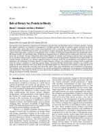
Cohort patient profiles showing CD4+ and CD8+ T cell counts and plasma viral loads for patients A, B and C, respectivelyFigure 1
Cohort patient profiles showing CD4+ and CD8+ T cell counts and plasma viral loads for patients A, B and C, respectively.
Patient B
1
10
100
1000
10000
100000
1000000
1.23.90
8.28.90
7.3.91
5.15.92
12.14.92
1.31.94
8.31.94
3.22.95
11.16.95
10.21.96
6.3.97
3.23.98
10.13.98
6.16.99
2.18.00
3.10.00
Sampling Date
Viral Load (copies / ml of blood)
0
200
400
600
800
1000
1200
1400
1600
CD4 and CD8 counts / u l
Viral
Load
CD4
CD8
Patient A
1
10
100
1000
10000
100000
1000000
5.3.90
2.27.92
4.29.92
6.1.92
8.26.92
12.16.92
4.7.93
7.28.93
11.17.93
3.9.94
12.22.94
4.16.96
2.6.98
9.13.99
Sampling Date
Viral Load (copies / ml of blood)
0
200
400
600
800
1000
1200
1400
1600
CD4 and CD8 counts / u l
Viral
Load
CD4
CD8
Patient C
1
10
100
1000
10000
100000
1000000
1.31.90
10.10.90
3.11.91
3.23.92
8.11.92
4.7.93
1.10.94
8.8.94
5.24.95
12.12.95
6.11.96
3.7.97
12.30.97
10.19.98
4.20.99
3.1.00
12.5.00
Sampling Date
Viral
Load
(copies
/
ml
of
blood)
0
200
400
600
800
1000
1200
1400
1600
CD4 and CD8 counts / ul
Viral
Loa
d
CD4
CD8
Retrovirology 2005, 2:41 />Page 4 of 10
(page number not for citation purposes)
Split graph of the cohort reconstructed using the Kimura-2-parameter corrected distancesFigure 2
Split graph of the cohort reconstructed using the Kimura-2-parameter corrected distances. The splits were refined since this
significantly improved the fit. Bootstrap values are indicated on the edges and were performed using the Neighbor-Joining
method on 1000 replicates (previously published in Mikhail et al., 2005). Bayesian trees were reconstructed in mrBayes v2.01.
Network analysis was performed in Splitstree v 1.0.1, 2.4; Huson 1998).
Retrovirology 2005, 2:41 />Page 5 of 10
(page number not for citation purposes)
recombination appears to be an inherent property in this
cluster, its exact biological association with progression
and non-progression of HIV disease in this cohort is only
partially clear, and the possible role of selection pressures
on disease progression is needed to be investigated.
Selective pressure and evolutionary rate analysis
To investigate the selective pressure exerted on the virus in
the cohort members, a non-synonymous/synonymous
substitution rate ratio scan was performed on the com-
plete genomes using a maximum likelihood estimation
procedure (Figure 3). The average dN/dS ratio shows con-
siderable variation across the genome, with the highest
ratios in the env gene, intermediate values in the accessory
genes and lower values in the pol gene, with fairly low val-
ues for the gag gene. A similar analysis using complete
genomes, representative for the HIV-1 diversity group M
found from the Los Alamos HIV Database, also resulted in
a similar plot, confirming previous reported results
[9,17,18]. With the methods at hand, we can quantify the
selective pressure across the genome for the complete
cohort but it is not possible to document differences in
selective pressure between cohort members due to param-
eter constraints of the mathematical models used. Thus,
although over time analyses do demonstrate that differen-
tial selective pressure is clearly present in this cohort, its
clear relationship with disease progression cannot be
unraveled due to the possible contributing role of recom-
bination. And since selection can result in heterogeneous
rates along sequences, conflicting phylogenetic signal in
this cohort might also have arisen from selection in addi-
tion to recombination. This is further confirmed by the
correlation of the log likelihood estimates of the overall
phylogenetic hypothesis plotted against the dN/dS ratios
obtained by the scanning window approach (data not
shown).
To investigate differences in evolutionary rate between
patients, molecular clock analysis was performed. Figure 4
shows the root-to-tip divergence in function of the sam-
pling time. Linear regression estimates for the evolution-
ary rates were 2.38 × 10
-3
(7.33 × 10
-4
-3.87 × 10
-3
), 7.75 ×
10
-3
(1.86 × l0
-3
-8.38 × 10
-3
) and 3.77 × 10
-3
(3.07 × 10
-3
-
4.44 × 10
-3
) nucleotide substitutions/site/year for patient
Table 1: Results of the Homoplasy Test and the Informative Sites Test
Homoplasy Test Informative Sites Test
P value HR P value ISI
complete genome P < 0.001 0.254 P < 0.001 0.34
gag P < 0.017 0.565 P < 0.098 0.38
pol P < 0.015 0.299 P < 0.007 0.41
env P < 0.043 0.152 P < 0.002 0.42
Non-synonymous : synonymous base rate ratio across the complete genome as estimated under a codon substitution model (MO) in a sliding window fashion with a step size of 81 bp and a window size of 801 bp, indicating the highest ratios within the env gene, followed by the pol, gag and nef genes, respectivelyFigure 3
Non-synonymous : synonymous base rate ratio across the
complete genome as estimated under a codon substitution
model (MO) in a sliding window fashion with a step size of 81
bp and a window size of 801 bp, indicating the highest ratios
within the env gene, followed by the pol, gag and nef genes,
respectively.
Retrovirology 2005, 2:41 />Page 6 of 10
(page number not for citation purposes)
A, B and C, respectively (Figure 4). By incorporating a glo-
bal molecular clock, constraining all branches with one
single evolutionary rate, and local molecular clocks,
accommodating for different rates among different
branch sets, evolutionary rates were obtained by maxi-
mum likelihood under the tip-dated model. Table 2
shows that allowing for different rates among the patients
provided a significantly better fit (P < 0.001) than the glo-
bal clock model, illustrating that the evolutionary rates
were significantly different for the three cohort members.
It should be noted however that the non-clock model,
allowing for a different rate for each branch in the phylog-
eny, still remained significantly better as determined by
the likelihood ratio test. Estimates of the evolutionary rate
show a slow evolution for patient A and much higher rates
in the two progressors (B and C), with the highest virus
evolutionary rate in recipient B in agreement with the lin-
ear regression analysis and also consistent with his recent
death with AIDS. Thus, from these analyses we have
strong evidence showing a considerable influence of viral
evolutionary rate on HIV disease progression.
Discussion
In this study we have carried-out detailed analyses of
molecular factors that might contribute to HIV disease
progression in an epidemiologically-linked cohort in
which a HIV-infected non-progressor transmitted virus to
recipients who gradually progressed to AIDS. With the
help of 15 full-length HIV-1 genomes derived from the
cohort members, where time and source of infection were
known, we are able to show how various genetic changes
following transmission of HIV from a non-progressor
(donor A) accompanied disease progression in two recip-
ients (B and C). Previously, Sydney Blood Bank Cohort
(SBBC) also identified a similar transmission of HIV-1
from a non-progressor to 5 other recipients, but in this
case patients did not progress as they were all infected
with a nef-deleted HIV-1 strain [19]. We have investigated
host-induced viral divergence, selection pressure, recom-
bination and viral evolutionary rates of HIV-1 strains in
this cohort.
It is apparent that following transmission of HIV-1 from
the donor A, the 2 recipients B and C gradually deterio-
rated over a 15-year period to low CD4
+
/CD8
+
T cell
counts and high viral loads despite the continuation of
HAART since 1997. These data suggest a possible role of in
vivo viral divergence and host selection pressure over time,
in the transition of a virus associated with non-progres-
sion in the donor, to a virus associated with gradual
progression of HIV in the 2 recipients B and C of the
cohort. To investigate this, the contribution of recombina-
tion to the genetic diversity and consequently disease pro-
gression evident in these cohort members was assessed
using IST and the Homoplasy test. As our cohort is epide-
miologically-linked, classical techniques such as Simplot,
which uses a scanning window approach to detect con-
flicting topologies, are unreliable. Our methods capture
conflicting phylogeny signal at the third codon positions
and fourfold degenerate sites, which is unlikely to have
resulted from selective pressure, thus indicating recombi-
nation. For the complete genomes, similar recombination
indices were obtained using both tests. Some differences
were observed when individual major genes were consid-
ered which could be attributed to different methodology
and/or different parameters used by the two different
algorithms.
Host-imposed immune selection was investigated by
scanning dN/dS ratios across the genome. The variation
found across the genome was consistent with that found
for HIV-1 group M. Of particular interest was the fairly
Linear regression plot for root to tip divergence versus sam-pling date within each patient of the cohortFigure 4
Linear regression plot for root to tip divergence versus sam-
pling date within each patient of the cohort. All regressions
had an R
2
value above 0.92. This graph indicates the highest
slope and thus evolutionary rate for recipient B, followed by
recipient C and lowest evolutionary rate for non-progressing
donor A.
Retrovirology 2005, 2:41 />Page 7 of 10
(page number not for citation purposes)
low ratios obtained for the gag gene which has been
extensively implicated in CTL escape [3,20]. Further inves-
tigations of our analysis also indicates which genome
regions have high dN/dS ratios. Though various reports
have documented the evolutionary constraints placed by
overlapping reading frames and secondary structures on
RNA viruses such as HIV-1 [21,22], it is important to note
that the exact number and location of the identified posi-
tively selected sites are not under investigation. Rather this
study focuses on attributing the discordant phylogenetic
patterns detected over time between cohort members by
the possible contribution of positive selection. Differen-
tial selective pressure was found to have substantially con-
tributed to virus evolution within these three cohort
members.
Furthermore, it is noteworthy that while recombination
in addition to selection forces may have contributed to the
formation of the virus causing the gradual progression of
HIV in the 2 recipients, it is possible that the HIV status of
these individuals is associated with their HLA types, and
not only due to the possible emergence of CTL escape
mutations or other host factors as described previously
[7,15,23].
In addition, by investigating the divergence of the serially
sampled sequences using linear regression [24], we ana-
lyzed the rate of viral evolution. Although this analysis is
suggestive of higher evolutionary rates in both progres-
sors, the overlapping confidence intervals do not allow us
to conclude significant differences. Earlier reports con-
ducted by Ganeshan et al., and Essajee and colleagues
based their HIV diversity studies on only partial segments
of the env gene [25,26], conducting similar phylogenetic
analysis but assessing viral heterogeneity either through
heteroduplex assays or nucleotide based distance matri-
ces, respectively. Despite both reports depending only on
the env gene, which is naturally variable, both indicate
that early quasispecies diversification may be associated
with a favorable clinical outcome, with limited heteroge-
neity correlating to slower HIV disease, and a lack of ver-
tical transmission from mother child pairs, respectively
[25,26]. Taken together, literature suggests that an inverse
relationship exists between viral diversity and disease pro-
gression [25,26], however other studies inclusive of ours
also indicate the contrary [15,27]. Moreover, as our
analysis relies on predetermined mathematical algo-
rithms the assumption of data independence by linear
regression estimates is violated as sequences share a phyl-
ogenetic history. Therefore, we estimated the evolutionary
rates using a maximum likelihood framework that takes
this into account and allows us to test different hypothe-
ses using local clock models imposed onto the genealogy
[28,29]. This molecular clock analysis, confirmed a higher
rate of evolution in progressors B and C, as opposed to a
lower rate in non-progressing donor A. The fact that HIV
evolutionary rate could be patient-specific and influenced
by immunologic control or even therapy-induced control
[30], has major implications for evolutionary and vaccine
studies. In our study it is difficult to assess the role of
therapy-induced control of HIV-evolution as both patient
B and C, who received therapy, had intermittent changes
in drug regimen, which usually comprises of a cocktail of
drugs and makes it impossible to dissect the role of each
drug on the virus. Previous studies have indicated that
combinations of RT drugs can act together to further
increase HIV-1 mutation frequencies [30]. Thus, although
we believe that therapy may have partially influenced viral
evolution of HIV-1 strains in cohort patients, it is difficult
to assess contribution of individual drugs in affecting viral
evolutionary rates. Nonetheless, it is important to reiterate
that it does not bias our overall interpretation of HIV dis-
ease progression as both recipients prior to initiation of
therapy (pre 1997) were showing a gradual decline in T
cell counts and rising plasma viremia.
Thus, the most unique aspect of our study the demonstra-
tion of patient-specific evolutionary rates as a major con-
tributor to the general lack of a molecular clock in HIV. To
date no molecular clock model accommodates for recom-
bination and one can dispute the relevance of the evolu-
tionary rates obtained. However, the genealogy-based
estimates are in good agreement with the linear regression
estimates, which were based on the viral divergence for
each patient separately. Simulations have shown that
recombination, even in small amounts, can disturb the
Table 2: Parameter estimates and log likelihoods under different clock models
Model p Log L Evolutionary rate
Different Rates 34 -24119 n.a.
Global clock 21 -24218 ABC: 2.928 × l0
-
3 (± 0.72 × l0
-
3)
Local clock for A and (BC) 22 -24164 A: 1.308 × l0
-
3 (± 0.19 × 10
-
3), BC: 5.08810
-
3 (± 0.41 × 10
-
3)
Local clock for A, B and C 23 -24156 A: 1.008 × l0
-
3 (± 0.16 × 10
-
3), B: 1.2 × l0
-
2 (± 1.86 × 10
-
3), C: 4.8 × l0
-
3 (± 0.38 × 10
-
3)
p
The amount of parameters used in the model.
LogL
The log likelihoods.
Retrovirology 2005, 2:41 />Page 8 of 10
(page number not for citation purposes)
molecular clock [31,32], and hence why the more general
non-clock model provides a better fit to this data.
Overall, our studies raise the possibility that non-progres-
sors, in some cases may harbor both pathogenic and non-
pathogenic variants. Host genetics may act as driving force
for positive selection of infecting strains [33]. Although
viral recombination and differential selective pressure
were found to have significantly affected virus variability
in all 3 cohort members, there was striking correlation
between faster viral evolutionary rate with accelerated dis-
ease progression.
Materials and methods
Cohort patient profiles
By using the well-described approaches of both Lookback
and Traceback, clusters of distant HIV transmissions can
be identified [34]. One such cluster was identified with
the donor A, who likely acquired infection in 1982 and
infected 2 recipients B (in 1983 autumn) and C (in 1983
summer) through blood transfusion. These infections
were confirmed serologically in late 1990. The donor has
remained well for over twenty years without requiring
antiretroviral therapy and has maintained CD4
+
T cell
count above 550 cells/mm3 and CD8
+
T cell count over
600 cells/mm3 and a viral load consistently less than
10000 copies /ml. In contrast, both recipients (B and C)
have required the use of highly active antiretroviral
therapy (HAART) which was initiated in 1995 and 1997
respectively (consisting of ddl/3TC/IMD) with recipient B
still alive. On the other hand recipient C experienced a
dramatic decline in CD4
+
T cell count in 1997 down to
CD4
+
T cell count of 7 cells / mm
3
(Figure 1A, IB and 1C)
and has recently died of AIDS-related illness after 14 years
post-infection. HLA typing was also conducted revealing
patient A to be type A2, A3, B57, B65 and unknown for
locus C, patient B showed to be HLA A2, A11, B56, B62
and CW1, while patient C was similariy found to be HLA
A2, A24, B7, B13 and unknown for locus C. For a detailed
description of patient clinical profiles, patient HLA types
and phylogenetic evidence confirming epidemiological
linkage refer to Mikhail et al., 2005.
Full Length genome amplification of HIV-1 strains
Gene-Amp XL PCR kit (Perkin – Elmer Emerville Ca, USA)
together with nested internal PCR reactions were used to
amplify near full-length HIV genomes (8766 base pairs,
the LTR domains were amplified separately) as previously
published [5,15]. Population sequencing was conducted
on a total of four longitudinal cohort samples obtained
from donor A, termed Al, A3, A5, and A6 and corre-
sponded to years 1992, 1997, 1998 and 2000. Similarity
4 time points from patient B were termed B3, B4, B5 and
B6 correspond to years: 1992, 1997, 1998 and 2000 for
sample collection, with C2, C3, C5, C6, C8, C10 and C11
representing patient C samples obtained from 1993,
1994, 1996, 1993, 1997, 1998 and 2000. To investigate
the presence of patient mutations within a known CTL
epitope, a database search was conducted within the Los
Alamos (NM, USA) immunology database [18]. HIV-1
near full length sequences derived from cohort patients
were consequently used to confirm epidemiological link-
age and investigate molecular gene by gene comparisons
as previously published [15].
Sequencing and phylogenetic analysis of cohort patients
Population nucleotide sequences and peptide sequences
were aligned using CLUSTAL W [35] and manually edited
in Se-AI according to their reading frame. The best-fitting
nucleotide-substitution model was selected using
Modeltestv3.06 [36], Phylogenetic trees were recon-
structed in PAUP4.0bl0, starting from a Neighbor-Joining
tree under a heuristic maximum likelihood search that
implemented both nearest-neighbor interchange (NNI)
and subtree pruning-regrafting (SPR). Bootstrap analysis
was performed using the Neighbor-Joining method on
1000 replicates (previously published in Mikhail et al.,
2005). Bayesian trees were reconstructed in mrBayes
v2.01. Network analysis was performed in Splitstree 2.4.
Recombination analysis
Since the detection of specific recombination patterns and
breakpoints in closely related sequences might be unreli-
able, evidence for recombination was investigated on a
non-overlapping DNA concatemer or in single gene
regions using two different tests: (a) the Informative Sites
Test (IST) as implemented in PIST on the third codon
positions [16], and (b) the Homoplasy Test on the
fourfold degenerate sites [16]. The Homoplasy Test deter-
mines if there is a statistically significant excess of homo-
plasies in the phylogenetic tree derived from the data set,
compared to an estimate of the number of homoplasies
expected by repeated mutation in the absence of recombi-
nation [37]. An index of greater than zero indicates link-
age equilibrium or recombination, but a value of zero or
less indicates pure clonal evolution [34], The IST test
detects whether the proportion of two-state parsimony-
informative sites to all polymorphic sites is greater than
expected from clonally generated data [16].
Selective pressure
Non-synonymous to synonymous substitution rate ratio's
(dN/dS) were estimated in a sliding-window fashion
under a probabilistic model of codon substitution that
restricts all sites to a single dN/dS (M0) index across the
complete genome [28]. All calculations were performed
using the codeml program from the PAML package.
Retrovirology 2005, 2:41 />Page 9 of 10
(page number not for citation purposes)
Evolutionary rate analysis
Root-to-tip divergences were calculated in VirusRates v.0,
provided by Andrew Rambaut [24]. Confidence intervals
for the linear regression estimates were obtained by boot-
strapping the original alignment. Maximum likelihood
analysis and local clock modeling was performed in
PAML v 3.13 b, provided by Ziheng Yang, which imple-
ments a tip-date model estimated as additional parame-
ters under the constraint that the positions of the tips are
proportional to the sampling date [28].
Genbank accession numbers
Near full length HIV-1 genomes derived from cohort
patient's PBMCs have been allocated Genebank accession
numbers AY779550
-AY779564.
List of abbreviations used
HIV-l human immunodeficiency virus type 1
AIDS acquired immunodeficiency syndrome
PBMC peripheral blood mononuclear cells
IST Informative site test
HR homoplasy ratio
SBBC Sydney blood bank cohort
CTL cytotoxic T lymphocyte
HLA human leukocyte antigen
NNI nearest neighbor interchange
Competing interests
The author(s) declare that they have no competing
interests.
Authors' contributions
M.M was assisted by B.W in carrying out the molecular
genetic studies, generating sequence alignments, and
drafting the paper. P.L conducted the evolutionary and
recombination studies, B.B together with M.J.G provided
the clinical samples, under analysis, while A-M.V partici-
pated in the design of the evolutionary study and its anal-
ysis. N.K.S conceived of the study, participated in its
supervision, design, complete coordination and conclu-
sion. All authors read and approved the final manuscript.
Acknowledgements
Authors would like to thank all members of the cohort for their participa-
tion. M.M was supported by the Australian Postgraduate Award (APA)
from the University of Sydney and a top-up grant from the Millennium
Foundation. P.L. was supported by the Flemish Institute for Scientific-tech-
nological Research in Industry (IWT).
References
1. Michael ML, Chang G, d'Arcy LA, Tseng CJ, Birx DL, Sheppard HW:
Functional characterization of human immunodeficiency
virus type 1 nef genes in patients with divergent rates of dis-
ease progression. J Virol 1995, 69:6758-6769.
2. Trachtenberg E, Korber B, Sollars C, Kepler TB, Hraber PL, Hayes E,
Funkhouser R, Fugate M, Theiler J, Hsu YS, Kunstman K, Wu S, Phair
J, Erlich H, Wolinsky S: Advantage of rare HLA supertype in
HIV disease progression. Nat Med 2003, 9:928-935.
3. Wang B, Dyer WB, Zaunders JJ, Mikhail M, Sullivan JS, Williams L,
Haddad DN, Harris G, Holt JA, Cooper DA, Miranda-Saksena M, Boa-
dle R, Kelleher AD, Saksena NK: Comprehensive Analyses of a
Unique HIV-1 -Infected Non-progressor Reveal a Complex
Association of Immunobiological Mechanisms in the Con-
text of Replication-Incompetent Infection. Virology 2000,
304:246-264.
4. Harrer T, Harrer E, Kalams SA: Cytotoxic T lymphocytes in
asymptomatic longterm non-progressing HIV-1 infection.
Breadth and specificity of the response and relation to in vivo
viral quasispecies in a person with prolonged infection and
low viral load. J Immunol 1996, 156:2616-2623.
5. Wang B, Mikhail M, Dyer WB, Zaunders JJ, Kelleher AD, Saksena NK:
First demonstration of lack of viral sequence evolution in a
non-progressor, defining replication-incompetent HIV-infec-
tion. Virology 2003, 312:315-350.
6. Wilson CC, Brown RC, Korber BT, Wilkes BM, Ruhl DJ, Sakamoto
D, Kunstman K, Luzuriaga K, Hanson 1C, Widmayer SM, Wiznia A,
Clapp S, Ammann AJ, Koup RA, Wolinsky SM, Walker BD: Frequent
detection of escape from cytotoxic T-lymphocyte recogni-
tion in perinatal human immunodeficiency virus (HIV) type
1 transmission: the ariel project for the prevention of trans-
mission of HIV from mother to infant. J Virol 1999,
73:3975-3985.
7. Migueles SA, Sabbaghian MS, Shupert WL, Bettinotti MP, Marincola
FM, Martino L, Hallahan CW, Selig SM, Schwartz D, Sullivan J, Con-
nors M: HLA B*5701 is highly associated with restriction of
virus replication in a subgroup of HIV-infected long term
nonprogressors. Proc Nafl Acad Sci U S A 2000, 97:2709-2714.
8. Kaslow RA, Carrington M, Apple R, Park L, Munoz A, Saah AJ, Goed-
ert JJ, Winkler C, O'Brien SJ, Rinaldo C, Detels R, Blattner W, Phair
J, Erlich H, Mann DL: Influence of combinations of human major
histocompatibility complex genes on the course of HIV-1
infection. Nat Med 1996, 2:405-411.
9. Yusim K, Kesmir C, Gaschen B, Addo MM, Altfeld M, Brunak S, Chi-
gaev A, Detours V, Korber BT: Clustering patterns of cytotoxic
T-lymphocyte epitopes in human immunodeficiency virus
type 1 (HIV-1) proteins reveal imprints of immune evasion
on HIV-1 global variation. J Virol 2002, 76:8757-8768.
10. Rosenberg ES, Billingsley JM, Caliendo AM, Boswell SL, Sax PE, Kalams
SA, Walker BD: Vigorous HIV-1 -specific CD4+ T cell
responses associated with control of viremia. Science 1997,
278:1447-1450.
11. Fang G, Burger H, Chappey C, Rowland-Jones S, Visosky A, Chen CH,
Moran T, Townsend L, Murray M, Weiser B: Analysis of transition
from long-term nonprogressive to progressive infection
identifies sequences that may attenuate HIV type 1. AIDS Res
Hum Retroviruses 2001, 17:1395-1404.
12. Saksena NK, Wang B, Dwyer WB: Biological and Molecular
Mechanisms in Progression and non-Progression of HIV
Disease. AIDS Rev 2001, 3:3-10.
13. Saksena NK, Ge YC, Wang B, Xiang SH, Ziegler J, Palasanthiran P,
Bolton W, Cunningham AL: RNA and DMA sequence analysis of
the nef gene of HIV type 1 strains from the first HIV type 1 -
infected long-term nonprogressing mother-child pair. AIDS
Res Hum Retroviruses 1997, 13:729-732.
14. Wang B, Ge YC, Palasanthiran P, Xiang SH, Ziegler J, Dwyer DE, Ran-
dle C, Dowton D, Cunningham A, Saksena NK: Gene defects clus-
tered at the C-terminus of the vpr gene of HIV-1 in long-
term nonprogressing mother and child pair: in vivo evolution
of vpr quasispecies in blood and plasma. Virology 1996,
223:224-232.
15. Mikhail M, Wang B, Lemey P, Beckholdt B, Vandamme AM, Gill JM,
Saksena NK: Full-Length HIV-1 Genome Analysis Showing
Evidence For HIV-1 Transmission From A Non-Progressor
To Two Recipients Who Progressed To AIDS. AIDS Res Hum
Retrov 2005 in press.
Publish with BioMed Central and every
scientist can read your work free of charge
"BioMed Central will be the most significant development for
disseminating the results of biomedical research in our lifetime."
Sir Paul Nurse, Cancer Research UK
Your research papers will be:
available free of charge to the entire biomedical community
peer reviewed and published immediately upon acceptance
cited in PubMed and archived on PubMed Central
yours — you keep the copyright
Submit your manuscript here:
/>BioMedcentral
Retrovirology 2005, 2:41 />Page 10 of 10
(page number not for citation purposes)
16. Posada D, Crandall KA: Evaluation of methods for detecting
recombination from DNA sequences: Computer
simulations. PNAS 2001, 98:13757-13762.
17. Maynard Smith J, Smith NH, O'Rouke , Spratt BG: How Clonal Are
Bacteria? Proc Natl Acad Sci 1993, 90:4384-4388.
18. Editors, Korber TMB, Brander C, Haynes BF, Koup R, Kuiken C,
Moore JP, Walker DB, Watkins ID: Theoretical Biology and Bio-
physics, Los Alamos, HIV Molecular Immunology 2. Pub-
lisher: Los Alamos National Laboratory. 2000:UR03-5816
[ />]. Los Ala-
mos, New Mexico
19. Birch MR, Learmont JC, Dyer WB, Deacon NJ, Zaunders JJ, Saksena
N, Cunningham AL, Mills J, Sullivan JS: An examination of signs of
disease progression in survivors of the Sydney Blood Bank
Cohort (SBBC). J Clin Virol 2001, 22(3):263-270.
20. Yang Z, Yoder AD: Estimation of the transition/transversion
rate bias and species sampling. J Mol Evol 1999, 48:274-283.
21. Holmes EC: Error thresholds and the constraints to RNA virus
evolution. Trends Microbiol 2003, 11(12):543-546.
22. Simmonds P, Smith DB: Structural constraints on RNA virus
evolution. J Virol 1999, 73(7):5787-5794.
23. Goulder PJ, Brander C, Annamalai K, Mngqundaniso N, Govender U,
Tang Y, He S, Hartman KE, O'Callaghan CA, Ogg GS, Altfeld MA,
Rosenberg ES, Cao H, Kalams SA, Hammond M, Bunce M, Pelton SI,
Burchett SA, Mclntosh K, Coovadia HM, Walker BD: Differential
narrow focusing of immunodominant human immunodefi-
ciency virus gag-specific cytotoxic T-lymphocyte responses
in infected African and caucasoid adults and children. J Virol
2000, 74:5679-5690.
24. Rambaut A: Estimating the rate of molecular evolution: incor-
porating non-contemporaneous sequences into maximum
likelihood phylogenies. Bioinformotics 2000, 16:395-399.
25. Ganeshan S, Dickover RE, Korber BT, Bryson YJ, Wolinsky SM:
Human immunodeficiency virus type 1 genetic evolution in
children with different rates of development of disease. J Virol
1997, 71(1):663-677.
26. Essajee SM, Pollack H, Rochford G, Oransky I, Krasinski K,
Borkowsky W: Early changes in quasispecies repertoire in
HIV-infected infants: correlation with disease progression.
AIDS Res Hum Retroviruses 2000, 16(18):1949-957.
27. Matala E, Crandall KA, Baker RC, Ahmad N: Limited heterogene-
ity of HIV type 1 in infected mothers correlates with lack of
vertical transmission. AIDS Res Hum Retroviruses 2000,
16(15):1481-1489.
28. Yang Z, Bielawski JP: Statistical methods for detecting molecu-
lar adaptation. Trends Ecol Evol 2000, 15:496-503.
29. Drummond A, Rodrigo AG: Reconstructing genealogies of serial
samples under the assumption of a molecular clock using
serial-sample UPGMA. Mol Biol Evol 2000, 17:1807-1815.
30. Mansky LM: HIV mutagenesis and the evolution of antiretro-
viral drug resistance. Drug Resist Updaf 2000, 5:219-223. Review
31. Schierup MH, Hein J: Recombination and the molecular clock.
Mol Biol Evol 2001, 17(10):1578-1579.
32. Maynard Smith J, Smith NH: Detecting recombination from
gene trees. Mol Biol Evol 1998, 15:590-599.
33. Deacon NJ, Tsykin A, Solomon A, Smith K, Ludford-Menting M,
Hooker DJ, McPhee DA, Greenway AL, Ellett A, Chatfield C:
Genomic structure of an attenuated quasi species of HIV-1
from a blood transfusion donor and recipients. Science 1995,
270:988-991.
34. Gill MJ, Towns D, Allaire S, Meyers G: Transmission of human
immunodeficiency virus through blood transfusion: the use
of look-back and trace-back approaches to optimize recipi-
ent identification in a regional population. Transfusion 1997,
37:513-516.
35. Thompson JD, Higgins DG, Gibson TJ: CLUSTAL W: improving
the sensitivity of progressive multiple sequence alignment
through sequence weighting, position-specific gap penalties
and weight matrix choice. Nucleic Acids Res 1994, 22:4673-4680.
36. Posada D, Crandall KA: MODELTEST: testing the model of
DMA substitution. Bioinformatics 1998, 14:817-818.
37. Worobey M: A novel approach to detecting and measuring
recombination: new insights into evolution in viruses, bacte-
ria, and mitochondria. Mol Biol Evol 2001, 18:1425-1434.