Báo cáo y học: "Ku protein as a potential human T-cell leukemia virus type 1 (HTLV-1) Tax target in clastogenic chromosomal instability of mammalian cells" ppt
Bạn đang xem bản rút gọn của tài liệu. Xem và tải ngay bản đầy đủ của tài liệu tại đây (1.15 MB, 10 trang )
BioMed Central
Page 1 of 10
(page number not for citation purposes)
Retrovirology
Open Access
Research
Ku protein as a potential human T-cell leukemia virus type 1
(HTLV-1) Tax target in clastogenic chromosomal instability of
mammalian cells
Franca Majone*
1
, Roberto Luisetto
1
, Daniela Zamboni
1
, Yoichi Iwanaga
2
and
Kuan-Teh Jeang
2
Address:
1
Department of Biology, University of Padua, Padua, Italy and
2
Laboratory of Molecular Microbiology, NIAID, NIH, Bethesda, Maryland,
20892-0460, USA
Email: Franca Majone* - ; Roberto Luisetto - ;
Daniela Zamboni - ; Yoichi Iwanaga - ; Kuan-Teh Jeang -
* Corresponding author
Abstract
The HTLV-1 Tax oncoprotein rapidly induces cytogenetic damage which can be measured by a
significant increase in the number of micronuclei (MN) in cells. Tax is thought to have both
aneuploidogenic and clastogenic effects. To examine the cellular target for Tax which might
mechanistically explain the clastogenic phenomenon, we tested the ability of Tax to induce MN in
rodents cells genetically defective for either the Ku80 protein or the catalytic subunit of DNA
protein kinase (DNAPKcs). We found that cells genetically mutated in Ku80 were refractory to
Tax's induction of MN while cells knocked-out for DNAPKcs showed increased number of Tax-
induced MN. Using a cytogenetic method termed FISHI (Fluorescent In Situ Hybridization and
Incorporation) which measures the number of DNA-breaks in cells that contained unprotected 3'-
OH ends, we observed that Tax increased the prevalence of unprotected DNA breaks in Ku80-
intact cells, but not in Ku80-mutated cells. Taken together, our findings suggest Ku80 as a cellular
factor targeted by Tax in engendering clastogenic DNA damage.
Background
We previously demonstrated that expression of the HTLV-
I Tax oncoprotein rapidly induces cytogenetic damage
which is reflected in a significant increase in the preva-
lence of micronuclei (MN) in cells [1-4]. To further char-
acterize the phenomenon of Tax associated clastogenic
DNA-damage, we wished to examine the status DNA-
breaks in the nucleus and in MN in the presence or
absence of Tax [4]. Using a cytogenetic method termed
FISHI (Fluorescent In Situ Hybridization and Incorpora-
tion), DNA-breaks in the nucleus and in MN with centric
or acentric DNA fragments could be characterized for the
presence or absence of free 3'-OH ends. In our definition,
free 3'-OH ends represent breaks which are accessible to
the in situ addition of digoxigenin (DIG) -labeled dUTP
using terminal deoxynucleotidyl transferase. On the other
hand, an absence of accessible 3'-OH ends suggests that
the breaks are protected and masked by a protein com-
plex. In vivo, unprotected free 3'-OH ends may progress to
larger lesions leading to increasingly serious chromo-
somal lesions which may eventually sow the seed for cel-
lular transformation.
Published: 13 July 2005
Retrovirology 2005, 2:45 doi:10.1186/1742-4690-2-45
Received: 13 June 2005
Accepted: 13 July 2005
This article is available from: />© 2005 Majone et al; licensee BioMed Central Ltd.
This is an Open Access article distributed under the terms of the Creative Commons Attribution License ( />),
which permits unrestricted use, distribution, and reproduction in any medium, provided the original work is properly cited.
Retrovirology 2005, 2:45 />Page 2 of 10
(page number not for citation purposes)
In earlier studies, we had observed that Tax increased the
frequency of MN containing centric DNA fragments with
unprotected free 3'-OH ends and that Tax decreased the
frequency of MN containing DNA fragments with incor-
poration-inaccessible (i.e. protected) 3'-OH ends. Based
on an increase in free 3'-OH containing ends/breaks, we
hypothesized that Tax interfered with a protective cellular
mechanism(s) that may normally recruit a protein com-
plex to newly created DNA breaks. Subsequent to the pub-
lication of our report [4], Gabet et al. [5] showed that in
some settings Tax can repress the expression of the cata-
lytic subunit of human telomerase (hTert).
Telomerase is a ubiquitously-expressed multi-protein
complex composed of a catalytic subunit (hTert), two
associated proteins (TP-1 and HSP 90), and a highly con-
served RNA (hTR) component of ~400 nucleotides. hTert
acts as a reverse transcriptase, and normally catalyses the
addition of short repetitive sequences to the ends of chro-
mosomes using an RNA-template embedded within the
hTert holoenzyme. Telomerase is expressed in proliferat-
ing stem cells, in germ cells, in activated lymphocytes and
in many neoplastic cells such as gastric and colorectal car-
cinoma, breast tumours and adrenal tumours [6,7], and in
some pre-neoplastic growths [8]. It is generally assumed
that telomerase is silent in most primary somatic cells.
Interestingly, because of the manner by which eukaryotic
cells replicate DNA, when a cell does not have active tel-
omerase, telomeres at the ends of chromosomes shorten
progressively after every cellular division. Once the telom-
eric repeats have reached a critically abbreviated state, fur-
ther cell division cannot ensue. This constraint may
explain the senescence seen for normal somatic cells.
Telomeric repeats at the ends of chromosomes also appear
to serve an end-protective function. Chromosomal ends
which lack telomeric repeats are labile for end-to-end
chromosome fusion and exonucleolytic degradation
which can progress to further genetic rearrangements/
damages. Provocatively, such gross rearrangements/dam-
ages can, at a low frequency, fortuitously alter the genome
in a way to actually induce telomerase activity in the
genetically altered cells. Once induced, such telomerase
activity could endow the cells with the capacity to prolif-
erate indefinitely, and this event could represent a first
step towards malignant transformation [9].
We previously hypothesized [4] that proteins such as Ku,
Sir, and the DNA protein kinase catalytic subunit (DNAP-
Kcs) which are normally found at telomeric ends of chro-
mosomes could be recruited rapidly to de novo interstitial
chromosomal breaks. We had proposed that de novo inter-
stitial breaks may be recognized by hTert and be stabilized
by the transient addition of telomeric repeats which could
then recruit Ku, Sir and DNAPKcs proteins [4]. Of note,
Ku and DNAPKcs are also components of the non-homol-
ogous end-joining (NHEJ) DNA repair pathway. NHEJ is
important for the repair of double-stranded DNA breaks.
Knock-out mice and cultured cells deficient for one or
more components of the Ku-DNAPKcs complex show
genome instability phenotypes [10-16].
Because Tax interferes with the stability of de novo DNA
breaks [4] and because Ku and DNAPKcs proteins appar-
ently contribute protection to DNA breaks, we wish to
understand how Tax influences double stranded DNA-
breaks in cells (e.g. hamster xrs-6 cells) which are either
genetically mutated for the Ku80 protein [10,11] or
knocked out for the DNAPKcs gene (e.g. mouse embryo
DNAPKcs -/- fibroblasts) [12]. We reasoned that if Tax acts
to subvert the Ku protein, then cells (i.e. xrs-6) already lost
for Ku80 would not incur increased DNA-break instability
when Tax is over-expressed. On the other hand, if Tax tar-
gets DNAPKcs function, then we would expect that
DNAPKcs-/- cells would not show enhanced frequency of
cytogenetic damage when Tax is over-expressed, while xrs-
6 cells would. Here, we used xrs-6 cells, DNAPKcs-/- cells,
and the technique of in situ DIG-dUTP incorporation to
distinguish between Ku80 and DNAPKcs as a DNA-break
stabilizing factor targeted by Tax.
Results
MN induction by Tax in hamster and mouse cells
Clastogenic and aneuploidogenic agents increase the fre-
quency of micronuclei (MN) because they disturb
genome stability control mechanisms [1,4]. The fre-
quency of MN can be viewed as being proportional to the
cell's (in)efficiency at maintaining its genomic integrity.
The NHEJ (Non-Homologous End Joining) pathway is
one of the major pathways which eukaryotes use to repair
double-stranded DNA breaks. Ku and DNAPKcs subunits
are important NHEJ protein components.
To check Tax's effect in cells impaired for NHEJ, we first
monitored the ambient frequency of micronuclei in ham-
ster xrs-6 cells which have a mutated Ku80 gene [10,17].
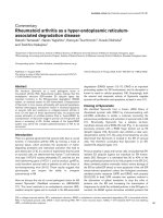
We observed that MN frequency was significantly higher
in xrs-6, than control CHO (Chinese hamster ovary) cells
(Fig. 1). To the extent that MN reflects DNA-damage, this
result suggests that under normal tissue culture conditions
xrs-6 cells have a higher proclivity for cytogenetic damage.
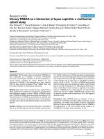
We next investigated mouse embryo fibroblasts (MEFs)
engineered to be DNA-PKcs-/- [12]. We found that DNAP-
Kcs-/- cells had a ten fold higher ambient frequency of MN
when compared to wild type MEFs (DNA-PKcs+/+); and
we also saw that DNAPKcs heterozygous MEFs (DNA-
PKcs+/-) showed a five fold increase in MN over control
MEFs (Fig. 2). Taken together, the results in figures 1 and
2 argue that both DNAPKcs and Ku proteins are important
Retrovirology 2005, 2:45 />Page 3 of 10
(page number not for citation purposes)
for the normal genomic homeostasis that prevents MN.
Inactivation of either of these two NHEJ components
appears to predispose the cell to increased cytogenetic
damage.
We next compared MN frequencies in CHO, xrs-6, DNAP-
Kcs+/+ and DNAPKcs-/- cells after transfection with a Tax-
expression plasmid. Interestingly, after Tax transfection,
the frequency of micronuclei in the xrs-6 cells did not sig-
nificantly change from that seen in the same cells without
Tax (Fig. 1). By contrast, Tax-transfected CHO cells
showed a three fold increase in MN compared to mock
transfected cells (Fig. 1). When we checked DNAPKcs+/+
and DNAPKcs-/- cells, we also found that both cell types
showed increases in micronuclei after Tax-expression (Fig.
2).
We interpret the above results to mean that in Ku-intact
cells (i.e. DNAPKcs+/+, DNAPKcs-/-, and CHO cells), Tax
can increase cytogenetic damage above ambient levels. By
contrast, Tax does not increase the extent of genetic dam-
age in Ku defective cells (i.e. xrs-6 cells) (Fig. 1, 2). The
two findings can be explained if Ku80 is specifically tar-
geted by Tax. If so, because xrs-6 cells are already lost for
Ku80, its already high baseline level of MN cannot be fur-
ther aggravated by Tax. On the other hand, Tax could tar-
get the still intact Ku function in DNAPKcs+/+, DNAPKcs-
/-, and CHO cells to increase MN numbers.
DIG(digoxigenin)-dUTP incorporation in nuclei and MN of
hamster and mouse cells
We next investigated the status of DNA breaks in the
nuclei and MN of xrs-6, DNA-PKcs-/- and control cells
using the previously described in situ DIG-dUTP incorpo-
ration assay [4]. This method incorporates in situ a tagged-
dUTP which can be used to identify and quantify broken
and unprotected 3'-OH DNA ends. We were curious to
compare how Tax affects the protection of 3'-OH DNA
ends in Ku80-/- (i.e. xrs-6) and DNAPKcs-/- cells.
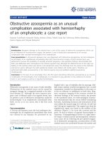
We found that the frequency of incorporated DIG-dUTP
in nuclei and MN was significantly increased in xrs-6 cells
compared to control CHO cells (Fig. 3). Under normal
culturing conditions, xrs-6 cells showed robust and
numerous in situ DIG-dUTP signals in nuclei and MN
(Fig. 4A). These findings suggest that loss of Ku-function
Frequency (%) of micronuclei containing cells in xrs-6 and CHO cell cultures without or with transfection by TaxFigure 1
Frequency (%) of micronuclei containing cells in xrs-6 and CHO cell cultures without or with transfection by
Tax. *** indicates significantly different value (P < 0.001, G test) from that found in CHO cells. ** indicates significantly differ-
ent value (P < 0.01, G test) from that found in CHO cells.
Retrovirology 2005, 2:45 />Page 4 of 10
(page number not for citation purposes)
significantly increases the prevalence of unprotected freely
accessible 3'-OH DNA ends. Interestingly, when we trans-
fected Tax into xrs-6 cells, no further increase in DIG-
dUTP incorporation in either the nuclei or MN was appar-
ent (Fig. 4B). Thus, Tax expression in cells already lost for
Ku80 failed to change further the number of unprotected
3'OH-DNA ends.
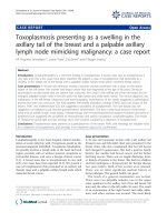
We also checked DNAPKcs-/- MEFs. These cells are
knocked out for the DNAPKcs gene but have intact Ku80
protein. Here, we found that the ambient incorporation of
DIG-dUTP into DNAPKcs-/- nuclei and MN was low (Fig.
3; Fig. 5A). Indeed, the DIG-dUTP incorporation fre-
quency in DNAPKcs-/- cells was not significantly different
from that in control DNAPKcs+/+ or in DNAPKcs+/- het-
erozygote cells (Fig. 3). After transfection with a Tax-plas-
mid, both DNAPKcs +/+ (Fig. 3) and DNA PKcs-/- (Fig. 3;
Fig. 5B) showed significant increases in the incorporation
of DIG-dUTP into nuclei and MN. Unlike xrs-6 cells,
DNAPKcs-/- and DNAPKcs+/+ cells have intact Ku80; we
interpret their DIG-dUTP incorporation results to mean
that Tax targeted the Ku80 protein in these cells and that
such targeting increased the number of DIG-dUTP acces-
sible unprotected 3'OH DNA ends.
Reduced Ku80 expression in HTLV-1 transformed cells
The above findings suggested Ku80 as a Tax-target. To ask
if Tax affects Ku80 in HTLV-1 transformed human cells,
we investigated the expression of this protein in Jurkat,
MT-4, and C81 cells (Fig. 6). Jurkat is a spontaneously
transformed T-cell line unrelated to HTLV-1; while both
MT-4 and C81 cells are HTLV-1 transformed cells that
highly express Tax. Using anti-Ku antibody which recog-
nizes both the Ku70 and Ku80 proteins, we found that
constitutive expression of Ku80 was reduced in both cells
that express Tax, MT-4 and C81 (Fig. 6, lanes 5 and 9),
Frequency (%) of micronuclei in primary cultures of mouse embryo fibroblasts with indicated genotypes of DNAPKcs +/+, DNAPKcs +/-, or DNAPKcs -/- assayed without or with transfection of a Tax plasmidFigure 2
Frequency (%) of micronuclei in primary cultures of mouse embryo fibroblasts with indicated genotypes of
DNAPKcs +/+, DNAPKcs +/-, or DNAPKcs -/- assayed without or with transfection of a Tax plasmid. *** indi-
cates significantly different value (P < 0.001, G test) from that in DNAPKcs +/+ cells, with or without transfect with Tax plas-
mid. ** indicates significantly different value (P < 0.01, G test) from that in DNAPKcs +/+ cells without transfection with Tax
plasmid.
Retrovirology 2005, 2:45 />Page 5 of 10
(page number not for citation purposes)
when compared to Jurkat (Fig. 6, lane 1). Interestingly,
when cells were treated with mitomycin C (a DNA-dam-
aging agent), Ku80 expression remained inducible in both
MT-4 and C81 cells. Thus targeting of Ku80 by Tax appears
not to be an irreversible process.
Discussion
Tax has been reported to cause both aneuploidogenic and
clastogenic effects. Here we have explored the clastogenic
property of Tax. We posed a simple question: in cells
respectively defective for either Ku80 or DNAPKcs, which
cell type remains responsive to Tax-induction of MN and
DIG-dUTP incorporation? Based on our results that cells
genetically mutated in Ku80 were no longer responsive to
Tax's induction of MN and DIG-dUTP incorporation, we
posit that Ku80, but not DNAPKcs, is a functional Tax
target.
Both Ku and DNAPKcs are important for NHEJ. The cur-
rent thinking is that Ku protein binds to DNA discontinu-
ously and in a sequence independent manner, carrying
out a DNA-protective role [18]. Once bound to DNA, Ku
proteins recruit and activate the catalytic DNAPKcs subu-
nit which can phosphorylate Ku and other neighboring
DNA-bound proteins [19]. It has also been reported that
DNAPKcs self-phosphorylates to inactivate the holo-
kinase complex and then dissociates itself from Ku and
the DNA. In this manner, the helicase activity of Ku is
inactivated, allowing base pairing to occur between
micro-homologous regions. DNAPKcs further recruits the
Comparison of the frequency of in situ incorporation of digoxigenin (DIG)-dUTP in nuclei of hamster and mouse cells in the absence or presence of TaxFigure 3
Comparison of the frequency of in situ incorporation of digoxigenin (DIG)-dUTP in nuclei of hamster and
mouse cells in the absence or presence of Tax. *** indicates significantly different value (P < 0.001, G test) from that
found in the respective control (comparison between the paired columns). ## or ### indicates significantly different value (P <
0.01, or P < 0.001, G test) from that of the respective controls in the absence of Tax.
&+2
;56
&+27D[
;567D[
'LJ G873 LQFRUSRUDWLRQ
VF.3$1'
;$7VF.3$1'
VOOHFHVXRPVOOHFUHWVPDK
Retrovirology 2005, 2:45 />Page 6 of 10
(page number not for citation purposes)
Visualization of in situ incorporation of DIG-dUTP in xrs-6 cells in the absence (A) or presence (B) of transfected TaxFigure 4
Visualization of in situ incorporation of DIG-dUTP in xrs-6 cells in the absence (A) or presence (B) of trans-
fected Tax. Counterstaining with propidium iodide is shown as red fluorescence while incorporation of DIG-dUTP is shown
as yellow-green fluorescence. Multiple views show that in situ incorporation signals in nuclei and micronuclei do not increase
substantially after transfection with a Tax-expressing plasmid.
!
"
823
Retrovirology 2005, 2:45 />Page 7 of 10
(page number not for citation purposes)
Visualization of in situ incorporation of DIG-dUTP in PKcs-/- cells in the absence (A) or presence (B) of transfected Tax plasmidFigure 5
Visualization of in situ incorporation of DIG-dUTP in PKcs-/- cells in the absence (A) or presence (B) of trans-
fected Tax plasmid. Counterstaining with propidium iodide is shown as red fluorescence while incorporation of DIG-dUTP
is shown as yellow-green fluorescence. Multiple views show that in the presence of the Tax (B) the incorporation signals are
far greater than those in the absence of Tax (A). Note that many MN are seen to contain in situ incorporation signals.
!
"
$.!0+CS
Retrovirology 2005, 2:45 />Page 8 of 10
(page number not for citation purposes)
XRCC4/ligase IV protein, which provides the DNA-ligase
function needed to complete repair [20]. This intimate
interplay between DNAPKcs and Ku explains why an
absence of one or the other protein results in increased
cytogenetic aberrations in cells.
Ku and DNAPKcs are commonly found at the telomeric
ends of chromosomes. One view is that these proteins
with other factors assemble a telomeric "cap" which con-
tributes to the stability of chromosome ends [21]. Of
note, there is evidence which suggests that telomeric
repeats may also be transiently added to de novo interstitial
chromosomal breaks leading to their stabilization and
preventing further exacerbation of damage [22]. Accord-
ingly, DNA-ends or DNA-breaks not capped by telomeric
sequences and their associated proteins are unstable and
labile to aberrant fusions [23,13]. Interestingly, studies
have shown that upon DNA damage, PARP-1 (a nuclear
enzyme which catalyzes the polyADP-ribosylation of tar-
get proteins in response to DNA damage) and Ku proteins
are rapidly activated and compete for binding to DNA-
ends [24], suggesting a general activity conferred by these
proteins in stabilizing damaged DNA [25]. PARP-1 and
Ku proteins can be co-immunoprecipitated [26],
indicating that the two DNA end-sensing molecules inter-
act in response to DNA strand breakages. Moreover, Ku
function can be modulated by PARP-1 [27,28]. Thus,
PARP-1 polyADP-ribosylates itself and also Ku70/80, and
the polyADP-ribosylated Ku 70/80 is reduced in its DNA
binding affinity, and becomes attenuated in its ability to
stimulate Werner syndrome (WRN) exonuclease [28].
Our current data add the viral Tax oncoprotein to the list
of complex interactors with Ku. We report here that cells
genetically knocked out for Ku80 are refractory to the
induction by Tax of MN and DIG-dUTP incorporation.
Interestingly, in cells intact for Ku80, Tax expression
reduced the ambient expression of this protein. It remains
to be resolved how Tax mechanistically affects Ku80-
expression; however, adding our current to our previous
demonstration that Tax interferes with the protective cel-
lular mechanisms used normally for stabilizing DNA
breaks [4,29], we propose that Ku80 likely represents a
crucial DNA end-protective protein targeted by Tax. Tar-
geting of DNA end-protective proteins by oncoproteins
may attenuate the functions of these factors and could
lead to increased DNA structural instability and progres-
sion of damage. Progression of DNA structural damage
may ultimately contribute to and mechanistically explain
the process of cellular transformation. Our views on the
implications of protecting de novo DNA-breaks with telo-
meric-caps for cellular transformation are in part
Reduced constitutive expression of Ku80 in MT-4 and C81-6645 (C81) cells compared to Jurkat cellsFigure 6
Reduced constitutive expression of Ku80 in MT-4 and C81-6645 (C81) cells compared to Jurkat cells. Total cell
lysates were prepared from the indicated cell lines and probed with anti-serum which recognizes both Ku70 and 80 proteins.
Where indicated the cells were also treated with 1 µM mitomycin C (MMC) for the stated time period before harvesting.
Note that constitutively reduced Ku80 expression remains inducible by MMC in the two Tax expressing cell lines (MT-4 and
C81).
Retrovirology 2005, 2:45 />Page 9 of 10
(page number not for citation purposes)
consistent with recent findings that telomeric fusion to
breaks reduces oncogenic translocations and tumor for-
mation [30].
Materials and methods
Cells and transfection
Hamster xrs-6 (genetically mutated for Ku 80) cells, CHO
wild type cells, mouse embryo fibroblasts knocked out for
the PKcs gene, and PKcs +/- or PKcs+/+ MEFs, were cul-
tured as monolayers in Dulbecco's minimal essential
medium (Invitrogen) supplemented with 10% fetal calf
serum. Where indicated, cells were transiently transfected
using calcium phosphate with a wild-type Tax expression
plasmid (HPx). The cells were surveyed 48 hours later for
cytogenetic effects.
Micronuclei (MN) assay
For MN assay, suspensions of cells were prepared by
trypsinization of cultured cells in log-phase. Cells were
divided into 40 mm dishes with each dish receiving 8 × 10
5
cells in 10 ml of medium. The cells were collected 48 h
later by trypsinization and were washed in phosphate-
buffered saline and fixed for 15 minutes in paraformalde-
hyde (1% in PBS) for in situ incorporation analysis. Inter-
phase preparations were obtained following the
procedures previously described [1].
Fluorescence in situ incorporation
Fluorescence in situ incorporation was carried out using
terminal transferase (TdT) which catalyses the addition of
deoxyribonucleotide triphosphates to the 3'-OH ends of
single or double-stranded DNA. To the substrates of TdT,
digoxigenin-11-dUTP (the digoxigenin is bound to posi-
tion 5 of the pyrimidine by an arm of 11 carbon atoms)
was added to the 3'-OH ends. Antibody detection of DIG-
dUTP labelling employed a specific antibody linked to
fluoresceine, a fluorochrome which when stimulated at
494 nm wavelength emits a green signal (λ = 523 nm).
The experimental protocol for fluorescent in situ incorpo-
ration used 2 washes with HBS (NaCl 280 mM, Na
2
PO
4
×
7H
2
O, 1.5 mM, Hepes 50 mM). The TdT incorporation
reaction of DIG-11-dUTP used the following: 10 µl of a
solution (Boheringer) containing potassium cocodylate 1
M, Tri-HCl 125 mM (pH 6.6, 4°C), Bovine serum albu-
min (BSA) 1.25 mg/ml, CoCl2 10 mM; 0.2 µl of a solution
(Boheringer) containing TdT (25 units/µl), EDTA 1 mM,
2 mercaptoethanol 4 mM, glycerol 50% (v/v) (pH 6.6,
4°C); 1 µl of DIG-11-dUTP (1 mM) mixture (Boheringer).
Distilled water was added to a final volume of 50 µl. The
cells were incubated in this solution at 37°C for 1 hour in
an HBS-moist environment. At the end of the incubation
the slides were immersed into a basin containing 0.1%
Triton X-100 and 0.5% BSA in HBS to equilibrate the
slides with anti-DIG-11-dUTP (1:50) labelled with FITC
(Boheringer). Equilibration was conducted at room
temperature for 30 minutes in an HBS moist environ-
ment. The slides were subsequently washed 3 times for 5
minutes each with the same HBS solution. The slides were
then counterstained with propidium iodide (0.3 µg/ml).
Scoring of the slides
Fluorescent microscopy was performed on a Zeiss micro-
scope with different filters and equipped with an HBO
100 mercury lamp (Osram, Munchen, Germany). Photo-
graphs were taken on Kodak Ektachrome 166 ASA film. To
determine the number of MN per nucleus in slides, for
each experimental point, 3000 cells were counted, using
at least two independent slides for each experimental
point. Differences between data from spontaneous and
Tax induced cytogenetic effects were tested for significance
using the G test [31].
Competing interests
The author(s) declare that they have no competing
interests.
Acknowledgements
We thank Claudio Friso and Renzo Mazzaro (Department of Biology,
Padua) for technical assistance in the preparation of figures, members of the
Jeang laboratory for critical reading of manuscript, and Anthony Elmo for
preparation of manuscript.
References
1. Majone F, Semmes OJ, Jeang KT: Induction of micronuclei by
HTLV-I Tax. Virology 1993, 193:456-459.
2. Semmes OJ, Majone F, Cantemir C, Turchetto L, Hjelle B, Jeang KT:
HTLV-I and HTLV-II Tax: differences in induction of micro-
nuclei in cells and transcriptional activation of viral LTRs.
Virology 1996, 217:373-379.
3. Jeang KT, Majone F: Aneuploidogenic and clastogenic DNA
damages induced by the HTLV-1 Tax protein. In Molecular
pathogenesis of HTLV-1 Edited by: Semmes OJ, Hammarskjöld ML.
Arlington, Va, USA: ABI Professional Publications; 1999:43-48.
4. Majone F, Jeang KT: Clastogenic effect of the human T-cell
leukemia virus type I Tax oncoprotein correlates with unsta-
bilized DNA breaks. J Biol Chem 2000, 275:32906-32910.
5. Gabet A, Mortreux F, Charneau P, Riou P, Duc Dodon M, Wu Y,
Jeang KT, Wattel E: Inactivation of hTERT transcription by
Tax. Oncogene 2003, 22:3734-3741.
6. Mannelli M, Gelmini S, Arnaldi G, Becherini L, Bemporad D, Crescioli
C, Pazzagli M, Mantero F, Serio M, Orlando C: Telomerase activity
is significantly enhanced in malignant adrenocortical tumors
in comparison to benign adrenocortical adenomas. J Clin
Endocrinol Metab 2000, 85:468-470.
7. Boltze C, Mundschenk J, Unger N, Schneider-Stock R, Peters B,
Mawrin C, Hoang-Vu C, Roessner A, Lehnert H: Expression profile
of the telomeric complex discriminates between benign and
malignant pheochromocytoma. J Clin Endocrinol Metab 2003,
88:4280-4286.
8. Zhu X, Kumar R, Mandal M, Sharma N, Sharma HW, Dhingra U,
Sokoloski J, Hsiao R, Narayanan R: Cell cycle dependent modula-
tion of telomerase activity in tumor cells. Proc Natl Acad Sci USA
1996, 93:6091-6095.
9. Musutomi K, Hahn W: Telomerase and tumorigenesis. Cancer
Lett 2003, 194:189-197.
10. Jeggo PA, Kemp LM: X-ray-sensitive mutants of Chinese ham-
ster ovary cell line, isolation and cross-sensitivity to other
DNA-damaging agents. Mutat Res 1983, 112:313-327.
11. Taccioli GE, Gottlieb TM, Blunt T, Priestley A, Demengeot J, Mizuta
R, Lehmann AR, Alt FW, Jackson SP, Jeggo PA: Ku80: Product of
the XRCC5 gene and its role in DNA repair and V(D)J
recombination. Science 1994, 265:1442-1445.
Publish with BioMed Central and every
scientist can read your work free of charge
"BioMed Central will be the most significant development for
disseminating the results of biomedical research in our lifetime."
Sir Paul Nurse, Cancer Research UK
Your research papers will be:
available free of charge to the entire biomedical community
peer reviewed and published immediately upon acceptance
cited in PubMed and archived on PubMed Central
yours — you keep the copyright
Submit your manuscript here:
/>BioMedcentral
Retrovirology 2005, 2:45 />Page 10 of 10
(page number not for citation purposes)
12. Kurimasa A, Ouyang H, Wang S, Cordon Cardo G, Li G: Catalytic
subunit of DNA dependent protein kinase impact on lym-
phocyte development and tumorigenesis. Proc Nat Acad Sci USA
1999, 96:1403-140.
13. Bailey SM, Meyne J, Chen DJ, Kurimasa A, Li GC, Lehnert BE, Good-
win EH: DNA double-strand break repair proteins are
required to cap the ends of mammalian chromosomes. Proc
Natl Acad Sci USA 1999, 96:14899-14904.
14. Ferguson DO, Sekiguchi J, Frank K, Gao Y, Sharpless NE, Gu Y, Manis
J, Depinho RA, Alt FW: The interplay between NHEJ and cell
cycle checkpoint factors in development genomic instability
and and tumorigenesis. Cold Spring Harbor Symposia on Quantitative
Biology 2000, LXV:395-403.
15. Pastink A, Eeken JC, Lohman PH: Genomic integrity and the
repair of double-strand DNA breaks. Mutat Res 2001, 480–
481:37-50.
16. Sekiguchi J, Ferguson DO, Yang E, Frank K, Gu Y, Nussennzweig A,
Alt FW: Genetic interactions between ATM and non homol-
ogous end joining factors in genomic stability and
development. Proc Natl Acad Sci USA 2001, 23:3243-3248.
17. Getts RC, Stamato TD: Absence of a Ku-like DNA end binding
activity in the xrs double-strand DNA repair-deficient
mutant. J Biol Chem 1994, 269:15981-15984.
18. Rathmel WK, Chu G: Involvment of the Ku autoantigen in the
cellular response to DNA double-strand breaks. Proc Natl Acad
Sci USA 1994, 91:7623-7627.
19. Chu G: Double strand break repair. J Biol Chem 1997,
272:24097-24100.
20. Carlsson P, Wateman ML, Jones KA: The hLEF/TCF 1 alpha HMG
protein contains a context dependent transcriptional activa-
tion domain that induces the TCR alpha enhancer in T cells.
Genes Dev 1993, 7:2418-2430.
21. Weaver DT: Telomeres: moonlighting by DNA repair
proteins. Curr Biol 1998, 8:492-494.
22. Wilkie AOM, Lamb J, Harris PC, Finney RD, Higgs DR: A truncated
human chromosome 16 associated with α thalassaemia is
stabilized by addition of telomeric repeat (TTAGGG)
n
.
Nature 1990, 346:868-871.
23. Boulton SJ, Jackson SP: Identification of a Saccharomyces cere-
visiae Ku80 homologue: roles in DNA double strand break
rejoining and in telomeric maintenance. Nucleic Acids Research
1996, 24:4639-4648.
24. D'Silva I, Pelletier JD, Lagueux J, D'Amours D, Chandhry MA, Wein-
feld M, Lees-Miller SP, Poirier GG: Relative affinities of
poly(ADP-ribose) polymerase and DNA-dependent protein
kinase for DNA strand interruptions. Biochem Biophys Acta 1999,
1430:119-126.
25. Herceg Z, Wang ZQ: Functions of poly (ADP-ribose) polymer-
ase (PARP) in DNA repair, genomic integrity and cell death.
Mutat Res 2001, 477:97-110.
26. Ariumi Y, Masutani M, Copeland TD, Mimori T, Sugimura T, Shimo-
tohno K, Ueda K, Hatanaka M, Noda M: Suppression of the
poly(ADP-ribose) polymerase activity by DNA-dependent
protein kinase in vitro. Oncogene 1999, 18:4615-4625.
27. Tong WM, Cortes U, Hande MP, Ohgaki H, Cavalli LR, Landsdorp
PM, Haddad BR, Wang ZQ: Synergistic role of Ku80 and
Poly(ADP-ribose) Polymerase in suppressing chromosomal
aberrations and liver cancer formation. Cancer Research 2002,
62:6990-6996.
28. Li B, Navarro S, Kasahara N, Comai L: Identification and bio-
chemical characterization of a Werner syndrome protein
complex with Ku70/80 and PARP-1. J Biol Chem 2004,
279:13659-13667.
29. Jeang KT, Giam G, Majone F, Aboud M: Life, death, and Tax: role
of HTLV-1 oncoprotein in genetic instability and cellular
transformation. J Biol Chem 2004, 279:31991-31994.
30. Qi L, Strong MA, Karim BO, Huso DL, Greider CW: Telomere
fusion to chromosome breaks reduces oncogenic transloca-
tions and tumour formation. Nature Cell Biol 2005, 7:706-711.
31. Sokal P, Rolph FJ: Biometry San Francisco: Freemann; 1991.