Báo cáo y học: " Silencing of human T-cell leukemia virus type I gene transcription by epigenetic mechanisms" pps
Bạn đang xem bản rút gọn của tài liệu. Xem và tải ngay bản đầy đủ của tài liệu tại đây (1.3 MB, 16 trang )
Retrovirology
BioMed Central
Open Access
Research
Silencing of human T-cell leukemia virus type I gene transcription
by epigenetic mechanisms
Yuko Taniguchi1, Kisato Nosaka1,2, Jun-ichirou Yasunaga1,
Michiyuki Maeda3, Nancy Mueller4, Akihiko Okayama5 and
Masao Matsuoka*1
Address: 1Laboratory of Virus Immunology, Institute for Virus Research, Kyoto University, Kyoto 606-8507, Japan, 2Department of Hematology,
Kumamoto University School of Medicine, Kumamoto 860-8556, Japan, 3Laboratory of Infection and Prevention, Institute for Virus Research,
Kyoto University, Kyoto 606-8507, Japan, 4Department of Epidemiology, Harvard School of Public Health, Boston, Massachusetts 02115, USA
and 5Department of Laboratory Medicine, Faculty of Medicine, University of Miyazaki, Miyazaki 889-1692, Japan
Email: Yuko Taniguchi - ; Kisato Nosaka - ; Junichirou Yasunaga - ; Michiyuki Maeda - ;
Nancy Mueller - ; Akihiko Okayama - ;
Masao Matsuoka* -
* Corresponding author
Published: 22 October 2005
Retrovirology 2005, 2:64
doi:10.1186/1742-4690-2-64
Received: 31 August 2005
Accepted: 22 October 2005
This article is available from: />© 2005 Taniguchi et al; licensee BioMed Central Ltd.
This is an Open Access article distributed under the terms of the Creative Commons Attribution License ( />which permits unrestricted use, distribution, and reproduction in any medium, provided the original work is properly cited.
Abstract
Background: Human T-cell leukemia virus type I (HTLV-I) causes adult T-cell leukemia (ATL) after
a long latent period. Among accessory genes encoded by HTLV-I, the tax gene is thought to play a
central role in oncogenesis. However, Tax expression is disrupted by several mechanims including
genetic changes of the tax gene, deletion/hypermethylation of 5'-LTR. To clarify the role of
epigenetic changes, we analyzed DNA methylation and histone modification in the whole HTLV-I
provirus genome.
Results: The gag, pol and env genes of HTLV-I provirus were more methylated than pX region,
whereas methylation of 5'-LTR was variable and 3'-LTR was not methylated at all. In ATL cell lines,
complete DNA methylation of 5'-LTR was associated with transcriptional silencing of viral genes.
HTLV-I provirus was more methylated in primary ATL cells than in carrier state, indicating the
association with disease progression. In seroconvertors, DNA methylation was already observed
in internal sequences of provirus just after seroconversion. Taken together, it is speculated that
DNA methylation first occurs in the gag, pol and env regions and then extends in the 5' and 3'
directions in vivo, and when 5'-LTR becomes methylated, viral transcription is silenced. Analysis of
histone modification in the HTLV-I provirus showed that the methylated provirus was associated
with hypoacetylation. However, the tax gene transcript could not be detected in fresh ATL cells
regardless of hyperacetylated histone H3 in 5'-LTR. The transcription rapidly recovered after in
vitro culture in such ATL cells.
Conclusion: These results showed that epigenetic changes of provirus facilitated ATL cells to
evade host immune system by suppressing viral gene transcription. In addition, this study shows the
presence of another reversible mechanism that suppresses the tax gene transcription without DNA
methylation and hypoacetylated histone.
Page 1 of 16
(page number not for citation purposes)
Retrovirology 2005, 2:64
Background
Human T-cell leukemia virus type I (HTLV-I) is associated
with a neoplastic disease, adult T-cell leukemia (ATL), and
inflammatory diseases, such as HTLV-I-associated myelopathy (HAM)/tropical spastic paraparesis (TSP) and
HTLV-I-associated uveitis [1,2]. Among HTLV-I carriers, a
part of infected individuals develop ATL after a long latent
period. During the leukemogenesis by HTLV-I, Tax protein is considered to play a critical role through its pleiotropic actions, which include transactivation of NF-κB,
CREB and SRF pathways, transrepression of lck, p18 and
DNA polymerase β gene transcriptions, and functional
inactivation of p53 and MAD1 [3-6]. Through these
actions, Tax induces the proliferation of HTLV-I infected
cells and inhibits their apoptosis, resulting in an increase
in the number of infected cells. However, since Tax protein is the major target of cytotoxic T-lymphocytes (CTLs)
in vivo, the expression also has a negative effect on the survival of ATL cells [7-9]. In some ATL cells, tax gene expression is inactivated by genetic and epigenetic changes,
which include deletion, insertion or mutation of the tax
gene, and DNA methylation or deletion of 5'-LTR [10-13].
Such inactivation of Tax expression is considered to allow
ATL cells to escape from the host immune system.
DNA methylation of retroviruses is regarded as a host
defense mechanism for inactivating retrovirus expression
[14]. However, it is also recognized as a mechanism for
virus-infected cells to escape from the host immune system and establish the latent state. In contrast, human
immunodeficiency virus (HIV) is resistant to silencing in
vivo. It is because HIV is frequently integrated into active
transcriptional unit in vivo [15]. These findings coincide
with the fact that HIV vigorously replicates in vivo. On the
other hand, DNA methylation accumulated in HTLV-I 5'LTR has been shown to silence viral gene transcription in
leukemic cells [12,13]. In addition, the frequency of integration of HTLV-I provirus into transcriptional units was
equivalent to that calculated based on random integration
[16], which also increased the silencing. It remains
unclear where and when DNA methylation occurs within
the HTLV-I provirus genome.
In this study, we analyzed DNA methylation and histone
modification in the whole HTLV-I provirus, and observed
the progressive accumulation of DNA methylation. In
addition, another reversible mechanism silenced viral
gene transcription regardless of hyperacetylated promoter
region.
Results
Analyses of DNA methylation of HTLV-I provirus
To reveal DNA methylation status within the HTLV-I provirus, we analyzed the DNA methylation by sodium
bisulfite sequencing and combined bisulfite restriction
/>
analysis (COBRA). Initially, DNA methylation in 5'-LTR,
gag, pol, env, pX and 3'-LTR was identified by sodium
bisulfite sequencing. In an ATL case (Fig. 1A), the internal
regions of the HTLV-I provirus, including gag, pol and
env, were heavily methylated. On the other hand, 5'-LTR
and pX were partially methylated, and 3'-LTR was not
methylated at all. In an ATL cell line, ATL-48T (Fig. 1A),
the internal sequences of the HTLV-I provirus were partially methylated, whereas both LTRs were not methylated. Since the analyses by sodium bisulfite sequencing
were time-consuming, we established the COBRA method
to detect and analyze DNA methylation in a large number
of samples, and then compared the results obtained with
the two methods. After amplification of sodium bisulfite
treated DNAs with each primer sets, the products were
digested with TaqI or AccII, which contain one (TaqI) or
two (AccII) CpG site(s) within the recognition sequences.
When CpG site is methylated, the products retain CpG
site, resulting in digestion by these enzymes. On the other
hand, CpG is converted to UG when it is unmethylated.
Therefore, PCR products are resistant to restriction
enzymes (Fig. 1B). With the COBRA method, the extent of
DNA methylation was quantified in eight CpG sites
throughout the HTLV-I provirus: 5'-LTR (620 according to
the numbering by Seiki et al. [17]), gag (1753), pol (2988,
4187 and 5151), env (6113), pX (7258) and 3'-LTR
(8342) (Fig. 1C). The extent of DNA methylation detected
by the COBRA method was well correlated with that
obtained by sodium bisulfite sequencing in both cases
studied, as shown in Fig. 1A and 1C.
DNA methylation throughout the HTLV-I provirus in
HTLV-l-transformed and ATL cell lines
Using the COBRA method, we analyzed the DNA methylation throughout the whole HTLV-I provirus of the cell
lines (Fig. 2B and 2C). In addition, we also analyzed the
tax gene transcription by RT-PCR (Fig. 2A) and the
number of integrated HTLV-I proviruses in each cell lines
by Southern blot method. Among the tax gene-expressing
cell lines (ATL-35T, MT-2, Sez627, MT-4, ATL-55T, MT-1
ATL-48T and ATL-2) (Fig. 2A), internal sequences from
gag to pX were variably methylated. However, 5'-LTR was
not methylated or partially methylated, while 3'-LTR was
not methylated in all cell lines (Fig. 2B). In ATL-43T and
TL-Oml, which did not show tax gene transcription (Fig.
2A), 5'-LTR and the internal sequences were heavily methylated (Fig. 2C), indicating the close correlation between
the extents of DNA methylation of the provirus, particularly 5'-LTR, and tax gene transcription. As previously
reported, the treatment by 5-aza-deoxy-cytidine can
recover the tax gene expression of these cell lines, indicating that the latent state by DNA methylation of 5'-LTR is
reversible [13].
Page 2 of 16
(page number not for citation purposes)
Retrovirology 2005, 2:64
/>
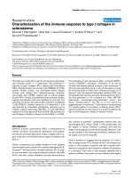
Figure 1
DNA methylation of the HTLV-I provirus assessed by sodium bisulfite sequencing and COBRA
DNA methylation of the HTLV-I provirus assessed by sodium bisulfite sequencing and COBRA. A. DNA methylation in the HTLV-I provirus was analyzed by sodium bisulfite sequencing in a case of acute ATL and a tax gene-expressing cell
line, ATL-48T. Eight DNA regions, which were represented as bars in A, were amplified with sodium bisulfite treated DNA.
The PCR products were subcloned into plasmid DNA, and then the sequences of each clone were determined for at least ten
clones of each region. Arrowheads indicate the CpG sites that were target sites for COBRA. Closed circle indicates methylated CpG, and open circle means unmethylated CpG. The number of integrated provirus has been shown in parenthesis. B.
Representative data of COBRA has been shown. PCR products, which were amplified with sodium bisulfite treated DNAs,
were digested with TaqI or AccII. The extent of methylation in each CpG site was measured as described in Methods, and presented as percentages of methylated CpG. The number in parenthesis represents the position of cytidine residue in analyzed
CpG site by COBRA according to Seiki et al. [41]. C. DNA methylation studied by COBRA at eight points in the provirus as
shown by arrowheads. Each bar represented the extent of DNA methylation at the points shown by arrowhead. The analyses
by COBRA were performed three times independently, and the extents of DNA methylation are shown by the mean ± SD.
The number in parenthesis shows the position of cytidine residue of CpG site analyzed by COBRA.
Page 3 of 16
(page number not for citation purposes)
Retrovirology 2005, 2:64
/>
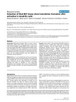
Figure 2
DNA methylation in ATL cell lines, HTLV-I carriers and ATL cases
DNA methylation in ATL cell lines, HTLV-I carriers and ATL cases. The tax gene transcription in ATL cell lines was
studied by RT-PCR (A), and the expression of GAPDH gene has been used as a control. DNA methylation throughout the
HTLV-I provirus was studied by COBRA in tax gene-expressing (B) and non-expressing cell lines (C). Furthermore, DNA
methylation was also analyzed in 20 carriers and 20 ATL cases by COBRA, and representative patterns of DNA methylation
are shown in D. The number of HTLV-I provirus has been analyzed by Southern blot method, and shown in the parenthesis (B,
C and D). Each bar indicates the extent of DNA methylation that was calculated by COBRA.
Page 4 of 16
(page number not for citation purposes)
Retrovirology 2005, 2:64
/>
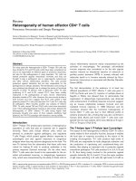
Figure 3
Comparison of the DNA methylation in carriers and ATL cases
Comparison of the DNA methylation in carriers and ATL cases. A. DNA methylation at eight different regions in the
HTLV-I provirus was compared between carriers (C) and ATL cases (A). DNA methylation was quantified by COBRA in 20
carriers and 20 ATL cases. Each sample was analyzed three times by COBRA at each site, and circles indicate mean values of
DNA methylation. The differences of DNA methylation are statistically significant in the gag, pol and env regions by the MannWhitney's U-test. Horizontal bars represent median of DNA methylation in each group. B. The relation between tax gene transcription and DNA methylation of 5'-LTR in the fresh ATL cells has been shown. DNA methylation of 5'-LTR was quantified by
COBRA assay and the tax gene transcripts were detected by RT-PCR.
Page 5 of 16
(page number not for citation purposes)
Retrovirology 2005, 2:64
/>
Seroconverter 1
60
40
20
0
80
3’-LTR
60
40
20
0
13 years
20
0
ATL-21C
100
80
% methylation
3 years
60
40
B
6 months
60
40
20
0
100
80
60
40
20
% methylation
0
5’-LTR
gag
pol 1
pol 2
pol 3
env
pX
3’-LTR
% methylation
100
40
20
100
80
% methylation
4 years
80
60
pol 2
3’-LTR
env
pX
pol 2
40
20
at seroconversion
env
pX
60
0
100
% methylation
80
100
80
% methylation
at seroconversion
5’-LTR
gag
% methylation
100
Seroconverter 2
5’-LTR
gag
A
9 years
0
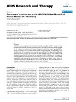
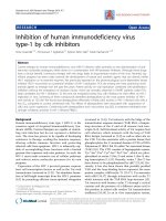
Figure 4
Sequential analyses of the DNA methylation in seroconverters and a cell line
Sequential analyses of the DNA methylation in seroconverters and a cell line. DNA methylation was analyzed by
COBRA in sequential samples from seroconverters (A) and in a cell line, ATL-21C, (B) cultured in vitro for more than 9 years.
DNA methylation was analysed by COBRA three times, and each bar indicates mean ± SD.
Page 6 of 16
(page number not for citation purposes)
Retrovirology 2005, 2:64
/>
A
B
-1400
-1000
-600 -200 +64 +620
-1400
-1000
-600 -200 +64 +620
5'LTR
5'LTR
Acute ATL 2
Acute ATL 1
N
N
C
D
5'LTR
Acute ATL 3
5'LTR
Acute ATL 21
N
N
E
F
5'LTR
5'LTR
Acute ATL 22
N
Chronic ATL 1
N
Figure 5
DNA methylation of provirus is not associated with methylated CpG sites in the genome
DNA methylation of provirus is not associated with methylated CpG sites in the genome. Integration sites of
HTLV-I provirus in leukemic cells have been determined by inverse PCR, and then DNA methylation in genome has been analyzed by sodium bisulfite sequencing. DNA methylation of 5'-LTR was also analyzed by sodium bisulfite sequencing method.
Vertical bars represent CpG sites. Open circle indicates unmethylated CpG site, and closed one means methylated CpG site.
N: normal PBMCs from non-carrier donor.
Page 7 of 16
(page number not for citation purposes)
Retrovirology 2005, 2:64
Among cell lines, HTLV-I provirus tends to be not so
methylated in cell lines with higher copy number of provirus (Fig. 2). The finding that cell lines with higher integrated provirus number contain hypomethylated provirus
is speculated to reflect the higher transcription of viral
genes.
DNA methylation of the HTLV-I provirus in ATL and HTLVI carrier states
Next, we analyzed the DNA methylation of the whole
HTLV-I provirus in ATL patients and HTLV-I carriers.
Although 5'-LTR is frequently deleted in ATL cells [10], we
omitted such ATL cases lacking 5'-LTR in this study. In Fig.
2D, we showed the representative pattern of DNA methylation of whole HTLV-I provirus in carriers and ATL
patients. In ATL samples, the gag, pol and env regions
were heavily methylated, whereas 5'-LTR was not methylated or partially methylated (Fig. 2D and 3A). On the
other hand, 5'-LTR was scarcely methylated and the gag,
pol and env regions seemed to be less methylated in
HTLV-I carriers (Fig. 2D and 3A). We compared DNA
methylation of these different eight regions between 20
carriers and 20 ATL cases (Fig. 3A). These differences in
DNA methylation were statistically significant in the gag,
pol and env regions between the ATL cases and HTLV-I
carriers by the Mann-Whitney's U-test. These data suggested that DNA methylation initially occurred in the gag,
pol, and env regions, and that DNA methylation of the
provirus accumulated during disease progression from the
carrier state to the leukemic stage. The frequency of DNA
methylation of 5'-LTR did not differ between carriers and
ATL patients. However, the extent of DNA methylation
among methylation-positive cases was higher in ATL cases
than in carriers (p = 0.001). Among ATL cases, the relationship between DNA methylation of 5'-LTR and tax
gene transcription was analyzed (Fig. 3B), and the transcript was detected in six cases. In four cases with relative
higher amount of tax gene transcripts (Case 1, 9, 12, 20),
5'-LTR was not methylated or slightly methylated. This
finding suggests that higher expression of tax gene is associated with unmethyalted or slightly methylated 5'LTR,
however, other mechanism(s) silences the tax gene transcription in ATL cells. There is no statistical correlation
between the tax gene transcription and DNA methylation
of 5'-LTR
DNA methylation of HTLV-I provirus after seroconversion
The analyses of DNA methylation suggest that it first
occurs around the gag, pol and env regions, and then
progresses in both the 5' and 3' regions. To study the
changes in DNA methylation after infection, we analyzed
sequential DNA samples from seroconverters. As shown
in Fig. 4A, DNA methylation already existed in the gag,
pol and env regions at the seroconversion. In seroconverter 1, DNA methylation was slightly increased at 4 and
/>
13 years after the seroconversion. Increase of DNA methylation at pol region (4187) is statistically significant 13
years later in seroconverter 1 (p = 0.02, by a Student's ttest). On the other hand, there was little change in the
DNA methylation in seroconverter 2, although the HTLVI provirus was already heavily methylated at the seroconversion. When DNA methylation of seroconverters was
compared with that in carriers (Fig. 3A), provirus of carriers was more methylated in carriers than that of seroconverters (p < 0.01 by a Student's t-test) except for pol2 in
seroconverter 2, and pX region. It suggests that DNA
methylation of provirus accumulates during a latent
period after seroconversion.
We established an HTLV-I-transformed cell line, ATL-21C,
and cultured for over 9 years in vitro, and analyzed the
DNA methylation of the HTLV-I provirus. Slight DNA
methylation was detected in the pol, env and pX regions at
6 months after culture, however, it did not increase after 9
years. This indicates that the DNA methylation of HTLV-I
provirus did not change after long-term in vitro culture
(Fig. 4B). On the other hand, the p16 gene in this cell line
was not methylated at 6 months after culture, but heavily
methylated after 9 years (data not shown). A comparison
with the data from the seroconverters suggests that DNA
methylation of the HTLV-I provirus tends to accumulate
in vivo.
Association with DNA methylation in the neighboring host
genome
It is possible that the HTLV-I provirus integrated into the
heterochromatin or hypermethylated regions tends to be
silenced [18], and that such HTLV-I-infected cells are
selected in vivo. Therefore, we analyzed the DNA methylation of the host genome around the integration sites of
the HTLV-I provirus. We first determined the integration
sites of the HTLV-I provirus in ATL cells, and then analyzed the DNA methylation of genomic DNAs around the
integration sites in both ATL cells and normal PBMCs
from a non-carrier donor. When genomic DNAs neighboring integration sites were heavily methylated (Fig. 5),
5'-LTR was not methylated in three cases (acute ATL 1, 2
and 21) while they were methylated in two cases (acute
ATL 3 and chronic ATL 1). In acute ATL 22, both genomic
DNA and 5'-LTR were not so methylated. Thus, DNA
methylation in the neighboring genomic regions was not
correlated with the methylation status of the provirus
among these cases.
Histone modification of the HTLV-I provirus
It has been demonstrated that DNA methylation of 5'-LTR
is associated with histone deacetylation and silencing of
viral gene transcription in cell lines [13]. When ATL-43T,
in which tax gene transcription was silenced by hypermethylation of 5'-LTR, was compared with a tax gene-
Page 8 of 16
(page number not for citation purposes)
Retrovirology 2005, 2:64
/>
Figure modifications in ATL cell lines
Histone 6
Histone modifications in ATL cell lines. Acetylation of histone was analyzed in tax gene-expressing (ATL-48T) and nonexpressing (ATL-43T) cell lines by ChIP assays with anti-acetyl-Histone H3 or H4 (A and B) at four different regions (for 5'LTR, env, pX and 3'-LTR) of the provirus. Representative data has been shown in A. W.C.E.: whole cell extract. ChIP assay was
performed three times and quantified as described in Methods. Values are means ± SD(B). *:p < 0.002.
Page 9 of 16
(page number not for citation purposes)
Retrovirology 2005, 2:64
/>
Figure 7
DNA methylation and histone modifications in fresh ATL cases
DNA methylation and histone modifications in fresh ATL cases. A. The relationships among DNA methylation, tax
gene expression and histone modification in 5'-LTR were analyzed in three ATL cases. Cases 1 and 3 have one copy of the
complete HTLV-I provirus, while Case 2 has a defective provirus that lacks part of the pol gene. DNA methylation was analyzed
by COBRA. The tax gene transcripts could be detected in Case 1, but not in Cases 2 or 3, by RT-PCR. ChIP assays were also
performed using primers for 5'-LTR to analyze acetylation of histone H3 (Ac-H3) and H4 (Ac-H4). W.C.E.: whole cell extract.
B. Recovery of tax gene expression ex vivo. The PBMCs isolated from Case 3 were immediately cultured ex vivo for several
hours and tested the transcription of tax mRNA by RT-PCR.
Page 10 of 16
(page number not for citation purposes)
Retrovirology 2005, 2:64
expressing cell line, ATL-48T, a difference was found in
the acetylation of histone H3 in 5'-LTR (Fig. 6A and 6B).
The histone H3 of 5'-LTR was hypoacetylated in ATL-43T
compared with ATL-48T, whereas there were no differences in pX or 3'-LTR among these cell lines. Since the
number of HTLV-I provirus in ATL-43T and -48T is one
and two copies respectively, and acetylation of histone H3
in pX and 3'-LTR was similar in both cell lines, the
number of provirus was thought to have no influence on
the results of ChIP assay in 5'-LTR.
However, the tax gene transcription is silenced in about
20% of ATL cases despite no or partial methylation of 5'LTR (Fig. 3B) [13], suggesting that there is aother mechanism(s) for suppressing viral gene transcription. To
address this question, we studied the histone modification of 5'-LTR in fresh ATL cells with or without tax gene
transcription. In a case with tax gene expression, 5'-LTR
was not methylated and histone H3 was hyperacetylated
(Fig. 7A, Case 1). On the other hand, in Case 2 with heavily methylated 5'-LTR, histone H3 was hypoacetylated in
5'-LTR, which was consistent with the lack of detection of
tax gene transcription in this case. However, in Case 3, tax
gene transcription could not be detected regardless of 5'LTR hyperacetylation. After in vitro culture, such cells
showed tax gene transcription within one hour (Fig. 7B).
Although both Cases 1 and 3 exhibited hyperacetylation
of 5'-LTR, tax gene transcription was silenced in Case 3.
Discussion
DNA methylation is regarded as a host defense mechanism for inactivating transportable elements such as retroviruses to inhibit viral transcription and the generation of
new viruses. On the other hand, it also renders the provirus into a latent state, resulting in the establishment of
latent infection. However, it remained unclear how and
when the provirus was methylated, and whether DNA
methylation changed in vivo.
Tax has the remarkable potency to promote the proliferation of infected cells [3], however, it is also a major target
of CTL in vivo [8]. Therefore, HTLV-I controls tax gene
expression by own viral proteins, Rex [19], p30 [20,21]
and HBZ [22]. In the leukemic cells, several mechanisms
have been identified to suppress or abolish Tax expression, including genetic changes of tax gene, deletion of 5'LTR, and DNA methylation of 5'-LTR. In this study, DNA
methylation was shown to occur in internal provirus
sequences, such as the gag, pol and env regions, and then
extend to 5' (5'-LTR) and 3' (pX) regions. Since DNA
methylation of 5'-LTR is associated with tax gene transcription, the finding that 5'-LTR was more highly methylated in ATL cells than in carriers, among cases with
methylated 5'-LTR, suggests that such HTLV-I-infected
cells and ATL cells with the methylated provirus, which
/>
produce lower amounts of viral proteins, are selected in
vivo by the host immune system. In this regard, HTLV-I is
quite different from another human retrovirus, HIV-1.
HIV-1 vectors were resistant to gene silencing in vivo
[23,24]. It is noteworthy that the number of CpG sites in
the U3 region of HIV-1 LTR (9 sites in LTR of NL43) is
much fewer than that of HTLV-I (47 sites in LTR of ATK).
This is consistent to the previous report that transcriptional suppression was not associated with DNA methylation of HIV-1 provirus [25]. In addition, HIV-1 provirus is
frequently integrated within transcriptional units, which
encode the genes that are transcribed in T-cells [15,26]. In
such regions, it is possible that HIV-1 tends to escape from
transcriptional silencing that is observed in the heterochromatin region such as alphoid repetitive sequences
[18]. These data suggest that HIV-1 is more resistant to
gene silencing than HTLV-I. Alternatively, it is possible
that HTLV-I takes advantage of susceptibility to DNA
methylation to escape from the host immune system.
This study shows that 3'-LTR is unmethylated in carriers
and ATL cells while 5'-LTR is methylated in about half of
cases. In HTLV-I, HTLV-I bZIP (HBZ) gene is encoded by
minus strand of provirus [22,27]. We observed that HBZ
gene was transcribed in all ATL cells, suggesting that HBZ
gene play a critical role in growth of HTLV-I infected cells
and ATL cells (submitted for publication). The finding
that 3'-LTR is unmethylated in all ATL cases and carriers
suggests that HBZ gene transcription is important for proliferation of ATL and HTLV-I infected cells.
Why does DNA methylation occur from the internal
sequences of the HTLV-I provirus? Since CpG island is recognized as DNA region that is susceptible to DNA methylation, we analyzed HTLV-I provirus by the criterion by
Takai and Jones [28]. CpG islands are present throughout
the provirus in 5'-LTR-gag (1–1360), pol (3876–4509), env
(5648–6166), env-pX (6446–7561), and pX-3'-LTR
(8212–9045) regions. Therefore, the presence of CpG
island could not explain why DNA methylation occurred
in the internal region of HTLV-I provirus. Among tumorsuppressor genes, which are transcriptionally silenced by
DNA methylation, the exon regions are first methylated,
and then DNA methylation progresses to the promoter
region [29]. When the promoter region is heavily methylated, the transcription of the corresponding gene is
silenced. Since 5'-LTR is the promoter/enhancer for viral
gene transcription, there might be a similar scenario
between the exon/promoter and DNA methylation in
both virus and tumor-suppressor genes. Thus, it is possible that gene coding regions are first methylated and DNA
methylation spreads to the promoter region of provirus,
5'-LTR.
Page 11 of 16
(page number not for citation purposes)
Retrovirology 2005, 2:64
Transcriptional silencing of tax gene in spite of hyperacetylated histone H3 is recognized as another mechanism to suppress the viral gene transcription in addition
to DNA methylation. The prompt recovery of tax gene
expression after in vitro culture suggests the presence of an
inhibitory factor(s) that binds to 5'-LTR, and suppresses
the viral gene transcription in vivo. It is noteworthy that
this phenotype is very similar to that of a mouse T-cell line
transfected with an HTLV-I LTR-derived reporter plasmid
[30]. In that study, a green fluorescent protein-fused Tax
(Gax) gene was transfected into a mouse T-cell line, EL-4,
and the transduced cells were then injected into Taximmunized and non-immunized mice. Although Taxinduced cytotoxic T-cells suppressed the expression of the
Gax gene in vivo, its expression was shown to recover
within three hours when the transduced cells were transferred to in vitro culture. This phenotype resembles that
observed in Case 3 in Fig. 7. Considering that Tax is the
major target of CTL in vivo, and at the same time, confers
growth advantages on the infected cells, such reversible
suppression of tax gene expression is thought to be suitable for the survival of HTLV-I infected cells, and ATL cells.
In this regard, potentiation of anti-Tax immunity might
protect against the development of ATL when combined
with possible therapeutics to induce Tax expression [31].
For this purpose, the mechanism for silencing viral transcription regardless of histone H3 hyperacetylation
should be studied.
In general, gene silencing is associated with several different mechanisms. DNA methylation in the promoter
region silences the gene transcription, whereas gene
silencing is often not associated with DNA methylation
[32,33]. In such situations, methylation of H3K9 is linked
with loss of transcriptions [34]. It is possible that silencing
of viral gene transcription renders proviral DNA vulnerable to methylation. Once proviral DNA is methylated,
such silencing would be fixed unless such cells are treated
with demethylating agents such as 5-aza-deoxy-cytidine.
DNA methylation of the HTLV-I provirus did not accumulate in a cell line that was cultured in vitro for more than 9
years. The finding that the p16 gene was heavily methylated in this cell line excluded the possibility that hypermethylation did not occur in this cell line due to aberrant
methylation machinery. Among the seroconverters, the
provirus was heavily methylated in internal regions such
as gag, pol and env. Taken together, DNA methylation in
the provirus is considered to reflect the selection in vivo.
Since the growth of in vitro HTLV-I-transformed cell lines
depends on Tax expression, cells with suppressed expression of the tax gene do not have the growth advantage in
vitro. However, the immune system exerts selection of the
infected cells with suppressed tax gene expression in vivo.
/>
Recently, both 5'- and 3'-LTR have been reported to be
transcriptionally active, and transcriptional factors and
Tax bind equally to both [35]. 3'-LTR may activate the
transcription of cellular genes, which are located in the
downstream of integration sites. In addition, unmethylated 3'-LTR is critical for transcription of the HBZ gene.
Since 5'-LTR is a promoter/enhancer for viral gene transcription, selective methylation of 5'-LTR is considered to
silence the transcription of viral genes.
Conclusion
We have demonstrated how DNA methylation of HTLV-I
provirus occurred, and how it suppressed viral gene transcription. When 5'-LTR was heavily methylated, viral transcription was silenced, which is thought to reflect the
immune system selection in vivo. In addition, mechanisms other than DNA methylation suppresses viral gene
transcription regardless of histone H3 hyperacetylation.
The mechanism of such suppression requires further
investigation.
Methods
Cells
HTLV-I-associated cell lines (MT-1, MT-2, MT-4, ATL-2,
TL-Oml and Sez627) were cultured in RPMI1640 medium
supplemented with 10% fetal bovine serum and penicillin/streptomycin. For interleukin-2-dependent cell lines
(ATL-43T, 48T and 55T), 100 U/ml of recombinant interleukin-2 (Shionogi, Osaka) was added to the medium.
Peripheral blood mononuclear cells (PBMC) or lymph
node cells were isolated from HTLV-I carriers and ATL
patients after informed consent was obtained. The polyclonal integration of HTLV-I provirus in carriers has been
shown by inverse PCR [36], and provirus load was determined by real-time PCR as reported previously [37].
Sodium bisulfite treatment of genomic DNA
Sodium bisulfite treatment was performed as described
previously [29]. Briefly, 1–3 µg of genomic DNA was
denatured in 0.3 N NaOH at 37°C for 15 min, and 1 µg
of salmon sperm DNA was added to each sample as a carrier. Sodium bisulfite (pH 5.0) and hydroquinone were
added to each sample to final concentrations of 3 M and
0.05 mM, respectively. The reaction was performed at
55°C for 16 h and the samples were then desalted using
the Wizard DNA Clean-Up System (Promega, Madison,
WI). Finally, samples were desulfonated in 0.3 N NaOH at
37°C for 15 min.
Sequencing of sodium bisulfite-treated genomic DNA
The sodium bisulfite-treated DNA (200–500 ng) was used
as a template for PCR amplification of eight HTLV-I provirus regions. The PCR reactions were performed using
FastStart Taq DNA Polymerase (Roche, Mannheim, Germany). The PCR primer pairs and annealing temperatures
Page 12 of 16
(page number not for citation purposes)
Retrovirology 2005, 2:64
/>
Table 1: Primer sets for COBRA and ChIP assay
Site in HTLV-Ia
COBRA
620
(5'-LTR)
1753
(gag)
2988
(pol)
4187
(pol)
5151
(pol)
6113
(env)
7258
(pX)
8342
(3'-LTR)
5'-LTRb
env
pX
3'-LTR
Forward primer
1st
2nd
1st
2nd
1st
2nd
1st
2nd
1st
2nd
1st
2nd
1st
2nd
1st
2nd
Reverse primer
Anneal (°C)
Enzyme for COBRA
5'-TTTGGAGTTTATTTAGATTTAG-3'
5'-GTTTTGTTTGATTTTGTTTGT-3'
5'-GGGAGTGTTAAAGATTTTTTTTGGG-3'
5'-TTTATTTTTTAAGGTTTGGAGGAG-3'
5'-GTTAAAAAGGTTAATGGAATTTGG-3'
5'-GGGTTTTTTGATTTGTTTAGTTTG-3'
5'-GGGTGAAATTGTGTAGTTTTGTAGG-3'
5'-GTGATTAGTAGGGTATTTGTGAGAG-3'
5'-GGTATTATTTTAAGTTTTTTGG-3'
5'-GTTAGTGGAAAGGATTATAGGAGG-3'
5'-GGATTTATTGTTTTGATTTTTAG-3'
5'-GGATTTATTGTTTTGATTTTTAG-3'
5'-GAGGTGGYGTTTTTTTTTTTGG-3'
5'-AAGGATAGTAAATYGTTAAGTATAG-3'
5'-YGATGGTAYGTTTATGATTTTYGGG-3'
5'-YGATGGTAYGTTTATGATTTTYGGG-3'
5'-GCTTTGCCTGACCCTGCTTGC-3'
5'-TGCCAGCCTCTCCACTTGGCACG-3'
5'-AAGGATAGCAAACCGTCAAGCACAG-3'
5'-CCCCTCATTTCTACTCTCACACGGC-3'
5'-CCAATAATAAACRACCAACCC-3'
5'-AAAAAAATTTAACCCATTACC-3'
5'-ACTCCAATAACCTACTTTCCC-3'
5'-TTAAAAATCCAAATCTAACAAACCC-3'
5'-CCTCTAAAAATAATAATAAATCCTC-3'
5'-AAACTTACTAAAAAAATATCATCC-3'
5'-CCTATTTTCAAACGAATCTACCTCC-3'
5'-ATTATCACAAAAATCATTCCCCC-3'
5'-CTCCAATTATAAAAATACAACAAC-3'
5'-AACTTACCCATAATATTAAAAATC-3'
5'-CTTTACATAATCCTCCTTACTCCC-3'
5'-CCCAAAACAAAAAATCAAAACC-3'
5'-CCTTAAAAATCTTAAAAATTCTC-3'
5'-CCCAAATAATCTAATACTCTAAAC-3'
5'-ACCCCCTCCTAAACTATCTCC-3'
5'-AACTCCTACTAATTTATTAAACC-3'
5'-AAGATTTGGCCCATTGCCTAGGG-3'
5'-ATGGAGCCGGTAATCCCGCCAGC-3'
5'-CCCAGGTGATCTGATGCTCTGGAC-3'
5'-TGGGTGGTTCTTGGTGGCTTCCC-3'
45
49
55
55
52
51
57
52
46
51
51
53
47
50
57
52
63
64
63
64
TaqI
TaqI
TaqI
AccII
TaqI
TaqI
TaqI
TaqI
a Nucleotide
bFor
position corresponding to that of ATK. This number means the cytidine of CpG sites analysed.
ChIP assay, we used primers to amplify the indicated regions.
are shown in Table 1. The amplified PCR products were
purified and subcloned into pGEM-T Easy vectors
(Promega). For each region, at least 10 clones were
sequenced using Big Dye Terminator v3.1 Cycle Sequencing Kit (Applied BioSystems, Foster City, CA) and
ABI3100 autosequencer (Applied Biosystems).
Combined bisulfite restriction analysis (COBRA)
For COBRA, eight different regions of HTLV-I provirus
were amplified with sodium bisulfite treated genomic
DNAs using each primer sets as shown in Table 1. The
nested PCR reactions were performed using FastStart Taq
DNA Polymerase (Roche) with the following condition: 5
minutes at 95°C for denaturation, 40 cycles of 30 sec at
95°C, 30 sec at each annealing temperature (Table 1), 30
sec at 72°C, and 2 min at 72°C for final extension. The
PCR products were digested for at least 4 hrs with an
appropriate restriction enzyme (TaqI or AccII) that had a
single recognition site within each product [38]. When
CpG site within amplified region was methylated, it was
resistant to sodium bisulfite treatment, resulting in digestion by these enzymes. On the other hand, since unmethylated CpG was converted to UG by sodium bisulfite
treatment, these enzymes could not digest the amplified
DNAs. The digested PCR products were separated in a 3%
Nusieve 3:1 agarose (BMA, Rockland, ME) gel. The intensity of each fragment was determined using ATTO Densitograph Ver. 4.0 (ATTO, Tokyo, Japan), and the extent of
DNA methylation was calculated as follows: % methylation = 100 × (digested PCR products/undigested+ digested
PCR products).
Southern blot analyses
To determine the number of integrated HTLV-I provirus,
we performed Southern blot method using HTLV-I probe
as described previously [10]. In brief, 5 µg of DNA were
digested with EcoRI, separated by electrophoresis in a
0.7% agarose gel, and transferred to nylon membrane
(Hybond N+, Amersham Biosciences, Piscataway, NJ).
The membrane was hybridized to the alkaline phospatase
labeled pX probes. 0.9 kb PCR product of HTLV-I pX
region derived from HTLV-I clone λ23-3 was used as
probe [39]. DNA probe was labeled, and hybridized to the
membrane with Gene Images AlkPhos Direct Labelling
and Detection system (Amersham Biosciences).
Inverse-long PCR
To check the HTLV-I integration in PBMCs of carriers, we
analyzed the genomic DNAs from carriers by inverse-long
PCR method as described previously [36]. In brief,
genomic DNA was digested with EcoRI, and then ligated
with T4 DNA ligase. Circularized DNA was digested with
MluI that cut the provirus at pX region to prevent amplification of provirus itself. Then, treated genomic DNA was
amplified with primers as follows: Long-IPCR-F: 5'TGCCTGACCCTGCTTGCTCAACTCTACGTCTTTG-3',
Long-IPCR-R 5'-AGTCTGGGCCCTGACCTTTTCAGACTTCTGTTTC-3'. PCR condition was as follows: 2 min at
98°C for denaturation, 5 cycles (30 sec at 98°C, 10 min at
64°C), followed by 35cycles (30 sec at 94°C, 10 min at
64°C) and 15 min at 72°C for final extension. The PCR
products were subcloned into plasmid DNA and their
sequences were determined.
Page 13 of 16
(page number not for citation purposes)
Retrovirology 2005, 2:64
/>
Table 2: Primer sets and annealing temperatures for genome specific PCR
Case
5q11.1
8p23.1
Acute ATL 3
1q31.1
Acute ATL 21
15q24.3
Acute ATL 22
19q13.11
Chronic ATL 1
Primers for
human genome
Acute ATL 1
Acute ATL 2
Primers for case
Locus
1p22.1
5q11.1
8p23.1
1q31.1
15q24.3
19q13.11
1p22.1
Forward primer
Reverse primer
Anneal (°C)
1st
2nd
1st
2nd
1st
2nd
1st
2nd
1st
2nd
1st
2nd
1st
5'-TTTGGAGAGGGAATTTTATATTG-3'
5'-GGAGTGTAGAGATGTAGTTTTGG-3'
5'-GAGAAATTTGTGTTGATTTTATTAG-3'
5'-TTAGTGGTAGATTAAGTTAAAG-3'
5'-GGTAGAAATTATAGGTTTTTGTAGG-3'
5'-GTTATTTGTGAAGTAAGATGTTTTG-3'
5'-GAGGTGGATTTTTATTTTATTG-3'
5'-GGTTTTTGATTATATTTGGGGAG-3'
5'-GTTAGTTGTTAGAGAGTTTTTTGG-3'
5'-AAGATTATTTAGTTTTTTGGGG-3'
5'-GGGTTTGAAGTTTTTTTTGTAGG-3'
5'-AAGATTATTTAGTTTTTTGGGG-3'
5'-TTTGGAGAGGGAATTTTATATTG-3'
5'-ACCCCCTCCTAAACTATCTCC-3'
5'-ACCCCCTCCTAAACTATCTCC-3'
5'-ACCCCCTCCTAAACTATCTCC-3'
5'-ACCCCCTCCTAAACTATCTCC-3'
5'-ACCCCCTCCTAAACTATCTCC-3'
5'-ACCCCCTCCTAAACTATCTCC-3'
5'-ACCCCCTCCTAAACTATCTCC-3'
5'-ACCCCCTCCTAAACTATCTCC-3'
5'-ACCCCCTCCTAAACTATCTCC-3'
5'-ACCCCCTCCTAAACTATCTCC-3'
5'-ACCCCCTCCTAAACTATCTCC-3'
5'-ACCCCCTCCTAAACTATCTCC-3' (5'-LTR U3)
5'-CCCAAACTAATCTTCAACTCC-3'
55
50
47
45
51
53
52
54
52
54
53
50
52
2nd
1st
2nd
1st
2nd
1st
2nd
1st
2nd
1st
2nd
5'-GGAGTGTAGAGATGTAGTTTTGG-3'
5'-GAGAAATTTGTGTTGATTTTATTAG-3'
5'-TTAGTGGTAGATTAAGTTAAAG-3'
5'-GGTAGAAATTATAGGTTTTTGTAGG-3'
5'-GTTATTTGTGAAGTAAGATGTTTTG-3'
5'-GAGGTGGATTTTTATTTTATTG-3'
5'-GGTTTTTGATTATATTTGGGGAG-3'
5'-GTTAGTTGTTAGAGAGTTTTTTGG-3'
5'-GTTTTTTGGTTAAGGTTATGGG-3'
5'-GGGTTTGAAGTTTTTTTTGTAGG-3'
5'-AAGATTATTTAGTTTTTTGGGG-3'
5'-CCACCATAAAAAACCCTCCC-3'
5'-AATATCACTATAACAATAACCAC-3'
5'-CTCTCAACAAATTCCATCTTTCC-3'
5'-CACCATTAAACAAACTAAATTCTC-3'
5'-CACATAAAAAAACCCACACAATC-3'
5'-ATCTACCTAAAAAACCCACCC-3'
5'-AAAAACCCACCCAAACAAACC-3'
5'-CAACTCCCTAAACCCTCCTCC-3'
5'-CTCCTACCACGAACCTACTCC-3'
5'-CAACAAAAACAATAAACAAAACC-3'
5'-CTTTACACCAATAAATTTAATACC-3'
54
46
49
51
53
52
57
52
54
54
50
DNA methylation in neighboring regions of HTLV-I
integration sites
The integration sites of HTLV-I provirus has been determined by inverse long PCR, and DNA methylation of
genomic DNAs neighboring integration sites was determined in both ATL cells and PBMCs. The nested PCR reactions were performed using FastStart Taq DNA
Polymerase (Roche) with the following condition: 5 minutes at 95°C for denaturation, 40 cycles of 30 sec at 95°C,
30 sec at each annealing temperature (Table 2), 30 sec at
72°C, and 2 min at 72°C for final extension.
RT-PCR
Total RNA was isolated from PBMCs or lymph node cells
using TRIzol Reagent (Invitrogen, Carlsbad, CA) and RTPCR was performed using RNA LA PCR Kit (AMV) Ver. 1.1
(Takara Bio Inc., Otsu, Japan) according to the manufacturer's protocol. The tax and GAPDH gene transcripts were
amplified using the following primers: RPX2 5'-CCGGCGCTGCTCTCATCCCGGT-3' and RPX5 5'-GGCCGAACATAGTCCCCCAGAG-3' (for tax), GAPDH1 5'ATGGGGAAGGTGAAGGTCGGAGTC-3' and GAPDH1a
5'-CCATGCCAGTGAGCTTCCCGTTC-3' (for GAPDH)
under following conditions: 2 minutes at 95°C for denaturation, 35 cycles of 30 sec at 95°C, 30 sec at 62°C, 30
sec at 72°C (for tax), 25 cycles of 30 sec at 95°C, 30 sec at
55°C, 30 sec at 72°C (for GAPDH) and 2 min at 72°C for
final extension.
Chromatin immunoprecipitation (ChIP) assay
ChIP assays were performed as described previously [40].
Briefly, ATL cell lines and fresh ATL cells from ATL
patients (5 × l05 cells/antibody) were fixed with formaldehyde and then sonicated to obtain soluble chromatin. The
chromatin solutions were immunoprecipitated with antiacetyl-Histone H3 or anti-acetyl-Histone H4 (Upstate Biotechnology), or normal rabbit IgG, overnight at 4°C, and
the immunoprecipitates were then collected with 50%
protein A and G-Sepharose slurry preabsorbed with 0.1
mg/ml sonicated salmon sperm DNA. The resulting purified DNAs were subjected to PCR reactions using primer
sets specific for 5'-LTR, env, pX and 3'-LTR. The sequences
of the primers are shown in Table 1. To distinguish 5' and
3'-LTR, we used primers specific for gag and R region of
LTR for amplification of 5'-LTR, and primers for pX region
and U3 region were used for amplification of 3'-LTR. The
PCR reactions were performed using FastStart Taq DNA
Polymerase (Roche) with the following condition: 5 minutes at 95°C, 35 or 37 cycles of 30 sec at 95°C, 30 sec at
each annealing temperature (Table 1), 30 sec at 72°C, and
2 min at 72°C. The PCR products were electrophoresed in
an agarose gel and the results were analyzed using ATTO
Densitograph Ver. 4.0. Values were calculated as the signal
intensity of each sample normalized by that of the whole
cell extract.
Statistical analyses
Statistical analyses were performed using the Mann-Whitney's U-test and Student's t-test.
Page 14 of 16
(page number not for citation purposes)
Retrovirology 2005, 2:64
Competing interests
/>
16.
The author(s) declare that they have no competing interests.
17.
Authors' contributions
YT conceived this project and carriers out most of experiments in Figs. 1, 2, 3, 5 and 6. KN established COBRA
assay and performed experiments in Figs. 1 and 2. JY performed experiments in Fig. 7. MM established most of
HTLV-I transformed cell lines, and analyzed experiments
in Fig. 4. AO and NM provided sequential DNA samples
from seroconverters, and analyzed the data. M.Matsuoka
directed and supervised the experiments and interpretations All authors read and approved the final manuscript.
18.
19.
20.
21.
Acknowledgements
We thank Shinjiro Hino for valuable suggestions.
22.
References
1.
2.
3.
4.
5.
6.
7.
8.
9.
10.
11.
12.
13.
14.
15.
Takatsuki K, Yamaguchi K, Matsuoka M: ATL and HTLV-I-related diseases. In Adult T-cell leukemia Edited by: Takatsuki K. New York:
Oxford University Press; 1994:1-27.
Matsuoka M: Human T-cell leukemia virus type I and adult Tcell leukemia. Oncogene 2003, 22(33):5131-5140.
Yoshida M: Multiple viral strategies of HTLV-1 for dysregulation of cell growth control. Annu Rev Immunol 2001, 19:475-496.
Franchini G, Fukumoto R, Fullen JR: T-cell control by human Tcell leukemia/lymphoma virus type 1. Int J Hematol 2003,
78(4):280-296.
Jin DY, Spencer F, Jeang KT: Human T cell leukemia virus type 1
oncoprotein Tax targets the human mitotic checkpoint protein MAD1. Cell 1998, 93(1):81-91.
Jin DY, Giordano V, Kibler KV, Nakano H, Jeang KT: Role of
adapter function in oncoprotein-mediated activation of NFkappaB. Human T-cell leukemia virus type I Tax interacts
directly with IkappaB kinase gamma. J Biol Chem 1999,
274(25):17402-17405.
Jacobson S, Shida H, McFarlin DE, Fauci AS, Koenig S: Circulating
CD8+ cytotoxic T lymphocytes specific for HTLV-I pX in
patients with HTLV-I associated neurological disease. Nature
1990, 348(6298):245-248.
Bangham CR: Human T-lymphotropic virus type 1 (HTLV-1):
persistence and immune control.
Int J Hematol 2003,
78(4):297-303.
Kannagi M, Ohashi T, Harashima N, Hanabuchi S, Hasegawa A:
Immunological risks of adult T-cell leukemia at primary
HTLV-I infection. Trends Microbiol 2004, 12(7):346-352.
Tamiya S, Matsuoka M, Etoh K, Watanabe T, Kamihira S, Yamaguchi
K, Takatsuki K: Two types of defective human T-lymphotropic
virus type I provirus in adult T-cell leukemia. Blood 1996,
88(8):3065-3073.
Furukawa Y, Kubota R, Tara M, Izumo S, Osame M: Existence of
escape mutant in HTLV-I tax during the development of
adult T-cell leukemia. Blood 2001, 97(4):987-993.
Koiwa T, Hamano-Usami A, Ishida T, Okayama A, Yamaguchi K,
Kamihira S, Watanabe T: 5'-long terminal repeat-selective CpG
methylation of latent human T-cell leukemia virus type 1
provirus in vitro and in vivo. J Virol 2002, 76(18):9389-9397.
Takeda S, Maeda M, Morikawa S, Taniguchi Y, Yasunaga J, Nosaka K,
Tanaka Y, Matsuoka M: Genetic and epigenetic inactivation of
tax gene in adult T-cell leukemia cells. Int J Cancer 2004,
109(4):559-567.
Verma M: Viral genes and methylation. Ann N Y Acad Sci 2003,
983:170-180.
Han Y, Lassen K, Monie D, Sedaghat AR, Shimoji S, Liu X, Pierson TC,
Margolick JB, Siliciano RF, Siliciano JD: Resting CD4+ T cells from
human immunodeficiency virus type 1 (HIV-1)-infected individuals carry integrated HIV-1 genomes within actively transcribed host genes. J Virol 2004, 78(12):6122-6133.
23.
24.
25.
26.
27.
28.
29.
30.
31.
32.
33.
34.
Doi K, Wu X, Taniguchi Y, Yasunaga JI, Satou Y, Okayama A, Nosaka
K, Matsuoka M: Preferential selection of human T-cell leukemia virus type I (HTLV-I) provirus integration sites in leukemic versus carrier states. Blood 2005.
Seiki M, Eddy R, Shows TB, Yoshida M: Nonspecific integration of
the HTLV provirus genome into adult T-cell leukaemia cells.
Nature 1984, 309(5969):640-642.
Jordan A, Bisgrove D, Verdin E: HIV reproducibly establishes a
latent infection after acute infection of T cells in vitro. Embo
J 2003, 22(8):1868-1877.
Seiki M, Inoue J, Hidaka M, Yoshida M: Two cis-acting elements
responsible for posttranscriptional trans-regulation of gene
expression of human T-cell leukemia virus type I. Proc Natl
Acad Sci U S A 1988, 85(19):7124-7128.
Zhang W, Nisbet JW, Albrecht B, Ding W, Kashanchi F, Bartoe JT,
Lairmore MD: Human T-lymphotropic virus type 1 p30(II) regulates gene transcription by binding CREB binding protein/
p300. J Virol 2001, 75(20):9885-9895.
Nicot C, Dundr M, Johnson JM, Fullen JR, Alonzo N, Fukumoto R,
Princler GL, Derse D, Misteli T, Franchini G: HTLV-1-encoded
p30II is a post-transcriptional negative regulator of viral replication. Nat Med 2004, 10(2):197-201.
Gaudray G, Gachon F, Basbous J, Biard-Piechaczyk M, Devaux C,
Mesnard JM: The complementary strand of the human T-cell
leukemia virus type 1 RNA genome encodes a bZIP transcription factor that down-regulates viral transcription. J
Virol 2002, 76(24):12813-12822.
Miyoshi H, Smith KA, Mosier DE, Verma IM, Torbett BE: Transduction of human CD34+ cells that mediate long-term engraftment of NOD/SCID mice by HIV vectors. Science 1999,
283(5402):682-686.
Gatlin J, Padgett A, Melkus MW, Kelly PF, Garcia JV: Long-term
engraftment of nonobese diabetic/severe combined immunodeficient mice with human CD34+ cells transduced by a
self-inactivating human immunodeficiency virus type 1 vector. Hum Gene Ther 2001, 12(9):1079-1089.
Pion M, Jordan A, Biancotto A, Dequiedt F, Gondois-Rey F, Rondeau
S, Vigne R, Hejnar J, Verdin E, Hirsch I: Transcriptional suppression of in vitro-integrated human immunodeficiency virus
type 1 does not correlate with proviral DNA methylation. J
Virol 2003, 77(7):4025-4032.
Schroder AR, Shinn P, Chen H, Berry C, Ecker JR, Bushman F: HIV1 integration in the human genome favors active genes and
local hotspots. Cell 2002, 110(4):521-529.
Larocca D, Chao LA, Seto MH, Brunck TK: Human T-cell leukemia virus minus strand transcription in infected T-cells. Biochem Biophys Res Commun 1989, 163(2):1006-1013.
Takai D, Jones PA: Comprehensive analysis of CpG islands in
human chromosomes 21 and 22. Proc Natl Acad Sci U S A 2002,
99(6):3740-3745.
Nosaka K, Maeda M, Tamiya S, Sakai T, Mitsuya H, Matsuoka M:
Increasing methylation of the CDKN2A gene is associated
with the progression of adult T-cell leukemia. Cancer Res 2000,
60(4):1043-1048.
Furuta RA, Sugiura K, Kawakita S, Inada T, Ikehara S, Matsuda T, Fujisawa J: Mouse model for the equilibration interaction
between the host immune system and human T-cell leukemia virus type 1 gene expression. J Virol 2002, 76(6):2703-2713.
Hanabuchi S, Ohashi T, Koya Y, Kato H, Hasegawa A, Takemura F,
Masuda T, Kannagi M: Regression of human T-cell leukemia
virus type I (HTLV-I)-associated lymphomas in a rat model:
peptide-induced T-cell immunity. J Natl Cancer Inst 2001,
93(23):1775-1783.
Soengas MS, Capodieci P, Polsky D, Mora J, Esteller M, Opitz-Araya
X, McCombie R, Herman JG, Gerald WL, Lazebnik YA, et al.: Inactivation of the apoptosis effector Apaf-1 in malignant
melanoma. Nature 2001, 409(6817):207-211.
Yasunaga J, Taniguchi Y, Nosaka K, Yoshida M, Satou Y, Sakai T, Mitsuya H, Matsuoka M: Identification of aberrantly methylated
genes in association with adult T-cell leukemia. Cancer Res
2004, 64(17):6002-6009.
Bachman KE, Park BH, Rhee I, Rajagopalan H, Herman JG, Baylin SB,
Kinzler KW, Vogelstein B: Histone modifications and silencing
prior to DNA methylation of a tumor suppressor gene. Cancer Cell 2003, 3(1):89-95.
Page 15 of 16
(page number not for citation purposes)
Retrovirology 2005, 2:64
35.
36.
37.
38.
39.
40.
41.
/>
Lemasson I, Polakowski NJ, Laybourn PJ, Nyborg JK: Transcription
regulatory complexes bind the human T-cell leukemia virus
5' and 3' long terminal repeats to control gene expression.
Mol Cell Biol 2004, 24(14):6117-6126.
Etoh K, Tamiya S, Yamaguchi K, Okayama A, Tsubouchi H, Ideta T,
Mueller N, Takatsuki K, Matsuoka M: Persistent clonal proliferation of human T-lymphotropic virus type I-infected cells in
vivo. Cancer Res 1997, 57(21):4862-4867.
Yasunaga J, Sakai T, Nosaka K, Etoh K, Tamiya S, Koga S, Mita S, Uchino M, Mitsuya H, Matsuoka M: Impaired production of naive T
lymphocytes in human T-cell leukemia virus type I-infected
individuals: its implications in the immunodeficient state.
Blood 2001, 97(10):3177-3183.
Xiong Z, Laird PW: COBRA: a sensitive and quantitative DNA
methylation assay. Nucleic Acids Res 1997, 25(12):2532-2534.
Clarke MF, Gelmann EP, Reitz MS Jr: Homology of human T-cell
leukaemia virus envelope gene with class I HLA gene. Nature
1983, 305(5929):60-62.
Hino S, Fan J, Taguwa S, Akasaka K, Matsuoka M: Sea urchin insulator protects lentiviral vector from silencing by maintaining
active chromatin structure. Gene Ther 2004, 11(10):819-828.
Seiki M, Hattori S, Hirayama Y, Yoshida M: Human adult T-cell
leukemia virus: complete nucleotide sequence of the provirus genome integrated in leukemia cell DNA. Proc Natl Acad
Sci U S A 1983, 80(12):3618-3622.
Page 16 of 16
(page number not for citation purposes)