Báo cáo y học: " A balanced transcription between telomerase and the telomeric DNA-binding proteins TRF1, TRF2 and Pot1 in resting, activated, HTLV-1-transformed and Tax-expressing human T lymphocytes" pot
Bạn đang xem bản rút gọn của tài liệu. Xem và tải ngay bản đầy đủ của tài liệu tại đây (406.3 KB, 10 trang )
BioMed Central
Page 1 of 10
(page number not for citation purposes)
Retrovirology
Open Access
Research
A balanced transcription between telomerase and the telomeric
DNA-binding proteins TRF1, TRF2 and Pot1 in resting, activated,
HTLV-1-transformed and Tax-expressing human T lymphocytes
Emmanuelle Escoffier
†1
, Amélie Rezza
†1
, Aude Roborel de Climens
2
,
Aurélie Belleville
2
, Louis Gazzolo
1
, Eric Gilson
2
and Madeleine Duc Dodon*
1
Address:
1
Virologie Humaine INSERM-U412, Ecole Normale Supérieure de Lyon, IFR 128 BioSciences Lyon-Gerland, 46 Allée d'Italie 69364 Lyon
Cedex 07, France and
2
Laboratoire de Biologie Moléculaire de la Cellule, CNRS UMR 5161 Ecole Normale Supérieure de Lyon, IFR 128 BioSciences
Lyon-Gerland, 46, allée d'Italie 69364 Lyon Cedex 07, France
Email: Emmanuelle Escoffier - ; Amélie Rezza - ; Aude Roborel de
Climens - ; Aurélie Belleville - ; Louis Gazzolo - ;
Eric Gilson - ; Madeleine Duc Dodon* -
* Corresponding author †Equal contributors
Abstract
Background: The functional state of human telomeres is controlled by telomerase and by a
protein complex named shelterin, including the telomeric DNA-binding proteins TRF1, TRF2 and
Pot1 involved in telomere capping functions. The expression of hTERT, encoding the catalytic
subunit of telomerase, plays a crucial role in the control of lymphocyte proliferation by maintaining
telomere homeostasis. It has been previously found that hTERT activity is down-regulated by the
human T cell leukaemia virus type 1 (HTLV-1) Tax protein in HTLV-1 transformed T lymphocytes.
In this study, we have examined the effects of Tax expression on the transcriptional profile of
telomerase and of shelterin in human T lymphocytes.
Results: We first provide evidence that the up-regulation of hTERT transcription in activated
CD4+ T lymphocytes is associated with a down-regulation of that of TERF1, TERF2 and POT1 genes.
Next, the down-regulation of hTERT transcription by Tax in HTLV-1 transformed or in Tax-
expressing T lymphocytes is found to correlate with a significant increase of TRF2 and/or Pot1
mRNAs. Finally, ectopic expression of hTERT in one HTLV-1 T cell line induces a marked decrease
in the transcription of the POT1 gene. Collectively, these observations predict that the increased
transcriptional expression of shelterin genes is minimizing the impact on telomere instability
induced by the down-regulation of hTERT by Tax.
Conclusion: These findings support the notion that Tax, telomerase and shelterin play a critical
role in the proliferation of HTLV-1 transformed T lymphocytes.
Background
Human telomeres are specialized chromosomal structures
that consist of repetitive sequences and a protein complex
named shelterin that caps the ends of linear chromo-
somes [1-3]. Telomeric DNA is mostly composed of dou-
ble-stranded 5' TTAGGG-3' repeats and terminates with
Published: 15 December 2005
Retrovirology 2005, 2:77 doi:10.1186/1742-4690-2-77
Received: 05 October 2005
Accepted: 15 December 2005
This article is available from: />© 2005 Escoffier et al; licensee BioMed Central Ltd.
This is an Open Access article distributed under the terms of the Creative Commons Attribution License ( />),
which permits unrestricted use, distribution, and reproduction in any medium, provided the original work is properly cited.
Retrovirology 2005, 2:77 />Page 2 of 10
(page number not for citation purposes)
an overhang of single-stranded 3' DNA. In human cells,
telomere length is maintained by telomerase (hTERT), a
human reverse transcriptase that adds TTAGGG repeats
onto the 3' ends of telomeres [4]. hTERT is normally
expressed in stem cells and in germ cells, but is present at
much reduced levels in many adult somatic cells. As a con-
sequence, loss of telomeric DNA results in replicative
senescence through chromosome damage and decrease in
cell viability [5]. The shelterin complex is formed by six
telomere-specific proteins that provide capping functions
and that regulate telomere length [3]. The TRF1, TRF2 and
Pot1 subunits bind to telomeric DNA and to the other
subunits of the complex, namely the TIN2, TPP1 and
Rap1 proteins
Telomerase activity is negatively regulated in vivo, at the
level of telomere itself, by several shelterin subunits,
including TRF1, TIN2, TPP1, Pot1 and Rap1. For instance,
Pot1, a single-stranded telomeric DNA-binding protein,
behaves as a terminal transducer of the cis-inhibitory
effect of the TTAGGG-repeat-binding protein TRF1 [6].
The shelterin subunit TRF2 [7,8] is also involved in a neg-
ative regulation of telomere lengthening but by cis-activat-
ing rapid deletion events within the telomeric tract [9-11].
Although TRF1 and TRF2 do not directly interact, they are
engaged in a dynamic complex for telomere length home-
ostasis [12].
There is now compelling evidences that telomere modifi-
cations seemingly display antagonistic functions in tum-
origenesis. On one hand, overexpression of telomerase in
cancer cells appears to be crucial for tumor progression
thanks to a wealth of studies using mice and cellular mod-
els of malignant transformation [13-19]. This is in agree-
ment with the observation that more than 90 % of human
tumors overexpress telomerase as compared to the normal
matching tissue [20]. On another hand, studies on mice
lacking the telomerase RNA gene demonstrate that critical
telomere shortening can favor initial stages of cancer for-
mation and cooperates with p53 deficiency to favor car-
cinogenesis with age [21-23]. In human cells, a burst of
telomere instability could also favor tumor formation
[21,24-27].
Human T-cell leukemia virus type 1 (HTLV-1) is the etio-
logical agent of adult T-cell leukemia (ATL), which devel-
ops after a prolonged period of latency of several decades
during which HTLV-1 infected cells proliferate favoring in
accumulation of genetic defects and deregulated cell
growth [28,29]. Leukemic CD4+ T cells isolated from
patients with ATL have been shown to harbor an elevated
telomerase activity [30,31]. Likewise, a positive correla-
tion has been established between telomerase activity and
development and progression of leukemia [32,33]. Provi-
ral transcription is silent in ATL cells, indicating that viral
expression is not directly involved in telomerase activa-
tion of ATL cells. We have recently shown that HTLV-1 in
vitro infected T cells express a low level of telomerase activ-
ity and that this decrease is induced by the viral Tax pro-
tein [34]. Tax, a regulatory protein that alters the
expression or function of numerous genes involved in the
proliferation of T cells, is implicated in the initiation of
the leukemogenic process [35-39]. In spite of this low
level of telomerase activity, HTLV-1 in vitro infected T cells
and Tax-expressing primary T lymphocytes still continue
to proliferate, suggesting the induction of a compensatory
mechanism.
In the present study, we have examined the transcriptional
profile of the genes encoding hTERT, TRF1, TRF2 and Pot1
in normal T lymphocytes as well as in HTLV-1- trans-
formed and in Tax-expressing T lymphocytes. We
observed that the physiological activation of CD4+ T lym-
phocytes induces an up-regulation of hTERT transcription
that is correlated with a down-regulation of shelterin sub-
units (TRF1, TRF2 and Pot1) transcription. Conversely,
the down-regulation of hTERT transcription mediated by
Tax is associated with an up-regulation of TERF2 and/or
POT1 transcription. Furthermore, the ectopic expression
of hTERT in HTLV-1 transformed T lymphocytes is suffi-
cient to down-regulate the expression of Pot1. Therefore,
these results indicate that in normal as well as in HTLV-1
Table 1: Real-time PCR analysis of hTERT, POT1, TERF1 and TERF2 gene expression upon activation in freshly isolated CD4+ T
lymphocytes.
Unstimulated 48 h-Stimulated Fold Activation
hTERT 0.019* 0.43 ± 0.02 22.6
Pot1 10.16 ± 0.02 3.21 ± 0.05 (-) 3.1
TRF1 2.08 ± 0.08 0.70 ± 0.09 (-) 2.9
TRF2 16.69 ± 8.4 4.24 ± 0.03 (-) 3.9
*Values are corrected for the expression of the human housekeeping gene PBGD in cells of each population. Results are expressed as the amount
of indicated mRNA relative to PBGD and compared to that found in Jurkat cells referred as 1. Standard deviations are from two determinations
performed in triplicate.
Retrovirology 2005, 2:77 />Page 3 of 10
(page number not for citation purposes)
transformed T lymphocytes and in Tax-expressing lym-
phocytes, the transcriptional balance between hTERT and
the shelterin subunits TRF1, TRF2 and Pot1 are regulating
telomere homeostasis and cell proliferation.
Results
Transcriptional expression of hTERT, POT1, TERF1 and
TERF2 genes in resting and in vitro activated CD4+ T
lymphocytes
During in vitro as well as in vivo activation of T lym-
phocytes, telomerase expression and activity are known to
be tightly regulated [40-42]. To determine whether POT1,
TERF1 and TERF2 genes were submitted to a similar regu-
lation, we analyzed the transcriptional profile of these
shelterin genes together with that of hTERT in CD4+ T
lymphocytes. These cells were isolated from peripheral
blood of healthy individuals and either left unstimulated
or activated with anti CD3 plus anti CD28 antibodies con-
jugated beads for 48 hours. Total mRNAs extracted from
either resting or activated cells, were then reverse tran-
scribed and analyzed by qPCR with appropriate primers
for the expression of hTERT, POT1, TERF1 and TERF2
genes. For the sake of clarity, the quantitative PCRs assays
performed throughout this study were evaluated by using
cDNA from Jurkat cells as a standard. As expected, hTERT
transcription was very low in resting cells, whereas a sig-
nificant increase (22.6-fold) was observed in activated
cells (Table 1). Of note, the increase in the amounts of
hTERT mRNAs reaches 65 fold when the latter were culti-
vated for 10 days in presence of IL2 (data not shown). We
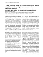
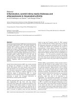
Real time quantitative PCR (qPCR) analysis of hTERT expression in HTLV-1-infected and uninfected T cellsFigure 1
Real time quantitative PCR (qPCR) analysis of hTERT expression in HTLV-1-infected and uninfected T cells. A) ATL cell lines
(HTLV-1 infected but negative for Tax expression); B) In vitro transformed cell lines (HTLV-1 infected and positive for Tax
expression); C) Jurkat T-cell clones positive for Tax expression (E12, C11 and C50); D) Uninfected primary CD4 T lym-
phocytes either activated or not (resting); T-cell line (DCH-4) immortalized with a lentivirus vector encoding a Tax-YFP fusion
protein; Infected T lymphocytes isolated from patient suffering from TSP/HAM. Cytoplasmic RNAs were isolated, reverse tran-
scribed and cDNA were analyzed by qPCR using primers for hTERT and PBGD. Results are expressed as indicated in legend of
table 1. Standard deviations are from at least three determinations performed in duplicate.
0
0,4
0,8
1,2
1,6
2
Jurkat JK-E12 JK-C11 JK-C50
0
0,4
0,8
1,2
1,6
2
MT1 TLom1 KK1
0
0,4
0,8
1,2
1,6
2
C8166 C91PL HUT102 MT2
0
0,4
0,8
1,2
1,6
2
Resting CD4 Activated
CD4
DCH-4 TSP/HAM
Relative hTERT mRNA
expression
AB
CD
Retrovirology 2005, 2:77 />Page 4 of 10
(page number not for citation purposes)
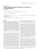
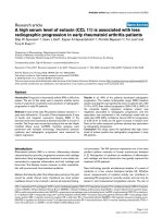
Analysis of TERF1, TERF2 and POT1 gene expression in T-cell lines expressing TaxFigure 2
Analysis of TERF1, TERF2 and POT1 gene expression in T-cell lines expressing Tax. Cytoplasmic RNAs were isolated, reverse
transcribed and cDNA were analyzed by qPCR using specific primers. Results are expressed as the amount of indicated mRNA
relative to PBGD. Each quantification was compared to that obtained with Jurkat cells, referred as 1. Standard deviations are
from at least three determinations performed in duplicate.
0
3
6
9
12
15
18
C91PL HUT-102 MT2 DCH-4 TSP/HAM
0
1
2
3
C91PL HUT-102 MT2 DCH-4 TSP/HAM
0
2
4
6
8
10
12
C91PL HUT-102 MT2 DCH-4 TSP/HAM
Pot 1
TRF1
TRF2
TRF2 mRNA expression
TRF1 mRNA expression
Pot1 mRNA
expression
Retrovirology 2005, 2:77 />Page 5 of 10
(page number not for citation purposes)
next performed a quantitative analysis of the expression of
POT1, TERF1 and TERF2 genes in both cell types. Resting
T lymphocytes expressed detectable levels of the respective
mRNAs. By contrast, activation of these cells was found to
induce a 2.9 to 3.9-fold decrease in the transcription of
these shelterin genes. Thus, in vitro physiological activa-
tion of CD4+ T cells reveals an inverse regulation in the
transcription of telomerase and that of POT1, TERF1 and
TERF2 genes.
Transcriptional expression of hTERT, POT1, TERF1 and
TERF2 genes in HTLV-1 transformed and Tax-expressing
T lymphocytes
It has been previously shown a decrease of telomerase
activity (assayed by TRAP assays) in HTLV-1 T cell lines
expressing Tax [34]. This decrease was associated with the
abitity of Tax to downregulate the hTERT promoter. To
further assess the inhibitory effect of Tax on the transcrip-
tion of the telomerase gene, the levels of hTERT mRNAs in
T cell lines, which do not express Tax were compared to
those in Tax-expressing T cells. The transcription of hTERT
was found to be significantly higher in the three cell lines
from ATL patients, (in which Tax is undetectable), than in
the three in vitro HTLV-1 transformed (IL2-independent)
T-cells (C91PL, HUT-102, MT2), which are expressing Tax
and producing viral particles (Fig. 1, compare A and B).
Likewise, hTERT transcription decreased in the three Jur-
kat T-cell clones expressing only Tax, when compared to
the parental cells (Fig. 1C). The transcription of the telom-
erase gene was next analyzed in IL2-dependent CD4+
DCH4 T-cells, obtained after transduction of activated pri-
mary human CD4+ cells with a lentivirus vector encoding
Tax and in peripheral blood T lymphocytes isolated from
a TSP/HAM patient, which are known to express Tax. In
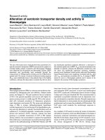
Characterization of C91PL cells over-expressing hTERT geneFigure 3
Characterization of C91PL cells over-expressing hTERT gene. Cells were transduced with a lentivirus vector encoding GFP
with or without hTERT A) Flow cytometry determination of the expression of GFP and p19
gag
. The percentage of cells in each
quadrant is indicated. B) Analysis of hTERT, and of telomere protein gene expression in hTERT/GFP transduced C91PL cells
(grey bars) and in control GFP transduced C91PL (white bars) was performed and quantified as in Figure 2.
GFP hTERT/GFP
GFP
Gag
B
0
10
20
30
40
GFP hTERT/ GFP
0
5
10
15
20
25
GFP hTERT/ GFP
hTERT Pot 1
hTERT mRNA
expression
Pot1
mRNA
expression
0
1
2
3
4
5
GFP hTERT/ GFP
0
1
2
3
4
5
GFP hTERT/ GFP
TRF1
mRNA
expression
TRF2
TRF2
mRNA
expression
TRF1
A
Retrovirology 2005, 2:77 />Page 6 of 10
(page number not for citation purposes)
both cell types, the amount of hTERT mRNA was ten-fold
less than that in normal activated CD4+ T lymphocytes
(Fig. 1D). Together, these observations underline a direct
correlation, between Tax expression and hTERT transcrip-
tional repression in T cells.
As indicated above, in resting T lymphocytes, a low level
of telomerase gene transcription correlates with a high
level of shelterin gene transcription. Therefore, these
results prompted us to test whether the inhibition of
hTERT transcription by Tax could lead to an enhancement
of the transcription of the shelterin genes. To that pur-
pose, we analyzed the transcriptional profile of TERF1,
TERF2 and POT1 in the HTLV-1 transformed T-cell lines
(C91PL, HUT-102, MT2). Remarkably, while the tran-
scription of TERF1 and of TERF2 underwent an increase of
1.2 to 2.4, and of 2.7 to 4.0, respectively, the transcription
of POT1 rose at a much higher level (Fig. 2). Indeed, the
amount of Pot1 in C91PL, HUT-102 and MT2 cells was
respectively 7.1-, 10.0- and 14.7-fold. In the DCH4 and in
the TSP/HAM cells, only a slight increase of TERF1 tran-
scription was observed, but lower than that in C91PL cells
(Fig. 2). Whereas the transcription of the TERF2 gene was
greatly enhanced in these cells (10.3 for the DCH4 cells
and 5.8 for the TSP/HAM cells), that of the POT1 gene
increased 8.9-fold in the TSP/HAM cells, but not in DCH4
cells. Overall these results underline that the down-regu-
lation of hTERT transcription in HTLV-1-T cell lines and in
Tax-expressing T lymphocytes correlates with an increased
transcription of shelterin genes, and more particularly
that of POT1 and TERF2 genes. Collectively, these results
validate the balanced transcription between telomerase
and the telomeric DNA-binding proteins TRF1, TRF2 and
Pot1.
Effect of ectopic expression of hTERT in HTLV-1
transformed T lymphocytes on shelterin gene expression
The above results showing an inverse correlation of hTERT
transcription with that of shelterin genes in T lymphocytes
proposes the possibility that hTERT negatively regulates
the transcription of the shelterin genes in these cells. To
test this hypothesis, we studied the effect of an ectopic
hTERT expression in C91PL cells, in which the low level of
hTERT transcription is associated with a high level of
POT1 transcription. To that purpose, C91PL cells were
transduced with either the pWPIR-hTERT-GFP or the con-
trol pWPIR-GFP lentiviral vectors. In these vectors, the
transcription of the hTERT-IRES-GFP and that of the IRES-
GFP sequences were under the control of an heterologous
promoter (EF1), which is not known to be down-regu-
lated by Tax. Ten days after transduction, cells were ana-
lyzed for GFP expression and for transcription of hTERT
and shelterin genes. First, FACS analysis showed that a sig-
nificant percentage (more than 50%) of both types of
transduced cells were GFP positive (Fig 3A). Next, quanti-
tative RT-PCR analysis showed that in hTERT-GFP-trans-
duced cells, a 47.6-fold increase of hTERT transcription
was observed, as compared to GFP control cells (Fig. 3B).
No significant modification in the proliferation rate of the
cell population over-expressing hTERT was observed.
Finally, a more than 2-fold decrease in the levels of TERF1,
TERF2 and POT1 transcripts were observed. Thus, Pot1
mRNAs, which are the most abundant in GFP-transduced
cells, were found to decrease by 2.7-fold in the hTERT-
GFP-transduced C91PL cells. These results confirm that
the transcriptional expression of shelterin genes appears
to be down-regulated by hTERT. They further indicate that
in HTLV-1 T cell lines Tax expression does not interfere
with this regulatory mechanism.
Discussion
In this study, we provide evidence that, during the activa-
tion of human CD4+ T lymphocytes, the increased tran-
scription of the gene encoding the telomerase catalytic
subunit is accompanied by a decreased transcription of
the genes encoding three subunits (TRF1, TRF2 and Pot1)
belonging to the shelterin complex. Since the products of
these genes are known to inhibit telomere length, tel-
omere homeostasis in activated T lymphocytes appears to
be regulated both by telomerase induction and by shel-
terin repression. We therefore propose that, during T cell
activation, the functional state of telomeres is regulated by
a change in the balance between the expression of hTERT
and that of shelterin genes (Figure 4, upper panel).
Human CD4+ T lymphocytes are the main targets of
HTLV-1 transformation and the viral regulatory Tax has
also been shown to inhibit telomerase activity [34]. We
indeed show that transcription of the telomerase gene is
inhibited in three in vitro HTLV-1 transformed T cell lines
as well as in Tax-expressing DCH4 T lymphocytes and in
TSP/HAM T lymphocytes. We next observe that the Tax-
induced decrease of hTERT mRNAs in these cells corre-
sponds to an increase in the overall amount of the TERF1,
TERF2 and Pot1 transcripts. Thus it appears that telomere
homeostasis in HTLV-1 T cell lines and in Tax-expressing
lymphocytes is regulated both by telomerase repression
and by shelterin induction (Fig. 4, lower panel). Collec-
tively, these results validate a balanced transcription
between telomerase and the telomeric DNA-binding pro-
teins TRF1, TRF2 and POT1 in normal, activated as well as
in HTLV-1 infected and in Tax-expressing T lymphocytes.
They therefore plead for a regulatory mechanism control-
ling this balance. Thus, the observation that ectopic
expression of hTERT in HTLV-1 T cells leads to a decrease
in the transcription of these genes, suggests that hTERT is
implicated in their negative transcription. It would be
worth to determine whether the ectopic expression of any
shelterin subunit would lead to a down-regulation of
hTERT transcription. Interestingly, HTLV-1 transformed T
cells and Tax expressing T lymphocytes are sharing with
Retrovirology 2005, 2:77 />Page 7 of 10
(page number not for citation purposes)
uninfected resting T lymphocytes, the same transcrip-
tional telomeric profile. It therefore appears that the tran-
scriptional balance between hTERT and the three shelterin
subunits is involved not only in regulating telomere
homeostasis, but also in sustaining HTLV-1-induced cel-
lular proliferation.
As indicated above, while the overall transcription of the
shelterin genes were found to increase in HTLV-1 T cell
lines and in Tax-expressing T lymphocytes, the transcrip-
tion of each shelterin subunit was not affected to a similar
extent in each cell type. Thus, the transcription of the
POT1 gene was enhanced to a higher level than that of the
TERF1 and TERF2 genes in the HTLV-1 T cell lines. Like-
wise, only TERF2 was found to be up-transcribed in
DCH4 cells, whereas the transcription of both TERF2 and
POT1 was significantly increased in TSP/HAM cells (Fig. 4,
lower panel). It is plausible that these differences might be
linked to their in vitro/in vivo derivation and/or to selec-
tion pressures in culture. Whatsoever, these data suggest
that Tax is not intervening in the modulation of this bal-
anced transcription. Indeed, the observation that Pot1
transcription decreased in C91PL cells over-expressing
hTERT implies that the activity of Tax on the telomeric
machinery is restricted to its inhibitory effect on hTERT
transcription.
Cancer cells commonly up-regulate telomerase, which is
consistent with telomerase conferring a strong selective
advantage for continued growth of malignant cells [43].
As a matter of fact, telomerase is highly expressed in
patients with the acute type of ATL [33]. In these ATL cells,
proviral transcription is silent, underlining that viral
genome expression is crucial only at the onset of the
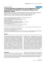
A model for hTERT and shelterin gene expression in T lymphocytesFigure 4
A model for hTERT and shelterin gene expression in T lymphocytes. In the upper panel, CD4+ T lymphocytes were either
resting or activated with a cocktail of anti CD3/anti CD28 antibodies. In the lower panel, three types of Tax-expressing T lym-
phocytes were represented. The model is based on the telomeric transcriptional profile defined as a balance between hTERT
on one hand, and shelterin (POT1, TERF1 and TERF2) gene expression on the other hand.
Primary T Lymphocytes
hTERT
Pot1
TRF1
TRF2
Activated
hTERT
Pot1
TRF1
TRF2
Resting
Tax-expressing T Lymphocytes
hTERT
hTERT
TRF2
DCH-4
hTERT
TSP/HAM
Pot 1
TRF2
Pot 1
Pot 1
TRF2
C91PL
Retrovirology 2005, 2:77 />Page 8 of 10
(page number not for citation purposes)
leukemogenic process. The present study suggests that in
infected T cells in which proviral expression is active, the
increased expression of TRF2 and/or Pot1, involved in tel-
omere capping functions, could trigger protective mecha-
nisms that compensate the decrease of telomerase
expression. Thus, by playing an important role in tel-
omere homeostasis, the shelterin proteins are allowing
the proliferation of HTLV-1 infected T cells or Tax-express-
ing T lymphocytes. Consequently, we anticipate that a
transcriptional decrease of these telomeric proteins cou-
pled with telomerase reactivation which might occur at a
time, when Tax is no more expressed, would contribute to
the emergence of telomerase-positive acute leukemic cells.
Interestingly, recent studies have provided new evidence
that telomerase enhances expression of growth-control-
ling genes to confer additional pivotal functions in tumor
progression other than telomere length maintenance [44].
Although more work is needed to elucidate the cellular
and molecular mechanisms of this telomere dysfunction
during the HTLV-1-induced leukemogenic process, the
present observations reveal new links between Tax, telom-
erase and shelterin, that might play a key role in the main-
tenance and in the proliferation of HTLV-1 infected T
lymphocytes.
Methods
Cells
The HTLV-1 T-cell lines (IL-2 independent) C91PL [45],
MT2 [46], HUT102 [47] and C8166 [48] have been
described elsewhere. The HTLV-1 T-cell lines either (IL-2
dependent) KK1, or (IL-2 independent), MT1 and TLom1
were generously provided by Dr. N. Mori [49]. The DCH4
cells, (kind gift by Dr. D. Derse), were established by
transduction of activated, primary human CD4+T cells
with a lentivirus vector encoding an HTLV-1 Tax-
enhanced yellow fluorescent protein fusion [50]. Three
clones of Jurkat T cells stably producing Tax (E12, C11,
C50) have been used in this study [51]. T lymphocytes iso-
lated from one TSP/HAM patient (CJ) were kindly pro-
vided by Dr. A. Gessain (Paris, France). These
lymphocytes and the cell lines KK1 and DCH4 were culti-
vated in RPMI 1640 (Invitrogen) with 10% heat-inacti-
vated fetal calf serum (FCS) 100 U/ml recombinant
human IL-2 (rhIL-2) and supplemented with 100 IU/ml
of penicillin and 50
µ
g/ml of streptomycin. The Jurkat
parental as well as the Jurkat Tax-expressing clones,
C91PL, MT2, HUT102, MT1 and TLom1 cell lines were
maintained in complete RPMI-1640 medium with 10%
FCS, without rhIL-2. The human 293T and rhabdomyosa-
rcoma TE cells were cultured in Dulbecco's minimum
Eagle medium (DMEM, Invitrogen) supplemented with
10% FCS and antibiotics.
Primary peripheral blood lymphocytes from healthy vol-
unteers were isolated by Ficoll density gradient centrifuga-
tion. Then, CD4+ T cell subsets were negatively selected
using magnetic beads (Stem Cell Technologies, Vancou-
ver, BC) according to the manufacturer instructions. Puri-
fied CD4+ T cells were activated with anti CD3/anti CD28
antibody coated beads (Dynal Biotech, Lake Success, NY)
and maintained in RPMI-1640 with 10% FCS and rhIL-2.
Lentiviral constructs and hTERT transduction
The pWPIR-GFP HIV-derived vector, obtained from
Didier Trono, contained the IRES-GFP (enhanced GFP as
a marker gene) under the control of the EF1 (human elon-
gation factor 1 alpha) promoter [52]. The pWPIR-hTERT-
GFP construct was generated by inserting the hTERT
cDNA upstream of the IRES in order to allow individual
translation of the bicistronic mRNA containing both
hTERT and GFP (hTERT-IRES-GFP). Helper-free recom-
binant lentiviruses were produced after transfection of
293T cells with the three following constructs (1) a pack-
aging plasmid, pCMVR8.91; (2) a transfer vector, pWPIR-
hTERT-GFP or pWPIR-GFP; (3) an envelope expression
plasmid, pCMV-VSVG. Twenty to 30 hours later, the
supernatant was harvested, filtered through a 0.45-
µ
m
membrane and aliquots were stored at -80°C. Titres of
virus stocks (from 3 to 5 × 10
5
transducing units per ml)
were determined by transduction of human rhabdomy-
osarcoma TE cells with serially diluted viral supernatant
and analysis five days later on a FACScan instrument (Bec-
ton Dickinson, Mountain View, CA). C91PL cells cultured
for 1–2 hours in presence of polybrene (8 µg/ml) were
then incubated overnight with virus supernatant at a mul-
tiplicity of infection of 2. Analysis of GFP-expressing cells
was performed on a FACScan. Data were analyzed with
the Cell Quest program ((Becton Dickinson).
Real-time polymerase chain reaction amplification
Total cellular RNAs were isolated from cells using Qiagen
RNeasy purification kits (Qiagen, Alameda, CA) accord-
ing to the manufacturer's instructions. To reduce the
amount of DNA originating from lysis, samples were
treated with RNase-free DNase (10 U/
µ
l, Qiagen) for 30
min at 20°C and then for 15 min at 65°C. Five-hundred
ng of RNA sample were reverse transcribed by using
oligo(dT)12–18 and Superscript II (InVitrogen Life tech-
nologies, Frederick, MD). Reverse transcription was per-
formed for 50 min at 42°C. The total cDNA volume of 20
µ
l was frozen until real-time quantitative PCR was per-
formed. After thawing for PCR experiments, the cDNA
was diluted in distilled water and 2
µ
l of diluted cDNA
was used for each PCR reaction. The real-time quantitative
PCR (qPCR) was performed in special lightcycler capillar-
ies (Roche) with a lightcycler Instrument (Roche), by
using the LightCycler-FastStart reaction Mix SYBR-Green
kit (Roche). The following specific primers were used to
detect: PBGD, sense 5'-GGAATGCATGTATGCTGTGG-3'
and antisense, 5'-CAGGTACAGTTGCCCATCC-3', Tax-
Retrovirology 2005, 2:77 />Page 9 of 10
(page number not for citation purposes)
HTLV-1
sense, 5'-GTTGTATGAGTGATTGGCGGGGTAA-3'
and antisense, 5'-TGTTTGGAGACTGTGTACAAGGCG-3',
hTERT sense, 5'-TGTTTCTGGATTTGCAGGTG-3' and anti-
sense, 5'-GTTCTTGGCTTTCAGGATGG-3', Pot1 sense, 5'-
TGGGTATTGTACCCCTCCAA-3' and antisense, 5'-GAT-
GAAGCATTCCAACCACGG-3'. TRF1 sense,5'-GCTGTTT-
GTATGGAAAATGGC-3' and antisense: 5'-
CCGCTGCCTTCATTAGAAAG-3', TRF2 sense, 5'-GACCT-
TCCAGCAGAAGATGC-3' and antisense, 5'-GTTGGAG-
GATTCCGTAGCTG-3'. The thermal cycling conditions
consisted of 40 cycles at 95°C for 10 sec, 61°C for 5 sec,
72°C for 10 sec. The fluorescence signal increase of SYBR-
GREEN was automatically detected during the 72°C
phase of the PCR. Omission of reverse transcriptase in the
RT-PCR protocol led to a failure of target gene amplifica-
tion in the positive controls. Light cycler PCR data were
analyzed using LightCycler Data software (Idaho Technol-
ogy). The software first normalizes each sample by back-
ground subtraction of initial cycles. A fluorescence
threshold is then set at 5% full scale, and the software
determines the cycle number at which each sample
reached this threshold. The fluorescence threshold cycle
number correlates inversely with the log of initial tem-
plate concentration. A standard calibration curve was per-
formed by using cDNA from Jurkat cells. The levels of
PBGD transcripts were used to normalize the amount of
cDNA in each sample.
Competing interests
The author(s) declare that they have no competing inter-
ests.
Acknowledgements
We thank D. Derse, N. Mori and A. Gessain for providing cells. This study
was supported by ARC (Association pour la Recherche sur le Cancer
n°5669 to LG.), by the Ligue Nationale contre le Cancer (to EG) and by
EPIMED program of the Cancéropole Lyon Auvergne Rhône-Alpes
(CLARA).
References
1. de Lange T, Shiue L, Myers RM, Cox DR, Naylor SL, Killery AM, Var-
mus HE: Structure and variability of human chromosome
ends. Mol Cell Biol 1990, 10:518-527.
2. de Lange T: Protection of mammalian telomeres. Oncogene
2002, 21:532-540.
3. de Lange T: Shelterin: the protein complex that shapes and
safeguards human telomeres. Genes Dev 2005, 19:2100-2110.
4. Cong YS, Wright WE, Shay JW: Human telomerase and its reg-
ulation. Microbiol Mol Biol Rev 2002, 66:407-425.
5. Blackburn EH: Switching and signaling at the telomere. Cell
2001, 106:661-673.
6. Loayza D, De Lange T: POT1 as a terminal transducer of TRF1
telomere length control. Nature 2003, 423:1013-1018.
7. Broccoli D, Smogorzewska A, Chong L, de Lange T: Human telom-
eres contain two distinct Myb-related proteins, TRF1 and
TRF2. Nat Genet 1997, 17:231-235.
8. Bilaud T, Brun C, Ancelin K, Koering CE, Laroche T, Gilson E: Telo-
meric localization of TRF2, a novel human telobox protein.
Nat Genet 1997, 17:236-239.
9. Ancelin K, Brunori M, Bauwens S, Koering CE, Brun C, Ricoul M,
Pommier JP, Sabatier L, Gilson E: Targeting assay to study the cis
functions of human telomeric proteins: evidence for inhibi-
tion of telomerase by TRF1 and for activation of telomere
degradation by TRF2. Mol Cell Biol 2002, 22:3474-3487.
10. Karlseder J, Smogorzewska A, de Lange T: Senescence induced by
altered telomere state, not telomere loss. Science 2002,
295:2446-2449.
11. Wang RC, Smogorzewska A, de Lange T: Homologous recombi-
nation generates T-loop-sized deletions at human telom-
eres. Cell 2004, 119:355-368.
12. Houghtaling BR, Cuttonaro L, Chang W, Smith S: A dynamic
molecular link between the telomere length regulator TRF1
and the chromosome end protector TRF2. Curr Biol 2004,
14:1621-1631.
13. Gonzalez-Suarez E, Samper E, Ramirez A, Flores JM, Martin-Caballero
J, Jorcano JL, Blasco MA: Increased epidermal tumors and
increased skin wound healing in transgenic mice overex-
pressing the catalytic subunit of telomerase, mTERT, in
basal keratinocytes. Embo J 2001, 20:2619-2630.
14. Gonzalez-Suarez E, Samper E, Flores JM, Blasco MA: Telomerase-
deficient mice with short telomeres are resistant to skin
tumorigenesis. Nat Genet 2000, 26:114-117.
15. Greenberg RA, Chin L, Femino A, Lee KH, Gottlieb GJ, Singer RH,
Greider CW, DePinho RA: Short dysfunctional telomeres
impair tumorigenesis in the INK4a(delta2/3) cancer-prone
mouse. Cell 1999, 97:515-525.
16. Artandi SE, Alson S, Tietze MK, Sharpless NE, Ye S, Greenberg RA,
Castrillon DH, Horner JW, Weiler SR, Carrasco RD, et al.: Consti-
tutive telomerase expression promotes mammary carcino-
mas in aging mice. Proc Natl Acad Sci U S A 2002, 99:8191-8196.
17. Hahn WC, Stewart SA, Brooks MW, York SG, Eaton E, Kurachi A,
Beijersbergen RL, Knoll JH, Meyerson M, Weinberg RA: Inhibition
of telomerase limits the growth of human cancer cells. Nat
Med 1999, 5:1164-1170.
18. Dong CK, Masutomi K, Hahn WC: Telomerase: regulation, func-
tion and transformation. Crit Rev Oncol Hematol 2005, 54:85-93.
19. Elenbaas B, Spirio L, Koerner F, Fleming MD, Zimonjic DB, Donaher
JL, Popescu NC, Hahn WC, Weinberg RA: Human breast cancer
cells generated by oncogenic transformation of primary
mammary epithelial cells. Genes Dev 2001, 15:50-65.
20. Kim NW, Piatyszek MA, Prowse KR, Harley CB, West MD, Ho PL,
Coviello GM, Wright WE, Weinrich SL, Shay JW: Specific associa-
tion of human telomerase activity with immortal cells and
cancer. Science 1994, 266:2011-2015.
21. Rudolph KL, Millard M, Bosenberg MW, DePinho RA: Telomere
dysfunction and evolution of intestinal carcinoma in mice
and humans. Nat Genet 2001, 28:155-159.
22. Chin L, Artandi SE, Shen Q, Tam A, Lee SL, Gottlieb GJ, Greider CW,
DePinho RA: p53 deficiency rescues the adverse effects of tel-
omere loss and cooperates with telomere dysfunction to
accelerate carcinogenesis. Cell 1999, 97:527-538.
23. Artandi SE, DePinho RA: A critical role for telomeres in sup-
pressing and facilitating carcinogenesis. Curr Opin Genet Dev
2000, 10:39-46.
24. O'Sullivan JN, Bronner MP, Brentnall TA, Finley JC, Shen WT, Emer-
son S, Emond MJ, Gollahon KA, Moskovitz AH, Crispin DA, et al.:
Chromosomal instability in ulcerative colitis is related to tel-
omere shortening. Nat Genet 2002, 32:280-284.
25. Zhang A, Wang J, Zheng B, Fang X, Angstrom T, Liu C, Li X, Erlands-
son F, Bjorkholm M, Nordenskjord M, et al.: Telomere attrition
predominantly occurs in precursor lesions during in vivo car-
cinogenic process of the uterine cervix. Oncogene 2004,
23:7441-7447.
26. Chin K, de Solorzano CO, Knowles D, Jones A, Chou W, Rodriguez
EG, Kuo WL, Ljung BM, Chew K, Myambo K, et al.: In situ analyses
of genome instability in breast cancer. Nat Genet 2004,
36:984-988.
27. Brunori M, Mathieu N, Ricoul M, Bauwens S, Koering CE, Roborel de
Climens A, Belleville A, Wang Q, Puisieux I, Decimo D, et al.: TRF2
inhibition promotes anchorage-independent growth of tel-
omerase-positive human fibroblasts. Oncogene 2006 in press.
28. Matsuoka M: Human T-cell leukemia virus type I and adult T-
cell leukemia. Oncogene 2003, 22:5131-5140.
29. Mortreux F, Gabet AS, Wattel E: Molecular and cellular aspects
of HTLV-1 associated leukemogenesis in vivo. Leukemia 2003,
17:26-38.
30. Sinha-Datta U, Horikawa I, Michishita E, Datta A, Sigler-Nicot JC,
Brown M, Kazanji M, Barrett JC, Nicot C: Transcriptional activa-
Publish with BioMed Central and every
scientist can read your work free of charge
"BioMed Central will be the most significant development for
disseminating the results of biomedical research in our lifetime."
Sir Paul Nurse, Cancer Research UK
Your research papers will be:
available free of charge to the entire biomedical community
peer reviewed and published immediately upon acceptance
cited in PubMed and archived on PubMed Central
yours — you keep the copyright
Submit your manuscript here:
/>BioMedcentral
Retrovirology 2005, 2:77 />Page 10 of 10
(page number not for citation purposes)
tion of hTERT through the NF-kappaB pathway in HTLV-I-
transformed cells. Blood 2004, 104:2523-2531.
31. Tsumuki H, Nakazawa M, Hasunuma T, Kobata T, Kato T, Uchida A,
Nishioka K: Infection of synoviocytes with HTLV-I induces tel-
omerase activity. Rheumatol Int 2001, 20:175-179.
32. Uchida N, Otsuka T, Arima F, Shigematsu H, Fukuyama T, Maeda M,
Sugio Y, Itoh Y, Niho Y: Correlation of telomerase activity with
development and progression of adult T-cell leukemia. Leuk
Res 1999, 23:311-316.
33. Kubuki Y, Suzuki M, Sasaki H, Toyama T, Yamashita K, Maeda K, Ido
A, Matsuoka H, Okayama A, Nakanishi T, et al.: Telomerase activ-
ity and telomere length as prognostic factors of adult T-cell
leukemia. Leuk Lymphoma 2005, 46:393-399.
34. Gabet AS, Mortreux F, Charneau P, Riou P, Duc-Dodon M, Wu Y,
Jeang KT, Wattel E: Inactivation of hTERT transcription by
Tax. Oncogene 2003, 22:3734-3741.
35. Hanon E, Hall S, Taylor GP, Saito M, Davis R, Tanaka Y, Usuku K,
Osame M, Weber JN, Bangham CR: Abundant tax protein
expression in CD4+ T cells infected with human T-cell lym-
photropic virus type I (HTLV-I) is prevented by cytotoxic T
lymphocytes. Blood 2000, 95:1386-1392.
36. Jin DY, Spencer F, Jeang KT: Human T cell leukemia virus type 1
oncoprotein Tax targets the human mitotic checkpoint pro-
tein MAD1. Cell 1998, 93:81-91.
37. Hollsberg P: Mechanisms of T-Cell Activation by Human T-
Cell Lymphotropic Virus Type I. Microbiol Mol Biol Rev 1999,
63:308-333.
38. Grassmann R, Berchtold S, Radant I, Alt M, Fleckenstein B, Sodroski
JG, Haseltine WA, Ramstedt U: Role of human T-cell leukemia
virus type 1 X region proteins in immortalization of primary
human lymphocytes in culture. J Virol 1992, 66:4570-4575.
39. Gatza ML, Watt JC, Marriott SJ: Cellular Transformation by the
HTLV-I Tax Protein, a Jack-of-All-Trades. Oncogene 2003,
22:5141-5149.
40. Liu K, Schoonmaker MM, Levine BL, June CH, Hodes RJ, Weng NP:
Constitutive and regulated expression of telomerase reverse
transcriptase (hTERT) in human lymphocytes. Proc Natl Acad
Sci U S A 1999, 96:5147-5152.
41. Hodes RJ, Hathcock KS, Weng NP: Telomeres in T and B cells.
Nat Rev Immunol 2002, 2:699-706.
42. Roth A, Yssel H, Pene J, Chavez EA, Schertzer M, Lansdorp PM, Spits
H, Luiten RM: Telomerase levels control the lifespan of human
T lymphocytes. Blood 2003, 102:849-857.
43. Shay JW, Bacchetti S: A survey of telomerase activity in human
cancer. Eur J Cancer 1997, 33:787-791.
44. Smith LL, Coller HA, Roberts JM: Telomerase modulates expres-
sion of growth-controlling genes and enhances cell prolifera-
tion. Nat Cell Biol 2003, 5:474-479.
45. Popovic M, Lange-Wantzin G, Sarin PS, Mann D, Gallo RC: Transfor-
mation of human umbilical cord blood T cells by human T-
cell leukemia/lymphoma virus. Proc Natl Acad Sci U S A 1983,
80:5402-5406.
46. Miyoshi I, Kubonishi I, Yoshimoto S, Akagi T, Ohtsuki Y, Shiraishi Y,
Nagata K, Hinuma Y: Type C virus particles in a cord T-cell line
derived by co-cultivating normal human cord leukocytes and
human leukaemic T cells. Nature 1981, 294:770-771.
47. Poiesz BJ, Ruscetti FW, Mier JW, Woods AM, Gallo RC: T-cell lines
established from human T-lymphocytic neoplasias by direct
response to T-cell growth factor. Proc Natl Acad Sci U S A 1980,
77:6815-6819.
48. Salahuddin SZ, Markham PD, Wong-Staal F, Franchini G, Kalyanara-
man VS, Gallo RC: Restricted expression of human T-cell
leukemia – lymphoma virus (HTLV) in transformed human
umbilical cord blood lymphocytes. Virology 1983, 129:51-64.
49. Mori N, Fujii M, Iwai K, Ikeda S, Yamasaki Y, Hata T, Yamada Y, Tan-
aka Y, Tomonaga M, Yamamoto N: Constitutive activation of
transcription factor AP-1 in primary adult T-cell leukemia
cells. Blood 2000, 95:3915-3921.
50. Chung HK, Young HA, Goon PK, Heidecker G, Princler GL, Shimo-
zato O, Taylor GP, Bangham CR, Derse D: Activation of inter-
leukin-13 expression in T cells from HTLV-1-infected
individuals and in chronically infected cell lines. Blood 2003,
102:4130-4136.
51. Lemasson I, Robert-Hebmann V, Hamaia S, Duc Dodon M, Gazzolo
L, Devaux C: Transrepression of lck gene expression by
human T-cell leukemia virus type 1-encoded p40tax. J Virol
1997, 71:1975-1983.
52. Salmon P, Kindler V, Ducrey O, Chapuis B, Zubler RH, Trono D:
High-level transgene expression in human hematopoietic
progenitors and differentiated blood lineages after transduc-
tion with improved lentiviral vectors. Blood 2000,
96:3392-3398.