cardiovascular complications of cirrhosis 2008
Bạn đang xem bản rút gọn của tài liệu. Xem và tải ngay bản đầy đủ của tài liệu tại đây (415.14 KB, 12 trang )
doi:10.1136/gut.2006.112177
2008;57;268-278 Gut
S Møller and J H Henriksen
Cardiovascular complications of cirrhosis
/>Updated information and services can be found at:
These include:
References
/>This article cites 81 articles, 17 of which can be accessed free at:
service
Email alerting
the top right corner of the article
Receive free email alerts when new articles cite this article - sign up in the box at
Notes
/>To order reprints of this article go to:
/> go to: GutTo subscribe to
on 11 August 2008 gut.bmj.comDownloaded from
Cardiovascular complications of
cirrhosis
S Møller, J H Henriksen
Department of Clinical
Physiology 239, Hvidovre
Hospital, University of
Copenhagen, Copenhagen,
Denmark
Correspondence to:
Associate Professor S Møller,
Department of Clinical
Physiology, 239, Hvidovre
Hospital, DK-2650 Copenhagen,
Denmark; soeren.moeller@
hvh.regionh.dk
Revised 2 August 2007
Accepted 2 August 2007
ABSTRACT
Cardiovascular complications of cirrhosis include cardiac
dysfunction and abnormalities in the central, splanchnic
and peripheral circulation, and haemodynamic changes
caused by humoral and nervous dysregulation. Cirrhotic
cardiomyopathy implies systolic and diastolic dysfunction
and electrophysiological abnormalities, an entity that is
different from alcoholic heart muscle disease. Being
clinically latent, cirrhotic cardiomyopathy can be
unmasked by physical or pharmacological strain.
Consequently, caution should be exercised in the case of
stressful procedures, such as large volume paracentesis
without adequate plasma volume expansion, transjugular
intrahepatic portosystemic shunt (TIPS) insertion, perito-
neovenous shunting and surgery. Cardiac failure is an
important cause of mortality after liver transplantation, but
improved liver function has also been shown to reverse
the cardiac abnormalities. No specific treatment can be
recommended, and cardiac failure should be treated as in
non-cirrhotic patients with sodium restriction, diuretics,
and oxygen therapy when necessary. Special care should
be taken with the use of ACE inhibitors and angiotensin
antagonists in these patients. The clinical significance of
cardiovascular complications and cirrhotic cardiomyopathy
is an important topic for future research, and the initiation
of new randomised studies of potential treatments for
these complications is needed.
The course in most cirrhotic patients is dominated
by complications to portal hypertension, such as
bleeding from oesophageal varices and ascites with
the development of spontaneous bacterial perito-
nitis, renal impairment and encephalopathy. Some
patients, however, seem to die of causes unrelated
to these complications. A closer clinical look shows
that a number of these patients display signs of
cardiovascular disturbances secondary to vasodila-
tation, with palmar erythema and reddish skin,
raised and bounding pulse, and a low systemic
blood pressure indicating a hyperdynamic circula-
tion.
12
The hyperdynamic syndrome was first
described .50 years ago and comprises increased
heart rate, cardiac output and plasma volume, and
reduced systemic vascular resistance and arterial
blood pressure.
1–3
The hyperdynamic syndrome is
today a well-characterised element in the clinical
appearance of the cardiovascular complications of
cirrhosis and portal hypertension of various aetiol-
ogies.
12
Experimental and clinical findings of
impaired cardiac function have led to the introduc-
tion of the new clinical entity, cirrhotic cardiomyo-
pathy, but cardiac dysfunction is not seen in all
patients, especially not in those with less advanced
disease, and its clinical significance is still under
discussion.
4
This review will primarily centre on
clinical and pathophysiological aspects of the
circulatory and cardiac complications of advanced
cirrhosis.
THE CIRCULATION IN CIRRHOSIS
An increase in cardiac output can be attributed to
an increase in venous return, heart rate and
myocardial contractility, all of which are controlled
by the autonomic nervous system. Vasodilatation
(low systemic vascular resistance), the presence of
arteriovenous communications, expanded blood
volume and increased sympathetic nervous activity
may further raise the cardiac output; most of these
pathophysiological mechanisms are active in
advanced cirrhosis.
12
In the early stages, the
presence of a hyperdynamic circulation is often
not apparent. However, with the progression of
the liver disease, there is an overall association
between the severity of the cirrhosis and the degree
of hyperdynamic circulation. Studies on circulatory
changes with posture suggest that the patients are
mostly hyperdynamic in the supine position.
356
Blood and plasma volumes are raised in advanced
cirrhosis, but the distribution between central and
non-central vascular areas is unequal.
78
Thus, by
different techniques it has been established that
the central and arterial blood volume—that is, the
blood volume in the heart, lungs and central
arterial tree—is most often decreased, whereas
the non-central blood volume, in particular the
splanchnic blood volume, is increased in animals
and patients with cirrhosis (see table 1).
27910
The
effective arterial blood volume (ie, the circulatory
compartment sensed by baroreceptors) and the
central circulation time (ie, central blood volume
relative to cardiac output) are substantially
reduced and bear a significant relationship to
poorer survival in advanced cirrhosis.
11
Total vascular compliance as well as arterial
compliance (ie, an increase in intravascular volume
relative to an increase in transmural blood pres-
sure) are increased in cirrhosis with the degree of
decompensation.
12 13
The altered static and
dynamic characteristics of the wall of large arteries
are closely associated with the circulatory and
homoeostatic derangement.
71314
Arterial compli-
ance depends on the properties of the elastic and
smooth muscle of the arterial wall and represents
an important coupling between the heart and the
arterial system with respect to relocation of
intravascular volume.
14
The changes in arterial
mechanics are reversible at least in part.
13
An
element in the elevated arterial compliance in
Recent advances in clinical practice
268 Gut 2008;57:268–278. doi:10.1136/gut.2006.112177
on 11 August 2008 gut.bmj.comDownloaded from
advanced cirrhosis is the reduced arterial blood
volume and blood pressure.
7
Arterial compliance
expresses the stroke volume relative to the pulse
pressure and is directly related to the severity of
cirrhosis.
13 14
However, in contrast to the systemic
vascular resistance, arterial compliance may be
determined independently of flow and pressure by
pulsewave velocity.
14
In addition, the arterial
compliance in cirrhosis seems to be affected by
vasoactive forces as it correlates directly with the
vasodilator calcitonin gene-related peptide (CGRP)
and inversely with catecholamines.
13
The arteriolar
tone adjusts the level of the blood pressure and
may thereby influences large artery compliance.
Recent data suggest that the hyperdynamic circu-
lation is mainly caused by circulatory alterations in
the splanchnic area.
15
Thus, arteriolar vasodilata-
tion would be a more localised event, whereas the
elevation in arterial compliance may be more
systemic.
7
Arterial compliance may therefore be
an integral variable for vascular responsiveness,
together with the systemic vascular resistance.
Arterial compliance is easy to determine and
elevated in advanced cirrhosis. Besides a relation-
ship to age, body size, sex and the level of arterial
blood pressure, arterial compliance is directly
related to the severity of cirrhosis, the hyperdy-
namic circulatory derangement and abnormal
volume distribution. Its role in clinical hepatology,
however, remains to be established.
The pulmonary vascular resistance is often
decreased in cirrhosis, except in the 2–4% of the
patients with portopulmonary hypertension.
16
Some patients exhibit characteristic vascular
abnormalities with arteriovenous shunts and
intrapulmonary dilatations.
116
Ventilatory lung
function and diffusion are impaired in the majority
of the patients, and the combination of vascular
abnormalities, reduced transfer factor and low
arterial oxygen saturation has been termed the
hepatopulmonary syndrome.
16
In patients with
cirrhosis, the reduced transfer factor correlates
with the low pulmonary blood volume, which
suggests that central underfilling also plays a role
in the impairment of pulmonary function.
17
Pathophysiology of splanchnic arteriolar
vasodilatation
Arteriolar vasodilatation in cirrhosis and portal
hypertension may be brought about by a combina-
tion of overproduction of circulating vasodilators,
vasodilators of intestinal or systemic origin, vaso-
dilators that escape degradation in the diseased
liver or bypass the liver through portosystemic
collaterals, reduced resistance to vasoconstrictors
and increased sensitivity to vasodilators.
12
According to ‘‘the arterial vasodilation hypoth-
esis’’, splanchnic arteriolar vasodilation leads to
reduction of the systemic vascular resistance,
central arterial underfilling with effective hypovo-
laemia, activation of vasoconstrictor systems, such
as the sympathetic nervous system (SNS), the
renin–angiotensin–aldosterone system (RAAS),
vasopressin, endothelins (ETs) and neuropeptide
Y, and hence development of a hyperkinetic
circulatory state.
1818
Thus, most of the haemody-
namic changes summarised in table 1 and figure 1
can be explained by this theory. The predomi-
nantly splanchnic vasodilation in cirrhosis precedes
the increase in cardiac output and heart rate, and it
has recently been shown experimentally that mild
increases in portal pressure upregulate nitric oxide
synthase (eNOS).
19
With the progression of the
disease, the splanchnic vasodilatation becomes
more pronounced and the hyperdynamic circula-
tion may no longer be sufficient to correct the
effective hypovolaemia.
20 21
The splanchnic circula-
tion is less sensitive to the effects of angiotensin II,
noradrenaline and vasopressin because of the
surplus of vasodilators which may play a role in
the development of the vascular hyporesponsive-
ness to vasoconstrictors.
22
The arterial blood
pressure is mainly maintained by vasoconstriction
in the renal, cerebral and hepatic vascular beds
Table 1 Circulatory changes in specific vascular beds in
cirrhosis
Systemic circulation
Plasma volume q
Total blood volume q
Non-central blood volume q
Central and arterial blood volume Q (R)
Arterial blood pressure Q (R)
Systemic vascular resistance Q
Cutaneous and skeletal muscle circulation
Skeletal muscular blood flow* qRQ
Cutaneous blood flow* qRQ
Heart
Heart rate q
Cardiac output q
Left atrial volume q
Left ventricular volume R (q)
Right atrial volume RqQ
Right atrial pressure Rq
Right ventricular end-diastolic pressure R
Pulmonary artery pressure Rq
Pulmonary capillary wedge pressure R
Left ventricular end-diastolic pressure R
Total vascular compliance q
Arterial compliance q
Hepatic and splanchnic circulation
Hepatic blood flow{ QR(q)
Hepatic venous pressure gradient q
Postsinusoidal resistance q
Renal circulation
Renal blood flow Q
Glomerular filtration rate (q)QR
Cerebral circulation
Cerebral blood flow QR
Pulmonary circulation
Pulmonary blood flow q
Pulmonary vascular resistance Q (q{)
Pulmonary blood volume Q
Pulmonary transit time Q
qRQdenote: increased, unchanged and decreased, respectively.
Arrows in parentheses describe early/less typical changes.
*Available data are highly dependent on the applied technique.
{Changes in intrahepatic blood flow due to variable co-determination of
portosystemic shunts.
{Increased in portopulmonary hypertension.
Recent advances in clinical practice
Gut 2008;57:268–278. doi:10.1136/gut.2006.112177 269
on 11 August 2008 gut.bmj.comDownloaded from
where there seems to be a diminished release of
nitric oxide (NO) from endothelial cells.
15 23
To explain the vasodilatation in the systemic
circulation, recent research has focused especially
on substances such as NO, CGRP and adrenome-
dullin, but natriuretic peptides, interleukins,
hydrogen sulphide, ETs and endocannabinoids
have also been implicated (table 2).
1
Blockade of
NO formation in animal models and cirrhotic
patients significantly increases arterial blood pres-
sure and decreases plasma volume, sodium reten-
tion and forearm blood flow.
24 25
Taken together,
there is a growing body of evidence that systemic
NO production is increased and precedes the
development of the hyperdynamic circulation in
cirrhosis, thereby playing a major role in the
arteriolar and splanchnic vasodilation and vascular
hyporeactivity.
15
In addition, vascular endothelial
growth factor (VEGF) seems to stimulate angio-
genesis and the development of portosystemic
collaterals, and blockade of the VEGF receptor-2
has been shown experimentally to inhibit this
process and revert portal hypertension and the
hyperdynamic circulation.
19 26
In addition, recent
studies have suggested that the haem oxygenase–
carbon monoxide pathway mediates hyporeactiv-
ity to phenylephrine in splanchnic vessels.
27
CGRP
and adrenomedullin are powerful vasodilating
peptides, which are both elevated in cirrhosis,
especially in those patients with ascites and the
hepatorenal syndrome correlating with markers of
central hypovolaemia.
1
Hydrogen sulphide is a
gaseous transmitter with potent vasodilating
properties, which has recently been implicated in
vascular abnormalities in cirrhosis.
28
New experi-
mental data suggest that defective rho-kinase
signalling may also contribute to the hypocontrac-
tility in cirrhosis.
29
Thus, the excess of vasodilators
combined with an inadequate haemodynamic
response to vasoconstrictors may explain the
vasodilatatory state and vascular hyporeactivity
in cirrhosis combined with a hyperdynamic circu-
lation, but the pathophysiological mechanisms
behind the development of the hyperdynamic
circulation in cirrhosis may be multifarious, as
listed in table 3.
The hepatic circulation
From a haemodynamic point of view, the hepatic
vascular resistance and portal inflow determine the
level of portal pressure. Factors that determine the
hepatic vascular resistance include both structural
and dynamic components. Among the structural
components are histological characteristics such as
steatosis, fibrosis and regeneration nodules.
Dynamic structures include cells with contractile
properties such as hepatic stellate cells, myofibro-
blasts and smooth muscle cells.
30
Portal venous
inflow is mainly determined by the degree of
splanchnic vasodilation. In healthy subjects, the
hepatic blood flow equals the splanchnic blood
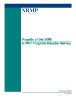
Figure 1 Splanchnic and peripheral arteriolar vasodilation with reduced systemic and splanchnic vascular resistance leads to a reduced effective
arterial blood volume (CBV), and hence to activation of vasoconstrictor systems. The haemodynamic and clinical consequences are increases in portal
pressure (HVPG), cardiac output (CO), heart rate (HR), and plasma (PV) and blood volumes (BV), and increased renal vascular resistance (RVR) and
decreased renal blood flow (RBF), low systemic vascular resistance (SVR) and arterial blood pressure (MAP), and fluid and water retention. The
development of the hyperdynamic circulation may increase portal inflow and further aggravate portal hypertension in a vicious cycle. SNS, sympathetic
nervous system; RAAS, renin–angiotensin–aldosterone system; AVP, arginine vasopressin; ET, endothelin.
Recent advances in clinical practice
270 Gut 2008;57:268–278. doi:10.1136/gut.2006.112177
on 11 August 2008 gut.bmj.comDownloaded from
flow, but patients with portal hypertension have a
substantial portosystemic collateral circulation,
and an increased mesenteric inflow of up to several
litres per minute has been reported (table 1). Thus,
a large part of the increased cardiac output is
returned through portosystemic collaterals. The
azygous blood flow is especially important, as the
azygous vein drains oesophageal varices and an
increase in azygous flow is associated with an
increased risk of variceal bleeding.
11
b-Blockers,
nitrates, octreotide, terlipressin, etc. can reduce the
increased splanchnic blood flow pharmacologically,
and infusion of these drugs may in some patients
partially reverse the hyperkinetic mesenteric circu-
lation. As outlined above, there seems to be a
defective sinusoidal eNOS-derived production of
NO.
15
In addition, recent investigations of endo-
genous vasoactive substances have focused espe-
cially on ET-1, angiotensin II, catecholamines and
leukotrienes in the increased hepatic–sinusoidal
resistance.
130
The haemodynamic imbalance with
a predominant sinusoidal constriction may con-
tribute significantly to the development of portal
hypertension and be an important target for
treatment.
Volume distribution and circulatory dysfunction
Imbalance between vasodilating and vasoconstrict-
ing forces in cirrhosis contributes to an abnormal
distribution of volume, vascular resistance and
flow. Although the cardiac output is increased,
thereby reflecting substantial vasodilatation, it
covers hyperperfused, normoperfused and hypo-
perfused vascular beds. Thus, in the kidney,
vasoconstriction prevails and plays a pivotal role
along with the development of hepatic decom-
pensation. Liver dysfunction, central hypovolae-
mia, arterial hypotension and neurohumoral
activation of particularly the RAAS and SNS with
renal vasoconstriction is of major importance.
120
The increased plasma volume in cirrhosis should
therefore be considered secondary to the activation
of neurohumoral mechanisms consequent on
mainly splanchnic vasodilatation, low arterial
blood pressure and reduced central and arterial
blood volume.
Central hypovolaemia and arterial hypotension
may be ameliorated by infusion of plasma expan-
ders. During volume expansion, most cirrhotic
patients respond with a further reduction in
systemic vascular resistance rather than an increase
in arterial blood pressure.
79
The infusion of
hyperosmotic solutions or albumin in cirrhosis
results initially in a shift of fluid from the
interstitial space into the plasma volume, with
expansion of the latter.
79
Irrespective of severity,
volume expansion produces a rise in stroke volume
and cardiac output. In early cirrhosis there is a
proportional expansion of the central and non-
central parts of the blood volume, whereas in late
cirrhosis, expansion is mainly confined to the non-
central part, with a proportionally smaller increase
in cardiac output, probably because of cardiac
dysfunction and abnormal vascular compliance.
931
Similar effects are seen after infusion of a plasma
protein solution, whereas infusion of packed red
blood cells may be less efficient possibly because of
a difference in the trapping of NO and shear stress.
1
When therapeutic paracentesis is done in decom-
pensated cirrhosis without administration of
plasma expanders, about 75% of patients will
develop what is termed paracentesis-induced cir-
culatory dysfunction.
32
This condition is charac-
terised by a pronounced activation of the RAAS
Table 2 Potential vasodilating and vasoconstricting
forces involved in disturbed haemodynamics in cirrhosis
(alphabetic order). Substances mentioned in the text are
written in italics
Vasodilator systems
Adenosine
Adrenomedullin
Atrial natriuretic peptide (ANP)
Bradykinin
Brain natriuretic peptide (BNP)
Calcitonin gene-related peptide (CGRP)
Carbon monoxide (CO)
Endocannabinoids
Endothelin-3 (ET-3)
Endotoxin
Enkephalins
Glucagon
Histamine
Hydrogen sulphide
Interleukins
Natriuretic peptide of type C (CNP)
Nitric oxide (NO)
Prostacyclin (PGI
2
)
Substance P
Tumour necrosis factor-a (TNF-a)
Vasoactive intestinal polypeptide (VIP)
Vasoconstrictor systems
Angiotensin II
Adrenaline and noradrenaline
Sympathetic nervous system (SNS)
Endothelin-1 (ET-1)
Neuropeptide Y
Renin–angiotensin–aldosterone system (RAAS)/
Vasopressin (ADH)
Table 3 Possible pathophysiological components in the
hyperdynamic circulation and cardiovascular dysfunction
in cirrhosis
Peripheral and splanchnic arterial vasodilatation
Baroreceptor-induced increase in heart rate
Autonomic dysfunction
Increased sympathetic nervous activity
Vagal impairment
Alterations in cardiac preload
Increased portosystemic shunting
Increased blood volume
Effects of posture
Decreased blood viscosity
Alterations in oxygen exchange
Anaemia
Hypoxaemia
Hepatopulmonary syndrome
Portopulmonary hypertension
Recent advances in clinical practice
Gut 2008;57:268–278. doi:10.1136/gut.2006.112177 271
on 11 August 2008 gut.bmj.comDownloaded from
and SNS, which reflects central hypovolaemia. It is
mainly caused by a paracentesis-induced splanch-
nic arteriolar vasodilatation and brings about a
further reduction in the systemic vascular resis-
tance.
33
Intravenous infusion of albumin has been
shown to prevent complications caused by circu-
latory dysfunction and may prevent development
of renal failure and rapid occurrence of ascites, and
prolong survival.
32
Recent studies have shown,
however, that administration of vasoconstrictors
such as terlipressin or noradrenaline may be
effective alone or especially in combination with
albumin.
34 35
Paracentesis-induced circulatory dys-
function is thus an example of a cirrhotic condition
where complications attributable to a potentially
reduced effective blood volume can be prevented
by a specific volume expansion.
The deterioration of the liver function is
followed by a decreased renal blood flow and
glomerular filtration rate, and increased sodium
and water reabsorption, and may progress into the
hepatorenal syndrome, a functional and reversible
renal impairment in severely ill patients (table 1).
20
However, glomerular hyperfiltration has been
described in some patients with preascitic cirrho-
sis.
36
Recently, a new concept has been put forward
in the pathophysiological explanation of renal
dysfunction as a circulatory dysfunction charac-
terised by insufficient cardiac output leading to
effective hypovolaemia.
20 21
This concept is sup-
ported by data from a longitudinal study in non-
azotaemic cirrhotic patients suggesting that circu-
latory dysfunction with a decrease in cardiac
output
combined with splanchnic arterial vasodi-
latation and activation of the RAAS contribute to
renal dysfunction and the hepatorenal syn-
drome.
20 37
Angiotensin II mainly acts on the
efferent arteriole, and a low dose of an ACE
inhibitor may induce a significant reduction in
glomerular filtration and a further reduction in
sodium excretion, even in the absence of a change
in arterial blood pressure. This suggests that the
integrity of the RAAS is important for the
maintenance of renal function in cirrhotic patients
and that RAAS overactivity does not solely
contribute to the adverse renal vasoconstriction.
Treatment of the hepatorenal syndrome is directed
towards improving liver function by liver trans-
plantation, arterial hypotension and central hypo-
volaemia, and reducing renal vasoconstriction, for
instance with the combined use of splanchnic
vasoconstrictors such as terlipressin and plasma
expanders such as human albumin.
20
The circulation of the extremities
The cutaneous and muscular circulations may be
increased in patients with cirrhosis.
1
Palmar
erythema, spider naevi and potatory face were
early recognised as clinical signs of cutaneous
hyperperfusion. These types of circulatory
abnormalities illustrate capillary hyperperfusion
and the presence of arteriovenous fistulae.
Muscular circulation is reported to be increased,
normal and reduced in patients with cirrhosis.
38 39
Evaluation of brachial and femoral artery blood
flow by Doppler techniques has failed to disclose a
clear hyperdynamic perfusion of the limbs.
38 39
Recently, however, it has been shown that block-
ade of NOS causes peripheral vasoconstriction in
the forearm in cirrhosis and that this system
contributes in the regulation of the peripheral
vascular tone and to the hyperdynamic state.
25
Estimates of skin blood flow by nuclear medicine
techniques have shown normal capillary skin blood
flow in cirrhotic patients.
40
The techniques used are hampered by various
caveats relating to the methods in use and
experimental circumstances. Venous occlusion
plethysmography with forearm and leg measure-
ments may give a combination of cutaneous and
muscular blood flow, but this method has also
given identical baseline values in patients and
controls.
41
We still have only a faint impression of
the haemodynamics of the peripheral circulation in
cirrhosis, and the cutaneous and muscular circula-
tions in cirrhosis are important topics for further
research. At present it can be concluded that the
increased cardiac output in patients with cirrhosis
covers systemic vascular beds with various degrees
of perfusion, owing to an imbalanced state of
vasoconstriction and vasodilatation. The exact
distribution of the increased cardiac output to the
different organs, tissues and types of vessels
remains to be clarified.
ABNORMALITIES IN THE REGULATION OF THE
CIRCULATION
Autonomic dysfunction
Cirrhosis is often associated with autonomic
neuropathy which has become evident from
studies of haemodynamic responses to standard
cardiovascular reflex tests, such as heart rate
variability and isometric exercise.
3542
Most studies
on these issues have found a high prevalence of
autonomic dysfunction in cirrhosis with associa-
tions with liver dysfunction and survival.
43 44
The
autonomic dysfunction may be temporary, arises
as a consequence of liver dysfunction and seems
reversible after liver transplantation.
45
Most studies
have focused on defects in the SNS, but the
importance of vagal impairment for sodium and
fluid retention has been shown.
34243
Sympathetic
responses to exercise are clearly impaired.
46 47
Similarly, blood pressure responses to orthostasis
are impaired, probably because of a blunted
baroreflex function in advanced cirrhosis.
548
Abnormal cardiovascular responses to vasocon-
strictors have been reported in patients with
cirrhosis,
1
and there is experimental evidence that
haem oxygenase mediates hyporeactivity to phe-
nylephrine in the mesenteric vessels of cirrhotic
rats with ascites.
27
Administration of captopril
partly corrects the parasympathetic dysfunction
in cirrhosis, which indicates that the vagal compo-
nent is to a certain extent caused by neuromodula-
tion with angiotensin II.
43
Involvement of the
RAAS is also supported by data that show
normalisation of cardiac responses to postural
Recent advances in clinical practice
272 Gut 2008;57:268–278. doi:10.1136/gut.2006.112177
on 11 August 2008 gut.bmj.comDownloaded from
changes after administration of canrenone, an
aldosterone antagonist, to compensated cirrhotic
patients.
48
Interestingly, the vasoconstrictor hypor-
eactivity seems to be reversible by such antiox-
idants as vitamin C, which indicates that oxidative
stress plays a role in vascular hyporeactivity and
that antioxidant therapy could possibly have a role
in these complications in cirrhosis.
49
The pathophysiological basis underlying the
autonomic dysfunction in cirrhosis is unknown,
but relationships to the severity of the liver disease,
mortality and reversibility after liver transplanta-
tion point to hepatic metabolism and increased NO
production, and reduced vasoconstrictor sensitivity
with postreceptor defects. This provides some
explanation for the vascular hyporeactivity in
cirrhosis (fig 2).
Arterial blood pressure and baroreceptor function in
cirrhosis
The level of the arterial blood pressure, which
depends on the cardiac output and the systemic
vascular resistance, is kept low normal in cirrhosis
as a circulatory compromise between the vasodila-
tating and counter-regulatory vasoconstricting
forces affecting both vascular resistance and
arterial compliance. There is a relationship
between the degree of arterial hypotension in
cirrhosis and the severity of disease, signs of
decompensation, and survival.
111
SNS, RAAS,
vasopressin and ET-1 are all important vasocon-
strictors involved in the maintenance of the arterial
blood pressure in cirrhosis.
150
The impact of potent
vasodilators has been mentioned above. NOS
blockade causes higher arterial blood pressure in
cirrhotic rats and reduces forearm blood flow in
cirrhotic patients.
25
Inhibition of the endocannabi-
noid CB1 receptor raises arterial blood pressure and
cardiac contractility in experimental cirrhosis, and
anandamide increases the splanchnic vessel dia-
meter, flow and cardiac output and may thus
contribute to the hyperkinetic state and arterial
hypotension in cirrhosis.
51–53
The arterial blood
pressure possesses a circadian variation. In cirrho-
sis, the arterial blood pressures are reduced during
the day, whereas at night the values are normal,
which indicates an abnormal blood pressure
regulation.
54
A resetting of the baroreceptors is still
discussed in human conditions in relation to wall
tension of the fibroelastic tissues in the vessels and
stretch-induced activation of the sodium–potas-
sium channels.
8
Whereas the baroreflex sensitivity
(BRS) may be normal in early cirrhosis,
55
there is
substantial evidence that BRS is impaired in
patients with advanced disease.
56 57
Recently, we
have described relationships of the reduced BRS to
determinants of the central circulation and the
RAAS. Together with a flat blood pressure/heart
rate slope as found during 24 h ambulatory blood
pressure monitoring, this indicates that low BRS
contributes to the dysregulation of the arterial
blood pressure, although the precise mechanism is
unknown.
54 57
CARDIAC DYSFUNCTION IN CIRRHOSIS
The expanded blood volume in advanced cirrhosis
contributes to a persistent increase in cardiac
output, which may overload the heart.
58
In other
circumstances, increased cardiac output and aug-
mented cardiac work would cause cardiac failure
but, because of the decreased afterload, as reflected
by reduced systemic vascular resistance and
increased arterial compliance, left ventricular fail-
ure may be latent in cirrhosis.
41359
Cardiac failure
may become manifest under strain or treatment
with vasoconstrictors. This type of cardiac dys-
function has been termed ‘‘cirrhotic cardiomyo-
pathy’’ and was for years erroneously attributed to
alcoholic heart muscle disease. At the 2005 World
Congress of Gastroenterology at Montreal, a
working party of experts in the field was set up
to work out a classification system for cirrhotic
cardiomyopathy. Essentials in the definition are a
chronic cardiac dysfunction in cirrhotic patients,
characterised by blunted contractile responsiveness
to stress, and/or altered diastolic relaxation with
electrophysiological abnormalities in the absence of
other known cardiac disease (table 4), and a
consensus working group is developing a specific
definition to be published in 2008. Elements in
Figure 2 Cardiovascular hyporeactivity in cirrhosis may
originate in the central nervous system, the autonomic
nervous system, from local mediators or within the
smooth muscle cell/heart muscle cell. Autonomic
dysfunction acts at cardiac, arterial and arteriolar levels.
The balance between vasodilators and vasoconstrictors is
different in different vascular beds. At the smooth cellular
level, hyporeactivity is caused by increased
concentrations of vasodilators, such as nitric oxide (NO)
and most probably by calcitonin gene-related peptide
(CGRP), atrial natriuretic peptide (ANP), C-type natriuretic
peptide (CNP), tumour necrosis factor-a (TNFa),
endocannabinoids, carbon monoxide (CO), hydrogen
sulphide (H
2
S), and/or decreased sensitivity to
vasoconstrictors from the sympathetic nervous system
(SNS) and endothelin-1 (ET-1).
Recent advances in clinical practice
Gut 2008;57:268–278. doi:10.1136/gut.2006.112177 273
on 11 August 2008 gut.bmj.comDownloaded from
cirrhotic cardiomyopathy include impaired cardiac
contractility with a systolic dysfunction, diastolic
dysfunction and electromechanical abnormalities
with a prolonged Q–T interval.
459
Various electro-
physiological mechanisms for the conductance
abnormalities and impaired cardiac contractility
have been suggested and include changes in the
cardiomyocyte plasma membrane with an
increased cholesterol/phospholipid ratio, attenu-
ated function of the b-adrenergic pathway and
greater activity of inhibitory systems.
4
Other
studies have focused on negative inotropic effects
of NO, nitration of cardiac proteins, CO, endogen-
ous cannabinoids, bile acids, endotoxins and other
systems.
59 60
Cannabinoids are endogenous ligands
including anandamide that binds to cannabinoid
receptors CB
1
and CB
2
.
451
The production may
increase in response to stress such as tachycardia
and overload.
61
Experimental studies have shown a
negative inotropic effect of anadamide in cirrhotic
rats, which suggests that this system is involved in
cirrhotic cardiomyopathy.
462
The haem oxyge-
nase–CO pathway has also been shown to play a
role in the pathogenesis of abnormal cardiac
contractility in cirrhotic cardiomyopathy.
427
Systolic dysfunction
In cirrhotic cardiomyopathy, the left ventricular
end-diastolic pressure increases after exercise, but
the expected increases in cardiac stroke index and
left ventricular ejection fraction (LVEF) are absent
or subnormal, which indicates an inadequate
response of the ventricular reserve to a rise in
ventricular filling pressure.
63
A vasoconstrictor-
induced increase of 30% in the left ventricular
afterload results in an approximate doubling in
pulmonary capillary wedge pressure, with no
change in cardiac output.
31
Recently, we have
shown by myocardial perfusion imaging that
infusion of terlipressin suppresses myocardial
function, whereas the myocardial perfusion is left
unaffected.
64
This response may be useful in
diagnosing cirrhotic cardiomyopathy. A similar
pattern is seen after insertion of a transjugular
intrahepatic portosystemic shunt (TIPS), but the
raised cardiac pressures after TIPS tend to normal-
ise with time.
65 66
Some of these patients (12%)
may develop manifest cardiac failure in association
with the TIPS insertion.
67
Similar effects are seen
after infusion of plasma expanders. Infusion of a
plasma protein solution, however, increases cardiac
output, as well as right atrial pressure, pulmonary
arterial pressure and pulmonary capillary wedge
pressure, whereas infusion of packed red blood cells
may not produce a change in these variables.
1
The LVEF reflects systolic function, even though
it is very much influenced by preload and afterload.
It has been reported to be normal at rest in some
studies and reduced in one study of a subgroup of
patients with ascites.
31 63 68
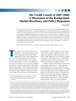
After exercise, LVEF
increases less in cirrhotic patients than in controls
(fig 3).
59 63 69
The reduced functional capacity may
be attributed to a combination of blunted heart
rate response to exercise, reduced myocardial
reserve and profound skeletal muscle wasting with
impaired oxygen extraction.
46 47
In patients with
advanced cirrhosis and severe vasodilatation, acti-
vation of the RAAS, impaired renal function and a
reduced systolic function (a decrease in cardiac
output) appear to be major determinants for the
development of the hepatorenal syndrome.
37
Spontaneous bacterial peritonitis is a well-known
Table 4 Proposal for diagnostic and supportive criteria
for cirrhotic cardiomyopathy
A working definition of cirrhotic cardiomyopathy
A cardiac dysfunction in patients with cirrhosis characterised by
impaired contractile responsiveness to stress and/or altered diastolic
relaxation with electrophysiological abnormalities in the absence of
other known cardiac disease
Diagnostic criteria
Systolic dysfunction
c Blunted increase in cardiac output on exercise, volume challenge
or pharmacological stimuli
c Resting ejection fraction ,55%
Diastolic dysfunction
c E/A ratio ,1.0 (age-corrected)
c Prolonged deceleration time (.200 ms)
c Prolonged isovolumetric relaxation time (.80 ms)
Supportive criteria
c Electrophysiological abnormalities
c Abnormal chronotropic response
c Electromechanical uncoupling/dys-synchrony
c Prolonged Q–T
c
interval
c Enlarged left atrium
c Increased myocardial mass
c Increased BNP and pro-BNP
c Increased troponin I
BNP, brain natriuretic peptide; E/A ratio, ratio of early to late (atrial)
phases of ventricular filling.
Figure 3 Illustration of systolic dysfunction in patients
with cirrhosis and controls. The changes in heart rate
(dHR), cardiac index (dCI) and left ventricular ejection
fraction (dEF) after stress ventriculography are
significantly reduced in cirrhotic patients, most
pronounced in decompensated patients. Mean and SEM.
*p,0.05 versus controls. The figure is based on data from
Torregrosa et al.
69
Recent advances in clinical practice
274 Gut 2008;57:268–278. doi:10.1136/gut.2006.112177
on 11 August 2008 gut.bmj.comDownloaded from
risk factor for the development of the hepatorenal
syndrome, and after resolution of the infection
suppression of systolic function appears to be more
pronounced in patients who develop renal failure.
Maintenance of cardiac contractility thus appears
to be an important factor in the prevention of renal
failure.
70
Diastolic dysfunction
Many patients with cirrhosis exhibit various
degrees of diastolic dysfunction, which implies
changes in myocardial properties that affect left
ventricular filling. Diastolic dysfunction may pro-
gress to systolic dysfunction, although this has not
been directly shown in cirrhotic patients.
31 71
The
pathological basis of the increased stiffness of the
left ventricle seems to be cardiac hypertrophy,
patchy fibrosis and subendothelial oedema.
43169
Determinants of a diastolic dysfunction on a
Doppler echocardiogram are decreased E/A ratio
(the ratio of early to late (atrial) phases of
ventricular filling) and delayed early diastolic
transmitral filling with prolonged deceleration
and isovolumetric relaxation times (table 4).
31 68 72
In a number of studies, A wave and E wave
velocities and deceleration times are much
increased and the E/A ratio is decreased in cirrhotic
patients, especially in those with ascites.
68 72
Recent
studies of ventricular diastolic filling in cirrhosis
support the presence of a subclinical myocardial
disease with diastolic dysfunction, which, in ascitic
patients, improves after paracentesis and can be
aggravated after TIPS.
65 68 72
In these decompen-
sated patients, paracentesis seems to ameliorate
diastolic, but not systolic, function.
68
Patients with
TIPS with an E/A ratio ,1 seem to have a poorer
survival rate than patients without signs of
diastolic dysfunction.
73
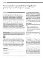
Liver transplantation has
recently been shown to reverse cardiac changes,
including diastolic dysfunction (fig 4).
69
It has been
proposed that diastolic dysfunction precedes
systolic dysfunction in early heart disease and that
anti-aldosterone treatment improves cardiac func-
tion. Pozzi et al recently demonstrated that anti-
aldosterone treatment with K-Canrenoate in cir-
rhosis ameliorated cardiac structure by reducing
left ventricular wall thickness and volume, but had
almost no effects on systolic and diastolic func-
tions.
74
It is also possible that anti-aldosterone
treatment may have beneficial effects on catecho-
lamine-induced cardiac fibrosis, as described in
heart failure.
75
The clinical significance of diastolic dysfunction
and its importance in cirrhotic cardiomyopathy has
been questioned, as overt cardiac failure is not a
prominent feature of cirrhosis. However, there are
several reports of unexpected death from heart
failure following liver transplantation, surgical
portocaval shunts and TIPS.
67 76
These procedures
involve a rapid increase in cardiac preload. In a less
compliant heart, the diastolic dysfunction could be
enough to cause pulmonary oedema and heart
failure. This is consistent with the findings of
Huonker et al,
65
who reported an increase in
pulmonary artery pressure, preload and diastolic
dysfunction after TIPS. In patients with the
hepatopulmonary syndrome and in children with
chronic hepatitis, an isolated right ventricular
diastolic dysfunction has been described and may
play a role in the right cardiac function and course
of these patients.
77
Thus, both left and right
diastolic dysfunction could account for part of
the cardiac dysfunction in cirrhotic cardiomyopa-
thy.
Electromechanical abnormalities
There is a large body of evidence for electrophy-
siological abnormalities in cirrhosis primarily com-
prising prolonged repolarisation time and increased
dispersion of the electromechanical time inter-
val.
78 79
The sympathetic nervous activity influ-
ences the heart rate and electromechanical
coupling by several mechanisms: noradrenaline
binding to b-receptors, receptor-mediated G pro-
tein interaction and, consequently, stimulation of
adenylcyclase, activation of cAMP-dependent
Figure 4 Illustration of reversibility of systolic
dysfunction in patients with cirrhosis and controls. The
change in heart rate (dHR), cardiac index (dCI) and left
ventricular ejection fraction (dEF) after stress
ventriculography significantly improved after liver
transplantation (Ltx). Mean and SEM. *p,0.05;
**p,0.01. The figure is based on data from Torregrosa et
al.
69
Figure 5 Q–T
c
in controls and in patients with cirrhosis
at baseline and during 80 mg propranolol treatment. b-
Adrenergic blockade (BB) significantly reduced the
prolonged Q–T
c
internal. Mean and SEM. *p,0.01 versus
controls. #p,0.01 versus baseline. Data from Henriksen
et al.
82
Recent advances in clinical practice
Gut 2008;57:268–278. doi:10.1136/gut.2006.112177 275
on 11 August 2008 gut.bmj.comDownloaded from
phosphokinase A and channel phosphorylation.
Several receptor and postreceptor defects have been
described in cirrhosis with reduced b-receptor
density and sensitivity, and altered G protein and
calcium channel functions.
480
All these defects may
explain both impaired chronotropic responses and
electromechanical uncoupling. The coupling
between cardiac contractions and the arterial
system is of major importance for the amount of
work performed by the left ventricular myocar-
dium, and thereby for the strain on the heart.
14 46
In
addition, Ward et al have shown a decrease in K
+
currents in ventricular cardiomyocytes from cir-
rhotic rats, which prolongs the Q–T interval.
81
The
prolonged repolarisation time is reflected by a
prolonged Q–T interval in a substantial fraction of
the patients with cirrhosis, which could lead to
ventricular arrhythmias and sudden cardiac death,
but the evidence from clinical studies is sparse.
459
In
cirrhotic patients, the prolonged Q–T interval is
significantly related to the severity of the liver
disease, portal hypertension, portosystemic shunts,
elevated brain-type natriuretic peptide (BNP) and
pro-BNP, elevated plasma noradrenaline and reduced
survival.
79 82 83
The prolongation of the Q–T interval
is partly reversible after liver transplantation and b-
blocker treatment (fig 5).
45 82
The prolonged Q–T
interval in cirrhosis should be considered an element
in the cirrhotic cardiomyopathy and may be of
potential use in identifying patients at risk.
CONCLUDING COMMENTS
Cardiovascular complications in cirrhosis may arise
on the basis of combined humoral, nervous and
haemodynamic changes. Cirrhotic cardiomyopathy
suggests a systolic and diastolic dysfunction and
electrophysiological abnormalities. It is different
from alcoholic heart muscle disease and appears to
be unmasked by procedures that stress the heart,
such as pharmacological vasoconstriction, exercise,
and by insertion of TIPS (Box 1).
59
Potential
diagnostic tools primarily include echocardiography
and ECG (table 5). The cardiovascular complications
in cirrhosis and cirrhotic cardiomyopathy may be
part of a multiorgan syndrome that affects the
patients’ prognosis.
12
No specific treatment can be
recommended, and is largely empiric and supportive.
Caution should be exercised with respect to stressful
procedures, such as large volume paracentesis with-
out adequate plasma volume expansion, TIPS inser-
tion, peritoneovenous shunting and surgery.
4
Cardiac
failure is an important cause of mortality after liver
transplantation. On the other hand, liver transplan-
tation has been shown to reverse systolic and
diastolic dysfunction and the prolonged Q–T inter-
val.
69
Thus, although the post-transplant pathophy-
siological mechanisms are complex, liver
transplantation appears to be an effective treatment
of the cardiovascular complications of cirrhosis.
Improvement of left ventricular contractility
with ACE inhibitors should be done with care, as
this may provoke severe arterial hypotension. b-
Blockers have been shown to reduce acutely the
prolonged Q–T interval and may, in addition to the
cardioprotective effects, be of benefit.
79 82
However,
effects on morbidity and mortality remain to be
shown in longitudinal studies.
Future studies should be directed towards a
delineation of the clinical importance of cardiovas-
cular complications and cirrhotic cardiomyopathy,
and randomised to examine benefits of the treat-
ments outlined above.
Competing interests: None.
Box 1: Key points of cirrhotic cardiomyopathy
c Present in the face of a hyperkinetic circulation
with a combined systolic and diastolic
dysfunction together with electrophysiological
abnormalities.
c Different from alcoholic heart muscle disease
c Systolic dysfunction demasked by physical or
pharmacological stress
c Diastolic dysfunction detected by
echocardiographic measurement of the E/A ratio
c Q–T interval prolongation assessed on the ECG
and adjusted adequately
c Treatment is non-specific and directed towards
the left ventricular heart failure
Table 5 Diagnostic tools in the assessment of systolic
and diastolic dysfunction
Systolic function
c Echocardiography/MRI:
c Volumes
c Fractional shortening
c Velocity of fractional shortening
c Ejection fraction (planimetry)
c Response to stress (dobutamine)
c Wall motion
c Exercise ECG:
c Exercise capacity
c Oxygen consumption*
c Pressure6heart rate product
c Radionuclide angiography (MUGA):
c Ejection fraction
c Cardiac volumes
c Pattern of contractility
c Myocardial perfusion imaging with gating:
c Regional myocardial perfusion
c Cardiac volumes
c Ejection fraction
c Wall motion and wall thickening
Diastolic function
c Echocardiography/MRI/MUGA:
c E/A ratio
c Deceleration time
c A and E waves
c Relaxation times
Most of the techniques mentioned have been validated in normal subjects
and patients with cardiac failure.
*Oxygen consumption (ml/min) = 126effect (W)/body weight (kg)
+3.5).
E/A ratio, ratio of early to late (atrial) phases of ventricular filling; MUGA,
multigated acquisition.
Recent advances in clinical practice
276 Gut 2008;57:268–278. doi:10.1136/gut.2006.112177
on 11 August 2008 gut.bmj.comDownloaded from
REFERENCES
1. Møller S, Henriksen JH. The systemic circulation in cirrhosis. In:
Gines P, Arroyo V, Rodes J, Schrier RW, eds. Ascites and renal
dysfunction in liver disease. Malden: Blackwell, 2005:139–55.
2. Iwakiri Y, Groszmann RJ. The hyperdynamic circulation of chronic
liver diseases: from the patient to the molecule. Hepatology
2006;43:S121–31.
3. Trevisani F, Sica G, Mainqua P, et al. Autonomic dysfunction and
hyperdynamic circulation in cirrhosis with ascites. Hepatology
1999;30:1387–92.
4. Liu H, Gaskari SA, Lee SS. Cardiac and vascular changes in
cirrhosis: pathogenic mechanisms. World J Gastroenterol
2006;12:837–42.
5. Laffi G, Barletta G, Lavilla G, et al. Altered cardiovascular
responsiveness to active tilting in nonalcoholic cirrhosis.
Gastroenterology 1997;113:891–8.
6. Møller S, Nørgaard A, Henriksen JH, et al. Effects of tilting on
central hemodynamics and homeostatic mechanisms in cirrhosis.
Hepatology 2004;40:811–9.
7. Brinch K, Møller S, Bendtsen F, et al. Plasma volume expansion
by albumin in cirrhosis. Relation to blood volume distribution,
arterial compliance and severity of disease. J Hepatol 2003;39:24–
31.
8. Schrier RW. Water and sodium retention in edematous disorders:
role of vasopressin and aldosterone. Am J Med 2006;119:S47–
S53.
9. Møller S, Bendtsen F, Henriksen JH. Effect of volume expansion
on systemic hemodynamics and central and arterial blood volume
in cirrhosis. Gastroenterology 1995;109:1917–25.
10. Kiszka-Kanowitz M, Henriksen JH, Møller S, et al. Blood volume
distribution in patients with cirrhosis: aspects of the dual-head
gamma-camera technique. J Hepatol 2001; 35:605–12.
11. Møller S, Bendtsen F, Christensen E, et al. Prognostic variables in
patients with cirrhosis and oesophageal varices without prior
bleeding. J Hepatol 1994;21:940–6.
12. Hadengue A, Moreau R, Gaudin C, et al. Total effective vascular
compliance in patients with cirrhosis: a study of the response to
acute blood volume expansion. Hepatology 1992;15:809–15.
13. Henriksen JH, Møller S, Schifter S, et al. High arterial compliance
in cirrhosis is related to elevated circulating calcitonin gene-related
peptide (CGRP) and low adrenaline, but not to activated
vasoconstrctor systems. Gut 2001;49:112–8.
14. Henriksen JH, Fuglsang S, Bendtsen F, et al. Arterial compliance
in patients with cirrhosis. High stroke volume/pulse pressure ratio
as an index of elevated arterial compliance. Am J Physiol
2001;280:G584–94.
15. Wiest R, Groszmann RJ. The paradox of nitric oxide in cirrhosis
and portal hypertension: too much, not enough. Hepatology
2002;35:478–91.
16. Rodriguez-Roisin R, Krowka MJ, Herve P, et al. Pulmonary–
hepatic vascular disorders (PHD). Eur Respir J 2004;24:861–80.
17. Møller S, Burchardt H, Ogard CG, et al. Pulmonary blood volume
and transit time in cirrhosis: relation to lung function. Liver Int
2006;26:1072–8.
18. Wiest R, Jurzik L, Herold T, et al. Role of NPY for vasoregulation in
the splanchnic circulation during portal hypertension. Peptides
2007;28:396–404.
19. Abraldes JG, Iwakiri Y, Loureiro-Silva M, et al. Mild increases in
portal pressure upregulate vascular endothelial growth factor and
endothelial nitric oxide synthase in the intestinal microcirculatory
bed, leading to a hyperdynamic state. Am J Physiol Gastrointest
Liver Physiol 2006;290:G980–7.
20. Arroyo V, Terra C, Gines P. Advances in the pathogenesis and
treatment of type-1 and type-2 hepatorenal syndrome. J Hepatol
2007;46:935–46.
21. Salerno F, Gerbes A, Gines P, et al. Diagnosis, prevention and
treatment of the hepatorenal syndrome in cirrhosis. A consensus
workshop of the International Ascites Club. Gut 2007;56:1310–8.
22. Helmy A, Newby DE, Jalan R, et al. Enhanced vasodilatation to
endothelin antagonism in patients with compensated cirrhosis and
the role of nitric oxide. Gut 2003;52:410–5.
23. Langer DA, Shah VH. Nitric oxide and portal hypertension:
interface of vasoreactivity and angiogenesis. J Hepatol
2006;44:209–16.
24. La Villa G, Barletta G, Pantaleo P, et al. Hemodynamic, renal, and
endocrine effects of acute inhibition of nitric oxide synthase in
compensated cirrhosis. Hepatology 2001;34:19–27.
25. Ferguson JW, Dover A, Chia S, et al. Inducible nitric oxide
synthase activity contributes to the regulation of peripheral
vascular tone in patients with cirrhosis and ascites. Gut
2005;55:542–6.
26. Fernandez M, Mejias M, Angermayr B, et al. Inhibition of VEGF
receptor-2 decreases the development of hyperdynamic splanchnic
circulation and portal–systemic collateral vessels in portal
hypertensive rats. J Hepatol 2005;43:98–103.
27. Bolognesi M, Sacerdoti D, Di Pascoli M, et al. Haeme oxygenase
mediates hyporeactivity to phenylephrine in the mesenteric vessels
of cirrhotic rats with ascites. Gut 2005;54:1630–6.
28. Ebrahimkhani MR, Mani AR, Moore K. Hydrogen sulphide and
the hyperdynamic circulation in cirrhosis: a hypothesis. Gut
2005;54:1668–71.
29. Hennenberg M, Biecker E, Trebicka J, et al. Defective RhoA/Rho-
kinase signaling contributes to vascular hypocontractility and
vasodilation in cirrhotic rats. Gastroenterology 2006;130:838–54.
30. Rockey DC. Hepatic blood flow regulation by stellate cells in
normal and injured liver. Semin Liver Dis 2001;21:337–49.
31. Møller S, Henriksen JH. Cardiovascular dysfunction in cirrhosis.
Pathophysiological evidence of a cirrhotic cardiomyopathy.
Scand J Gastroenterol 2001;36:785–94.
32. Gines P, Guevara M, De Las HD, et al. Review article: albumin for
circulatory support in patients with cirrhosis. Aliment Pharmacol
Ther 2002;16(Suppl 5):24–31.
33. Sola-Vera J, Minana J, Ricart E, et al. Randomized trial comparing
albumin and saline in the prevention of paracentesis-induced
circulatory dysfunction in cirrhotic patients with ascites.
Hepatology 2003;37:1147–53.
34. Moreau R, Asselah T, Condat B, et al. Comparison of the effect of
terlipressin and albumin on arterial blood volume in patients with
cirrhosis and tense ascites treated by paracentesis: a randomised
pilot study. Gut 2002;50:90–4.
35. Singh V, Kumar B, Nain CK, et al. Noradrenaline and albumin in
paracentesis-induced circulatory dysfunction in cirrhosis: a
randomized pilot study. J Intern Med 2006;260:62–8.
36. Wong F, Logan A, Blendis L. Hyperinsulinemia in preascitic
cirrhosis: effects on systemic and renal hemodynamics, sodium
homeostasis, forearm blood flow, and sympathetic nervous
activity. Hepatology 1996;23:414–22.
37. Ruiz-Del-Arbol L, Monescillo A, Arocena C, et al. Circulatory
function and hepatorenal syndrome in cirrhosis. Hepatology
2005;42:439–47.
38. Maroto A, Gines P, Arroyo V, et al. Brachial and femoral artery
blood flow in cirrhosis—relationship to kidney dysfunction.
Hepatology 1993;17:788–93.
39. Luca A, Garcia-Pagan JC, Feu F, et al. Noninvasive measurement
of femoral blood flow and portal pressure response to propranolol
in patients with cirrhosis. Hepatology 1995;21:83–8.
40. Carella M, Hunter JO, Fazio S, et al. Capillary blood flow to the
skin of forearm in cirrhosis. Angiology 1992;43:969–74.
41. Helmy A, Newby DE, Jalan R, et al. Nitric oxide mediates the
reduced vasoconstrictor response to angiotensin II in patients with
preascitic cirrhosis. J Hepatol 2003;38:44–50.
42. Hendrickse MT, Triger DR. Vagal dysfunction and impaired
urinary sodium and water excretion in cirrhosis. Am J Gastroenterol
1994;89:750–7.
43. Dillon JF, Nolan J, Thomas H, et al. The correction of autonomic
dysfunction in cirrhosis by captopril. J Hepatol 1997;26:331–5.
44. Ates F, Topal E, Kosar F, et al. The relationship of heart rate
variability with severity and prognosis of cirrhosis. Dig Dis Sci
2006;51:1614–8.
45. Mohamed R, Forsey PR, Davies MK, et al. Effect of liver
transplantation on QT interval prolongation and autonomic
dysfunction in end-stage liver disease. Hepatology 1996;23:1128–
34.
46. Grose RD, Nolan J, Dillon JF, et al. Exercise-induced left
ventricular dysfunction in alcoholic and non-alcoholic cirrhosis.
J Hepatol 1995;22:326–32.
47. Epstein SK, Ciubotaru RL, Zilberberg MD, et al. Analysis of
impaired exercise capacity in patients with cirrhosis. Dig Dis Sci
1998;43:1701–7.
48. Villa GL, Barletta G, Romanelli RG, et al. Cardiovascular effects of
canrenone in patients with preascitic cirrhosis. Hepatology
2002;35:1441–8.
49. Ferlitsch A, Pleiner J, Mittermayer F, et al. Vasoconstrictor
hyporeactivity can be reversed by antioxidants in patients with
advanced alcoholic cirrhosis of the liver and ascites. Crit Care Med
2005;33:2028–33.
50. Tripathi D, Therapondos G, Ferguson JW, et al. Endothelin-1
contributes to maintenance of systemic but not portal
haemodynamics in patients with early cirrhosis: a randomised
controlled trial. Gut 2006;55:1290–5.
51. Ros J, Claria J, To-Figueras J, et al. Endogenous cannabinoids: a
new system involved in the homeostasis of arterial pressure in
experimental cirrhosis in the rat. Gastroenterology 2002;122:85–
93.
Recent advances in clinical practice
Gut 2008;57:268–278. doi:10.1136/gut.2006.112177 277
on 11 August 2008 gut.bmj.comDownloaded from
52. Moezi L, Gaskari SA, Liu H, et al. Anandamide mediates
hyperdynamic circulation in cirrhotic rats via CB(1) and VR(1)
receptors. Br J Pharmacol 2006;149:898–908.
53. Batkai S, Mukhopadhyay P, Harvey-White J, et al.
Endocannabinoids acting at CB1 receptors mediate the cardiac
contractile dysfunction in vivo in cirrhotic rats. Am J Physiol Heart
Circ Physiol 2007;293:H1689–95.
54. Møller S, Wiinberg N, Henriksen JH. Noninvasive 24-hour
ambulatory arterial blood pressure monitoring in cirrhosis.
Hepatology 1995;22:88–95.
55. Wong F, Logan A, Blendis L. Systemic hemodynamic, forearm
vascular, renal, and humoral responses to sustained
cardiopulmonary baroreceptor deactivation in well-compensated
cirrhosis. Hepatology 1995;21:717–24.
56. Laffi G, Lagi A, Cipriani M, et al. Impaired cardiovascular
autonomic response to passive tilting in cirrhosis with ascites.
Hepatology 1996;24:1063–7.
57. Møller S, Iversen JS, Henriksen JH, et al. Reduced baroreflex
sensitivity in alcoholic cirrhosis: relations to hemodynamics and
humoral systems. Am J Physiol Heart Circ Physiol
2007;292:G2966–72.
58. Møller S, Søndergaard L, Møgelvang J, et al. Decreased right
heart blood volume determined by magnetic resonance imaging:
evidence of central underfilling in cirrhosis. Hepatology
1995;22:472–8.
59. Møller S, Henriksen JH. Cirrhotic cardiomyopathy: a
pathophysiological review of circulatory dysfunction in liver
disease. Heart 2002;87:9–15.
60. Mani AR, Ippolito S, Ollosson R, et al. Nitration of cardiac proteins
is associated with abnormal cardiac chronotropic responses in rats
with biliary cirrhosis. Hepatology 2006;43:847–56.
61. Pacher P, Batkai S, Kunos G. Cirrhotic cardiomyopathy: an
endocannabinoid connection? Br J Pharmacol 2005;146:313–4.
62. Gaskari SA, Liu H, Moezi L, et al. Role of endocannabinoids in the
pathogenesis of cirrhotic cardiomyopathy in bile duct-ligated rats.
Br J Pharmacol 2005;146:315–23.
63. Wong F, Girgrah N, Graba J, et al. The cardiac response to
exercise in cirrhosis. Gut 2001;49:268–75.
64. Krag A, Bendtsen F, Henriksen JH, et al. Cardiac effects of
terlipressin in cirrhosis. Unmasking a cirrhotic cardiomyopathy.
J Hepatol 2007;46:S96.
65. Huonker M, Schumacher YO, Ochs A, et al. Cardiac function and
haemodynamics in alcoholic cirrhosis and effects of the
transjugular intrahepatic portosystemic stent shunt. Gut
1999;44:743–8.
66. Merli M, Valeriano V, Funaro S, et al. Modifications of cardiac
function in cirrhotic patients treated with transjugular intrahepatic
portosystemic shunt (TIPS). Am J Gastroenterol 2002;97:142–8.
67. Gines P, Uriz J, Calahorra B, et al. Transjugular intrahepatic
portosystemic shunting versus paracentesis plus albumin for
refractory ascites in cirrhosis. Gastroenterology 2002;123:1839–
47.
68. Pozzi M, Carugo S, Boari G, et al. Evidence of functional and
structural cardiac abnormalities in cirrhotic patients with and
without ascites. Hepatology 1997;26:1131–7.
69. Torregrosa M, Aguade S, Dos L, et al. Cardiac alterations in
cirrhosis: reversibility after liver transplantation. J Hepatol
2005;42:68–74.
70. Ruiz-Del-Arbol L, Urman J, Fernandez J, et al. Systemic, renal,
and hepatic hemodynamic derangement in cirrhotic patients with
spontaneous bacterial peritonitis. Hepatology 2003;38:1210–8.
71. Pozzi M, Redaelli E, Ratti L, et al. Time-course of diastolic
dysfunction in different stages of chronic HCV related liver
diseases. Minerva Gastroenterol Dietol 2005;51:179–86.
72. Finucci G, Desideri A, Sacerdoti D, et al. Left ventricular diastolic
function in liver cirrhosis. Scand J Gastroenterol 1996;31:279–84.
73. Cazzaniga M, Salerno F, Pagnozzi G, et al. Diastolic dysfunction is
associated with poor survival in cirrhotic patients with transjugular
intrahepatic portosystemic shunt. Gut 2007;56:869–75.
74. Pozzi M, Grassi G, Ratti L, et al. Cardiac, neuroadrenergic, and
portal hemodynamic effects of prolonged aldosterone blockade in
postviral child A cirrhosis. Am J Gastroenterol 2005;100:1110–6.
75. Bos R, Mougenot N, Findji L, et al. Inhibition of catecholamine-
induced cardiac fibrosis by an aldosterone antagonist. J Cardiovasc
Pharmacol 2005;45:8–13.
76. Myers RP, Lee SS. Cirrhotic cardiomyopathy and liver
transplantation. Liver Transpl 2000;6:S44–52.
77. Karabulut A, Iltumur K, Yalcin K, et al. Hepatopulmonary
syndrome and right ventricular diastolic functions: an
echocardiographic examination. Echocardiography 2006;23:271–8.
78. Henriksen JH, Fuglsang S, Bendtsen F, et al. Dyssynchronous
electrical and mechanical systole in patients with cirrhosis.
J Hepatol 2002;36:513–20.
79. Zambruni A, trevisani F, Caraceni P, et al. Cardiac
electrophysiological abnormalities in patients with cirrhosis.
J Hepatol 2006;44:994–1002.
80. Zavecz JH, Bueno O, Maloney RE, et al. Cardiac excitation–
contraction coupling in the portal hypertensive rat. Am J Physiol
2000;279:G28–39.
81. Ward CA, Ma Z, Lee SS, et al. Potassium currents in atrial and
ventricular myocytes from a rat model of cirrhosis. Am J Physiol
Gastrointest Liver Physiol 1997;273:G537–44.
82. Henriksen JH, Bendtsen F, Hansen EF, et al. Acute non-selective
beta-adrenergic blockade reduces prolonged frequency-adjusted
Q–T interval (QTc) in patients with cirrhosis. J Hepatol
2004;40:239–46.
83. Henriksen JH, Go¨tze JP, Fuglsang S, et al. Increased circulating
pro-brain natriuretic peptide (proBNP) and brain natriuretic peptide
(BNP) in patients with cirrhosis: relation to cardiovascular
dysfunction and severity of disease. Gut 2003;52:1511–7.
Recent advances in clinical practice
278 Gut 2008;57:268–278. doi:10.1136/gut.2006.112177
on 11 August 2008 gut.bmj.comDownloaded from