Báo cáo y học: "Clinical relevance of and risk factors for HSV-related tracheobronchitis or pneumonia: results of an outbreak investigation" doc
Bạn đang xem bản rút gọn của tài liệu. Xem và tải ngay bản đầy đủ của tài liệu tại đây (569.98 KB, 11 trang )
Open Access
Available online />Page 1 of 11
(page number not for citation purposes)
Vol 11 No 6
Research
Clinical relevance of and risk factors for HSV-related
tracheobronchitis or pneumonia: results of an outbreak
investigation
Ilka Engelmann
1
, Jens Gottlieb
2
, Astrid Meier
3
, Dorit Sohr
4
, Arjang Ruhparwar
5,6
, Cornelia Henke-
Gendo
1
, Petra Gastmeier
3
, Tobias Welte
2
, Thomas Friedrich Schulz
1
and Frauke Mattner
3
1
Institute of Virology, Medizinische Hochschule Hannover, Carl-Neuberg-Strasse 1, 30625 Hannover, Germany
2
Department of Pneumology, Medizinische Hochschule Hannover, Carl-Neuberg-Strasse 1, 30625 Hannover, Germany
3
Institute of Medical Microbiology and Hospital Epidemiology, Medizinische Hochschule Hannover, Carl-Neuberg-Strasse 1, 30625 Hannover,
Germany
4
Institute of Hygiene and Hospital Epidemiology, Charité, Hindenburgdamm 27 12203 Berlin, Germany
5
Division of Thoracic and Cardiovascular Surgery, Medizinische Hochschule Hannover, Carl-Neuberg-Strasse 1, 30625 Hannover, Germany
6
Department of Cardiac Surgery, University of Heidelberg, Im Neuenheimer Feld 110, 69120 Heidelberg, Germany
Corresponding author: Ilka Engelmann,
Received: 5 Jun 2007 Revisions requested: 9 Jul 2007 Revisions received: 24 Aug 2007 Accepted: 8 Nov 2007 Published: 8 Nov 2007
Critical Care 2007, 11:R119 (doi:10.1186/cc6175)
This article is online at: />© 2007 Engelmann et al, licensee BioMed Central Ltd.
This is an Open Access article distributed under the terms of the Creative Commons Attribution License ( />2.0), which permits unrestricted use, distribution, and reproduction in any medium, provided the original work is properly cited.
Abstract
Introduction Herpes simplex virus (HSV) type 1 was identified
in respiratory specimens from a cluster of eight patients on a
surgical intensive care unit within 8 weeks. Six of these patients
suffered from HSV-related tracheobronchitis and one from HSV-
related pneumonia only. Our outbreak investigation aimed to
determine the clinical relevance of and risk factors associated
with HSV-related tracheobronchitis or pneumonia in critically ill
patients, and to investigate whether the cluster was caused by
nosocomial transmission.
Methods A retrospective cohort study was performed to identify
risk factors for the outcomes of HSV-related tracheobronchitis
or pneumonia and death using univariable analysis as well as
logistic regression analysis. Viruses were typed by molecular
analysis of a fragment of the HSV type 1 glycoprotein G.
Results The cohort of patients covering the outbreak period
comprised 53 patients, including six patients with HSV-related
tracheobronchitis and one patient with pneumonia only. HSV-
related tracheobronchitis or pneumonia was associated with
increased mortality (100% in patients with versus 17.8% in
patients without HSV-related tracheobronchitis or pneumonia; P
< 0.0001). The interaction of longer duration of ventilation and
tracheotomy was associated with HSV-related
tracheobronchitis or pneumonia in multivariable analysis.
Identical HSV type 1 glycoprotein G sequences were found in
three patients and in two patients. The group of three identical
viral sequences belonged to a widely circulating strain. The two
identical viral sequences were recovered from bronchoalveolar
lavages of one patient with HSV-related tracheobronchitis and
of one patient without clinical symptoms. These viral sequences
showed unique polymorphisms, indicating probable nosocomial
transmission.
Conclusion HSV-related tracheobronchitis or pneumonia is
associated with increased mortality in critically ill patients. Care
should be taken to avoid nosocomial transmission and early
diagnosis should be attempted.
Introduction
Herpes simplex virus (HSV) is a double-stranded DNA virus
occurring in two types, HSV-1 and HSV-2. Transmission usu-
ally occurs by contact with infected saliva or cutaneous lesions
[1]. After primary infection, HSV-1 establishes a life-long latent
infection through persistence in neurons of the dorsal root
ganglia and the autonomic nervous system [2]. Reactivation
can be triggered by local stimuli (ultraviolet irradiation, tissue
damage) or by systemic stimuli (fever, menstruation, surgery,
physical or emotional stress, hormonal imbalance,
bp = base pair; HSV = herpes simplex virus; SICU = surgical intensive care unit; PCR = polymerase chain reaction.
Critical Care Vol 11 No 6 Engelmann et al.
Page 2 of 11
(page number not for citation purposes)
immunosuppression) [3]. Clinical manifestations of HSV-1
infection include gingivostomatitis (primary infection), herpes
labialis, encephalitis, and keratoconjunctivitis; infections of the
respiratory or gastrointestinal tract have been described pre-
dominantly in immunosuppressed patients [2].
Asymptomatic shedding of HSV in healthy individuals has
been reported to occur in 2–10% of infected individuals [4,5].
HSV-1 can be detected in the upper respiratory tract and in
the lower respiratory tract of intensive care unit patients in 22–
23% and 16% of cases, respectively [5,6]. Whether these
proportions represent clinically relevant HSV infection or,
rather, are an indicator of severe disease favouring reactivation
without clinical significance is the subject of ongoing debate
[5,7-9]. Tracheobronchitis due to HSV has been described in
critically ill patients [10,11].
As more than 90% of adults have antibodies specific for HSV-
1 [11], infections in adulthood are usually assumed to be reac-
tivation of endogenous virus, although reinfection with a differ-
ent HSV-1 strain that is immunologically distinct is also
possible [12].
Eight patients in a surgical intensive care unit (SICU) had
HSV-1 detected in their respiratory tract within 8 weeks. Tra-
cheobronchitis was associated with HSV-1 detection in six
patients and with pneumonia in four patients. This cluster
prompted us to investigate the clinical impact of HSV-related
tracheobronchitis or pneumonia and to identify risk factors
predisposing to HSV-related tracheobronchitis or pneumonia
and fatal outcome. As the cluster suggested the possibility of
nosocomial transmission, molecular epidemiological studies
were performed to type all viruses recovered from the patients.
Materials and methods
Setting and patients
When a cluster of six patients with HSV-1-related tracheo-
bronchitis occurred on a 15-bed cardiothoracic SICU (Figure
1), the present outbreak investigation was initiated. Medical
records of the SICU and the database of the Department of
Virology were reviewed to identify all patients who were hospi-
talized on this SICU during the time period when the cluster
occurred and who had HSV-1 detected (by antigen detection,
virus isolation or PCR) in respiratory fluids.
Demographic data as well as underlying diseases, clinical
course, any severe clinical presentations in addition to the
HSV-1-associated ones and outcome were recorded (Tables
1 and 2). All records of bronchoscopic and radiologic exami-
nations were reassessed, focusing on endobronchial bleed-
ings and lesions or infiltrates compatible with HSV infection.
Microbiological and mycological findings were reviewed to
determine whether concurrent infections with pathogens other
than HSV were present. To assess the clinical relevance of
HSV-1 detection in respiratory fluids, the clinical presentations
and outcomes of HSV-1-positive patients were analysed
(Tables 1 and 2).
Bronchoscopies were sampled after routine disinfection. DNA
was isolated and used as the input in the diagnostic HSV PCR
and in the typing PCR (see below).
The institutional review board approved the outbreak
investigation.
Cohort study
A retrospective cohort study was performed including the 8-
week period that entirely covered the cluster episode. During
this period all patients admitted to the SICU with a stay longer
than 72 hours were included (n = 53). The analysed outcomes
were death and HSV-related tracheobronchitis or pneumonia.
The latter was defined as HSV detection in the respiratory
tract concomitant with the presence of tracheobronchitis or
pneumonia and an absence of other respiratory pathogens
even though histopathology was not performed. Sampling for
HSV detection in respiratory fluids was performed if clinically
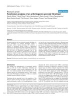
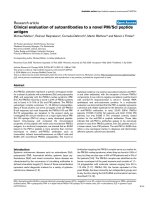
Figure 1
Patient cluster with herpes simplex virus in respiratory specimens on the surgical intensive care unitPatient cluster with herpes simplex virus in respiratory specimens on the surgical intensive care unit. Grey bars, period of stay on the surgical inten-
sive care unit; black bars, day when herpes simplex virus type 1 was detected in the respiratory tract of the patient.
Available online />Page 3 of 11
(page number not for citation purposes)
indicated (that is, in case of unexplained deterioration of respi-
ratory function, tracheobronchial bleeding or suspicious
mucosal lesions on bronchoscopic examination).
Of the 53 patients, seven fulfilled the criteria for the outcome
HSV-related tracheobronchitis or pneumonia. One of the
patients with HSV detection in bronchoalveolar lavage was
excluded from the analysis of the cohort study (Patient C3)
because he did not show symptoms related to HSV (Tables 1
and 2). The HSV glycoprotein G sequence of this patient was
included in the molecular epidemiological analysis (see
below). None of the remaining 45 patients presented with
symptoms of tracheobronchitis. Eight of the 45 remaining
patients were confirmed negative for HSV in their lower respi-
ratory tract secretions, whereas the other patients were not
tested because testing was only performed if clinical symp-
toms were evocative.
The variables analysed as risk factors for HSV-related trache-
obronchitis or pneumonia and fatal outcome are presented in
Tables 3 and 4. The variables pneumonia and HSV-related tra-
cheobronchitis or HSV-related pneumonia were included
Table 1
Demographic and clinical characteristics of patients with herpes simplex virus type 1 detection in respiratory specimens
Cluster
Patient/Isolate
Gender Age (years) Underlying disease Surgical procedure Herpes simplex virus-associated
infection
C1 Female 81 Aortic stenosis Replacement of the aortic valve
by biological graft
Pneumonia, haemorrhagic
tracheobronchitis
C2 Male 74 Infection of the aortic Y-
prosthesis
Replacement of the aorta Haemorrhagic tracheobronchitis
C3 Female 18 Cystic fibrosis Lung transplantation -
C4 Male 78 Coronary heart disease Coronary artery bypass graft Haemorrhagic tracheobronchitis
C5 Male 61 Coronary heart disease Coronary artery bypass graft Pneumonia, haemorrhagic
tracheobronchitis
C6 Male 51 Coronary heart disease Extracorporeal membrane
oxygenation implantation
Haemorrhagic tracheobronchitis
C7 Male 67 Covered rupture of an aortic
aneurysm
Replacement of the aorta Pneumonia, haemorrhagic
tracheobronchitis
C8 Male 77 Coronary heart disease Coronary artery bypass graft Pneumonia
Table 2
Clinical and virologic characteristics of patients with herpes simplex virus type 1 detection in respiratory specimens
Cluster
Patient/
Isolate
Clinical presentation besides herpes simplex virus-
associated presentations
Herpes simplex virus detection Outcome
Specimen type Direct
immunofluorescence
testing
Virus culture PCR
C1 Right-sided heart failure Tracheal aspirate - - + Death
C2 Infection of the aortic Y-prosthesis, intraabdominal
bleedings
Bronchoalveolar lavage + + + Death
C3 - Bronchoalveolar lavage + - + Survival
C4 Adult respiratory distress syndrome, internal carotid
artery stenosis
Tracheal aspirate + + + Death
C5 Adult respiratory distress syndrome Nasopharyngeal swab + + + Death
C6 - Bronchoalveolar lavage + + + Death
C7 Peritonitis with coagulase-negative Staphylococci Bronchoalveolar lavage + + + Death
C8 Sepsis Bronchoalveolar lavage + + + Death
Critical Care Vol 11 No 6 Engelmann et al.
Page 4 of 11
(page number not for citation purposes)
Table 3
Frequency of outcome herpes simplex virus type 1 (HSV-1)-related tracheobronchitis or pneumonia depending on patient
characteristics and extrinsic risk factors
Risk factor Number (%) of patients Number (%) of patients with HSV-1-
related tracheobronchitis or pneumonia
P value
a
Relative risk
Without risk factor With risk factor Without risk factor With risk factor
Age ≥ median (64 years) 27 (51.9) 25 (48.1) 2 (7.4) 5 (20.0) 0.24 2.70
Gender, male 15 (28.8) 37 (71.2) 1 (6.6) 6 (16.2) 0.66 2.43
Simplified Acute Physiology Score >
median (31)
27 (51.9) 25 (48.1) 2 (7.4) 5 (20.0) 0.24 2.70
Time at risk on SICU > median
(8.5 days)
26 (50.0) 26 (50.0) 1 (3.9) 6 (23.1) 0.10 6.00
Ventilation time > median (4.7 days)
b
26 (50.0) 26 (50.0) 0 (0) 7 (26.9) 0.01 -
Bronchoscopy 24 (46.1) 28 (53.9) 3 (12.5) 4 (14.3) 1.00 1.14
Number of bronchoscopies > median
(1)
37 (71.1) 15 (28.9) 4 (10.8) 3 (20.0) 0.40 1.85
Tracheotomy 46 (88.5) 6 (11.5) 4 (8.7) 3 (50.0) 0.03 5.75
Reintubation 37 (71.1) 15 (28.9) 4 (10.8) 3 (20.0) 0.40 1.85
Reanimation 44 (84.6) 8 (15.4) 5 (11.4) 2 (25.0) 0.29 2.20
Underlying disease
Cardiomyopathy
b
48 (92.3) 4 (7.7) 7 (14.6) 0 (0) 1.00 -
Valvular heart disease 35 (67.3) 17 (32.7) 6 (17.1) 1 (5.9) 0.40 0.34
Cystic fibrosis
b
51 (98.1) 1 (1.9) 7 (13.7) 0 (0) 1.00 -
Infrarenal aortic aneurysm 47 (90.4) 5 (9.6) 6 (12.8) 1 (20.0) 0.53 1.57
Suprararenal aortic aneurysm 47 (90.4) 5 (9.6) 6 (12.8) 1 (20.0) 0.53 1.57
Coronary heart disease 37 (71.1) 15 (28.9) 3 (8.11) 4 (26.7) 0.17 3.29
Congenital valvular heart disease
b
48 (92.3) 4 (7.7) 7 (14.6) 0 (0) 1.00 -
Surgical intervention
Solid organ transplantation
b
44 (84.6) 8 (15.4) 7 (19.9) 0 (0) 0.58 -
Left ventricular assist device 43 (82.7) 9 (17.3) 5 (11.6) 2 (22.2) 0.59 1.91
Coronary artery bypass graft 38 (73.1) 14 (26.9) 4 (10.5) 3 (21.4) 0.37 2.04
Aortic surgery 41 (78.8) 11 (21.2) 5 (12.2) 2 (18.2) 0.63 1.49
Valve surgery 37 (71.1) 15 (28.9) 6 (16.2) 1 (6.7) 0.66 0.41
Immunosuppressive medication 27 (51.9) 25 (48.1) 5 (18.5) 2 (8.0) 0.42 0.43
Steroids 28 (53.8) 24 (46.2) 5 (17.9) 2 (8.3) 0.43 0.47
Cyclosporin
b
46 (88.5) 6 (11.5) 7 (15.2) 0 (0) 0.58 -
Mycophenolate mofetil
b
44 (84.6) 8 (15.4) 7 (15.9) 0 (0) 0.58 -
Basiliximab
b
46 (88.5) 6 (11.5) 7 (15.2) 0 (0) 0.58 -
Tacrolimus 48 (92.3) 4 (7.7) 6 (12.5) 1 (25.0) 0.45 2.00
Blood products
b
2 (3.8) 50 (96.2) 0 (0) 7 (14.0) 1.00 -
Erythrocyte concentrates
b
8 (15.4) 44 (84.6) 0 (0) 7 (15.9) 0.58 -
Fresh frozen plasma
b
4 (7.7) 48 (92.3) 0 (0) 7 (14.6) 1.00 -
Thrombocyte concentrates 15 (28.8) 37 (71.2) 2 (13.3) 5 (13.5) 1.00 1.01
Number of erythrocyte concentrates
> median (7)
27 (51.9) 25 (48.1) 1 (3.7) 6 (24.0) 0.05 6.48
Number of fresh frozen plasma units
> median (11.5)
26 (50.0) 26 (50.0) 1 (3.9) 6 (23.1) 0.09 6.00
Number of thrombocyte
concentrates > median (3)
26 (50.0) 26 (50.0) 3 (11.5) 4 (15.4) 1.00 1.33
Clotting factor substitution 31 (59.6) 21 (40.4) 2 (6.5) 5 (23.8) 0.10 3.69
C1 esterase inhibitor
b
49 (94.2) 3 (5.8) 7 (14.3) 0 (0) 1.00 -
Interactions
Ventilation time > median with
tracheotomy
48 (92.3) 4 (7.7) 4 (8.3) 3 (75.0) 0.006 9.00
a
Fisher's exact test.
b
Variable could not be included in the logistic regression model for mathematical reasons.
Available online />Page 5 of 11
(page number not for citation purposes)
Table 4
Frequency of outcome death depending on patient characteristics and extrinsic risk factors
Risk factor Number (%) of patients Number (%) of patients
with outcome death
P value
a
Relative risk
Without risk factor With risk factor Without risk factor With risk factor
Age ≥ median (64 years) 27 (51.9) 25 (48.1) 8 (29.6) 7 (28.0) 1.00 0.95
Gender, male 15 (28.8) 37 (71.2) 4 (26.7) 11 (29.7) 1.00 1.12
Simplified Acute Physiology Score >
median (31)
27 (51.9) 25 (48.1) 6 (22.2) 9 (36.0) 0.36 1.62
Time at risk on SICU > median (8.5 days) 26 (50.0) 26 (50.0) 5 (19.2) 10 (38.5) 0.22 2.00
Ventilation time > median (4.7 days) 26 (50.0) 26 (50.0) 2 (7.7) 13 (50.0) 0.002 6.50
Bronchoscopy 24 (46.1) 28 (53.9) 3 (12.5) 12 (42.9) 0.03 3.43
Number of bronchoscopies > median (1) 37 (71.1) 15 (28.9) 10 (27.0) 5 (33.3) 0.74 1.23
Tracheotomy 46 (88.5) 6 (11.5) 11 (23.9) 4 (66.7) 0.05 2.79
Reintubation 37 (71.1) 15 (28.9) 10 (27.0) 5 (33.3) 0.74 1.23
Reanimation 44 (84.6) 8 (15.4) 12 (27.3) 3 (37.5) 0.68 1.38
Cardiovascular disease
Cardiomyopathy
b
48 (92.3) 4 (7.7) 15 (31.3) 0 (0) 0.31 -
Valvular heart disease 35 (67.3) 17 (32.7) 11 (31.4) 4 (23.5) 0.75 0.75
Cystic fibrosis
b
51 (98.1) 1 (1.9) 15 (29.4) 0 (0) 1.00 -
Infrarenal aortic aneurysm 47 (90.4) 5 (9.6) 13 (27.7) 2 (40.0) 0.62 1.45
Suprararenal aortic aneurysm 47 (90.4) 5 (9.6) 14 (29.8) 1 (20.0) 1.00 0.67
Coronary heart disease 37 (71.1) 15 (28.9) 8 (21.6) 7 (46.7) 0.10 2.16
Congenital valvular heart disease 48 (92.3) 4 (7.7) 14 (29.2) 1 (25.0) 1.00 0.86
Surgical intervention
Solid organ transplantation 44 (84.6) 8 (15.4) 14 (31.8) 1 (12.5) 0.41 0.39
Left ventricular assist device 43 (82.7) 9 (17.3) 12 (27.9) 3 (33.3) 0.71 1.19
Coronary artery bypass graft 38 (73.1) 14 (26.9) 9 (23.7) 6 (42.9) 0.19 1.81
Aortic surgery 41 (78.8) 11 (21.2) 12 (29.3) 3 (27.3) 1.00 0.93
Valve surgery 37 (71.1) 15 (28.9) 12 (32.4) 3 (20.0) 0.51 0.62
Immunosuppression 27 (51.9) 25 (48.1) 9 (33.3) 6 (24.0) 0.55 0.72
Steroids 28 (53.8) 24 (46.2) 10 (35.7) 5 (20.8) 0.36 0.58
Cyclosporin 46 (88.5) 6 (11.5) 14 (30.4) 1 (16.7) 0.66 0.55
Mycophenolate mofetil 44 (84.6) 8 (15.4) 14 (31.8) 1 (12.5) 0.41 0.39
Basiliximab 46 (88.5) 6 (11.5) 14 (30.4) 1 (16.7) 0.66 0.55
Tacrolimus 48 (92.3) 4 (7.7) 14 (29.2) 1 (25.0) 1.00 0.86
Blood products
b
2 (3.8) 50 (96.2) 0 (0) 15 (30.0) 1.00 -
Erythrocyte concentrates 8 (15.4) 44 (84.6) 1 (12.5) 14 (31.8) 0.41 2.55
Fresh frozen plasma
b
4 (7.7) 48 (92.3) 0 (0) 15 (31.3) 0.31 -
Thrombocyte concentrates 15 (28.8) 37 (71.2) 2 (13.3) 13 (35.1) 0.18 2.64
Number of erythrocyte concentrates >
median (7)
27 (51.9) 25 (48.1) 4 (14.8) 11 (44.0) 0.03 2.97
Number of fresh frozen plasma units >
median (11.5)
26 (50.0) 26 (50.0) 4 (15.4) 11 (42.3) 0.06 2.75
Number of thrombocyte concentrates >
median (3)
26 (50.0) 26 (50.0) 4 (15.4) 11 (42.3) 0.06 2.75
Clotting factors 31 (59.6) 21 (40.4) 6 (19.4) 9 (42.9) 0.12 2.21
C1 esterase inhibitor 49 (94.2) 3 (5.8) 13 (26.5) 2 (66.7) 0.20 2.51
Herpes simplex virus-related
tracheobronchitis or pneumonia
b
45 (86.5) 7 (13.5) 8 (17.8) 7 (100) <0.000
1
-
Pneumonia 46 (88.5) 6 (11.5) 11 (23.9) 4 (66.7) 0.05 2.79
a
Fisher's exact test.
b
Variable could not be included in the logistic regression model for mathematical reasons.
Critical Care Vol 11 No 6 Engelmann et al.
Page 6 of 11
(page number not for citation purposes)
additionally as risk factors for fatal outcome. As appropriate to
analyse the time period at risk for developing HSV-related tra-
cheobronchitis or pneumonia, variables were only regarded as
positive if they occurred prior to HSV detection (for patients
with HSV-related tracheobronchitis or pneumonia) or prior to
SICU discharge (for patients without HSV-related tracheo-
bronchitis or pneumonia).
Statistical analysis
Univariable analysis and logistic regression with stepwise (for-
ward and backward) variable selection were performed using
SAS software (SAS Institute, Inc., Cary, NC, USA). P < 0.05
was regarded as significant for univariable analysis. A signifi-
cance level of 0.1 was chosen for inclusion of variables into a
logistic regression model and for remaining included in the
model.
The area under the receiver operating characteristic curve (c
index) is used to evaluate the predictive power of the logistic
regression model. The c index represents the probability that
the regression model equation assigns randomly chosen
patients with HSV higher probabilities of acquiring HSV than
randomly chosen patients without HSV [13].
Viral diagnostics and molecular typing
Respiratory specimens (bronchoalveolar lavage or tracheal
aspirates in most cases, nasopharyngeal swab in one patient)
were submitted to direct immunofluorescence staining with
antibodies specific for HSV. A sample (1–2 ml) of the speci-
men was added to cell cultures of Hep-2 and Vero cells, and
was monitored twice weekly for up to 3 weeks for emergence
of a cytopathic effect. DNA was isolated and a real-time PCR
was performed, detecting a 254 bp fragment of the HSV UL27
gene (Engelmann, I., Petzold, D.R., Kosinska A., Hepkema
B.G., Schulz, T. F. and Heim, A., accepted for publication in
Journal of Medical Virology; Title: Rapid quantitative PCR
assays for the simultaneous detection of herpes simplex virus,
varicella zoster virus, cytomegalovirus, Epstein Barr virus and
human herpesvirus 6 DNA in blood and other clinical speci-
mens). Melting curve analysis was used to differentiate HSV-1
and HSV-2.
All viruses recovered from patients on the SICU during the
time period of the cluster (designated C; Figure 1) – including
patients with HSV-related tracheobronchitis or pneumonia
and one patient with asymptomatic HSV detection – were
typed. Additionally, HSV isolates from patients of the same
SICU outside the cluster period (designated I) and HSV iso-
lates from patients on other wards (designated M) were typed.
A hypervariable part of the HSV-1 glycoprotein G was ampli-
fied as described by Rekabdar and colleagues [14]. Direct
sequencing was performed using the PCR primers and the
dRhodamine Terminator Cycle Sequencing Ready Reaction
Kit (Applied Biosystems, Foster City, CA, USA) according to
the manufacturer's instructions on the ABI PRISM 310
Genetic Analyzer (Applied Biosystems, Foster City, CA, USA).
If direct sequencing was unsuccessful the samples were sub-
jected to a nested PCR. The first round involved the amplifica-
tion of a 600 bp fragment using the outer primers HSV-N1-For
(5'-GGGTTCCCACCAACGTCTCC) and HSV-N1-Rev (5'-
GGGTGTGTGCGTCGCCCGC). The resulting PCR product
was used as template in the PCR described above.
The nucleotide sequences have been submitted to the NCBI
database GenBank (accession numbers EF376300
–
EF376333
).
Phylogenetic analysis of the 309 bp sequence of the PCR
product was conducted with Phylip software (version 3.63)
and with the MEGA Software package (version 3.1) [15]. The
phylogenetic tree was constructed using the neighbour-join-
ing method (Kimura two-parameter matrix) with a transition/
transversion ratio of 2.0. Bootstrapping was performed with
1,000 replicates and values above 80% are indicated. The
tree is presented as an unrooted tree because the use of HSV-
2 as an outgroup was impossible due to highly divergent
sequences between HSV-1 and HSV-2 in the analysed region.
Results
HSV-related tracheobronchitis was diagnosed in six patients
on a SICU within 8 weeks. Endobronchial bleeding was life-
threatening in three patients. HSV-1 was detectable in the res-
piratory specimens of all patients (Tables 1 and 2) whereas no
concurrent bacterial or fungal pathogens were isolated. Fur-
thermore, HSV-1 was detected in bronchoalveolar lavages of
two additional patients who did not suffer from HSV-related
tracheobronchitis, one of whom had pneumonia (Figure 1).
The only surviving patient (C3) had neither clinical nor bron-
choscopic signs of HSV-related tracheobronchitis but did
receive antiviral medication.
All patients were HSV IgG-positive at the time of HSV detec-
tion, suggesting that none of them suffered from primary
infection.
Cohort study
In order to confirm that HSV-related tracheobronchitis or
pneumonia was associated with a higher mortality, and to
identify risk factors for this entity, a cohort study including 52
patients (after exclusion of one patient with HSV detection in
bronchoalveolar lavage but without symptoms related to HSV)
was performed. Tables 3 and 4 present the results of univaria-
ble analysis for the outcomes HSV-related tracheobronchitis
or pneumonia and death. Multiple logistic regression analysis
revealed statistical association of HSV-related tracheobron-
chitis or pneumonia with the interaction between longer dura-
Available online />Page 7 of 11
(page number not for citation purposes)
tion of ventilation and tracheotomy, and statistical association
of HSV-related tracheobronchitis or pneumonia with the trans-
fusion of a higher number of erythrocyte concentrates (Table
5).
Fatal outcome was associated with longer duration of ventila-
tion, with bronchoscopy and with higher number of thrombo-
cyte concentrate transfusions in multiple logistic regression
analysis (Table 5). For mathematical reasons HSV-related tra-
cheobronchitis or pneumonia could not be included as a risk
factor in the logistic regression model, although it was the
most significant risk factor in univariable analysis (Table 4).
No other examined variable showed a significant association
with fatal outcome or with HSV-related tracheobronchitis or
pneumonia in multiple logistic regression analysis.
Molecular epidemiology
To address the question of endogenous reactivation versus
nosocomial transmission, all cluster patients' viruses were
subjected to molecular typing. Sequencing of the hypervaria-
ble region of the viral glycoprotein G revealed that patients'
viruses fell into two groups, with viral sequences of patients
C2 and C3 grouping together and with viral sequences of
patients C1, C7 and C8 grouping together (Figures 2 and 3).
Three cluster patients' viral isolates showed HSV-1
sequences that were not detected in other cluster patients'
viral isolates (isolates C4, C5 and C6; Figure 2).
As some viral genotypes are known to predominate even in
epidemiologically unrelated samples [16], analysis was
extended to other HSV isolates obtained during approximately
the same time period in the same hospital. Epidemiologically
unrelated virus isolates (isolates I2, I6, I8, M4, M6, M7, M9,
M12 and M13) that showed sequences identical to those of
the cluster patients were found in the group represented by
viral sequences C1, C7 and C8 (Figures 2 and 3). Viral
sequences of patients C2 and C3, however, showed a unique
sequence that was not identified in any of the epidemiologi-
cally unrelated isolates and could not be found in the NCBI
database. The C2/C3 viral sequence was characterized by a
deletion of six base pairs in a repeat region and a point muta-
tion at position 110 (of the alignment of partial glycoprotein G
sequences) (Figure 3). Isolate C2 was obtained from a patient
with HSV-related tracheobronchitis. In contrast to that, the
patient from whom the viral sequence C3 was recovered did
not show symptoms associated with HSV detection. Those
two patients had overlapping stays on the SICU, and HSV was
detected in their bronchoalveolar lavages on consecutive days
(Figure 1).
Two out of 11 bronchoscope samples tested positive in the
HSV real-time PCR and were also subjected to the HSV glyc-
oprotein G PCR and the nested glycoprotein G PCR. A PCR
product and sequence was obtained in only one case (viral
sequence B1; Figures 2 and 3). The viral sequence B1 clus-
tered with patient isolate C6 and a group of other isolates not
epidemiologically related.
Discussion
HSV-1 detection in respiratory fluids of critically ill patients is
frequently reported but its clinical relevance is often uncertain
[5,7-9]. In our report the clinical relevance of HSV-1 detection
in the respiratory tract is clearly shown by the fact that six out
of eight patients with HSV-1 detection in their respiratory tract
presented with haemorrhagic tracheobronchitis and four of
the eight patients showed radiologic evidence of pneumonia.
Only one out of eight patients had no symptoms or signs con-
sistent with HSV-1 infection (patient C3); this patient received
Table 5
Risk factors for herpes simplex virus-related tracheobronchitis or pneumonia and fatal outcome: results of multiple logistic
regression analysis with stepwise variable selection
Variable Adjusted odds ratio 95% confidence interval
Outcome herpes simplex virus-related tracheobronchitis or pneumonia
Interaction of ventilation time > median with tracheotomy 32.8 2.72–1000
Number of erythrocyte concentrates > median (7) 8.16 0.96–226.4
c index 0.867
Outcome death
Ventilation time > median (4.7 days) 30.7 3.3–892.7
Bronchoscopy 19.8 2.2–536.6
Number of thrombocyte concentrates > median (3) 16.3 2.1–388.7
c index 0.899
Significance level for inclusion of variables in the model and for remaining included in the model was 0.1. (For mathematical reasons, herpes
simplex virus-related tracheobronchitis or pneumonia could not be included in the model for outcome death.)
Critical Care Vol 11 No 6 Engelmann et al.
Page 8 of 11
(page number not for citation purposes)
ganciclovir prophylaxis as part of a postlung-transplant care
regimen, which might have prevented development of a
clinically relevant HSV-1 infection and might be the reason
why virus culture was negative. Furthermore, the clinical
relevance of HSV-related tracheobronchitis or pneumonia in
critically ill patients is shown by its association with signifi-
cantly increased mortality compared with patients without
HSV-related tracheobronchitis or pneumonia (100% versus
17.8%, P < 0.0001; Table 4). We therefore conclude that
HSV-1 reactivation in the respiratory tract accompanied by tra-
cheobronchitis or pneumonia is clinically relevant and is asso-
ciated with high mortality. It is not possible, however, to prove
to which extent HSV-related tracheobronchitis or pneumonia
contributed to the fatal outcome in our patients because all
patients suffered from other severe medical conditions.
In agreement with our results a recent study showed that lower
respiratory tract infection with HSV was associated with
poorer outcome (prolonged duration of mechanical ventilation
and intensive care unit stay as well as more episodes of bac-
terial ventilator-associated pneumonia), although it was not
associated with increased mortality in their patient cohort [17].
Interestingly, the clinical picture was different, as tracheobron-
chial bleeding was not described as one of the predominant
symptoms [17].
Antiviral therapy with acyclovir initiated after diagnosis of HSV-
1 had been established in five of eight patients on average 2.9
days before death (data not shown). The therapy initiation,
however, seems to have been too late to improve outcome. In
case of endobronchial bleedings or bronchoscopic signs of
haemorrhagic tracheobronchitis, therefore, acyclovir treatment
should be initiated directly after bronchoscopy and specimen
sampling (prior to diagnosis of HSV-1 infection), which would
have been on average 8.6 days prior to death in our collective
(data not shown).
Surgical procedures and critical illness result in immune dys-
function [18,19], which can be regarded as a predisposing
factor for HSV reactivation in the patients described here. The
vagal ganglia are thought to be the source of lower respiratory
and oesophageal HSV reactivation [20]. Mucosal damage
caused by intubation and mechanical ventilation, thoracic sur-
gery or aspiration has been hypothesized to favour HSV reac-
tivation [5,11,17,21,22]. Our observation that the interaction
between longer duration of ventilation and tracheotomy (which
was performed under bronchoscopic assistance) was a risk
factor for HSV-related tracheobronchitis or pneumonia in
logistic regression analysis may be explained by inoculation of
HSV-1-positive fluids from the upper respiratory tract via bron-
choscopically assisted tracheotomy or by mechanical airway
irritation as a possible stimulus of reactivation. Whether longer
duration of ventilation or tracheotomy alone represent signifi-
cant risk factors for HSV-related tracheobronchitis or pneumo-
nia remains unclear because they were not identified as
independent risk factors in logistic regression analysis, and we
cannot exclude that the interaction between longer duration of
ventilation and tracheotomy indicates the severity of disease.
The most significant risk factor for fatal outcome identified in
univariable analysis was HSV-related tracheobronchitis or
pneumonia (P < 0.0001, Table 4). Owing to the fact that all
patients with HSV-related tracheobronchitis or pneumonia
died, this factor could not be included in the logistic regres-
sion model for mathematical reasons. Longer duration of ven-
tilation, bronchoscopy and higher number of thrombocyte
concentrate transfusions were associated with fatal outcome
in the logistic regression analysis. These variables might be
surrogate markers for more severe illness rather than risk
factors.
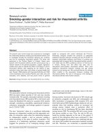
Figure 2
Neighbour-joining tree of partial herpes simplex virus type 1 glycopro-tein G sequencesNeighbour-joining tree of partial herpes simplex virus type 1 glycopro-
tein G sequences. Numbers indicate percentage bootstrap values (only
shown if >80). Scale bar indicates 1% genetic divergence. C, cluster
patients' viral sequences; I, surgical intensive care unit patients' viral
sequences; M, viral sequences from patients from other wards; B, viral
sequence recovered from brochoscope; R, reference strains (R1 =
17+, R2 = F, R3 = Kos).
Available online />Page 9 of 11
(page number not for citation purposes)
Our investigation has the following limitations. Owing to initia-
tion of the analysis in a suspected outbreak situation, system-
atic testing of respiratory samples for HSV was not performed
for all patients. The outcome was therefore defined clinically
(that is, presence or absence of HSV-related
tracheobronchitis or pneumonia) and not virologically
(absence of HSV detection). HSV pneumonia or HSV-related
tracheobronchitis was not confirmed by histopathology
because, clinically, the presence of pneumonia or tracheo-
bronchitis concurrent with HSV detection in respiratory fluids
Figure 3
Partial alignment of herpes simplex virus type 1 glycoprotein G sequencesPartial alignment of herpes simplex virus type 1 glycoprotein G sequences. Black circles, unique polymorphisms in viral sequences C2 and C3. C,
cluster patients' viral sequences; I, SICU patients' viral sequences; M, viral sequences from patients from other wards; B, viral sequence recovered
from brochoscope ; R, reference strains (R1 = 17+, R2 = F, R3 = Kos).
Critical Care Vol 11 No 6 Engelmann et al.
Page 10 of 11
(page number not for citation purposes)
in the absence of other respiratory pathogens was regarded
as sufficient for diagnosis and initiation of antiviral treatment.
The size of the cohort is rather small, a limitation that is inher-
ent to outbreak investigations.
Worthy of note is the fact that two bronchoscope samples had
HSV-1 DNA detected after routine disinfection procedures.
Typing was successful in only one case (viral sequence B1),
which had a sequence identical to patient isolate C6 and to a
number of epidemiologically unrelated virus isolates (Figures 2
and 3). The bronchoscope sample B1, however, was col-
lected more than 2 months later than the positive specimen of
patient isolate C6. An epidemiological association is therefore
unlikely. Furthermore, as no viable virus was recovered in virus
culture, the significance of this finding concerning the possibil-
ity of transmission is uncertain. Testing of bronchoscope sam-
ples was initiated when the cluster was already recognized
and first preventive measures had already been implemented.
We therefore could not clarify whether bronchoscopes were
the source of the suspected nosocomial transmission. Bron-
choscopes, however, have been identified as sources of noso-
comial transmission of other pathogens [23-26].
Studies examining HSV-1 detection in the respiratory tract of
intensive care unit patients have assumed HSV-1 positivity to
be caused by endogenous reactivation, and have not consid-
ered transmission although viruses were not typed [5,7-9].
HSV infections caused by nosocomial transmission, however,
have been reported in different settings [27-35]. In those stud-
ies, viruses were typed by restriction fragment length polymor-
phism and almost identical restriction patterns have been
interpreted as proof of transmission. That approach, however,
does not take into account that some viral genotypes predom-
inate in epidemiologically unrelated virus isolates [16]. Taking
this fact into consideration we extended our sequence-based
typing to epidemiologically unrelated virus isolates from the
same hospital.
We found three cluster patients with HSV-1 sequences that
were not detected in other cluster patients' viral isolates (iso-
lates C4, C5 and C6; Figure 2). This finding favours endog-
enous reactivation as an underlying mechanism.
The group of three cluster patients with identical viral
sequences also showed identical sequences to nine epidemi-
ologically unrelated isolates and to the reference strain F (Fig-
ure 2). Furthermore, the epidemiological association between
these three patients is weak (patient C1 stayed on the SICU
at the beginning of the cluster, and patients C7 and C8 at the
end; Figure 1). These findings favour the hypothesis that their
viruses are part of the predominating genotypes. One patient
pair (patients C2 and C3) that had a strong epidemiological
association (overlapping stay in the SICU, HSV diagnosis on
consecutive days; Figure 1) showed unique polymorphisms in
the HSV glycoprotein G sequence that were not identified in
any other sequenced isolates nor in the NCBI database. In this
case we assume transmission to be likely. As both patients
were IgG-positive prior to HSV detection this could represent
nosocomial reinfection with a different virus strain. Patient C3
did not present with HSV-related tracheobronchitis but rather
with asymptomatic HSV infection, possibly because the gan-
ciclovir prophylaxis he received prevented development of a
clinically relevant HSV infection.
As HSV isolates are not routinely typed it is possible that noso-
comial transmission is not a rare event but might often not be
recognized as such.
Conclusion
In the present article we have shown that HSV-related trache-
obronchitis or pneumonia represents an important infectious
complication in critically ill patients. This differential diagnosis
has to be considered especially in the case of tracheobron-
chial haemorrhage or if the patient's condition does not
improve with antibacterial and antifungal therapy. Whether
acyclovir prophylaxis or early treatment in cases suspicious for
HSV-related tracheobronchitis or pneumonia in critically ill
patients has the potential of preventing fatal outcome should
be addressed in future prospective studies.
Competing interests
The authors declare that they have no competing interests.
Authors' contributions
IE participated in the design of the outbreak investigation, car-
ried out the molecular genetic studies and the phylogenetic
analysis, and wrote the manuscript. JG participated in the col-
lection, interpretation and analysis of clinical data. AM partici-
pated in the collection, interpretation and analysis of clinical
data. DS performed the statistical analysis. AR participated in
the collection, interpretation and analysis of clinical data. CH-
G participated in the phylogenetic analysis. PG participated in
the study design, interpretation of data and drafting of the
manuscript. TW participated in the interpretation of data and
drafting of the manuscript. TFS participated in the study
design, interpretation of the data and drafting of the manu-
script. FM initiated the study, participated in the design of the
study and in the interpretation and analysis of clinical data, and
contributed to the writing of the manuscript.
Key messages
• HSV-related tracheobronchitis or pneumonia is associ-
ated with high mortality in critically ill patients.
• Tracheobronchial haemorrhage should prompt diagnos-
tics for HSV.
• Molecular typing of virus isolates revealed one event of
probable nosocomial transmission.
Available online />Page 11 of 11
(page number not for citation purposes)
Acknowledgements
The author(s) would like to acknowledge the excellent technical assist-
ance of Heidi Pommer and Marlies Werhane.
References
1. Adler SP: Herpes simplex virus. In Hospital Epidemiology and
Infection Control Edited by: Marshall G. Philadelphia, PA: Lippin-
cott Williams & Wilkins; 2004.
2. Whitley RJ, Roizman B: Herpes simplex virus infections. Lancet
2001, 357:1513-1518.
3. Whitley RJ: Herpes simplex viruses. In Virology Edited by: Knipe
DM, Howley PM. Philadelphia, PA: Lippincott Williams & Wilkins;
2001.
4. Hatherley LI, Hayes K, Jack I: Herpes virus in an obstetric hospi-
tal. II: asymptomatic virus excretion in staff members. Med J
Aust 1980, 2:273-275.
5. Bruynseels P, Jorens PG, Demey HE, Goossens H, Pattyn SR,
Elseviers MM, Weyler J, Bossaert LL, Mentens Y, Ieven M: Herpes
simplex virus in the respiratory tract of critical care patients: a
prospective study. Lancet 2003, 362:1536-1541.
6. Cook CH, Martin LC, Yenchar JK, Lahm MC, McGuinness B, Dav-
ies EA, Ferguson RM: Occult herpes family viral infections are
endemic in critically ill surgical patients. Crit Care Med 2003,
31:1923-1929.
7. Cook CH, Yenchar JK, Kraner TO, Davies EA, Ferguson RM:
Occult herpes family viruses may increase mortality in criti-
cally ill surgical patients. Am J Surg 1998, 176:357-360.
8. Ong GM, Lowry K, Mahajan S, Wyatt DE, Simpson C, O'Neill HJ,
McCaughey C, Coyle PV: Herpes simplex type 1 shedding is
associated with reduced hospital survival in patients receiving
assisted ventilation in a tertiary referral intensive care unit. J
Med Virol 2004, 72:121-125.
9. van den Brink JW, Simoons-Smit AM, Beishuizen A, Girbes AR,
Strack van Schijndel RJ, Groeneveld AB: Respiratory herpes
simplex virus type 1 infection/colonisation in the critically ill:
marker or mediator? J Clin Virol 2004, 30:68-72.
10. Sherry MK, Klainer AS, Wolff M, Gerhard H: Herpetic
tracheobronchitis. Ann Intern Med 1988, 109:229-233.
11. Klainer AS, Oud L, Randazzo J, Freiheiter J, Bisaccia E, Gerhard H:
Herpes simplex virus involvement of the lower respiratory
tract following surgery. Chest 1994, 106:8S-14S.
12. Roest RW, Carman WF, Maertzdorf J, Scoular A, Harvey J, Kant M,
van der Meijden WI, Verjans GM, Osterhaus AD: Genotypic anal-
ysis of sequential genital herpes simplex virus type 1 (HSV-1)
isolates of patients with recurrent HSV-1 associated genital
herpes. J Med Virol 2004, 73:601-604.
13. Hanley JA, McNeil BJ: The meaning and use of the area under
a receiver operating characteristic (ROC) curve. Radiology
1982, 143:29-36.
14. Rekabdar E, Tunback P, Liljeqvist JA, Bergstrom T: Variability of
the glycoprotein G gene in clinical isolates of herpes simplex
virus type 1. Clin Diagn Lab Immunol 1999, 6:826-831.
15. Kumar S, Tamura K, Nei M: MEGA3: integrated software for
molecular evolutionary genetics analysis and sequence
alignment. Brief Bioinform 2004, 5:150-163.
16. Sakaoka H, Kurita K, Iida Y, Takada S, Umene K, Kim YT, Ren CS,
Nahmias AJ: Quantitative analysis of genomic polymorphism of
herpes simplex virus type 1 strains from six countries: studies
of molecular evolution and molecular epidemiology of the
virus. J Gen Virol 1994, 75(Pt 3):513-527.
17. Luyt CE, Combes A, Deback C, Aubriot-Lorton MH, Nieszkowska
A, Trouillet JL, Capron F, Agut H, Gibert C, Chastre J: Herpes sim-
plex virus lung infection in patients undergoing prolonged
mechanical ventilation. Am J Respir Crit Care Med 2007,
175:935-942.
18. Howard RJ, Simmons RL: Acquired immunologic deficiencies
after trauma and surgical procedures. Surg Gynecol Obstet
1974, 139:771-782.
19. Slade MS, Simmons RL, Yunis E, Greenberg LJ: Immunodepres-
sion after major surgery in normal patients. Surgery 1975,
78:363-372.
20. Tuxen DV: Prevention of lower respiratory herpes simplex virus
infection with acyclovir in patients with adult respiratory dis-
tress syndrome. Chest 1994, 106:28S-33S.
21. Hanley PJ, Conaway MM, Halstead DC, Rhodes LV III, Reed J III:
Nosocomial herpes simplex virus infection associated with
oral endotracheal intubation. Am J Infect Control 1993,
21:310-316.
22. Camps K, Jorens PG, Demey HE, Pattyn SR, Ieven M: Clinical sig-
nificance of herpes simplex virus in the lower respiratory tract
of critically ill patients. Eur J Clin Microbiol Infect Dis 2002,
21:758-759.
23. Corne P, Godreuil S, Jean-Pierre H, Jonquet O, Campos J, Jumas-
Bilak E, Parer S, Marchandin H: Unusual implication of biopsy
forceps in outbreaks of Pseudomonas aeruginosa infections
and pseudo-infections related to bronchoscopy. J Hosp Infect
2005, 61:20-26.
24. Bou R, Aguilar A, Perpinan J, Ramos P, Peris M, Lorente L, Zuniga
A: Nosocomial outbreak of Pseudomonas aeruginosa infec-
tions related to a flexible bronchoscope. J Hosp Infect 2006,
64:129-135.
25. Sorin M, Segal-Maurer S, Mariano N, Urban C, Combest A, Rahal
JJ: Nosocomial transmission of imipenem-resistant Pseu-
domonas aeruginosa following bronchoscopy associated with
improper connection to the Steris System 1 processor. Infect
Control Hosp Epidemiol 2001, 22:409-413.
26. Michele TM, Cronin WA, Graham NM, Dwyer DM, Pope DS, Har-
rington S, Chaisson RE, Bishai WR: Transmission of Mycobac-
terium tuberculosis by a fiberoptic bronchoscope.
Identification by DNA fingerprinting. JAMA 1997,
278:1093-1095.
27. Francis DP, Herrmann KL, MacMahon JR, Chavigny KH, Sanderlin
KC: Nosocomial and maternally acquired herpesvirus hominis
infections. A report of four fatal cases in neonates. Am J Dis
Child 1975, 129:889-893.
28. Linnemann CC Jr, Buchman TG, Light IJ, Ballard JL: Transmission
of herpes-simplex virus type 1 in a nursery for the newborn.
Identification of viral isolates by D.N.A. 'fingerprinting'. Lancet
1978, 1:964-966.
29. Adams G, Stover BH, Keenlyside RA, Hooton TM, Buchman TG,
Roizman B, Stewart JA: Nosocomial herpetic infections in a
pediatric intensive care unit. Am J Epidemiol 1981,
113:126-132.
30. Hammerberg O, Watts J, Chernesky M, Luchsinger I, Rawls W: An
outbreak of herpes simplex virus type 1 in an intensive care
nursery. Pediatr Infect Dis 1983, 2:290-294.
31. Manzella JP, McConville JH, Valenti W, Menegus MA, Swierkosz
EM, Arens M: An outbreak of herpes simplex virus type I gingi-
vostomatitis in a dental hygiene practice. JAMA 1984,
252:
2019-2022.
32. Sakaoka H, Aomori T, Ozaki I, Ishida S, Fujinaga K: Restriction
endonuclease cleavage analysis of herpes simplex virus iso-
lates obtained from three pairs of siblings. Infect Immun 1984,
43:771-774.
33. Van Dyke RB, Spector SA: Transmission of herpes simplex
virus type 1 to a newborn infant during endotracheal suction-
ing for meconium aspiration. Pediatr Infect Dis 1984,
3:153-156.
34. Sakaoka H, Saheki Y, Uzuki K, Nakakita T, Saito H, Sekine K, Fuji-
naga K: Two outbreaks of herpes simplex virus type 1 nosoco-
mial infection among newborns. J Clin Microbiol 1986,
24:36-40.
35. Perl TM, Haugen TH, Pfaller MA, Hollis R, Lakeman AD, Whitley RJ,
Nicholson D, Hunter GA, Wenzel RP: Transmission of herpes
simplex virus type 1 infection in an intensive care unit. Ann
Intern Med 1992, 117:584-586.