Báo cáo y học: "The influence of venous admixture on alveolar dead space and carbon dioxide exchange in acute respiratory distress syndrome: computer modelling" pot
Bạn đang xem bản rút gọn của tài liệu. Xem và tải ngay bản đầy đủ của tài liệu tại đây (670.59 KB, 7 trang )
Open Access
Available online />Page 1 of 7
(page number not for citation purposes)
Vol 12 No 2
Research
The influence of venous admixture on alveolar dead space and
carbon dioxide exchange in acute respiratory distress syndrome:
computer modelling
Lisbet Niklason, Johannes Eckerström and Björn Jonson
Department of Clinical Physiology, University Hospital, Getingevägen 4, SE-221 85 Lund, Sweden
Corresponding author: Lisbet Niklason,
Received: 4 Oct 2007 Revisions requested: 7 Nov 2007 Revisions received: 28 Feb 2008 Accepted: 18 Apr 2008 Published: 18 Apr 2008
Critical Care 2008, 12:R53 (doi:10.1186/cc6872)
This article is online at: />© 2008 Santos et al.; licensee BioMed Central Ltd.
This is an open access article distributed under the terms of the Creative Commons Attribution License ( />),
which permits unrestricted use, distribution, and reproduction in any medium, provided the original work is properly cited.
Abstract
Introduction Alveolar dead space reflects phenomena that
render arterial partial pressure of carbon dioxide higher than that
of mixed alveolar gas, disturbing carbon dioxide exchange.
Right-to-left shunt fraction (Qs/Qt) leads to an alveolar dead
space fraction (VdA
S
/VtA; where VtA is alveolar tidal volume). In
acute respiratory distress syndrome, ancillary physiological
disturbances may include low cardiac output, high metabolic
rate, anaemia and acid-base instability. The purpose of the
present study was to analyze the extent to which shunt
contributes to alveolar dead space and perturbs carbon dioxide
exchange in ancillary physiological disturbances.
Methods A comprehensive model of pulmonary gas exchange
was based upon known equations and iterative mathematics.
Results The alveolar dead space fraction caused by shunt
increased nonlinearly with Qs/Qt and, under 'basal conditions',
reached 0.21 at a Qs/Qt of 0.6. At a Qs/Qt of 0.4, reduction in
cardiac output from 5 l/minute to 3 l/minute increased VdA
S
/VtA
from 0.11 to 0.16. Metabolic acidosis further augmented the
effects of shunt on VdA
S
/VtA, particularly with hyperventilation.
A Qs/Qt of 0.5 may increase arterial carbon dioxide tension by
about 15% to 30% if ventilation is not increased.
Conclusion In acute respiratory distress syndrome, perturbation
of carbon dioxide exchange caused by shunt is enhanced by
ancillary disturbances such as low cardiac output, anaemia,
metabolic acidosis and hyperventilation. Maintained
homeostasis mitigates the effects of shunt.
Introduction
In acute respiratory distress syndrome (ARDS), dead space is
often high [1,2]. This impedes gas exchange and efforts to
ventilate at low tidal volume in order to provide lung protective
ventilation. Airway dead space is increased by connecting
tubes, often including a humidifying filter, and by limiting time
for equilibration between airway and alveolar space [3]. In a
complex relationship, dead space at the alveolar level reflects
uneven ventilation/perfusion among lung compartments. Ven-
tilated compartments with nearly zero perfusion may result
from microthrombosis. Other compartments may have a broad
distribution of ventilation/perfusion relationships. In a ground
breaking study, West [4] showed that this impedes gas
exchange by increasing alveolar dead space.
In ARDS intrapulmonary shunt depends on collapsed lung
units that are perfused but not ventilated. Part of venous blood
thereby passes the lung without exchanging carbon dioxide
and then mixes with arterial blood. Venous blood has a higher
carbon dioxide content than does arterialized blood from ven-
tilated and perfused lung units, and a shunt thereby leads to
an increase in arterial carbon dioxide tension (PaCO
2
). There-
fore, a right-to-left shunt widens the difference between alveo-
lar carbon dioxide tension (PaCO
2
) and PaCO
2
, which defines
the alveolar dead space (see Equation 1, below). Accordingly,
it contributes to the classical concept physiological dead
space [5,6]. Such a shunt may reach 50% of cardiac output or
more and increases the need for alveolar ventilation (V'A) and
total ventilation (V'tot) [7]. The effect of a shunt on arterial oxy-
genation is routinely considered in critical care and can easily
ARDS = acute respiratory distress syndrome; CvCO
2
= venous carbon dioxide content; FiO
2
= fraction of inspired oxygen; PaCO
2
= arterial carbon
dioxide tension; PaCO
2
= alveolar carbon dioxide tension; PcCO
2
= partial end-capillary carbon dioxide tension; Qs = blood flow to shunt; Qs/Qt =
shunt fraction; Qt = total cardiac output; SaO
2
= arterial oxygen saturation; VdA
S
= alveolar dead space caused by shunt; VdA
VQ
= alveolar dead
space caused by uneven ventilation/perfusion; V'A = alveolar ventilation; VtA = alveolar tidal volume; V'tot = total ventilation.
Critical Care Vol 12 No 2 Niklason et al.
Page 2 of 7
(page number not for citation purposes)
be estimated by using the shunt equation [8]. The effect of
shunt on dead space and carbon dioxide exchange reflects
complex relationships between content and partial carbon
dioxide tension and oxygen saturation in venous, arterial and
pulmonary end-capillary blood. Applying a simplified lung
model, Mecikalski and coworkers [9] calculated the extent to
which shunt affects alveolar dead space under specific cir-
cumstances. Later, Giovannini and colleagues [10] developed
a model that allows accurate calculations of difference in car-
bon dioxide concentration between venous and arterial blood.
We amended this model to calculate effects of shunt on car-
bon dioxide exchange under different conditions.
The purpose of the present study was to analyze the extent to
which intrapulmonary shunt contributes to alveolar dead
space, thereby perturbing carbon dioxide exchange, at varying
physiological conditions that are relevant in ARDS. The effects
of varying cardiac output, metabolic rate, respiratory and met-
abolic acid-base status, haemoglobin concentration and hae-
matocrit were analyzed, as were the effects of combinations of
these factors.
Materials and methods
Conventionally, the abbreviations used to denote partial pres-
sure, saturation, content, arterial, venous, pulmonary end-cap-
illary and alveolar include the letters P, S, C, a, v, c and A,
respectively. Total cardiac output (Qt) is distributed to venti-
lated alveoli and shunt (blood flow to shunt [Qs]; Figure 1).
Shunt fraction is denoted Qs/Qt.
At steady state, we assumed the following: equilibrium of dif-
fusion between alveolar gas and pulmonary end-capillary
blood and homogeneity of ventilation/perfusion among venti-
lated alveoli. Accordingly, PaCO
2
was regarded to be equiva-
lent to partial end-capillary carbon dioxide tension (PcCO
2
).
The part of alveolar dead space that is caused by shunt is
denoted VdA
S
. The fraction of alveolar tidal volume (VtA) rep-
resenting alveolar dead space caused by shunt (VdA
S
/VtA)
was calculated using the following equation:
VdA
S
/VtA = (PaCO
2
- PaCO
2
)/PaCO
2
= (PaCO
2
- PcCO
2
)/
PaCO
2
PaCO
2
and PcCO
2
were determined for Qs/Qt from 0 to 0.6
by simulating various physiological conditions.
The simulation can in detail be followed in Additional file 1 and
is outlined here with reference to Figure 2. Input parameters
from which the simulation was initiated ('basal conditions')
were as follows: haemoglobin 145 g/l, haematocrit 0.445, Qt
5 l/minute, oxygen consumption 250 ml/minute STPD (stand-
ard temperature and dry gas at standard barometric pressure),
and respiratory quotient 0.8. Basal metabolic acid base bal-
ance was defined as venous pH 7.37, which according to Sig-
gaard-Andersen [11] yields a base excess of zero. Venous
carbon dioxide tension was for most simulations chosen so as
to obtain a PaCO
2
of 5.33 kPa. Fraction of inspired oxygen
(FiO
2
) was increased from 0.4 to 0.7 or 1.0 to maintain an arte-
rial oxygen saturation (SaO
2
) above 95%, if possible. Baro-
metric pressure was 101.3 kPa, body temperature 37°C and
the concentration of 2,3-diphosphoglycerate was 5 mmol/l in
all simulations.
Intermediate parameters were calculated by adding 250 ml
oxygen/minute to cardiac output and eliminating 200 ml car-
bon dioxide/minute from the same blood volume. For that we
used the alveolar gas equation [12], equations describing the
oxyhaemoglobin dissociation curve [13] and Fick's equation
(Figure 2a).
Venous oxygen saturation was iteratively calculated from oxy-
gen content in venous blood using the haemoglobin dissocia-
tion curve defined by input parameters. SaO
2
was similarly
derived. Venous carbon dioxide content (CvCO
2
) was calcu-
lated in accordance with the method reported by Giovannini
and coworkers [10] (Figure 2b).
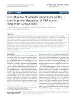
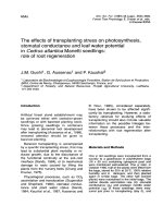
Figure 1
Simplified lung modelSimplified lung model. Total cardiac output (Qt) was distributed to ven-
tilated capillaries (QA) and to a right-to-left shunt (Qs). At steady state
the alveolar carbon dioxide tension (PaCO
2
) is the same as in the end-
capillary blood (PcCO
2
). V'CO
2
, eliminated carbon dioxide (ml/minute);
pHv, venous pH; PvCO
2
, venous carbon dioxide tension; PaCO
2
, arte-
rial carbon dioxide tension.
Available online />Page 3 of 7
(page number not for citation purposes)
PaCO
2
and PcCO
2
were then obtained in order to calculate
VdA
S
/VtA using Equation 1 (Figure 2c). This was done by sim-
ulating gas exchange in two iterated loops: one simulating the
path from mixed venous blood to pulmonary end-capillary
blood, and another simulating the path from mixed venous
blood to arterial blood. The latter loop (Figure 2) began with an
arbitrary, temporary PaCO
2
. The amount of carbon dioxide
eliminated from cardiac output (Qt) while venous oxygen satu-
ration changed to SaO
2
and venous carbon dioxide tension
changed to the temporary PaCO
2
was calculated in accord-
ance with Giovannini and coworkers. The temporary value of
PaCO
2
was iteratively adjusted until the calculated amount of
carbon dioxide eliminated equalled 200 ml/minute (Solver in
the Newton mode, Excel 2002; Microsoft Corp., Redmond,
WA, USA). The other loop began with an arbitrary PcCO
2
; its
value was iteratively adjusted until 200 ml carbon dioxide/
minute was eliminated, but from the blood flowing through
ventilated alveolar capillaries. VdA
S
/VtA was finally calculated
using Equation 1. For each loop, iterations continued until dif-
ference from the desired carbon dioxide elimination was under
0.001 ml/minute. Carbon dioxide elimination that depended
on increased oxygen saturation within the pulmonary capillar-
ies (the Haldane effect) was separated from the carbon diox-
ide elimination that depended on reduction in carbon dioxide
tension.
One purpose of ventilation is to effect carbon dioxide
exchange and achieve and maintain the target PaCO
2
, what-
ever that may be. Accordingly, it may be necessary to increase
V'A and V'tot in response to augmented VdA
S
/VtA. Alterna-
tively, one may allow PaCO
2
to increase. The VdA
S
/VtA values
obtained from the simulations above with different Qs/Qt were
used to calculate increases in V'A or PaCO
2
(using Equations
2 to 4, below). Increases in V'tot were also calculated at con-
stant PaCO
2
.
PaCO
2
= V'CO
2
/V'A × k = V'CO
2
/(RR × [Vt - Vd
phys
]) × k
Vd
phys
= Vd
aw
+ VdA
VQ
+ VdA
S
VdA
S
= VdA
S
/VtA × (Vt - Vd
aw
- VdA
VQ
)
where V'CO
2
is carbon dioxide elimination, k is barometric
pressure, RR is the respiratory rate, Vt is the tidal volume,
Vd
phys
is the physiological dead space, Vd
aw
is the airway dead
space, and VdA
VQ
is the part of alveolar dead space that is
caused by alterations in the ventilation/perfusion relationships
other than shunt. The calculations were based upon the mean
airway dead space of 0.2 l from the study conducted by Bey-
don and coworkers [1] and a deduced mean value for VdA
VQ
of 0.06 l from the same study. The calculations can be fol-
lowed by reference to Additional file 1 (Figures 6 and 7 in
Additional file 1).
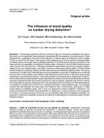
Results
All iterative calculations efficiently met the convergence crite-
rion. When, at basal conditions, Qs/Qt was increased to 0.5,
VdA
S
/VtA reached 0.15 (Figure 3). At reduced Qt the VdA
S
/
VtA paralleled the difference between CvCO
2
and arterial car-
bon dioxide content (CaCO
2
); specifically, CvCO
2
- CaCO
2
increased from 40 ml/l at a Qt of 5 l/minute to 67 ml/l at a Qt
of 3 l/minute. When CvCO
2
- CaCO
2
was increased to the
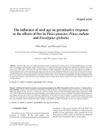
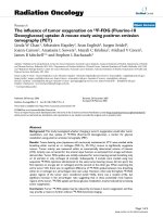
Figure 2
Outline of calculations further detailed in Additional file 1Outline of calculations further detailed in Additional file 1. (a) Starting
out from input parameters, analytical calculations of intermediate
parameters were performed using standard equations. (b) Input param-
eters, together with venous oxygen content (CvO
2
) and arterial oxygen
content (CaO
2
), define unique values of venous oxygen saturation
(SvO
2
) and arterial oxygen saturation (SaO
2
), which were iteratively
determined. Venous carbon dioxide content (CvCO
2
) was calculated in
accordance with the method reported by Giovannini and coworkers
[10]. (c) In an extensive system of iterations, arterial carbon dioxide ten-
sion (PaCO
2
) was iteratively adjusted until veno-arterial difference in
carbon dioxide content (ΔC [v-a]CO
2
) multiplied by total cardiac output
(Qt) became equal to carbon dioxide elimination (V'CO
2
). In a step par-
allel to that shown in panel c, end-capillary carbon dioxide tension
(PcCO
2
) was iteratively determined employing the value of QA (blood
flow to ventilated alveoli) instead of Qt.
Critical Care Vol 12 No 2 Niklason et al.
Page 4 of 7
(page number not for citation purposes)
same extent as when Qt was reduced, but via increased met-
abolic rate, the effects on VdA
S
/VtA were similar.
To maintain SaO
2
at 95%, at basal conditions FiO
2
was
increased to 0.7 at a Qs/Qt of 0.3 and further to 1.0 at a Qs/
Qt of 0.4. When FiO
2
was increased to 1.0 at a Qs/Qt of 0.3,
although SaO
2
was above 95%, VdA
S
/VtA increased from
0.071 to 0.079. In contrast, if FiO
2
was maintained at 0.4 while
SaO
2
fell to 91%, VdA
S
/VtA decreased to 0.063.
Normochromic anaemia (proportional decrease in haematocrit
and haemoglobin) or hypochromic anaemia (constant haema-
tocrit) was simulated by reducing haemoglobin from 145 to 97
and to 60 g/l. In both cases, at a Qs/Qt of 0.5 the VdA
S
/VtA
increased from 0.15 to 0.17 and 0.19, respectively. Variation
in haematocrit and respiratory quotient had only trivial effects
on VdA
S
/VtA.
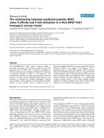
Respiratory acidosis, simulated by higher PaCO
2
at zero base
excess, led to lower VdA
S
/VtA (Figure 4). Metabolic acidosis
had the opposite effect. For the conditions shown in Figure 4
and at a Qs/Qt of 0.4, the fraction of carbon dioxide exchange
caused by the Haldane effect varied between 0.2 and 0.3.
Low CvCO
2
, as occurs in metabolic acidosis or hyperventila-
tion, was associated with low Haldane effect. At a Qs/Qt of
0.5, the VdA
S
/VtA correlated with the logarithm of CvCO
2
(VdA
S
/VtA = -0.10 × ln [CvCO
2
] + 0.55; R = 0.997).
In critically sick patients, physiological aberrations are often
combined. A patient in traumatic shock may have high Qs/Qt,
and low haemoglobin and Qt. Tissue hypoxia may lead to
metabolic acidosis. Figure 5 shows how VdA
S
/VtA would
increase as a consequence of these successive or parallel
phenomena. Particularly high values of VdA
S
/VtA were
observed in compensated metabolic acidosis, characterized
by hyperventilation to reduce PaCO
2
so as to partially normal-
ize pH.
More detailed data underlying Figures 3 to 5 are presented in
Additional file 1.
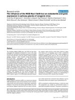
Figure 3
Alveolar dead space fraction versus shunt fraction at varying cardiac outputAlveolar dead space fraction versus shunt fraction at varying cardiac
output. Shown is the alveolar dead space fraction (VdA
S
/VtA) versus
shunt fraction (Qs/Qt) at varying cardiac output (Qt).
Figure 4
Alveolar dead space fraction versus shunt fraction at varying acid base statusAlveolar dead space fraction versus shunt fraction at varying acid base
status. Alveolar dead space fraction (VdA
S
/VtA) versus shunt fraction
(Qs/Qt) at varying acid-base status. Respiratory acidosis I and II refer
to arterial carbon dioxide tension (PaCO
2
) values of 9.1 kPa and 15.8
kPa, respectively, yielding arterial pH (pHa) values of 7.25 and 7.09,
respectively. Metabolic acidosis I and II refer to base excess (BE) val-
ues of -9.0 mmol/l and -17 mmol/l, yielding pHa values of 7.25 and
7.10, respectively.
Figure 5
Alveolar dead space fraction versus shunt fraction at additive ancillary pathologyAlveolar dead space fraction versus shunt fraction at additive ancillary
pathology. Alveolar dead space fraction (VdA
S
/VtA) versus shunt frac-
tion (Qs/Qt) at additive ancillary pathology. Step-wise analyses of
effects of a low haemoglobin (Hb; 97 g/l), low Hb and Qt (3.5 l/minute),
low Hb and Qt and metabolic acidosis (base excess [BE] -13 mmol/l),
and the latter case after respiratory compensation for acidosis by
hyperventilation (arterial carbon dioxide tension [PaCO
2
] 2.1 kPa).
Available online />Page 5 of 7
(page number not for citation purposes)
Depending upon which strategy is chosen to balance gas
exchange with lung protection, one can increase ventilation or
allow PaCO
2
to increase in response to the effect of shunt. At
basal conditions, at reduced Qt (3 l/minute) and at reduced Qt
combined with low haemoglobin and metabolic acidosis, a
Qs/Qt of 0.5 would result in increases in V'A (or in PaCO
2
) of
18%, 29% and 39%, respectively (Figure 6). Regarding dead
space of a non-VdA
S
origin, the increase in total ventilation
needed to maintain PaCO
2
would be 8.5%, 14% and 19% at
the same conditions as above (Figure 7). In the setting of a
tidal volume of 450 ml and a respiratory rate of 20 breaths/
minute, a Qs/Qt of 0.5 would be accompanied by increases in
tidal volume by 38 ml, 62 ml and 84 ml for the three conditions.
Discussion
The present study focuses on one of many factors to consider
when balancing adequate gas exchange with minimal ventila-
tor-induced lung injury during mechanical ventilation in ARDS
patients (alveolar dead space related to intrapulmonary shunt).
The rationale underpinning this approach is that increased
understanding of all such factors may form the basis for
improved treatment, and not only with respect to how the
patient should be ventilated. The effect on dead space of
shunt was studied under varied physiological circumstances,
such that may occur in ARDS; notable among these are
variation in metabolic rate, respiratory quotient, cardiac output,
haemoglobin and acid base status. By incorporating into our
model the parameters reported by Mecikalski and coworkers
[9], we were largely able to corroborate their findings,
although our values for VdA
S
/VtA are slightly higher. However,
in relation to the work conducted by Mecikalski and coworkers
[9], our findings regarding the effects of variation in haemo-
globin, acid base status and combinations of physiological
aberrations are novel. Additional file 1 can be used to verify
and expand upon the results by entering alternative input
parameters. The study did not incorporate diffusion limitation
or uneven ventilation/perfusion – factors that are more impor-
tant than shunt in other groups of critically ill patients.
Our analysis of VdA
S
/VtA was based upon well validated
equations, which together describe the highly complex proc-
ess of gas exchange. We employed iterative mathematics, as
first applied by West [4], and the algorithms developed by
Giovannini and coworkers [10] allow modelling of carbon diox-
ide exchange with particular precision.
Transport of carbon dioxide and the mechanisms underlying
its turnover depend on several factors, each of which are medi-
ated by nonlinear relationships between two or more factors.
The physiological background to the effects of shunt on dead
space and on carbon dioxide exchange at differing physiolog-
ical conditions is therefore complex. In each situation it is nev-
ertheless possible to recognize the primary mechanism
underlying the effects of shunt. A low cardiac output or a high
metabolic rate augments the venous content of carbon diox-
ide, and thereby the effect on shunt on PaCO
2
. The effect of
anaemia can be attributed to the fact that fewer haemoglobin
molecules are available to absorb the excess carbon dioxide
transferred to arterialized blood via shunted blood. Therefore,
Figure 6
Increase in PaCO
2
or alveolar ventilation versus shunt fractionIncrease in PaCO
2
or alveolar ventilation versus shunt fraction. Increase
in arterial carbon dioxide tension (PaCO
2
; %) at constant alveolar venti-
lation versus shunt fraction. This is equivalent to required increase in
alveolar ventilation to maintain PaCO
2
. Examples are as follows: 'Basal':
Qt = 5 l/minute, haemoglobin (Hb) = 145 g/l and base excess (BE) =
0; 'Qt = 3': Qt = 3 l/minute, Hb = 145 g/l and BE = 0; and 'Metab. aci-
dosis': Qt = 3.5 l/minute, Hb = 97 g/l and BE = -13.
Figure 7
Increase in total ventilation versus shunt fraction at constant PaCO
2
Increase in total ventilation versus shunt fraction at constant PaCO
2
.
Required increase in total ventilation (%) at different shunt fractions
(Qs/Qt) to maintain arterial carbon dioxide tension (PaCO
2
) constant.
Examples are as follows: 'Basal': Qt = 5 l/minute, haemoglobin (Hb) =
145 g/l and base excess (BE) = 0; 'Qt = 3': Qt = 3 l/minute, Hb = 145
g/l and BE = 0; and 'Metab. acidosis': Qt = 3.5 l/minute, Hb = 97 g/l
and BE = -13. Airway dead space (Vd
aw
) and the alveolar dead space
caused by uneven ventilation/perfusion (VdA
VQ
) were assumed to be
0.2 l and 0.06 l, respectively.
Critical Care Vol 12 No 2 Niklason et al.
Page 6 of 7
(page number not for citation purposes)
in a state of anaemia, this excess will to an increased extent
appear as dissolved carbon dioxide. This leads to an
enhanced increase in PaCO
2
. A high FiO
2
increases oxygen
content and saturation in blood from ventilated lung compart-
ments and in arterial blood. Through the Haldane effect, hae-
moglobin will then carry less carbon dioxide, leading to a
surplus that will be carried as dissolved carbon dioxide, thus
increasing PaCO
2
. In acid-base perturbations, the effects of
shunt on VdA
S
/VtA were tightly and negatively related to
ln(CvCO
2
). At low CvCO
2
, such as occurs in respiratory alca-
losis, the Haldane effect is less efficient. This hampers alveolar
carbon dioxide exchange and contributes to alveolar dead
space.
Effects on VdA
S
/VtA of shunt fractions up to about 0.2 to 0.3
are small and are of minimal clinical significance. Higher
degrees of shunt, particularly when combined with complicat-
ing physiological aberrations, the effect of shunt on carbon
dioxide exchange merits attention. An example is metabolic
acidosis combined with hyperventilation. Increased VdA
S
/VtA
should be added to known harmful effects of hyperventilation.
Clinically relevant effects of increasing VdA
S
/VtA are permis-
sively increased PaCO
2
or, equivalently, increased alveolar
ventilation (Figure 6). Obviously, one may choose a compro-
mise between these two alternatives. Figure 7 shows that total
ventilation at high shunt fraction may need to be increased by
10% to 20%, depending upon concurrent pathophysiology.
This estimate was based upon values for other dead space
compartments regarded as typical for ARDS. One may reason
that this is an effect of limited clinical importance. On the other
hand, an awareness of all of factors that are of importance to
the magnitude of dead space fractions may allow us to
develop less traumatic ventilation strategies. Clearly, airway
dead space caused by connecting tubes and humidifiers is
one such factor. Mode of inspiration is another; dead space
can also be reduced by selecting a mode of inspiration that
lengthens the mean distribution time during which the alveolar
tidal volume is present in the respiratory zone [3,14].
The essence of intensive care is to support the patient by
maintaining homeostasis. In ARDS, adequate oxygenation may
be achieved by reducing intrapulmonary shunt using ventila-
tion patterns that favour lung recruitment [15,16]. Such strat-
egies have the additional benefits of reducing VdA
S
/VtA and
the associated perturbation in carbon dioxide exchange. Other
routines in intensive care serve to maintain adequate cardiac
output, to control metabolic rate, and to avoid anaemia and to
maintain a proper acid base balance. All of them lead to lower
VdA
S
/VtA and reduced requirements for ventilation. A high
FiO
2
may lead to toxicity and enhances alveolar derecruitment
in ARDS [17]. As shown, an unduly high FiO
2
also augments
alveolar dead space.
This study provides additional motivation to maintain homeos-
tasis in ARDS. It underscores how combinations of physiolog-
ical aberrations may lead to inefficient carbon dioxide
exchange related to intrapulmonary shunting of blood.
Conclusion
In ARDS, perturbation of carbon dioxide exchange caused by
high shunt fraction is enhanced by ancillary disturbances such
as low cardiac output, anaemia, metabolic acidosis and
hyperventilation. Maintained homeostasis mitigates the effects
of shunt.
Competing interests
The authors declare that they have no competing interests.
Authors' contributions
JE conducted preliminary analyses. LN and BJ together devel-
oped the calculation program, performed the analyses and
wrote the manuscript. All authors read and approved the final
manuscript.
Additional files
Acknowledgements
The authors gratefully acknowledge that Hanna Fager performed initial
theoretical studies as part of her medical education. This study was sup-
ported by the Swedish Heart Lung Foundation.
References
1. Beydon L, Uttman L, Rawal R, Jonson B: Effects of positive end-
expiratory pressure on dead space and its partitions in acute
lung injury. Intensive Care Med 2002, 28:1239-1245.
2. Nuckton TJ, Alonso JA, Kallet RH, Daniel BM, Pittet JF, Eisner MD,
Matthay MA: Pulmonary dead-space fraction as a risk factor for
death in the acute respiratory distress syndrome. N Engl J
Med 2002, 346:1281-1286.
3. Åström E, Uttman L, Niklason L, Aboab J, Brochard L, Jonson B:
Pattern of inspiratory gas delivery affects CO
2
elimination in
Key messages
• In ARDS intrapulmonary shunt perturbs carbon dioxide
exchange by increasing alveolar dead space, particu-
larly in the presence of low cardiac output, reduced
haemoglobin levels and metabolic acidosis.
• Maintained homeostasis mitigates these effects of
shunt.
The following Additional files are available online:
Additional file 1
Dead space caused by shunt. This executable Excel file
allows calculation of alveolar dead space fraction caused
by shunt under different physiological conditions.
See />supplementary/cc6872-S1.xls
Available online />Page 7 of 7
(page number not for citation purposes)
health and after acute lung injury. Intensive Care Med 2008,
34:377-384.
4. West JB: Ventilation-perfusion inequality and overall gas
exchange in computer models of the lung. Respir Physiol
1969, 7:88-110.
5. Fletcher R, Jonson B, Cumming G, Brew J: The concept of dead-
space with special reference to the single breath test for car-
bon dioxide. Br J Anaesth 1981, 53:77-88.
6. West JB, Jones NL: Effects of changes in topographical distri-
bution of lung blood flow on gas exchange. J Appl Physiol
1965, 20:825-835.
7. Eriksson L, Wollmer P, Olsson CG, Albrechtsson U, Larusdottir H,
Nilsson R, Sjögren A, Jonson B: Diagnosis of pulmonary embo-
lism based upon alveolar dead space analysis. Chest 1989,
96:357-362.
8. Riley RL, Permutt S: Venous admixture component of the
AaPO
2
gradient. J Appl Physiol 1973, 35:430-431.
9. Mecikalski MB, Cutillo AG, Renzetti AD Jr: Effect of right-to-left
shunting on alveolar dead space. Bull Eur Physiopathol Respir
1984, 20:513-519.
10. Giovannini I, Chiarla C, Boldrini G, Castagneto M: Calculation of
venoarterial CO
2
concentration difference. J Appl Physiol
1993, 74:959-964.
11. Siggaard-Andersen O: Blood acid-base alignment nomogram.
Scand J Clin Lab Invest 1963, 15:211-217.
12. Riley RL, Lilienthal JLJ, Proemmel D, Franke RE: On the determi-
nation of the physiologically effective pressures of oxygen and
carbon dioxide in alveolar air. Am J Physiol 1948, 147:191-198.
13. Siggaard-Andersen O, Wimberley PD, Fogh-Andersen N, Giovan-
nini I, Gøthgen IH: Measured and derived quantities with mod-
ern pH and blood gas equipment: calculation algorithms with
54 equations. Scand J Clin Lab Invest 1988, 48(suppl
189):7-15.
14. Aboab J, Niklason L, Uttman L, Kouatchet A, Brochard L, Jonson
B: CO
2
elimination at varying inspiratory pause in acute lung
injury. Clin Physiol Funct Imaging 2007, 27:2-6.
15. Richard JC, Brochard L, Vandelet P, Breton L, Maggiore SM, Jon-
son B, Clabault K, Leroy J, Bonmarchand G: Respective effects
of end-expiratory and end-inspiratory pressures on alveolar
recruitment in acute lung injury. Crit Care Med 2003, 31:89-92.
16. Richard JC, Maggiore SM, Jonson B, Mancebo J, Lemaire F, Bro-
chard L: Influence of tidal volume on alveolar recruitment.
Respective role of PEEP and a recruitment maneuver. Am J
Respir Crit Care Med 2001, 163:1609-1613.
17. Aboab J, Jonson B, Kouatchet A, Taille S, Niklason L, Brochard L:
Effect of inspired oxygen fraction on alveolar derecruitment in
acute respiratory distress syndrome. Intensive Care Med 2006,
32:1979-1986.