Báo cáo y học: "Are serum cytokines early predictors for the outcome of burn patients with inhalation injuries who do not survive" pot
Bạn đang xem bản rút gọn của tài liệu. Xem và tải ngay bản đầy đủ của tài liệu tại đây (455.51 KB, 8 trang )
Open Access
Available online />Page 1 of 8
(page number not for citation purposes)
Vol 12 No 3
Research
Are serum cytokines early predictors for the outcome of burn
patients with inhalation injuries who do not survive?
Gerd G Gauglitz
1,2
*, Celeste C Finnerty
1,2
*, David N Herndon
1,2
, Ronald P Mlcak
1
and
Marc G Jeschke
1,2
1
Shriners Hospitals for Children, 815 Market Street, Galveston, Texas, 77550, USA
2
Department of Surgery, University of Texas Medical Branch, 301 University Boulevard, Galveston, Texas, 77550, USA
* Contributed equally
Corresponding author: Marc G Jeschke,
Received: 7 Mar 2008 Revisions requested: 14 Apr 2008 Revisions received: 25 Apr 2008 Accepted: 18 Jun 2008 Published: 18 Jun 2008
Critical Care 2008, 12:R81 (doi:10.1186/cc6932)
This article is online at: />© 2008 Gauglitz et al.; licensee BioMed Central Ltd.
This is an open access article distributed under the terms of the Creative Commons Attribution License ( />),
which permits unrestricted use, distribution, and reproduction in any medium, provided the original work is properly cited.
Abstract
Introduction Severely burned patients suffering from inhalation
injury have a significantly increased risk for mortality compared
with burned patients without inhalation injury. Severe burn is
associated with a distinct serum cytokine profile and alterations
in cytokines that contribute to morbidity and mortality. The aim
of the present study was therefore to determine whether
severely burned pediatric patients with concomitant inhalation
injury who had a fatal outcome exhibited a different serum
cytokine profile compared with burn patients with inhalation
injury who survived. Early identification followed by appropriate
management of these high-risk patients may lead to improved
clinical outcome.
Methods Thirteen severely burned children with inhalation injury
who did not survive and 15 severely burned pediatric patients
with inhalation injury who survived were enrolled in the study.
Blood was collected within 24 hours of admission and 5 to 7
days later. Cytokine levels were profiled using multiplex antibody
coated beads. Inhalation injury was diagnosed by bronchoscopy
during the initial surgery. The number of days on the ventilator,
peak inspiratory pressure rates, arterial oxygen tension (PaO
2
)/
fraction of inspired oxygen (FiO
2
) ratio and incidence of acute
respiratory distress syndrome were recorded for those patients.
Results Significantly altered levels of IL-4, IL-6, IL-7, IL-10, and
IL-13 were detected within the first 7 days after admission in
serum from burn pediatric patients with concomitant inhalation
injury who did not survive when compared with similar patients
who did (P < 0.05). Alterations in these cytokines were
associated with increased incidence of acute respiratory
distress syndrome, number of days under ventilation, increased
peak inspiratory pressure, and lower PaO
2
/FiO
2
ratio in this
patient population. Multiple logistic regression analysis revealed
that patients with increased IL-6 and IL-10 as well as decreased
IL-7 serum levels had a significantly greater risk for mortality (P
< 0.05).
Conclusion Early alterations in serum levels of IL-6, IL-7 and IL-
10 may constitute useful predictive markers for identifying
patients those who have sustained a burn with concomitant
inhalation injury and who have high mortality.
Introduction
Mortality from major burns has significantly decreased during
the past 20 years. Inhalation injury, however, still constitutes
one of the most critical adverse factors after thermal insult and
has remained associated with a mortality rate of 25% to 50%
when patients require ventilator support for more than 1 week
after injury [1-3]. Although many organ systems are affected by
a burn, the pulmonary system often sustains the most damage
[4]. Because inhalation injury is a major contributor to mortality
in thermally injured patients [3-5], early diagnosis and treat-
ment are crucial for the prevention of complications. The arte-
rial oxygen tension (PaO
2
)/fraction of inspired oxygen (FiO
2
)
ratio is a parameter that is widely used to define acute respira-
tory distress syndrome (ARDS) and – along with age, underly-
ing disease, malnutrition, and infection – it has been proposed
to be a prospective clinical predictor of poor outcome after
inhalation injury [6].
ARDS = acute respiratory distress syndrome; FiO
2
= fraction of inspired oxygen; IL = interleukin; PaO
2
= arterial oxygen tension; PIP = peak inspir-
atory pressure; PMN = polymorphonuclear neutrophil; TNF = tumor necrosis factor.
Critical Care Vol 12 No 3 Gauglitz et al.
Page 2 of 8
(page number not for citation purposes)
Inhalation injury is caused by steam or toxic inhalants such as
fumes, gases, or mists. It results in increased pulmonary micro-
vascular hyperpermeability, leading to edema formation, atel-
ectasis, and tracheobronchitis [1,7]. Subsequently,
neutrophils undergo diapedesis from the pulmonary microvas-
culature and release enzymes (including elastase) and free
oxygen radicals, disrupting endothelial junctions and epithelial
integrity, thus permitting an exudate of protein-rich plasma to
enter the lungs [8]. The inhalation of toxic smoke leads to the
release of inflammatory mediators such as thromboxane,
which enhance pulmonary artery pressure and cause second-
ary damage to the respiratory epithelium and the release of
additional mediators, such as tumor necrosis factor (TNF)
[3,8]. Release of these inflammatory molecules into the sys-
temic vasculature may cause injury to other organs [9].
In a previous study [10] we demonstrated that a burn causes
marked alterations in various inflammatory cytokines 1 week
after thermal injury when compared with healthy children.
Alterations in inflammatory mediators, such as cytokines, are
main contributors to the incidence of multiple organ failure and
mortality in critically ill patients [11]. The aim of the present
study was therefore to assess, in a cohort of severely burned
pediatric patients with inhalation injury, whether those severely
burned children who had a fatal outcome exhibited a distinct
serum cytokine profile in comparison with those who survived;
such a profile could serve as a predictive marker.
Materials and methods
Thirteen severely burned children with inhalation injury who did
not survive burn trauma ('nonsurvivors') and 15 severely
burned children with inhalation injury who survived ('survivors')
were enrolled in the study. Permission for conducting the
study was obtained from the Institutional Review Board of the
University of Texas Medical Branch, Galveston, Texas, USA.
Before the study, for each participant the patient, parent, or
child's legal guardian signed a written informed consent form.
All patients were 16 years of age or younger and were admit-
ted within 7 days after injury to the Shriners Hospital for Chil-
dren, Galveston, Texas, USA. Every child was suffering from
burns to more than 40% of total body surface area with a third
degree component of more than 24%, and required at least
one surgical intervention for escharectomy and skin grafting.
Patients were excluded if there was any sign of infection, sep-
sis, concomitant major injuries, or complications at admission.
After admission, patients were treated according to the stand-
ard of burn care at our hospital, including early excision and
grafting of the burn wound, and fluid and caloric resuscitation
in accordance with the Galveston formulas [12].
Demographics
Age, burn size, depth of burn, and time to admission were
recorded in each group.
Inhalation injury
Inhalation injury was diagnosed by bronchoscopy, which was
performed in all patients within 24 hours after admission in
accordance with the following criteria (Figure 1): signs of
exposure to smoke in an enclosed space, including presence
of facial burns, singed nasal vibrissae, bronchorrhea, sooty
sputum, and wheezing or rales upon auscultation; hypoxemia
and/or elevated levels of carbon monoxide; and bronchoscopy
findings of airway edema, inflammation, mucosal necrosis,
presence of soot and charring in the airway, tissue sloughing,
or carbonaceous material in the airway.
PaO
2
/FiO
2
ratio
The PaO
2
/FiO
2
ratio was used to quantify the degree of abnor-
malities in pulmonary gas exchange. PaO
2
/FiO
2
ratio was
measured in all patients within 24 hours after admission. In
addition, the number of days on the ventilator and peak inspir-
atory pressure (PIP) rates were recorded, and presence or
absence of ARDS was documented in accordance with the
guidelines proposed by the American-European Consensus
Conference on ARDS [13].
Cytokine measurements
Blood was collected in serum separator collection tubes at the
time of admission and 5 to 7 days thereafter (Figure 1). Blood
was centrifuged at 1,320 rpm for 10 minutes, and serum was
removed and then stored at -70°C until assayed. IL-1β, IL-2, IL-
4, IL-5, IL-6, IL-7, IL-8, IL-10, IL-12p70, IL-13, IL-17, granulo-
cyte colony-stimulating factor, granulocyte-macrophage col-
ony-stimulating factor, interferon-γ, monocyte chemoattractant
protein-1, macrophage inflammatory protein-1β, and TNF were
measured using the Bio-Plex Human Cytokine 17-Plex panel in
combination with the Bio-Plex Suspension Array System (Bio-
Rad Laboratories Inc., Hercules, CA, USA). The assay was
performed in accordance with the manufacturer's instructions.
Briefly, serum samples were thawed, centrifuged at 4,500 rpm
for 3 minutes at 4°C, and incubated with micro beads labeled
with antibodies specific to one of the aforementioned
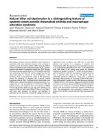
Figure 1
Outline of the studyOutline of the study. The arterial oxygen tension (PaO
2
)/fraction of
inspired oxygen (FiO
2
) ratio was measured in all patients within 24
hours after admission. Blood was drawn at hospital admission and 5 to
7 days afterward.
Available online />Page 3 of 8
(page number not for citation purposes)
cytokines for 30 minutes. After a wash step, the beads were
incubated with the detection antibody cocktail, each bead
specific to a single cytokine. After an additional wash step, the
beads were incubated with streptavidin-phycoerythrin for 10
minutes, washed, and placed in the array reader for determina-
tion of the respective cytokine concentration.
Statistical analysis
Unpaired Student's t-tests were used to compare differences
in cytokine expression, differences in length of ventilation, PIP,
and PaO
2
/FiO
2
ratio between groups. Demographics were
compared using t-tests or χ
2
tests. The Fisher's exact test was
used to compare baseline variables. Data are expressed as
percentages of means ± standard error of the mean, where
appropriate. Statistical significance was accepted at a P value
of less than 0.05. Statistics were run using SigmaStat 2004
(Systat Software Inc., Chicago, Illinois, USA). Multiple logistic
regression was used to develop a prediction equation for
determining the likelihood of mortality of burn patients with
concomitant inhalation injury from early serum cytokine pro-
files. (To calculate a probability from the logistic equation
shown in the Results section [below], transform the logit using
Prob[YG9group] = 1/[1 + ExpBurned].) To assess the good-
ness-of-fit for the regression, the likelihood ratio test statistic
and the mean, standard error of the mean, and Wald statistic
for each parameter were examined.
Results
Twenty-eight severely burned children with inhalation injury
were studied. Patient demographics are shown in Table 1.
Groups were of similar age, burn size, extent of third-degree
burn, and time from burn to admission, but there were signifi-
cantly more females than males in the survivor group.
Severely burned children with inhalation injury who did not sur-
vive exhibited lower PaO
2
/FiO
2
rates within 24 hours after hos-
pital admission when compared with children who survived
(Figure 2a). Burn patients with inhalation injury who had a fatal
outcome exhibited a significantly (P < 0.05) greater number of
days on the ventilator than did children who survived (Figure
2b). Significantly higher PIP rates were observed in nonsurvi-
vors than in survivors (P < 0.05; data shown in Figure 2c).
Severely burned children with concomitant inhalation injury
who did not survive had a higher incidence of ARDS as com-
pared with those who survived, but this difference was not sta-
tistically significant (55.6% versus 27.7%).
Seventeen cytokine serum levels were significantly increased
at the time of hospital admission, both in burned patients with
inhalation injury who did not survive and in those who survived
compared with levels in nonburned, normal pediatric patients
(data not shown).
By comparing severely burned children suffering from inhala-
tion injury who did not survive with those who survived, we
found significant differences in serum levels of IL-4, IL-6, IL-7,
IL-10, and IL-13 (Figure 3). Nonsurvivors exhibited a signifi-
cant increase in IL-4 serum levels upon hospital admission
when compared with the survivor group (P < 0.05; Figure 3a).
IL-6 serum levels were significantly elevated in the nonsurvivor
group at admission when compared with survivors (P < 0.05;
Figure 3b). Nonsurvivors exhibited significantly lower IL-7
serum levels 5 to 7 days after admission compared with the
survivor group (P < 0.05; Figure 3c). IL-10 serum levels were
significantly increased in the nonsurvivor group at admission
and 5 to 7 days after hospital admission compared with survi-
vors (P < 0.05; Figure 3d). Nonsurvivors showed a significant
increase in IL-13 serum levels at admission compared with the
survivor group (P < 0.05; Figure 3e).
Serum levels of IL-1β, IL-2, IL-4, IL-5, IL-6, IL-7, IL-8, IL-10, IL-
12p70, IL-13, IL-17, granulocyte colony-stimulating factor,
granulocyte-macrophage colony-stimulating factor, interferon-
γ, monocyte chemoattractant protein-1, macrophage inflam-
matory protein-1β, and TNF were not significantly different
between the two groups.
We found a panel including IL-6, IL-7, and IL-10 to exhibit
excellent predictive ability with respect to mortality (likelihood
ratio test statistic: 7.1 [P < 0.008] and 8.5 [P < 0.01] at
admission and 5 to 7 days after admission, respectively). The
Wald statistic was significant for each of these three variables
when this regression was run (P < 0.05). When any of the
other 15 cytokines measured was added to the multiple logis-
tic regression, the Wald statistic was not significant for the
added cytokine. The other 15 cytokines were therefore not
included in the multiple logistic regression analysis. A multiple
logistic regression was conducted with the variables of mortal-
ity (dependent variable), IL-6, IL-7, and IL-10, and the following
equations were obtained: Logit P = 1.551 - (0.343 × IL-10)
and Logit P = 0.690 - (0.00662 × IL-6) + (0.869 × IL-7), at
admission and 5 to 7 days after admission, respectively. The
coefficients for IL-6 and IL-10 were negative, indicating that
the risk for mortality increased as the levels of IL-6 and IL-10
Table 1
Patient demographics
Parameter Survivors Nonsurvivors
Number (n)1513
Age (years) 8 ± 2 9 ± 1
Sex (n; female/male) 3/12 9/4*
Burn to admittance (days) 3 ± 1 2 ± 1
TBSA (%) 65 ± 5 76 ± 4
Third degree (%) 53 ± 6 69 ± 5
Where applicable, data are presented as means ± standard
deviation. *P < 0.05. TBSA, total body surface area.
Critical Care Vol 12 No 3 Gauglitz et al.
Page 4 of 8
(page number not for citation purposes)
increased. The coefficient for IL-7 was positive, indicating that
the risk of mortality increased as the levels of IL-7 decreased.
The means, standard errors, and Wald statistics of the logistic
regression coefficients are as follows: IL-10, 3.883 ± 0.174 (P
= 0.049) upon admission; and IL-6, 4.570 ± 0.00289 (P =
0.022) and IL-7, 4.369 ± 0.416 (P = 0.037) 5 to 7 days after
admission. When the other parameters were added sequen-
tially, the following Wald statistics were obtained for the
added variable: IL-4 (P = 0.196) and IL-13 (P = 0.158) at
admission, and IL-10 (P = 0.143) 5 to 7 days after admission.
Because some of the cytokines are highly correlated, the logis-
tic regression is not improved by adding all five variables.
Discussion
Smoke inhalation may lead to release of mediators that
increase pulmonary artery pressure and cause secondary
damage to the respiratory epithelium and the release of addi-
tional inflammatory molecules [3,8]. Lung injury resulting from
smoke inhalation is associated with significant increases in the
incidence of pneumonia and ARDS in thermally injured
patients [14]. These may be exacerbated by early hemody-
namic instability and massive burn edema, both of which are
commonly observed in burn injury patients with smoke inhala-
tion. Severe pulmonary dysfunction resulting from smoke inha-
lation therefore remains one of the leading contributors to
mortality in patients with thermal injury [15]. Thus, early identi-
fication followed by appropriate management of those high-
risk patients may lead to improved clinical outcome.
Alterations in inflammatory mediators, such as cytokines, are
main contributors to the incidence of multiple organ failure and
mortality in critically ill patients. Thus, in the present study we
hypothesized that burned pediatric patients with inhalation
injury who had a fatal outcome exhibited a different serum
cytokine profile when compared with similar patients who sur-
vived. Patients divided into the two study groups were of sim-
ilar age, burn size, extent of third-degree burn, and time from
burn to admission. There were considerably more females
than males in the survivor group. This does not constitute a
concern, because we recently found that sex-specific differ-
ences in pediatric patients do not play a role in mortality rates
[16].
Here we found that increases in inflammatory cytokines IL-4,
IL-6, IL-7, IL-10, and IL-13 within the first 7 days after admis-
sion were strongly associated with the incidence of mortality
in these patients. Patient mortality correlated with the
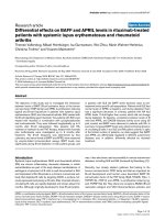
Figure 2
Nonsurviving pediatric patients with inhalation injury display more severe deterioration of lung function than their surviving counterpartsNonsurviving pediatric patients with inhalation injury display more severe deterioration of lung function than their surviving counterparts. (a) The arte-
rial oxygen tension (PaO
2
)/fraction of inspired oxygen (FiO
2
) ratio of severely burned children with inhalation injury who did not survive was lower
than in those who survived (220 ± 27 mmHg versus 282 ± 23 mmHg). (b) Burn patients with inhalation injury who had a fatal outcome had signifi-
cantly more ventilator days than children who survived (24 ± 5 days versus 5 ± 1 days). (c) Nonsurvivors exhibited significantly higher peak inspira-
tory pressure rates than survivors (71.5 ± 8.2 cmH
2
O versus 30.6 ± 2.1 cmH
2
O). (d) Severely burned children with concomitant inhalation injury
who did not survive had a higher incidence of acute respiratory distress syndrome (ARDS) than did those who survived, which was not statistically
significant (55.6% versus 27.7%). Bars represent means; error bars correspond to standard error of the mean. *P < 0.05.
Available online />Page 5 of 8
(page number not for citation purposes)
incidence of ARDS, the number of ventilation days, the peak
inspiratory pressure, and the PaO/FiO
2
ratio in this population
– parameters that are widely used to define inhalation injury.
Age, underlying disease, malnutrition, and infections have
been studied as prospective clinical predictors of poor out-
come after inhalation injury in addition to the PaO
2
/FiO
2
ratio
[6]. Despite its widespread use, the validity of the PaO
2
/FiO
2
ratio as a tool for assessing pulmonary gas exchange has
remained controversial [17]. González-Castro and colleagues
[6] found a value of PaO
2
/FiO
2
ratio above 100 mmHg 24
hours after admission to the intensive care unit to be associ-
ated with a lower mortality in patients who underwent lung
transplantation. Yilmaz and coworkers [18] successfully uti-
lized the PaO
2
/FiO
2
ratio on day 3 after the onset of acute lung
injury to assess hospital and 6-month mortality. In contrast, no
correlation could be established between outcome in patients
with severe lung injury and PaO
2
/FiO
2
ratio by Krafft and col-
leagues [19]. Our data suggest that levels of PaO
2
/FiO
2
below 220 mmHg at admission tend to be associated with
fatal outcome in burn patients with inhalation injury. However,
the higher values of PaO
2
/FiO ratio in nonsurvivors did not
reach statistical significance when compared with those of
patients who survived burn trauma with inhalation injury. This
could be a result of the relatively small number of patients in
our study. Pronounced deterioration in lung function in nonsur-
Figure 3
Cytokines are significantly altered in nonsurviving versus surviving patients who sustained inhalation injuryCytokines are significantly altered in nonsurviving versus surviving patients who sustained inhalation injury. (a) IL-4 serum levels were significantly
increased in the nonsurvivor group at admission compared with survivors (normal IL-4: 0 ± 0 pg/ml). (b) Nonsurvivors exhibited a significant increase
in IL-6 serum levels 5 to 7 days after admission compared with the survivor group (normal IL-6: 8.7 ± 5 pg/ml). (c) Nonsurvivors exhibited a signifi-
cant decrease in IL-7 serum levels 5 to 7 days after admission compared with the survivor group (normal IL-7: 3.8 ± 0.63 pg/ml). (d) IL-10 serum lev-
els were significantly increased in the nonsurvivor group at admission and 5 to 7 days after admission compared with survivors (normal IL-10: 1.4 ±
0.3 pg/ml). (e) Nonsurvivors exhibited a significant increase in IL-13 serum levels upon hospital admission when compared with the survivor group
(normal IL-13: 0.9 ± 0.2 pg/ml). Throughout the figure, histograms depict serum concentrations of the respective cytokine at steady state levels.
Bars represent means; error bars correspond to standard error of the mean. *P < 0.05. pAD, post-admission days.
Critical Care Vol 12 No 3 Gauglitz et al.
Page 6 of 8
(page number not for citation purposes)
vivors was subsequently revealed by their significantly greater
number of days on the ventilator and significantly higher PIP
rates. An increased incidence of ARDS was also observed,
but this was not statistically significant when compared with
that in similar patients who survived the injury.
The aim of the present study was therefore to develop a means
to predict the outcome of severely burned patients who sus-
tained inhalation injury sufficiently early after admission to
allow adequate measures to be implemented for their hospital
management. Because of the relevance of inflammatory medi-
ators as biomarkers in acute lung injury, we hypothesized that
the levels of various cytokines were elevated early during the
course of hospitalization in this patient population. Serum lev-
els of IL-6, IL-8, and IL-10 have been evaluated in several clin-
ical trials. Although not detectable in all patients at risk for
developing ARDS [20-22], increased levels of IL-6 and the
persistence of these levels have been strongly associated with
mortality [20,23]. Similarly, in our study the increased levels of
IL-6 determined during the first day after admission exhibited
a strong correlation with outcome in patients who did not sur-
vive. There is mounting evidence that the immunomodulatory
properties of IL-6 result in increased polymorphonuclear neu-
trophil (PMN)-mediated hyperinflammation, enhancing PMN
cytotoxic potential and influencing host immunosuppression
[11].
The proinflammatory cytokine IL-8 was also found to be
increased in patients at risk for developing ARDS, although
the association between plasma IL-8 levels and morbidity and
mortality in small clinical studies has not been consistent [24-
26]. In our study we found no correlation between elevated
serum IL-8 levels and fatal outcome, even though burned
patients with inhalation injury overall exhibited significantly
increased serum levels of IL-8 when compared with healthy
children. IL-8, mainly released from alveolar macrophages, is
one of the most important contributors to the complex events
that occur at reperfusion and is a key chemotactic factor for
PMNs [27]. It dramatically enhances neutrophil transmigration
through pulmonary endothelium and epithelium as well as
PMN chemoattraction and activation [28]. However, because
of its chemoattractant activity, it is likely that IL-8 contributes
more to acute inflammation within the lung than in the circula-
tion [25,26,29].
Unlike IL-8, we found IL-10 serum levels to be associated with
fatal outcome. IL-10 has been demonstrated to inhibit alveolar
macrophage production of proinflammatory mediators that are
involved in severe lung injury. It has been also shown to play a
role in downregulating HLA-DR expression on monocytes from
septic patients and may play a role in modulating the host
response to infections in these critically ill patients [30]. Sch-
neider and coworkers [31] found that IL-10 is a critical media-
tor of immunosuppression after traumatic injury. Studies by
Lyons and colleagues [32] indicated that increased IL-10 pro-
duction correlates with subsequent septic events, and in the
burn mouse IL-10 appears to induce decreased resistance to
infection. Plasma IL-10 levels did not predict the development
of ARDS in patients at risk but were found to be increased in
patients with ARDS who did not survive [33]. IL-7 was found
to have antiapoptotic effects on T cells via Bcl-2 expression,
indicating that this cytokine plays an important role in support-
ing cell survival [34]. In a recent study by our group [1], this
mediator was significantly decreased in pediatric burn patients
with inhalation injury compared with similar patients without
inhalation injury. In our study, decreases in IL-7 serum levels
correlated with increased incidence of mortality in these
patients. However, how this particular cytokine is modulated in
response to inhalation injury is not known. In contrast, we
found that the anti-inflammatory cytokines IL-4 and IL-13 were
significantly increased upon admission. These cytokines are
believed to be part of the underlying mechanisms for the devel-
opment of ARDS [35]. IL-13 induces multiple features of aller-
gic lung disease, including metaplasia and mucus
hypersecretion, contributing to airway obstruction [36].
Increases in IL-13 mRNA in pulmonary tissue correlated
closely with the incidence of ARDS in a rodent model [37]. An
association with mortality was not found in these studies.
Compelling evidence that causally links elevation in various
cytokine serum levels to poor patient outcome is lacking. How-
ever, it has been established that during the response follow-
ing severe illness, release of various cytokines is not properly
regulated [11,38]. Indeed, high blood levels of proinflamma-
tory cytokines may lead to a debilitating condition known as
autodestructive systemic inflammatory response syndrome
[11]. In this condition both proinflammatory cytokines and anti-
inflammatory cytokines appear in circulating blood, leading to
septic shock, multiple organ dysfunction and immunosuppres-
sion, ultimately contributing to increased mortality [11,38].
Conclusion
We believe that the results presented here from our relatively
small patient cohort indicate that serum cytokine levels may be
valuable outcome predictors in burn patients with inhalation
injury, even though it is presently unclear whether their eleva-
tion arises from local pulmonary inflammation or an associated
systemic inflammatory response. In contrast, despite its wide-
spread use, the validity of the PaO
2
/FiO
2
ratio as a tool for pre-
dicting mortality in patients with severe lung injury has
remained controversial [6,16-18]. Determination of serum IL-
6, IL-7, and IL-10 levels upon admission is convenient and sim-
ple, and may serve as an early indicator for identifying patients
who have a greater risk for mortality after a burn with concom-
itant inhalation injury.
Competing interests
The authors declare that they have no competing interests.
Available online />Page 7 of 8
(page number not for citation purposes)
Authors' contributions
GGG gathered data, helped conducting the statistics, and
wrote the manuscript. CCF performed experiments to obtain
data, conducted the statistical analysis, and reviewed the man-
uscript. DNH gathered data, reviewed the analysis, and helped
to write the manuscript. RPM helped to collect data and write
the manuscript. MGJ designed the study, gathered data, con-
ducted the statistical analyses, and reviewed the manuscript.
Acknowledgements
This study was supported by the American Surgical Association Foun-
dation, Shriners Hospitals for Children 8660, 8760, and 9145, NIH
R01-GM56687, T32 GM008256, and P50 GM60338, and NIDRR
H133A020102.
References
1. Finnerty CC, Herndon DN, Jeschke MG: Inhalation injury in
severely burned children does not augment the systemic
inflammatory response. Crit Care 2007, 11:R22.
2. Thompson PB, Herndon DN, Traber DL, Abston S: Effect on mor-
tality of inhalation injury. J Trauma 1986, 26:163-165.
3. Nugent N, Herndon DN: Diagnosis and treatment of inhalation
injury. In Total Burn Care 3rd edition. Edited by: Herndon DN.
London, UK: Saunders & Elsevier; 2007:262-272.
4. Weiss SM, Lakshminarayan S: Acute inhalation injury. Clin
Chest Med 1994, 15:103-116.
5. Demling RH: Smoke inhalation injury. New Horiz 1993,
1:422-434.
6. González-Castro A, Llorca J, Burón J, Suberviola B, Vallejo A,
Miñambres E: Evaluation of the oxygenation ratio as long-term
prognostic marker after lung transplantation. Transplant Proc
2007, 39:2422-2424.
7. Sheridan RL: Airway management and respiratory care of the
burn patient. Int Anesthesiol Clin 2000, 38:129-145.
8. Traber DL, Herndon DN, Enkhbaatar P, Maybauer MO, Maybauer
DM: The pathophysiology of inhalation injury. In Total Burn
Care 3rd edition. Edited by: Herndon DN. London, UK: Sounders
Elsevier; 2007:248-261.
9. Anonymous: Ventilation with lower tidal volumes as compared
with traditional tidal volumes for acute lung injury and the
acute respiratory distress syndrome. The Acute Respiratory
Distress Syndrome Network. N Engl J Med 2000,
342:1301-1308.
10. Finnerty CC, Herndon DN, Przkora R, Pereira CT, Oliveira HM,
Queiroz DM, Rocha AM, Jeschke MG: Cytokine expression pro-
file over time in severely burned pediatric patients. Shock
2006, 26:13-19.
11. Biffl WL, Moore EE, Moore FA, Peterson VM: Interleukin-6 in the
injured patient. Marker of injury or mediator of inflammation?
Ann Surg 1996, 224:647-664.
12. Herndon DN: Total Burn Care 3rd edition. London, UK: Saunders
Elsevier; 2007.
13. Bernard GR, Artigas A, Brigham KL, Carlet J, Falke K, Hudson L,
Lamy M, Legall JR, Morris A, Spragg R: The American-European
Consensus Conference on ARDS. Definitions, mechanisms,
relevant outcomes, and clinical trial coordination. Am J Respir
Crit Care Med 1994, 149:818-824.
14. Wright MJ, Murphy JT: Smoke inhalation enhances early alveo-
lar leukocyte responsiveness to endotoxin. J Trauma 2005,
59:64-70.
15. Shirani KZ, Pruitt BA Jr, Mason AD Jr: The influence of inhalation
injury and pneumonia on burn mortality. Ann Surg 1987,
205:82-87.
16. Barrow RE, Przkora R, Hawkins HK, Barrow LN, Jeschke MG,
Herndon DN: Mortality related to gender, age, sepsis, and eth-
nicity in severely burned children. Shock 2005, 23:485-487.
17. Karbing DS, Kjaergaard S, Smith BW, Espersen K, Allerod C,
Andreassen S, Rees SE: Variation in the PaO
2
/FiO
2
ratio with
FiO
2
: mathematical and experimental description, and clinical
relevance. Crit Care 2007, 11:R118.
18. Yilmaz M, Iscimen R, Keegan MT, Vlahakis NE, Afessa B, Hubmayr
RD, Gajic O: Six-month survival of patients with acute lung
injury: prospective cohort study. Crit Care Med 2007,
35:2303-2307.
19. Krafft P, Fridrich P, Pernerstorfer T, Fitzgerald RD, Koc D, Schnei-
der B, Hammerle AF, Steltzer H: The acute respiratory distress
syndrome: definitions, severity and clinical outcome. An anal-
ysis of 101 clinical investigations. Intensive Care Med 1996,
22:519-529.
20. Casey LC, Balk RA, Bone RC: Plasma cytokine and endotoxin
levels correlate with survival in patients with the sepsis
syndrome. Ann Intern Med 1993, 119:771-778.
21. Casey LC: Role of cytokines in the pathogenesis of cardiopul-
monary-induced multisystem organ failure. Ann Thorac Surg
1993, 56(5 Suppl):S92-S96.
22. Goldie AS, Fearon KC, Ross JA, Barclay GR, Jackson RE, Grant
IS, Ramsay G, Blyth AS, Howie JC: Natural cytokine antagonists
and endogenous antiendotoxin core antibodies in sepsis syn-
drome. The Sepsis Intervention Group. JAMA 1995,
274:172-177.
23. Pinsky MR, Vincent JL, Deviere J, Alegre M, Kahn RJ, Dupont E:
Serum cytokine levels in human septic shock. Relation to mul-
tiple-system organ failure and mortality. Chest 1993,
103:565-575.
24. Chollet-Martin S, Montravers P, Gibert C, Elbim C, Desmonts JM,
Fagon JY, Gougerot-Pocidalo MA: High levels of interleukin-8 in
the blood and alveolar spaces of patients with pneumonia and
adult respiratory distress syndrome. Infect Immun 1993,
61:4553-4559.
25. Miller EJ, Cohen AB, Nagao S, Griffith D, Maunder RJ, Martin TR,
Weiner-Kronish JP, Sticherling M, Christophers E, Matthay MA:
Elevated levels of NAP-1/interleukin-8 are present in the air-
spaces of patients with the adult respiratory distress syn-
drome and are associated with increased mortality. Am Rev
Respir Dis 1992, 146:427-432.
26. Pugin J, Verghese G, Widmer MC, Matthay MA: The alveolar
space is the site of intense inflammatory and profibrotic reac-
tions in the early phase of acute respiratory distress
syndrome. Crit Care Med 1999, 27:304-312.
27. Harada A, Sekido N, Akahoshi T, Wada T, Mukaida N, Matsushima
K: Essential involvement of interleukin-8 (IL-8) in acute
inflammation. J Leukoc Biol 1994, 56:559-564.
28. Rao JN, Clark SC, Ali S, Kirby J, Flecknell PA, Dark JH: Improve-
ments in lung compliance after pulmonary transplantation:
correlation with interleukin 8 expression. Eur J Cardiothorac
Surg 2003, 23:497-502.
29. Donnelly SC, Strieter RM, Kunkel SL, Walz A, Robertson CR,
Carter DC, Grant IS, Pollok AJ, Haslett C: Interleukin-8 and
Key messages
• Severely burned patients suffering from inhalation injury
have a significantly increased risk for mortality com-
pared with burned patients without inhalation injury.
• Alterations in inflammatory mediators, such as
cytokines, are main contributors to the incidence of mul-
tiple organ failure and mortality in critically ill patients.
• Age, underlying disease, malnutrition, infections, and
PaO
2
/FiO
2
ratio are commonly utilized tools that may be
used to predict poor outcome after inhalation injury.
• Alterations in IL-4, IL-6, IL-7, IL-10, and IL-13 appear to
be associated with increased incidence of ARDS,
number of days under ventilation, increased PIP, and
lower PaO
2
/FiO
2
ratio in this patient population.
• Early alterations in serum levels of IL-6, IL-7, and IL-10
may constitute useful predictive markers for identifying
patients with high mortality after burns with concomitant
inhalation injury.
Critical Care Vol 12 No 3 Gauglitz et al.
Page 8 of 8
(page number not for citation purposes)
development of adult respiratory distress syndrome in at-risk
patient groups. Lancet 1993, 341:643-647.
30. Fumeaux T, Pugin J: Role of interleukin-10 in the intracellular
sequestration of human leukocyte antigen-DR in monocytes
during septic shock. Am J Respir Crit Care Med 2002,
166:1475-1482.
31. Schneider CP, Schwacha MG, Chaudry IH: The role of inter-
leukin-10 in the regulation of the systemic inflammatory
response following trauma-hemorrhage. Biochim Biophys
Acta 2004, 1689:22-32.
32. Lyons A, Kelly JL, Rodrick ML, Mannick JA, Lederer JA: Major
injury induces increased production of interleukin-10 by cells
of the immune system with a negative impact on resistance to
infection. Ann Surg 1997, 226:450-458. discussion 458–460.
33. Parsons PE, Moss M, Vannice JL, Moore EE, Moore FA, Repine JE:
Circulating IL-1ra and IL-10 levels are increased but do not
predict the development of acute respiratory distress syn-
drome in at-risk patients. Am J Respir Crit Care Med 1997,
155:1469-1473.
34. Lee SK, Surh CD: Role of interleukin-7 in bone and T-cell
homeostasis. Immunol Rev 2005, 208:169-180.
35. Li Q, Qian G, Zhang Q, Gong J, Tang Z, Gao Z: Changes of
plasma interleukin-4, interleukin-10 and interleukin-13 in
patients with acute respiratory distress syndrome [in
Chinese]. Zhonghua Jie He He Hu Xi Za Zhi 2002, 25:661-664.
36. Wills-Karp M, Luyimbazi J, Xu X, Schofield B, Neben TY, Karp CL,
Donaldson DD: Interleukin-13: central mediator of allergic
asthma. Science 1998, 282:2258-2261.
37. Li Q, Qian G, Zhang Q, Xu J, Long Y, Tang Z, Gong J: The change
in IL-13 mRNA expression in rat lungs with acute pulmonary
injury induced by lipopolysaccharide [in Chinese]. Zhonghua
Shao Shang Za Zhi 2002, 18:145-148.
38. Aikawa N: Cytokine storm in the pathogenesis of multiple
organ dysfunction syndrome associated with surgical insults
[in Japanese]. Nippon Geka Gakkai Zasshi 1996, 97:771-777.